on some remarkable new monticellia-like and other cestodes … · 2006-05-18 · on some remarkable...
TRANSCRIPT

On some remarkable new Monticellia-like andother Cestodes from Sudanese Siluroids
By
W. N. F. Woodland,Wellcome Bureau of Scientific Research, Endsleigh Gardens,
London, N.W. 1.
With Plates 53 and 54 and 1 Text-figure.
Monticellia-like Cestodes.IN 1891 Monticelli first described from a European (pre-
sumably) unnamed species of Silurus a tapeworm which henamed Tetracotylus coryphicephala. This tape-worm—a Tetraphyllidean—was remarkable in that it differed,not only from all other Tetraphyllidea but from all otherCestoda then known to zoologists in having (1) the testessituated in the cortex, i.e. external to the inner layer of longi-tudinal muscles, on the dorsal side of the proglottis, and (2) thouterus and the ovary (in large part) also situated in the cortex—ventrally and dorsally respectively (see Text-figure on p. 713).This species Tetracotylus coryphicephala (and thopossibly closely - allied species Tetracotylus macro-cotylea from Silurus megacephalus and Tetra-cotylus diesingii from Silurus dargado) has been tosome extent redescribed by La Rue (1914) from Monticelli'sslides, and he substituted the name Monticellia for Monti-celli's ' Tetracotylus ' and the ' Ichthyotaenia ' laterapplied by Lonnberg, Riggenbach, and Braun. La Rue wasapparently unaware when he published his excellent mono-graph on the Proteocephalidae in 1914 that Riggenbach in1896 had described in a second species of ' Corallobo-t h r i u m '—' Corallobothrium' lobosum from a speciesof Silurus from the Rio Paraguay, South America—a similar

704 W. N. F. WOODLAND
dorsal cortical disposition of the testes, but associated in thiscase with an ovary (small lobules of which, however, accordingto Fuhrmann, 1916, project through the internal longitudinalmuscle-layer into the dorsal cortex) and uterus situated, inthe normal Proteocephalid manner, in the medulla. In bothMonticelli's and Eiggenbach's species the vitellaria are alsocortical in position (ventro-lateral), so contrasting with themedullary vitellaria of normal Proteocephalidae.
In 1916 Puhrmann described another example of theseaberrant Siluroid Cestodes also from South America (theAmazon), which he named Goezee l la s i l u r i , from Ce top-sis c a e c u t i e n s . This species resembled in all essentials theM o n t i c e l l i a c o r y p h i c e p h a l a originally described byMonticelli, and Fuhrmann only relegated his species to adifferent genus because it possesses a scolex of the ' Cor al l o-b o t h r i u m ' type and not of the simple unadorned kindcharacteristic of most species of Proteocephalidae. Fuhrmannfurther redescribed Eiggenbach's C o r a l l o b o t h r i u m lobo-sum from the original material and renamed it, on account ofthe aberrant situation of the testes and vitellaria, E u d o l -p h i e l l a l obosa (Eigg.), so differentiating it from theC o r a l l o b o t h r i u m s o l i d u m of Fritsch, the genitalia ofwhich conform to the normal Proteocephalid type.
The type of Tetraphyllidean Cestoda distinguished by thecortical situation of the testes (the other organs already men-tioned either being also cortical or retaining the normal medul-lary disposition) may be provisionally labelled the Monticelliatype. Of this type two different conditions have, as we haveseen, so far been described : (1) the condition found in La Eue'sM o n t i c e l l i a and Fuhrmann's Goezee l l a , in whichpractically all the genitalia are cortical in situation—testes,ovary, uterus, and vitellaria, and (2) the condition found inFuhrmann's E u d o l p h i e l l a , in which only the testes, verysmall projections of the ovary and the vitellaria are cortical.I now proceed to describe, from an altogether distinct zoologicalregion, two species showing a third condition of the Monticelliatype, viz. one in which only the testes are cortical, all other

CESTODES FROM SILUROIDS 705
organs—uterus, ovary, and even the vitellaria—being medul-lary. The significance, taxonomic position, and nomenclatureof the various species illustrating these three conditions willbe discussed subsequently.
M a r s y p o c e p h a l u s r e c t a n g u l u s Wedl, 1862.
This species and the following (Marsypocepha lush e t e r o b r a n c h u s ) are represented in the large collection ofHelminths made by the late Dr. A. J. Chalmers when Directorof the Wellcome Tropical Eesearch Laboratories at Khartoumand kindly presented to the Wellcome Bureau by his successor,Major E. G. Archibald. The present species was obtained fromthe intestine of Clar ias a n g u i l l a r i s Linn., a NileSiluroid, and of it I possess only one scolex of an adult wormand 13 mm. of attached anterior proglottids, and two otherportions of the posterior strobila mounted whole in balsam anda large number of excellent horizontal and transverse sections.I am unable therefore to state the total length of any oneworm, but, judging from the condition of the portions ofstrobila named, I should say that 40 mm. is a minimum lengthfor the strobila (this estimate was made before I was awarethat Wedl gives 3-4 cm. as the lengths of the worms hedescribed). The maximum breadth of mature proglottids is2-5 mm.
The scolex (fig. 1, PL 53) is of the ordinary .Proteocephalidtype, with two peculiarities. My single example from an adultworm is somewhat flattened and the suckers and apex slightlydistorted, but, so far as I can see, there is no terminal suckeror other apical organ. The scolex is large, measuring (slightlyflattened) 1-3 mm. in breadth and about 0-83 mm. in length.The entire surface of the scolex is divided longitudinally by fourpermanent grooves in the cuticle (thickened behind the apex)into quadrants, each bearing a sucker, and the grooves arecontinued on to the first two or three proglottids. The foursuckers are large (about 0-49 mm. in breadth) and thick-walled,with narrow openings when contracted, and their margins
706 W. N. F. WOODLAND
possess, in each case, a peculiar and conspicuous apparentlymuscular knob-like thickening at the point of the marginnearest the apex (figs. 1, 2, PI. 53). This muscular knob appearsto me to be a structure which aids prehension by the sucker,and it doubtless represents what Wedl described as an over-hanging retractile skin-flap. Personally I doubt Wedl's inter-pretation, not only because of the structure of this knob, asseen in sections (fig. 2, PI. 53), but also because I am whollyunable to conceive the utility of the sucker-opening beingwholly or partially closed. I could detect no spinelets.
The strobila, like the hind half of the scolex, is covered witha relatively very thick cuticle, which borders numerous well-marked transverse and longitudinal furrows unrelated tosegmentation (figs. 1, 3, 4, PI. 53). Transverse furrows of courseserve in all cases to demarcate the proglottids, but owing tothese additional intra-segmental transverse furrows it is onlypossible to ascertain the exact limits of the proglottids wherethe genital organs are formed, especially since the corners ofthe proglottids are not in any way prominent. In my toto-mounts the proglottids immediately posterior to the scolex(a neck being absent) measure 1-2 mm. in breadth and increasein width up to 1-5 mm. in the first thirteen millimetres of thestrobila length which contain no signs of genital rudiments.Mature proglottids with uterine pores measure 2-2 mm. broadand about 0-8 mm. or less in length, so that they are decidedlybroader than long. Gravid proglottids were not present inmy material. As already mentioned, a neck is absent.
The mature proglottids in transverse section (figs. 3, 4,PI. 53) show the characteristic feature of the Monticellia typeof Tetraphyllidea, viz. the dorsal single layer of testes lyingin the cortex, and the other feature characteristic of this genus,viz. the medullary situation of all the other genitalia. Apartfrom the first feature the genitalia of M a r s y p o c e p h a l u sr e c t a n g u l u s are distinctly Proteocephalid in character(fig. 5, PI. 53). The ovary is bilobed, the two lobes being unitedby a median isthmus, and as usual lies at the hind end of theproglottis. The genital pores open marginally and in front

CESTODES FROM SILUROIDS 707
of the middle transverse line of the proglottis and are irregu-larly alternate. Similarly, and differing from M o n t i c e l l i ac o r y p h i c e p h a l a (and Goezee 11 a si 1 ur i ?) in which thevagina is stated always to lie anterior to the cirrus sac, thecirrus sac and vagina irregularly alternate as to which lies infront. When the vagina lies anterior to the cirrus sac it passesventrally to the coils of the vas deferens and, whether anterioror posterior to the cirrus sac, the vagina opens into the genitalatrium on a level (i.e. horizontal level) with the cirrus sacopening and not dorsally to it, as La Hue supposes to be thecase in all species of P r o t e o c e p h a l u s and ' 0 p h i o -t a e n i a ' . The genital atrium, which is not situated on apapilla, is always covered over by a curious flap of the lateralbody-wall (fig. 3, PI. 53)—a circumstance which possiblyaccounts for Wedl not having found the openings of thecirrus and vaginal ducts. The cirrus sac in rny toto-rnountsmeasures 0-46-0-58 mm. in length and 0-19-0-22 mm. inbreadth, extends usually across less than a quarter of thebreadth of the proglottis, and contains several coils of theductus ejaculatorius. The vas deferens is extremely convolutedafter leaving the cirrus sac, and extends in transverse sectionsto near the middle of the proglottis. The cirrus was notextruded in any of my preparations. The vagina becomesconsiderably dilated (fig. 3, PI. 53) during its course to theovary. The testes are extremely numerous and lie, as alreadymentioned, in a single median dorsal field, one layer deep,between the two excretory vessels bordering the inner sidesof the lateral vitellarian strands when viewed in toto-prepara-tions. Individual testes measure, in raj toto-mounts, about43-9x36-6 microns as a maximum. The vitellaria are nothistologically very well defined in my slides, and I have nothingto say concerning them except to emphasize their medullaryposition, contrasting with their cortical position in M o n t i -cel l ia , Goezee l l a , and R u d o l p h i e l l a , and the fact thatthe vesicles are compacted together into the usual definite lateralstrand and not widely separate over a broad lateral area as inthe other three genera just named. The uterus in young

708 W. N. F. WOODLAND
mature proglottids consists solely of the central stem, witha number of median ventral openings to the exterior. Itoriginates behind the ovary lobes as a very thin convolutedduct, which passes anteriorly, dorsal to the ovarian isthmus.Later the uterus produces laterally a number (about 15-20on each side) of thin-walled narrow diverticula (fig. 6, PI. 53),which, however, rarely (in my preparations, in which howeverthe proglottids are quite young, no uterine eggs being present)approach the outer edge of the testis zone, though they probablydo in gravid segments. The whole of the uterus (central stemand diverticula) lies in the medullary region, save those ventralmedian projections which open to the exterior, and even in theseonly the extremities manage to penetrate the internal longitu-dinal muscle-layer (fig. 3, PI. 53). The uterus does not extendventral to the ovary.
I have not ascertained the details of structure of the fineducts situated between the two lobes of the ovary, but theyare presumably as in other Proteocephalidae.
No uterine eggs were observed in any of my material.I may mention that in one toto-preparation and one series
of sections I found in each case a plerocercoid larva of M a r -s y p o c e p h a l u s r e c t a n g u l u s attached by its suckersto the outer wall of the adult worm and not to the gut-wall ofthe fish. The one mounted whole was 1-1 mm. long, and theother (in section) was only represented by a scolex (the strobilahaving been torn off) and showed well the structure of thesuckers (fig. 2, PI. 53).
The diagnostic characters of the genus M a r s y p o c e p h a l u sand of the species M. r e c t a n g u l u s will be stated below.
It will be noticed that I have taken for granted that theabove-described new form of Cestoda is identical with theM a r s y p o c e p h a l u s r e c t a n g u l u s described by Wedlin 1862. The original description of this species is very scanty,and I am not sure but that its inadequacy would justify mein ignoring it. However, the facts that both Wedl's speciesand the form above described are found in the same or closely-allied species of fish (Wedl's form was found in the hind-gut

CESTODES FROM SILUEOIDS 709
oi ' H e t e r o b r a n c h u s a n g u i l l a r i s ' which, according toBoulenger (1911), is the Clar ias l azera of Cuvier andValenciennes), that both agree in general shape and size, andthat in both the scolex is divided into quadrants, the suckershave on their margins the peculiar knobs I have referred to,and the proglottids have dorsal and ventral median longitu-dinal superficial furrows, suffice I think to show that Wedl'sspecies is identical with the one I have just described. I preferto assume this rather than to create a new genus.
M a r s y p o c e p h a l u s h e t e r o b r a n c h u s , n.sp.
Of this species I possess two scolices and attached anteriorproglottids, fourteen other whole-mounted portions of thestrobila—anterior, middle, and hind regions—and a largenumber of excellent transverse and horizontal sections. Ihowever possess no entire worms, and consequently I am unableto state the total lengths of any examples of the species. Themaximum breadth of the strobila is 1-94 mm., in the region ofthe mature proglottids. This material was obtained fromthe gut of the Nile Siluroid, H e t e r o b r a n c h u s b ido r sa l i sI. Geoffr. in the neighbourhood of Khartoum. The generalstructure of the worm resembles that of M a r s y p o c e p h a l u sr e c t a n g u l u s , though it is a very distant species.
The scolex (fig. 7, PI. 53) is of the ordinary Proteocephalidtype. In one specimen the length is 0-83 mm. and the breadth1-49 mm., and in the other the length is 1-12 mm. and thebreadth 1-19 mm., but both specimens have been somewhatflattened and therefore distorted. The suckers are relativelylarge and, in my examples, have fairly wide openings. Theymeasure 0-56-0-74 mm. in external breadth. The margins ofthe suckers are uniform in character and spinelets are absent.There is no apical organ on the scolex, the extremity onlybeing distinguished by a slight prominence, and the surfaceof the scolex is not demarcated into four areas by cuticularfurrows. There is no neck.
The strobila is, as in M a r s y p o c e p h a l u s r e c t a n g u l u s ,covered with a thick cuticle, and the proglottid surface is
710 W. N. F. WOODLAND
often traversed by a number of secondary transverse furrows.The anterior proglottids with immature genitalia have amaximum breadth of 1-75 mm. and are about 0-29 mm. long,so that they are much broader than long. Mature proglottidsmeasure about 1-89 mm. broad (maximum of 1-94 mm.),with an average length of 0-83 mm. Gravid proglottids measurefrom 1-64 mm. (broad) by 0-49 mm. (long) up to 1-51 mm.(broad) by 209 mm. (long), and consequently are much longerthan broad when posteriorly situated. The genital aperturesare irregularly alternate, and are situated, in gravid proglottids,on distinct protuberances (fig. 8, PL 53), well anterior to themiddle transverse line. Also on the ventral surface of matureand gravid proglottids a median longitudinal shallow furrowexists, into which open the ventral uterine pores.
Transverse sections across mature proglottids (fig. 10,PI. 53) show the characteristic features of the genus, viz.the dorsal cortical situation of the single layer of testes, and themedullary situation of all the other organs. Apart from thecortical position of the testes the genitalia are typically Proteo-cephalid in arrangement, and it will suffice if I briefly state thechief characteristics, laying emphasis upon those features inwhich the present species differs from Marsypocepha lusr e c t a n g u l u s . The vagina and cirrus sac irregularly alternateas to which is anterior, and the apertures of both lie closetogether and on the same horizontal level (fig. 12, PL 53),opening into a small atrium, which, unlike that of Marsypo-cepha lus r e c t a n g u l u s , is freely exposed on the surface.The cirrus sac is large and measures, in gravid proglottids,0-33-046 mm. in length and 0-18-0-28 mm. in breadth, extendsover about one-quarter of the breadth of the proglottis, andcontains several coils of the ductus. The cirrus was notextruded in any of my preparations. The vagina is only veryslightly if at all dilated in the region of the cirrus sac. Thetestes are as in Mar sypocepha lus r e c t a n g u l u s , andmeasure, in my toto-preparations, on an average, about33 x 30 microns. The vitellaria are as in Marsypocepha lusr e e t a n g u l u s .

CESTODBS FEOM SILUROIDS 711
The uterus is chiefly distinguished from that of Marsypo-cepha lus r e c t a n g u l u s in having a much smaller (abouthalf) number of diverticula and in these being of a roundedshape and not narrow, and in most of my preparations are filledwith eggs (fig. 11, PI. 53). In other respects the uterus resemblesthat of Marsypocepha lus r e c t a n g u l u s . The sphericalembryos of the uterine eggs measure 14-6-16-4 microns indiameter.
The Signif icance of the Mont ice l l i a Type andTaxonomy of the Species be longing to it.
In Burger's classification of the Nemertinea this group isdivided up into four Orders according to the position of themain nerve-trunks relative to the circular and longitudinalmuscle-layers underlying the subcutaneous layer in Nemertineanatomy. In these four Orders the nerve-trunks can lie eitherentirely external to the muscle-layers (Protonemertini) orentirely internal to them (Metanemertini) or actually in thelongitudinal muscle-layer (Mesonemertini). Now the Nemer-tinea constitute a small isolated group, extremely uniform ingeneral anatomy and therefore almost certainly monophyletic,whence it follows that the varying position of the nervesrelative to the muscle-layers cannot be of any fundamentalsignificance. In other words, the exact path along which thelongitudinal nerves develop in the substance of the body-wallis a matter of as little importance as the development of anextra layer of muscles in this body-wall (Heteronemertini),and the Orders into which the Nemertinea are subdividedchiefly on this basis are only regarded as Orders in the sameway that Proteocephalids with testes situated in the cortexwould probably have come to be regarded as o rd ina l l ydistinct from. Proteocephalids with testes situated in themedulla, if all other Cestodes had become extinct. Further,it is to be recalled that the longitudinal nerves of Nemertinea,as of other animals, are organs of fundamental importance,intimately connected with and controlling all other organs.
If then the varying situation of important organs like the

712 W. N. F . WOODLAND
longitudinal nerves relative to the muscle-layers be but ofminor significance in the Nemertinea, the similar varyingsituation of comparatively separate movable organs like thetestes, ovary, uterus, and vitellaria relative to one deep-seatedmuscle-layer in Cestodes can be but of still less significance.This is especially well shown in the case of the discrete vitellariain the more primitive Cestodes (vide Text-figure). In theCaryophyllaeidae the vitellaria are medullary in Caryo-p h y l l a e u s and W e n y o n i a but cortical in L y t o c e s t u s . 1
In the majority of the Pseudophyllidea the vitellaria arecortical (as e.g. in C y a t h o c e p h a l u s ) , yet in a number ofgenera (sometimes both conditions occur in the same sub-family, e.g. the P t y c h o b o t h r i i n a e ) they are medullary(e.g. in the A b o t h r i i n a e , according to Nybelin, 1922), andin some genera (e.g. E u b o t h r i u m ) they lie actually insidethe substance of the muscle-layer. In the Tetraphyllidea alsoboth conditions occur, the vitellaria being cortical in theOnchobothriidae and Phyllobothriidae and medullary in mostProteocephalidae. It is evident, therefore, that the corticalor medullary situation of the vitellaria is a character of notmore than generic value. Additional evidence confirming thisconclusion, when applied to the disposition of the testes,ovary, and uterus, is forthcoming in the three conditions of theMonticellia type above described. In M a r s y p o c e p h a l u sonly the testes and extreme points of the uterine ducts havepenetrated the muscle-layer ; in E u d o l p h i e l l a the vitel-laria and small dorsal projections of the ovary have followedthe testes ; while in M o n t i c e l l i a all these organs, includingthe uterus, are entirely or almost entirely in the cortex.Further, in several species of ' I c h t h y o t a e n i a ' describedby Beddard (' Proc. Zool. Soc.', vol. i, 1913, pp. 10 et seq.)and in a new species of P r o t e o c e p h a l u s — P r o t e o c e -
1 I failed to recognize this genus (to be based on the cortical ovary andvitellaria and the non-extension of the latter behind the ovary ; videCohn, 1908) as a Caryophyllaeid in my previous papers (Woodland, 1923,1924 a), though I described two species belonging to it under the namesof C a r y o p h y l l a e u s f i l i f o r m i s and C a r y o p h a l l a e u sc h a l m e r s i u a .
CESTODES FROM SILUROIDS 713
phalus t igrinus, from the Indian frog Ban a t igrina,which I shall describe in another communication, an internal
TEXT-FIG. 1.
CYATHOCEPHALUS
ABOTHRIUM
,OV
UTMONTICELLIA
OV•<To o o o o o o\o o o o~o"--s
UTRUDOLPHIELLA
PHYLLOBOTHRIUM
t OOOOOOOOOOO^
NORMAL
PROTEOCEPHAUIDAE
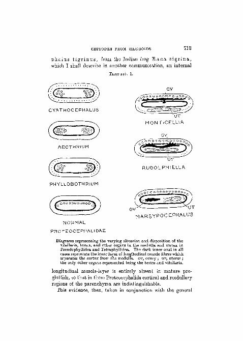
MARSYPOCEPHALUS
Diagrams representing the varying situation and disposition of thevitellaria, testes, and other organs in the medulla and cortex inPseudophyllidea and Tetraphyllidea. The dark inner oval in allcases represents the inner layer of longitudinal muscle-fibres whichseparates the cortex from the medulla, ov, ovary ; TJT, uterus ;the only other organs represented being the testes and vitellaria.
longitudinal muscle-layer is entirely absent in mature pro-glottids, so that in these Proteocephalids cortical and medullaryregions of the parenchyma are indistinguishable.
This evidence, then, taken in conjunction with the general

714 W. N. F. WOODLAND
Proteocephalid structure of these aberrant genera, compelsus to conclude that they all belong to the family Proteo-cephalidae, and that therefore there is no justification forsegregating them in a separate family, La Rue's Monticellidae,merely because the extension of the testes and other organs intothe cortex has only occurred in a few isolated cases insteadof being of common occurrence, like the cortical situation of thevitellaria.
Before providing diagnoses of the genus Marsypo-c e p h a l u s and its two species, I should like to refer to Fuhr-mann's new genus ' G o e z e e l l a ' . The new species describedby Fuhrmann—Goezeella' siluri—evidently belongs toLa Rue's genus Mont i ce l l i a , and Fuhrmann placed it ina distinct genus solely on the ground that it possesses a scolexpractically identical with that of O o r a l l o b o t h r i u msol idum (Fritsch). Now from the original description ofFritsch (1886), Riggenbach (1896), and Fuhrmann (1916), itis evident that the ' C o r a l l o b o t h r i u m ' type of scolex isfound in three genera—'Coral lobothrium' , R u d o l -ph i e 11 a, and 6 o e z e e 11 a—all of which are more distinct,as regards the disposition of the genitalia, from each otherthan any other three genera in the Proteocephalidae, whenceit is evident that the possession of any small modification ofthe scolex, however conspicuous, cannot be regarded as acharacter of more than specific value. I have demonstratedpreviously this same fact in the Caryophyllaeidae (Woodland,1923, 1924 a), in other Proteocephalidae and in the Pseudo-phyllidea (Woodland, 1924 b ; also see the P r o t e o c e p h a l u s .m a l o p t e r u r i and C l e s t o b o t h r i u m c l a r i a s in thepresent paper). If this be so then the Proteocephalid genusC o r a l l o b o t h r i u m must disappear as such (together withChoanosco lex , A c a n t h o t a e n i a , and my provisionalgenus Gangesia—Woodland, 1924 6—all of which generaare similarly founded on trivial scolex characters) and becomeP r o t e o c e p h a l u s , while Fuhrruann's Goezeel la s i lu r ibecomes Mont ice l l i a s i l u r i . Fuhrmann's Rudo l -p h i e l l a , on the other hand, remains, with the diagnosis as

CESTODES FROM SILUROIDS 715
given by him (only omitting or extending the specific referenceto the P r o t e o c e p h a l u s sol id us type of scolex), andLa Rue's M o n t i c e l l i a must be redefined simply as a Pro-teocephalid, with the ' testes, vitellaria, and uterus entirelyoutside the inner longitudinal muscle-sheath. Vitellaria com-posed of scattered follicles which form broad lateral fields.Testes numerous, forming a single broad dorsal field betweenvitellaria. Uterus ventral, with many lateral pouches.' Ovarybilobed and situated dorsally largely outside the longitudinalmuscle-sheath. Found in Siluridae, from Europe and SouthAmerica. The Proteocephalid genera remaining, after thiswholesale but necessary deletion, are then P r o t e o c e p h a l u s(as diagnosed by La Bue, but either omitting or generalizingall references to scolex characters and including statementsas to all organs being situated in the medulla, and the vitellariabeing grouped round a central duct, and extending the hoststo include Amphibia and Reptiles), M o n t i c e l l i a , R u d o l -ph i e 11 a, and M a r s y p o c e p h a l u s . 1
It remains to diagnose the revived genus M a r s y p o -c e p h a l u s and its two species above described. The defini-tion of the genus, stripped of the merely specific charactersstated by Braun (1894-1900, p. 1730), is : Proteocephalidin general structure but with the testes situated in the dorsalcortex in a single field ; all other organs medullary. Vitellariaas in P r o t e o c e p h a l u s . Siluridae, Egypt and Sudan.
The diagnoses of the two species of M a r s y p o c e p h a l u sare as follows :Marsypocepha lus r e c t a n g u l u s (type species). Scolex large (about
0-8 x 1-3 mm.) and superficially divided into quadrants by cuticularfurrows, each quadrant bearing a sucker. No apical organ. Suckers
1 La Rue's genus Oph io t aen i a (1911) must be considered to besynonymous with Monticelli's C r e p i d o b o t h r i u m (1889), if we agreewith Nybelin (1917) and Liihe (1899) that notched suckers do not forma sufficient basis on which to found a genus, since C r e p i d o b o t h r i u mger r a rd i i (Monticelli, 1899) has its testes distributed in two fields.As thus redefined I do not accept the genus C r e p i d o b o t h r i u m forreasons stated in another paper to be published shortly in ' Parasitology ',though it may perhaps remain as a sub-genus.
NO. 276 3 B

716 W. N. F. WOODLAND
large (about 0-49 mm. broad) and thick-walled, with a knob-like thicken-ing on the margin of the opening at the point nearest the apex of thescolex. No spinelets. Strobila 30-40 mm. in length, with a maximumbreadth of about 2-5 mm. and, like the hind half of the scolex, coveredwith a thick cuticle which often forms secondary furrows on the pro-glottids. Immature and mature proglottids broader than long. No neck.Genital pores marginal and irregularly alternate and in front of themiddle transverse line of the proglottid. The cirrus sac and vaginairregularly alternate as to which is anterior and both open on the samehorizontal level. The genital atrium is covered over by a dorso-ventralflap of the side wall of the proglottid. Cirrus sac extends over lessthan a quarter of the breadth of the proglottid and contains several coilsof the ductus. Vagina considerably dilated between its origin and itsapproach to the ovary. Testes (maximum measurements =43-9 x36-6 microns) numerous and occupy a single field, one layer deep,between the lateral vitellaria. Vitellaria compacted together to forma lateral strand. Fully formed uterus has between thirty and fortynarrow-pointed lateral diverticula and a number of ventral uterinepores opening into a furrow in the median ventral line. The uterus doesnot extend ventral to the ovary. Eggs thin-shelled (Wedl). Habitat:intestine ofClar ias angu i l l a r i s Linn. Egypt and Sudan.
Marsypocepha lus h e t e r o b r a n c h u s . Scolex large (about 1-12x1-19 mm.), not divided into quadrants. No apical organ or spinelets.Suckers large (0-56-0-74 mm. broad). The strobila reaches a maximumbreadth of about 2 mm. The strobila and hind half of the scolex arecovered with a thick cuticle which forms secondary transverse furrowson the proglottids. Immature and mature proglottids broader thanlong, but old gravid proglottids are one and a half times long as broad.Genital apertures irregularly alternate and situated in front of themiddle transverse line of the proglottid. The cirrus sac and vaginalapertures irregularly alternate as to which is anterior and both openinto a shallow atrium at the same horizontal level. The genital atriumis freely exposed on the edge of the proglottis. In gravid proglottids thecirrus-sac area swells into a tubercle. The cirrus aac extends abouta quarter of the breadth of the proglottis and contains several coils of theductus. The vagina is only very slightly dilated near its externalaperture. Testes (about 33x30 microns) and vitellaria as in M. r ec t -a n g u l u s . The fully formed uterus has relatively few (15-20) largelobose diverticula, and does not extend ventral to the ovary. Theembryos of uterine eggs measure 14'6-16-4 microns. Habitat: intestineof H e t e r o b r a n c h u s b ido r sa l i s I. Geoffr. Anglo-EgyptianSudan.

CESTODES FROM SILUROIDS 717
P r o t e o c e p h a 1 us m a l o p t e r u r i (syn. ' T a e n i am a l o p t e r u r i ' , Fritsch, 1886).
La Bue, with wholly insufficient data, provisionally relegatesthe ' T a e n i a m a l o p t e r u r i ' of Fritsch to his family Monti-cellidae and goes so far as to rename it M o n t i c e l l i a m a l o -p t e r u r i . In Dr. Chalmers's collection I find that I havea' considerable amount of material of what I believe to be thisspecies, and I shall briefly describe its structure both becausemy examination proves this form not to be of the Monticelliatype and because it is of interest as presenting such a combina-tion of characters as to make it impossible to refer it to anyone of the six genera into which La Bue subdivided the Proteo-cephalidae. In fact this species and the species recentlylabelled by me Ganges i a (Woodland, 1924b) prove, quiteapart from the evidence already offered, that La Rue's classifica-tion of the Proteocephalidae requires revision, and I shalldiscuss this subject in a paper shortly to be published in ' Para-sitology '.
The diagnosis of Fritsch's ' T a e n i a m a l o p t e r u r i ' ,based on the facts ascertained from my material (vide figs. 13-17, PI. 54), is as follows. Scolex quadrilobular, each lobebearing a sucker. The scolex measures 0-86-0-44 mm. inbreadth and 0-19-0-33 mm. in length. At the apex is a hemi-spherical or disc-shaped (according to state of contraction)protuberance representing a thick muscular rostellum (0-14-0-22 mm. broad) which bears round its greatest circumferencea broad band of numerous minute booklets, each hooklet con-sisting of a stout spine (2-6 microns long) planted upon a base(6-9 microns long). Numerous minute spinelets are presenton the lobes bearing the suckers, the suckers, and the neck.Suckers measure 0-12-0-17 mm. in maximum breadth. Theneck is nearly 2 mm. long, measures 0-16-0-24 mm. in minimumbreadth a short distance from the scolex, and then graduallyincreases in diameter to the first traces of segmentation.The length of a mature worm is at least 70 mm. and probablymuch longer. The maximum breadth of the strobila is about
3 B 2

718 W. N. F. WOODLAND
1-9 mm. Mature proglottids may be more than twice as longas broad, and gravid proglottids may be nearly three timeslonger than broad. The proglottids are very contractile. Thegenitalia are Proteocephalid in type, all the organs beingsituated in the medulla. The genital apertures are irregularlyalternate and the cirrus and vaginal apertures irregularlyalternate as to which is anterior. The genital apertures areusually situated half-way in the length of the proglottid, buton occasion may be a little behind or in front of this position.The cirrus pouch varies in shape and measures 0-24-0-29 mm.in length, with a maximum breadth of 0-16 mm., and in somecases stretches across one-quarter of the breadth of the pro-glottid but usually less (about one-fifth). The sac contains noductus coils. The coils of the vas deferens extend to themiddle of the proglottid. Testes numerous and separated intotwo lateral fields between the vitellaria and the ovary. Theovary is bilobed as usual, each lobe being almost spherical (insurface view) and very lobulated (follicular). Vitellaria com-pacted into strands. The uterus is, as usual, a median tubewhich in gravid proglottids bears on each side about twenty-four diverticula. The eggs (seen in sections) measure about25-6 x 18-3 microns. Habitat: intestine of M a l o p t e r u r u se l e c t r i c us Lacep., Anglo-Egyptian Sudan.
Assuming that the species just diagnosed is identical withthat described by Fritsch (1886) under the name of ' T a e n i am a l o p t e r u r i ' (and the numerous points of resemblance andidentity of host render this extremely probable), it is necessaryto make comments on several of the characters. Fritschfigured the rostellum as being uniformly covered with theminute hooklets, but this is not the case, the hooklets beingrestricted to a definite broad band (fig. 14, PI. 54). Fritsch'smeasurement of the width of the suckers (0-25 mm.) is toogreat, and I suspect that he measured the width of the lobewhich bears each sucker. Fritsch's statement that ripe pro-glottids are broader than long is difficult to understand excepton the supposition that his material was both very limited andvery contracted. The mature proglottids in my material

CESTODBS FROM SILUROIDS 719
(fig. 17, PL 54) measure from 1-7 mm. broad by 0-73 mm. longup to 1-02 mm. broad by 2-98 mm. long, and all my gravidproglottids are from two to nearly three times longer thanbroad. All the organs of the proglottid are medullary in positionand are so much of the usual Proteocephalid type that I havenot thought it worth while to provide a figure of a transversesection. La Rue's supposition that the testes are arranged inone continuous field between the vitellaria is erroneous (fig. 17,PI. 54), and Fritsch's drawing showing them to be arrangedin two lateral fields is correct.
Finally, I may remark upon the difficulty of assigning' T a e n i a m a l o p t e r u r i ' to any one of the seven or eightgenera at present recognized. It cannot be placed in P r o -t e o c e p h a l u s or in my recent provisional genus G a n g e s i a(Woodland, 1924 b) because the testes are arranged in twolateral fields ; it cannot be placed in C r e p i d o b o t h r i u m( = O p h i o t a e n i a , La Rue, 1914) because it possesses amuscular rostellum and both hooks and spines, and it obviouslydoes not belong to C h o a n o s c o l e x , C o r a l l o b o t h r i u m ,or A c a n t h o t a e n i a . I therefore, despite the two-fielddistribution of the testes and the presence of spinelets on thescolex, place the species, for the present, in the genus P r o -t e o c e p h a l u s , pending the full consideration of the subjectof genera in the Proteocephalidae which I shall shortly publishin ' Parasitology '.
C l e s t o b o t h r i u m c l a r i a s , n.sp.
My material for the description of this new and interestingBothriocephalid species consists of three whole worms andportions of about three others, all stained and mountedwhole, and a large number of transverse and horizontalwell-stained sections contained in Dr. Chalmers's collectionfrom the Sudan. This species was found in the intestines ofCla r i a s a n g u i l l a r i s Giinth. (a Nile Siluroid), and, likethe B o t h r i o c e p h a l u s p y c n o m e r u s , which I recently

720 W. N. F. WOODLAND
described from India (Woodland, 1924 b), is of interest becauseof its armed scolex and because it -presents a combination ofthis and other characters which make it impossible to insertit into the taxonomic system of Liihe (or any other author)without emending the diagnostic characters of the genus mostsuited to its reception. This, however, is a subject I shalldiscuss later. This new species is also of interest as being thethird described of the genus, assuming that the C les to -b o t h r i u m g l a c i a l e of Cholodkovsky (1915) is a species ofthis genus.
C l e s t o b o t h r i u m c l a r i a s is a small worm, my longestspecimen measuring 14-5 mm. with a maximum breadth of0-298 mm., but since this specimen is devoid of the scolexand a certain number of attached anterior proglottids andhas no gravid proglottids, we can only say that the bodyprobably does not exceed 20-25 mm. in maximum length andhas a maximum breadth (gravid proglottids) of about 0-415 mm.The strobila is thus very slender (fig. 18, PI. 54). I possesssix scolices which measure in length 0-249-0-431 mm. (fromapex of crown to anterior edge of the first proglottid) and inmaximum breadth 0-182-0-240 mm. Each scolex (fig. 19,PI. 54) consists anteriorly of a crown, apparently circular intransverse section and therefore devoid of lappets, which bearsdeeply embedded in its edge thirty-two to thirty-six rod-shapedspinelets, slightly curved in form and pointed at their outerand blunt at their inner extremities, and measuring about32-9-51-2 microns in length in different worms. In each •wormthe spinelets are uniform in length. In the interior of thecrown is a deeply staining ' nucleus ', probably muscular innature. Below the crown the scolex is columnar in form, thenarrow column bearing two very deep bothridial grooves,bordered by the broad free edges of the bothridia. The scolextherefore rather resembles that type which, according toLiihe, is characteristic of the genus P t y c h o b o t h r i u m ,though the bothridial edges do not project away from thecolumn anteriorly and posteriorly, and it is very dissimilarto the peculiar globose scolex of C l e s t o b o t h r i u m e r a s -

CESTODBS FROM SILUEOIDS 721
s iceps figured by Molin (1861), Ariola (1900), and otherauthors. The scolex of C l e s t o b o t h r i u m g l a c i a l e hasnot been described.
There is no unsegmented neck. The majority of anteriorimmature proglottids are, unless contracted, longer thanbroad (in three -worms they average 0-149-0-166 mm. longand 0-099-0132 mm. broad), though the first two or threeproglottids may be square or even slightly broader than long.On the other hand, immature proglottids containing distinctgenital rudiments are about square in outline, and matureproglottids are nearly always broader than long (0-215-0-381 mm. broad and 0-083-0-249 mm. long). Gravid pro-glottids, i.e. containing little else than the dilated uteri filledwith eggs, are also slightly broader than long (0-332-0-415 mm.broad and about 0-249 mm. long). The edges of the matureand gravid proglottids are only very slightly salient, and theposterior borders of anterior proglottids hardly at all overlapthe anterior borders of posterior proglottids. In transversesection (figs. 22-6, PI. 54) the mature and gravid proglottidsare remarkably deep dorso-ventrally, in some cases approachingthe circular, and for this reason worms mounted whole oftenshow the genital openings apparently displaced from themiddle line, even so as to become apparently marginal—adelusive appearance due to the strobila having been rotatedunder the coverslip.
The genitalia of C l e s t o b o t h r i u m c l a r i a s conform inthe main to the usual Ptychobothriine type, with, however,several distinctive features. The bilobed ovary is, as usual,situated near the hind limit of the proglottid, but, in thisspecies, is very large relative to the size of the proglottis (fig. 21,PI. 54), occupying rather more than a third of both breadthand depth and about half the length. The two lobes of theovary are united by a posterior ventral median ' isthmus '.The median dorsal cirrus aperture lies not, as is usual, in theanterior half of the segment, but about midway in the lengthof the proglottis and the vaginal aperture a short distancebehind this, the two apertures opening separately and not

722 • W. N. F. WOODLAND
into a common atrium. The uterine aperture on the ventralsurface is small, median (fig. 26, PI. 54), and, so far as I canascertain from imperfect series of sections, at a level slightlyanterior to the dorsal cirrus aperture. The uterine aperture isonly developed in gravid proglottids, i.e. in those with theuterus dilated so as to occupy nearly the whole of the pro-glottid volume ; in merely mature proglottids the uterus isonly a blind tubular structure more or less swollen anterior tothe ovary (fig. 21, PI. 54). Liihe (1899) bases the diagnosis ofhis genus C l e s t o b o t h r i u m chiefly upon the mode ofdevelopment of the uterus and the character of the scolex.With reference to the uterus he says : ' Anfangstheil desUterus ein gewundener Canal (Uteringang), welcher in eineausserordentlich geriiumige, in reifen Proglottiden alle anderenGenitalorgane verdriingende Uterushohle fuhrt', and this modeof development (figured by Liihe, 1902) alone occurs in thisgenus. In two portions of the strobila in my material gravidand merely mature proglottids are contiguous (fig. 20, PL 54),a fact which shows that in the hind end of the strobila eachproglottis becomes ripe without reference to the condition of itsneighbours : the proglottids do not necessarily gradually ripenseriatim, i. e. according to their position in the strobila, as inmost Cestodes. The testes are relatively few in number(rarely more than twenty in each proglottis) and are situated,chiefly laterally, in the medullary parenchyma. The vitellariaare numerous, sniall (7-3 x5-4 microns as a maximum), and,as usual, situated in the cortical parenchyma (figs. 22-4, PL 54).The material at my disposal does not enable me to describein detail the configuration of the ducts behind the ovary orall the organs (receptaculum seminis, shell-gland, &c.) con-nected with these ducts.
The eggs contained in the fully developed uterus are ovalin shape, thin-shelled, apparently without operculum, andmeasure (in balsam) on an average about 26x18 microns.The contents are granular, with a number of nuclei.
Though the dibothridiate type of scolex is usually or alwaysassociated with a more or less uniform type of genital system,

CESTODES FROM SILUROIDS 723
and therefore certainly serves as a good character for theprimary classification of Cestodes, yet it is now beginningto be recognized that the innumerable modifications of this typeof scolex possess no more than a specific value in more detailedclassification. The present new species—Clestobothriumclarias—provides a good illustration of this statement.C l e s t o b o t h r i u m is the only genus of Luhe's sub-family,the Ptychobothriinae, in which the uterus becomes so enor-mously dilated, after the genitalia as a whole have become mature,as to occupy practically the whole of the proglottis cavity,the other genital organs becoming suppressed. For this reason,and on account of the distinct segmentation and lack of a neck,I have placed the present new species in the genus Cles to -b o t h r i u m . On the other hand, Luhe's type-species of hisgenus—Clestobothrium c r a s s i c e p s (Eud.)—possessesa large globose scolex of a peculiarly modified type, and thistype of scolex forms part of Luhe's diagnosis of his genus.Since it is obvious, however, that minor scolex characterscannot be as reliable taxonomically as the characters of aninternal organ essential for the propagation of the speciessuch as the uterus, the only proper procedure is to amendLuhe's definition of this genus so as to include forms with otherforms of scolex. In the same way, when recently describing(Woodland, 1924b) a new species of B o t h r i o c e p h a l u s ,s.str., from India—Bothr iocephalus pycnomerus—which possessed a scolex with an armed four-lappeted crown,I was compelled to amend Luhe's definition of the genusB o t h r i o c e p h a l u s in a similar manner. Further, I maypoint out that, for the same reason, Lonnberg's genus P t y c h o -bo th r iu rn (1889) becomes synonymous with Diesing's genusP o l y o n c h o b o t h r i u m (1854: redefined by Klaptocz in1906), the only essential distinction between the two beingthat the latter possesses an armed four-lappeted scolex withshallow bothridia instead of an unarmed pillar-shaped scolexbearing deep bothridia with projecting margins. If we comparethe definitions of P t y c h o b o t h r i u m (Liihe, 1899) with thedefinition of P o l y o n c h o b o t h r i u m (Klaptocz, 1906),

724 W. N. F. WOODLAND
the synonymy of the two, when the characters of the scolexare disregarded, becomes evident.
P t y o h o b o t h r i u m Lonnberg, 1889. Scolex, in consequence of thestrong development of the bothridial grooves, arrow-shaped. Outersegmentation indefinite. An unsegmented neck absent. Vitellaria inthe cortex. Ovary median and consisting of a thin sheet of cells lyingtransversely at the hind end of the proglottis. The cell-sheet bends roundanteriorly slightly towards the ventral surface, but also reaches thedorsal limit of the medulla. The oviduct arises from the anterior surfaceof the ovary where it bends ventrally. The receptaculum seminis iscomparatively large and is considerably longer than broad. Uteruswithout dilated sac, and consisting of a few loose coils. Theuterus opening is approximately median, like the dorsal genitalopenings.
P o l y o n c h o b o t h r i u m Diesing, 1854. Scolex elongated, armed, withtwo shallow longitudinally disposed bothridial grooves. No neck.Segmentation indefinite. Mature segments broader than long. Vitel-laria in cortex, absent in the median as well as in the lateral parts of thecortex, so that they are disposed in four groups, two dorsal and twoventral. Ovary at hind end of proglottis, lying median in a transversedirection and forming a thin sheet of cells. Eeceptaculum seminis isa dilatation of the vagina, into which the oviduct opens. Uterus withoutsac, forming a long much-convoluted canal, which for the most partis situated towards one side of the proglottis, the vas deferens occupy-ing the other side. Genital openings median. Eggs indistinctlyoperculated (?).
SUMMARY.
1. Those species of Proteocephalid Cestodes in which thetestes are situated in the cortex may be described as of theMonticellia type. Of this type there are three conditions :(a) the M o n t i c e l l i a condition in which the testes, uterus,ovary, and vitellaria are all situated in the cortex; (b) theE u d o l p h i e l l a condition in which the testes and vitellariaalone are in the cortex, the other organs being entirely or almostentirely in the medulla ; and (c) the M a r s y p o c e p h a l u scondition in which the testes alone are in the cortex, all otherorgans being medullary.
Fuhrmann's genus Goezee l la is synonymous with Mont i -

CESTODBS FROM SILUROIDS 725
cellia if we ignore the characters of the scolex as featuresof generic value.
2. The anatomy of two species of Marsypocephalus isdescribed: Marsypocephalus r ec t angu lus Wedl,1862, and Marsypocephalus h e t e r o b r a n c h u s , n.sp.,from Nile Siluroid fishes.
3. It is concluded that the cortical situation of the testesand other organs is a taxonomic feature of generic valueonly (as in Pseudophyllidea in the case of the vitellaria)and La Eue's new family of the Monticellidae, created toinclude Monticellia-like forms, is not accepted. Monti-cell ia, Eudo lph ie l l a , and Marsypocephalus arethus regarded as new genera in the Proteocephalidae.
4. The facts that the ' C o r a l l o b o t h r i u m ' type ofscolex is found in all of the three genera Monticel l ia(as amended by me and including 'Goezee l l a ' s i lu r i ,Fuhrmann), Eudo lph ie l l a , and P ro t eocepha lus (asamended by me and including ' C o r a l l o b o t h r i u m 'sol idum, Pritsch), and that in the Caryophyllaeidae,Bothriocephalidae, and Cyclophyllidea (cf. e.g. Taeniasolium and Taenia saginata) minor scolex charactersare evidently only features of specific value, compel us todelete such genera as Cora l lobo thr ium, Choano-scolex, A c a n t h o t a e n i a , and my own recent genusGangesia and to regard them as synonyms of P ro teo -cephalus (La Eue's genus ' Ophio taenia ', syn. ' Crepi-dobo th r ium ' , not being accepted). Fuhrmann's Goe-zeella s i lur i becomes Monticel l ia s i lu r i , and Eritach'sCora l lobothr ium solidum becomes P ro t eocepha lu ssol idus . The genera of the Proteocephalidae are thus fourin number: P r o t e o c e p h a l u s , M o n t i c e l l i a , E u d o l -p h i e l l a , and Marsypocep,halus, and these are formallyor informally redefined. The two species of Marsypoce-phalus are diagnosed.
5. The ' Taenia m a l o p t e r u r i ' of Fritsch, 1886, is notof the Monticellia type, as suggested by La Eue. Its structureis of the usual Proteocephalid type, save that the scolexpossesses a rostellum and a broad band of hooklets and is

726 W. N. F. WOODLAND
covered with spinelets. It is renamed P r o t e o c e p h a l u sm a l o p t e r u r i .
6. A new species of Cles tobothr ium—Cles toboth-r ium c l a r i a s , from Clar ias angu i l l a r i s Giinth.—is described. It is of interest, not only as being the third(second ?) species known of the genus, but because it affordsone more illustration of the fact that the characters of thescolex cannot be used for diagnoses of genera. For this reasonalso, Lonnberg's genus P t y c h o b o t h r i u m (1889) becomessynonymous with Diesing's genus P o l y o n c h o b o t h r i u m(1S84).
Postscript.—Since the preceding, paper was written and dispatched tothe editor, Dr. 0. Fuhrmann has published an important communication l
which contains an account of a Cestode—L oennberg ia t a n g a n y i k a e ,g.n., sp.n., from Clar ias l aze ra , Lake Tanganyika—which, in essentialfeatures, is very similar to the Marsypocepha lus r e c t a n g u l u sdescribed above. Fuhrmann's species, however, differs from the speciesI have described in possessing folds of loose skin round the base of thescolex, in being devoid of knobs on the suckers, in having a distinct neck,in the uterus being placed ventral to the ovary,2 and in some other minorfeatures. I will here remark that jxist as Fuhrmann now identifies hisother recent new genus R u d o l p h i e l l a with Diesing's E p h e d r o -cepha lus so his present new genus Loennbe rg i a must be regardedas synonymous with WedPs M a r s y p o e e p h a l u s . Thus three speciesof Mar sypocepha lus are now known to exist: M. r e c t a n g u l u sWedl, M. h e t e r o b r a n c h u s Woodland, and M. t a n g a n y i k a eFuhrmann. I may also remark that I cannot accept Fuhrmann's reten-tion of La Rue's family, the Monticellidae, nor his suggestion that Fritsch's' T a e n i a ' m a l o p t e r u r i should be referred to my recent genusGanges ia , for reasons supplied above and to be supplied in anotherpaper which has already been sent to press and will shortly appear in' Parasitology '.
Dr. Fuhrmann's criticisms of my views concerning the Caryophyllaeidaewill receive attention in a paper now in preparation.
1 " Zoological Results of the Third Tanganyika Expedition, conductedby Dr. W. A. Cunnington, 1904-5. Report on the Cestoda ", ' Proc. Zool.Soc. London ', 1925 (published April 3), p. 79.
2 In my species of M a r s y p o c e p h a l u s the uterus is on the samelevel as and anterior to the ovary, as in most P r o t e o c e p h a l u s spp.Fuhrmann's diagrams A and B (on his p. 98) are therefore not strictlyaccurate for all species of P ro teocepha lusand Marsypocepha lus .

CESTODES FROM SILUEOIDS 727
LITERATURE E E F B R E N C E S .
Ariola, V. (1900).—" Revisione della Famiglia Bothriocephalidae s. str.",' Archives de Parasitologie', torn, iii, no. 1, p. 369.
Beddard, F. E. (1913).—" Contributions to the Anatomy and SystematicArrangement of the Cestoidea. IX. On a new Genus of Ichthyo-taeniids ", ' Proc. Zool. Soc. London ', Part I, p. 243.
Boulenger, G. A. (1911).—•' Catalogue of the Fresh-water Fishes of Africain the British Museum ', London.
Braun, M. (1894-1900).—In Bronn's ' Klassen und Ordnungen des Thier-reichs ', Bd. iv, Vermes, Abth. I. b. Cestodes.
Cholodkovsky, N. (1915).—" Cestodes nouveaux ou peu connus. TroisierneSerie ", ' Ann. Mus. Zool. Acad. Imp. Sciences Petrograd', torn, xix(1914), p. 516.
Diesing, C. M. (1854).—" Ueber eine naturgemasse Vertheihmg derCephalocotyleen", ' Sitzber. d. K. Akad. d. Wiss., math.-nat. Cl.\Bd. xiii, p. 556.
Fritsch, G. (1886).—" Die Parasiten des Zitterwelses " , ' Sitzungsb. d. k.preuss. Akad. Wiss. Berlin', Jahrg. 1886, p. 99.
Fuhrmann, 0. (1916).—" Eigentiimliche Fischcestoden ", ' Zool. Anzeig.',Bd. xlvi, 1915-16, p. 385.
Klaptocz, B. (1906).—" Polyonchobothrium polypteri (Leydig) ", ' Cen-tralbl. f. Bakt., Parasit. Infekt.', Bd. xli, I. Abth., Orig., p. 527.
La Rue, G. (1911).—" A Revision of the Cestode Family Proteoce-phalidae " , ' Zool. Anzeig.', Bd. xxxviii, p. 473.
(1914).—"A Revision of the Cestode Family Proteocephalidae ",' Illinois Biological Monographs ', University of Illinois. Vol. i, Urbana,Illinois, nos. 1 and 2.
Lonnberg, E. (1889).—" Bidrag till Kiinnedomen om i Sverige forekom-mande Cestoder", ' Bih. till Svenska Vet.-Akad. Handl.', Bd. xiv,Afd. iv, no. 9.
Liihe, M. (1899).—" Zur Anatomie und Systematik der Bothrioce-phaliden " , ' Verh. Deutsch. Zoologisch. Gesell.', ix. Jahrg., p. 30.
(1902).—" Revision meines Bothriocephalidensystemes " , ' Centralbl.f. Bakt., Parasit. Infekt.', Bd. xxxi, Abth. i, Orig., p. 318.
Molin, R. (1861).—" Prodromus Faunae Helminthologicae Venetae ",' Denkschr. d. k. Akad. d. Wissen.', Bd. xix, 2 Abth., Wien.
Monticelli, Fr. Sav. (1891).—" Notizie su di alcune Specie di Taenia ",' Boll. Soc. di Nat. in Napoli', ser. 1, vol. v, anno v, p. 151.
Nybelin, O. (1917).—•" Results of Dr. E. Mjoberg's Swedish ScientificExpeditions to Australia. XIV. Australische Cestoden", SvenskaVet.-Akad. Handl.', Bd. Hi, no. 14.
(1922).—" Anatomisch-systematische Studien iiber Pseudopliyl-lideen ", ' Goteborgs Kungl. Vetenskaps Handl.', 4, xxvi: I.

728 \V. N. F. WOODLAND
Riggenbach, E. (1896).—" Das Genus Ichtlryotaenia", 'Rev. SuisseZool. et Ann. Mus. d'Hist. Nat. Geneve ', torn, iv, p. 165.
Wed], K. (1862).—" Zur Helminthenfauna Agyptens ", ' Sitzungsb. d.Math.-Nat. Classe d. k. Akad. Wissen. Wien ', Bd. xliv, I. Abth., Jahrg.1861, p. 463.
Woodland, W. N. F. (1923).—" On some remarkable new forms of Caryo-phyllaeidae from the Anglo-Egyptian Sudan, and a revision of theFamilies of the Cestodaria", ' Quart. Journ. Micr. Soi.', vol. lxvii,Part III, p. 435.
(1924 a).—" On a new Species of the Cestodarian Genus Caryo-phyllaeus from an Egyptian Siluroid", ' Proe. Zool. Soc. London',Part II, p. 529.
(1924 6).—" On a new Bothriooephalus and a new Genus of Proteo-cephalidae from Indian freshwater Fishes ", ' Parasitology ', vol. xvi,p. 441.
DESCEIPTION OP PLATES 53 AND 54.
(All figures drawn under the camera luoida.)
LETTERING.
BWM, body-wall of adult worm ; CF, cuticular furrow ; cs, cirrus sac ;CSOP, cirrus sac aperture ; EXC, excretory canal; FL, lateral flap coveringgenital atrium ; GA, genital atrium ; GT, tubercle marking position ofcirrus sac ; HK, hooklets; ILML, internal layer of longitudinal muscles ;KN, knob on sucker rim ; N, lateral nerve ; ov, ovary ; HOS, rostellum ;SHGL, shell-gland; sp, spines on crown of scolex; TES, testes; UT,uterus ; TJTO, uterine aperture ; VAG, vagina ; VAGOF, vaginal aperture ;VD, vas deferens ; vrr, vitellaria; VITD, vitelline duet; VMF, ventralmedian groove.
PLATE 53.
Figs. 1-6. M a r s y p o c e p h a l u s r e c t a n g u l u s .Fig. 1.—Scolex in outline, showing the' knobs ' on the suckers, x 17-5.Fig. 2.—Sucker (in section) of a pleroceroid larva attached to the
strobila of an adult worm. x87-5.Fig. 3.—Transverse section of a proglottid in the region of the cirrus
sac. x 27-5.Fig. 4.—Transverse section (slightly oblique) of a proglottid in the
region of the ovary. x27-5.Fig. 5.—The principal organs in a mature proglottid. The diverticula
of the uterus are not yet formed. The testes are only shown on one side.X27-5.
Fig. 6.—Outline of the uterine diverticula seen in a horizontal sectionthrough a mature proglottid (no eggs were present). The lack of diverticula

CESTODBS FROM SILUROIDS 729
in the gap on the left was due to the presence of coils of the vas deferens.x39.
Pigs. 7-12. Mar sypocepha lus h e t e r o b r a n c h u s , n.sp.Kg. 7.—Scolex in outline, x 17-5.Pig. 8.—Ventral aspect of three old gravid proglottids. x 12.Fig. 9.—Transverse section of a mature proglottid through the region
of the ovary, x 39.Fig. 10.—Transverse section of a mature proglottid through the region
of the cirrus sac. x 39.Fig. 11.—The principal organs in a gravid proglottid. x 27-5.Fig. 12.—The genital atrium and the cirrus sac and vagina openings
seen in a horizontal section, x 39.
PLATE 54.
Pigs. 13—17. P r o t e o c e p h a l u s m a l o p t e r u r i (Fritsch).Fig. 13.—A worm, entire save for detached posterior proglottids.
Natural size.Fig. 14.—Scolex in outline, showing the muscular rostellum and band
of hooklets. x 56.Fig. 15.—The rostellum become disc-shaped with a terminal depression.
X87-5.Fig. 16.—One of the rostellar hooklets. x 840.Fig. 17.—A mature proglottid. x 27-5.
Figs. 18-26. C le s tobo th r ium c l a r i a s , n.sp.Fig. 18.—A young mature worm, x 12.Fig. 19.—The scolex, with crown of spines and the two deep bothridia.
X87-5.Fig. 20.—Mature and completely gravid proglottids in juxtaposition.
x39.Fig. 21.—Mature proglottid, from the dorsal aspect, x 180.Fig. 22.—Transverse section through a mature proglottid in the region
of the vagina opening and ovarian isthmus, x 180.Fig. 23.—Transverse section through the same mature proglottid as is
shown in fig. 22 (four sections intervene between the two sections figured)in the region of the cirrus-sac opening, x 180.
Fig. 24.—Transverse section through the same mature proglottid as isshown in figs. 22 and 23 (six sections intervene between the section offig. 23 and this section) in the region of the slightly dilated uterus andcoils of vas deferens. x 180.
Pig. 25.—Transverse section through a proglottid in which the uterusis about half dilated. Region of cirrus-sac opening, x 87-5.
Pig. 26.—Transverse section through a proglottid in which the uterusis about three-quarters dilated. Region of ventral uterine opening. Thecompletely dilated uterus is shown in fig. 20. X 87-5.

Quart. Jonrn. Micr. Sci. Vol. <>9, N. S., PI. 53
GT VMF
W. K. F, Woodland, del.

Quart. Joiirn. Micr. Scl. Vol. 69, N. S., PI. 54
OV VAOrOP
IV. N. F. Woodland, del.