online nanoflow rp−rp-ms reveals dynamics of multicomponent ku complex in response to
TRANSCRIPT

Online Nanoflow RP-RP-MS Reveals Dynamics of Multicomponent
Ku Complex in Response to DNA Damage
Feng Zhou,†,‡,⊥ Job D. Cardoza,†,‡,⊥ Scott B. Ficarro,‡,§,⊥ Guillaume O. Adelmant,‡,§,⊥
Jean-Bernard Lazaro,|,⊥ and Jarrod A. Marto*,‡,§,⊥
Department of Cancer Biology and Blais Proteomics Center, Dana-Farber Cancer Institute, Department ofSurgery, Brigham and Women’s Hospital, and Department of Biological Chemistry and Molecular
Pharmacology, Harvard Medical School, Boston, Massachusettes, United States
Received May 14, 2010
Tandem affinity purification (TAP) coupled with mass spectrometry has become the technique of choicefor characterization of multicomponent protein complexes. While current TAP protocols routinely providehigh yield and specificity for proteins expressed under physiologically relevant conditions, analyticalfigures of merit required for efficient and in-depth LC-MS analysis remain unresolved. Here weimplement a multidimensional chromatography platform, based on two stages of reversed-phase (RP)separation operated at high and low pH, respectively. We compare performance metrics for RP-RPand SCX-RP for the analysis of complex peptide mixtures derived from cell lysate, as well as proteincomplexes purified via TAP. Our data reveal that RP-RP fractionation outperforms SCX-RP primarilydue to increased peak capacity in the first dimension separation. We integrate this system withminiaturized LC assemblies to achieve true online fractionation at low (e5 nL/min) effluent flow rates.Stable isotope labeling is used to monitor the dynamics of the multicomponent Ku protein complex inresponse to DNA damage induced by γ radiation.
Keywords: tandem affinity purification • RP-RP • reversed phase-reversed phase • LC-MS • LC-MS/MS • 2D-LC • two-dimensional separation • online fractionation • SCX-RP • mudpit • Orbitrap • Qstar •protein complex • DNA damage
IntroductionNormal and aberrant biological phenotypes are driven by
numerous interactions of proteins, nucleotides, and smallmolecule metabolites. As a result, there is growing appreciationthat so-called systems level experimental strategies can aug-ment traditional hypothesis-driven approaches for character-ization of cellular response to stress, infection, disease, andother biological or environmental perturbations.1,2 Data derivedfrom large-scale protein characterization studies generally lagbehind those for DNA and RNA, both in terms of throughputand comprehensiveness. Generally speaking, the technicalhurdles that impede progress toward true proteome-scalestudies are based on the limited detection and dynamic rangeavailable on current mass spectrometry instruments.3 Hence,faced with the difficulties inherent to the generation of a“complete” proteome catalog, many researchers have optedinstead to pursue targeted analyses of protein subclasses orspecific post-translational modifications. Anecdotal evidence
suggests that enriched biological materials used in focusedstudies nevertheless span a wide range of sample complexityand can often yield a protein and peptide concentration rangethat exceeds the analytical capabilities of modern mass spec-trometry technology.4
Ongoing efforts to analyze complex biological samples haverevealed other analytical deficiencies in LC-MS technologies.For example, it is now generally accepted, although admittedlydifficult to quantify, that under typical electrospray conditions,a multitude of coeluting peptides will compete for charge andeffectively suppress the detected signal of low basicity species.5
In addition, the duty cycle of MS/MS, while steadily improving,6,7
is nonetheless often overwhelmed by the number of peptidespresented simultaneously for analysis. Collectively these dataand observations have catalyzed a plethora of work designedto improve overall peak capacity of peptide and proteinseparations, with the ultimate goal of reducing the number ofanalytes presented for MS/MS per unit time. Multidimensionalfractionation has emerged as the most compelling means tofacilitate in-depth analysis of complex peptide mixtures derivedfrom biological extracts.8 In this approach, two or moreseparation methods, each based upon different physiochemicalproperties, are used in tandem to obtain significantly improvedpeak capacity. To date, the combination of strong cationexchange (SCX) chromatography and reversed phase (RP)chromatography is the most widely used system.9-12 Despitethese successful studies, it is clear that, at the extreme,
* To whom correspondence should be addressed. Jarrod A. Marto,Department of Cancer Biology, Dana-Farber Cancer Institute, 44 BinneyStreet, Smith 1158A, Boston, MA, 02115-6084. Phone: (617) 632-3150 (office).Fax: (617) 582-7737. E-mail: [email protected].
† These authors contributed equally to this work.⊥ Department of Cancer Biology, Dana-Farber Cancer Institute.‡ Department of Biological Chemistry and Molecular Pharmacology,
Harvard Medical School.§ Blais Proteomics Center, Dana-Farber Cancer Institute.| Department of Surgery, Brigham and Women’s Hospital.
6242 Journal of Proteome Research 2010, 9, 6242–6255 10.1021/pr1004696 2010 American Chemical SocietyPublished on Web 09/27/2010

biological complexity often exceeds the peak capacity ofSCX-RP. As a result, numerous other multidimensional sepa-ration strategies, including SAX-RP,13-15 gel-based isoelectricfocusing coupled with RP,16-20 and CIEF-RP21-24 have beenutilized for shotgun proteomics. More recently Gilar et al.introduced a multidimensional fractionation platform basedon two stages of reversed phase operated at high and low pH,respectively.25 The RP-RP geometry provides high peak capac-ity in each dimension, while the change in pH between thefirst and second stages modulates peptide hydrophobicitythrough a switch in ion pairing at acidic (high buffer pH) andbasic (low buffer pH) side chains, respectively.26-29 Less wellunderstood is the relative benefit of multidimensional frac-tionation for analysis of samples across the full spectrum ofbiological complexity (e.g., whole cell lysate, modified peptides,protein complexes, etc.).In principle the analysis of discrete,multicomponent protein complexes represents an experimentalparadigm that is well-matched to the analytical performanceof mass spectrometry. For example, large-scale studies in yeastand human cell lines indicated that protein complexes consistof ∼30 proteins on average,30-34 a level of complexity veryamenable to analysis by LC-MS/MS. Moreover, the growingrepositories of affinity tag constructs, along with improvementsin robotics for cloning, expression, and cell culture, provide abasis for systematic production of reagents suitable for large-scale proteomics studies.35-39 Indeed, recent reports havedescribed the development and application of new affinityreagents and associated methods designed to improve bothyield and specificity of purified protein complexes.40-45 How-ever, the analytical figures of merit for subsequent LC-MSanalysis remain relatively unexplored.
In this report, we compare SCX-RP and RP-RP multidi-mensional chromatography platforms for analysis of peptidesderived from cell lysate and multicomponent protein com-plexes isolated via TAP. A modified Waters 2-D NanoACQUITYsystem allows convenient use of either SCX or RP in the firstdimension, and is readily interfaced with our recently describedminiaturized LC electrospray assemblies,46 to provide for trueonline, nanoflow (e5 nL/min) multidimensional fractionation.Comparison of these separation platforms for the analysis ofcell lysate revealed that RP-RP outperformed SCX-RP duelargely to improved first dimension peak capacity. We observedsimilar performance improvements for analysis of tandemaffinity purified protein complexes. Finally, we utilize RP-RPin conjunction with iTRAQ-based quantification to monitorchanges in protein membership of the multicomponent Kucomplex in response to DNA damage induced by γ radiation.
Experimental Procedures
Column Construction. The first dimension column (RP orSCX) consisted of 360 µm O.D. × 100 µm I.D. fused silicacapillary tubing capped with 0.5 µm inlet frits (Upchurch, OakHarbor, WA). Waters 3.5 µm Xbridge C18 silica resin and SepaxTechnology (Newark, DE) SCX media were used to pack 10 cmbeds for first dimension RP and SCX columns, respectively.Second dimension pre- (PC) and analytical columns wereconstructed as previously described.46 Briefly, silicate-basedfrits were cast in situ within 360 µm O.D. × 100 µm I.D. fusedsilica capillary tubing. Next, dense slurry of POROS 10R2 resinwas prepared in 1 mL of acetonitrile and pressure loadedbehind the frit to a final bed length of 5 cm. The AC wasconstructed from 360 µm O.D. × 25 µm I.D. fused silicacapillary tubing, and packed to a bed length of 12 cm with
Waters Xbridge 3.5 µm diameter silica resin. A laser-basedpipette puller (P-2000, Sutter Instruments, Novato, CA) wasused to generate an integrated electrospray emitter tip of ∼1µm diameter at the end of the capillary, ∼2 mm from the bedfrit.
Our 2D RP-RP platform was based on a Waters NanoAC-QUITY UHPLC system, equipped with a third, 6-port, 2-positionvalve (VICI Valco, Houston, TX). The autosampler was used to(i) load samples and (ii) inject first dimension elution buffers,consisting of varying concentrations of either KCl (SCX) oracetonitrile (RP). Discrete first dimension elutions were per-formed at 1 µL/min with 20 mM ammonium formate in 3%acetonitrile or 0.1% formic acid in 10% acetonitrile, for RP-RPand SCX-RP, respectively (Detailed concentrations are listedin Supplementary Table 1, Supporting Information). Truenanoflow rates in the second dimension were achieved throughuse of a passive split located prior to the second dimensionPC; the split line was configured in a vented column geometry.47,48
Flow is blocked in the split line during first dimension elutionto ensure that peptides are directed to the second dimensionPC. The valve then switches to engage the passive split suchthat a majority of LC effluent is diverted to waste prior to thesecond dimension PC.
Cell Culture and γ Radiation. HeLa S3 cells that expresstandem FLAG and HA peptide epitope tagged Ku86 (FLAG-HAKU86) were grown in BD Falcon 15-cm diameter tissue culturedishes, each containing 25 mL DMEM (Invitrogen, Carlsbad,CA) supplemented with 5% fetal bovine serum. Cells wereincubated for three days in a 37 °C, 5.0% CO2, after which theywere supplemented with an additional 3 mL of media, and thenincubated again for 24 h. A MDS Nordion Gammacell 40Exactor, equipped with two 137Cs sources, was used for γ-radia-tion of the HeLa S3 cells. In total twelve, 15 cm dishes ofconfluent cells were irradiated with 10 Gy, at a central doserate of 1.1 Gy/min. Cells were then placed back into theincubator, and then removed in groups of 4 plates after 30 min,2 h, and 5 h, respectively. Under these conditions, we did notobserve loss of cell viability or signs of apoptosis. One groupof 4 plates (the control) was processed in parallel but notexposed to γ-radiation.
Nuclear Preparation. All purifications were performed fromnuclear extracts, prepared as follows: Cells were transferred to50 mL conical tubes (2 plates/tube). Next, the cells werepelleted at 500× g for 3 min at 4 °C, in a refrigerated centrifuge(Eppendorf, Hamburg, Germany). The supernatant was dis-carded, and the cell pellets were resuspended in 50 mL of 1×PBS solution at 4 °C, (137 mM NaCl, 2.7 mM KCl, 4.3 mMNa2HPO4 ·7H2O, 1.4 mM KH2PO4) and then centrifuged againat 500× g for 3 min at 4 °C.
The four-plate equivalents of cells were combined, sus-pended in 10 mL 1× PBS, and centrifuged at 500× g for 3 minat 4 °C. The supernatant was discarded and the pellet wassuspended in ten times the pellet volume (typically 700 µL forfour plates of HeLa S3 cells) of hypotonic buffer (10 mM TrispH 7.3, 10 mM KCl, 1.5 mM MgCl2, 10 mM �-mercaptoethanol).The suspension was centrifuged at 800× g for 3 min at 4 °C.The supernatant was discarded and the pellet was suspendedin 1 mL of hypotonic buffer and transferred to a 7 mL Douncetissue homogenizer. Twelve strokes were used to disrupt cells,and then the homogenized lysate was divided equally betweentwo 1.5 mL microcentrifuge tubes. The cell lysates were thencentrifuged at 1000× g for 4 min at 4 °C, yielding a pellet ofnuclei with cytoplasm in the supernatant.
Multicomponent Ku Complex in Response to DNA Damage research articles
Journal of Proteome Research • Vol. 9, No. 12, 2010 6243

After centrifugation, the supernatants were transferred toseparate 1.5 mL microcentrifuge tubes. Each nuclear pellet wasthen resuspended in 1 mL of hypotonic buffer, and centrifugedat 1000× g for 10 min at 4 °C. The supernatants were discarded,and remaining nuclei were flash frozen with liquid nitrogenand stored at -80 °C.
Tandem Affinity Purification of FLAG-HA Tagged Ku86Complex. Frozen nuclear pellets were re- suspended in 2.5×the pellet volume of lysis buffer (30 mM NaCl, 10% glycerol,50 mM Tris pH 7.5, 0.2% NP-40, 1 mM EDTA, Roche EDTA-free protease inhibitor cocktail). Solutions were placed on arotisserie platform (Barnstead-Thermolyne, CA, USA) at 4 °Cfor 30 min. The resultant nuclear lysates were centrifuged at14 000× rpm for 10 min at 4 °C. The supernatants weretransferred to fresh tubes and remaining pellets were discarded.Supernatants for each time point (0, 30 min, 2 h, and 5 h) werecombined. Agarose beads (80 µL) conjugated with Sigma M2mouse antiflag antibody were washed three times in 800 µL oflysis buffer and then resuspended in 80 µL of lysis buffer.Aliquots of bead suspension (25 µL) were added to each of thefour nuclei samples (0, 30 min, 2 h, and 5 h) and the sampletubes were incubated on the rotisserie platform at 4 °C for 4 h.
Beads were washed three times with 200 µL lysis buffer andthen once with 200 µL HA buffer (30 mM NaCl, 10% glycerol,50 mM Tris pH 7.5, 0.05% NP-40, 1 mM EDTA, Roche EDTA-free protease inhibitor cocktail). Ku86 protein complexes werespecifically eluted by addition of 18.5 µL HA buffer with 1.5 µLconcentrated FLAG peptide (2 mM), followed by vortex at 1200rpm for 20 min at 4 °C. FLAG elution was repeated one time;the elutents were combined and filtered through a 0.45 µmPVDF membrane (Millipore, Billerica, MA) at 14 000× rpm for30 s at 4 °C.
Next, 80 µL HA conjugated agarose beads (Santa CruzBiotechnology) were washed three times in 800 µL HA bufferand resuspended in 80 µL HA buffer. Aliquots of HA beadsuspension (25 µL) were added to each FLAG elution, followedby gentle vortex at 1200 rpm overnight at 4 °C. Each samplewas specifically eluted two times with 18.5 µL HA buffer and1.5 µL concentrated HA peptide (2 mM) at 1200 rpm and 4 °C.Eluents were filtered as described above and stored at -80 °C.
Preparation of iTRAQ-Labeled Peptides. Frozen proteincomplex eluates for each of the four time points (four-plateequivalents per time points), were denatured and reduced byaddition of 0.1 wt % Rapigest SM (Waters Corporation, Milford,MA) and 5 mM dithiothreitol (Sigma). The solutions wereincubated at 60 °C for 30 min. After equilibration to roomtemperature, the denatured protein solutions were then di-gested with 750 ng sequencing grade trypsin (Promega) at 37°C in a CO2 incubator for 24 h.
After digestion, the Rapigest was cleaved by addition of 0.5%v/v trifluoroacetic acid. The samples were then incubated at37 °C for 30 min in a CO2 incubator. The Rapigest was pelletedby centrifugation (14 000× rpm) at 4 °C for 20 min. Thesupernatants were removed and desalted on a reversed phasemicroelution 96-well plate. Each well was prepared by rinsingonce with 400 µL 40% acetonitrile/0.1% TFA, followed by two400 µL washes with 0.1% TFA. The peptide solution was loadedonto the prepared well, washed twice with 400 µL 0.1% TFA,and eluted with two sequential aliquots of 50 µL 40% aceto-nitrile/0.1% TFA.
The samples were dried by centrifugal concentration andreconstituted in 14 µL 0.5 M triethylammonium bicarbonatepH 8.5 buffer. Simultaneously, a 40 µL aliquot of Activated Thiol
Sepharose 4B beads (GE Healthcare) was prepared by washingapproximately 20 µL dry beads five times with 1 mL aliquotsof ultrapure, dionized water. The beads were then washed onetime with 200 µL 0.5 M TEAB and reconstituted in 40 µL 0.5 MTEAB. From this stock solution of beads, 14 µL were added toeach of the four peptide samples and vortexed at 1400 rpm forone hour at room temperature.
After centrifugation, the supernatants were transferred tofresh tube, and the beads were discarded. At this stage, fourchannels of iTRAQ reagent [114, 115, 116 and 117] were eachreconstituted in 70 µL ethanol yielding a total volume of 90 µLfor each channel. To each of the four samples, 40 µL ethanoland 36 µL reconstituted iTRAQ reagent were added using thefollowing scheme: 114 - 0 min, 115 - 30 min, 116 - 120 min,117 - 300 min. The solutions were then incubated for one hourat room temperature.
Sample volumes were reduced to 30 µL each by centrifugalconcentration, and then further diluted by addition of 100 µL0.1% TFA. The four samples were combined and desalted on areversed phase microelution plate as described above. Thecombined sample was dried by centrifugal concentration andreconstituted in 65 µL 20 mM ammonium formate pH 10. Thesample was then divided into 10 µL aliquots, corresponding totwo-plate equivalents of starting cell material, and stored at-70 °C until LC-MS.
MS/MS Analysis. Both LCQ Decca XP (ThermoFisher Sci-entific, Waltham, MA) and QSTAR Elite (ABI-SCIEX, Toronoto,Ontario) were used for LC-MS acqusition. The QSTAR Elitewas operated in information dependent acquisition (IDA) modewith the following parameters: MS acquisition time ) 1 s.; MS/MS accumulation ) 2.5 s with Smart IDA activated; fragmentintensity multiplier value ) 5. The five most intense precursorswith charge state range of +2 to +4 were selected for CAD withthe precursor selection threshold set to 50 counts; selectedprecursors were excluded for 30 s. The LCQ Decca XP wasoperated in data dependent mode with the following param-eters: normalized collision energy ) 35%. One full-scan massspectrum was followed by three Data Dependent MS/MSspectra of the three most intense precursor ions. The dynamicexclusion was enabled (Repeat Counts: 2, Repeat Duration: 0.2min, Exclusion Duration: 5 min and Exclusion Mass width: 2Da).
Data Processing. MS/MS spectra were searched against aNCBI RefSeq database (released on 04/03/2006) for Escherichiacoli. K12 and Uniprot human datadase (release 14.0) for Ku86complexes using ProteinPilot, v3.0 (Applied Biosystems, FosterCity, CA). Peak lists were generated from .RAW and .WIFF filesusing Mascot Deamon and Protein Pilot, respectively. Thenumber of database entries for E. coli. was 4149 and thenumber of entries for Uniprot human was 20331. The searchparameters were set as follows: Instrument selection was setto “LTQ” for E. coli., and “QSTAR Elite” for analysis of Kuassociated proteins. Data were searched in Protein Pilot usingthe “Thorough” parameter setting. Only those peptide se-quences that passed a 1% FDR threshold (based on embeddedProtein Pilot “Global FDR” calculation) were considered for thedata reported herein. Protein quantification output fromProtein Pilot (ratios normalized to 114) was further calibratedaccording to Ku86 in each iTRAQ channel. SupplementaryTables (Supporting Information) provide complete lists ofprotein assignments based on parsimonious use of peptidesequences. Further analyses of peptide distributions across
research articles Zhou et al.
6244 Journal of Proteome Research • Vol. 9, No. 12, 2010

RP-RP and SCX-RP fractionation techniques were performedin our multiplierz desktop software environment.49,50
Permutation Test. The frequency of occurrence for aminoacids in peptide sequences identified by RP-RP and SCX-RP,respectively, was analyzed by counting the number of eachamino acid and then dividing by the total number of residuesin each data set. These data were plotted for each amino acidas the log ratio (base 2) of the frequency of occurrence foramino acid composition in peptide sequences detected inRP-RP and SCX-RP. The significance of each ratio wascalculated by combining the peptides detected in RP-RP andSCX-RP and then randomly permuting the assignments suchthat the number of sequences assigned as (RP-RP) and(SCX-RP) remained constant. After each permutation, theamino acid percent compositions and their log ratios were
recalculated as described above, for a total of 10 000 permuta-tions. One-sided p-values were calculated by summing thefrequency (probability) of all log ratios equal to or more extremethan the observed log ratios; these values were doubled to yieldtwo-sided p-values. Finally, two-sided p-values were Bonferronicorrected based on the number of amino acids. Frequency ofoccurrence was considered significant for p-val e0.01.
Western Blotting. Gels used for Western blot were preparedfresh and consisted of a 10% acrylamide running gel and a 4%acrylamide stacking gel. Each gel was run in a Mini Protean IIICell (BioRad) using SDS electrophoresis buffer (25 mM Tris,192 mM Glycine, 34.7 mM SDS). The 2 µL aliquots taken fromthe denatured silver stain aliquots (for each time step) wereloaded onto the gel, along with aliquots of Benchmark ProteinLadder solution (Invitrogen), and the gel was run at 150 V
Figure 1. Schematic diagram of automated, online nanoflow RP-RP platform. (A) Autosampler loads peptides and then is used toinject salt or organic eluents to generate first dimension SCX (red) or RP (blue) fractions, respectively. First dimension fractions arediluted (10×) and acidified with reversed phase A solvent (0.1% formic acid, 3% acetonitrile) introduced by an ultrahigh pressurebinary pump. An additional 6-port, 2-position valve provides a vented second dimension column configuration, and ensures efficientcapture of peptides onto the precolumn. (B) Ultrahigh pressure binary pump delivers an organic gradient to elute peptides from thesecond dimension column for MS/MS analysis. The vent valve generates an effluent split of ∼1000:1 to provide a stable column/ESIflow rate of ∼5 nL/min. Active solvent flow paths are highlighted in green. (Right) Computer-controlled positioning platform (DigitalPicoView) automatically moves the emitter tip beneath a wash station for sample loading in-between fractions.
Multicomponent Ku Complex in Response to DNA Damage research articles
Journal of Proteome Research • Vol. 9, No. 12, 2010 6245

(constant voltage mode) with a 40 mA current limit for 60 min.Proteins were transferred to a PVDF membrane using a semidryapparatus operated at 12 V (constant voltage mode) with a 300mA current limit for 50 min.
After transfer, the membrane was removed and incubatedin Ponceau S solution for two minutes and then rinsed twicein water for one minute. Next, the membrane was incubatedin a milk solution (2% w/v powdered milk/1× PBS/0.04%Tween 20 v/v) for 30 min on an orbital shaker. After blocking,the membrane was incubated overnight in HRP-conjugatedR-flag peptide antibody diluted 1:1000 v/v in milk solution ona rotisserie platform at 4 °C.
After overnight incubation, the membrane was washed (4×)with 1× PBS/0.04% Tween 20 v/v for 5 min on an orbitalshaker, with the membrane flipped after each wash. Blots weredeveloped with the SuperSignal West Pico Chemiluminescent
Substrate kit (Pierce, Rockford, IL), and imaged in a lumines-cence analyzer (Fujifild, LAS-4000). Quantitation of bandintensities was performed with Fujifilm Multi Gauge v3.1software. Load amounts for each gel lane were determined bynormalizing the amount of Ku per sample, as measured by thedetection of the FLAG epitope.
Validation immunoblots for Ku complex proteins, DDX21,HNRNPC, and RCC2, were performed as described above withthe following modifications. Primary stock antibody solutionswere diluted 1:2000 v/v (Rabbit R-DDX21, Bethyl Laboratories,Inc.), 1:2000 (Rabbit R-RCC2, Bethyl Laboratories, Inc.), and1:1000 (Mouse monoclonal R-hnRNP C1/C2, Abcam Inc.). Blotswere developed with the SuperSignal West Femto MaximumSensitivity Substrate kit (Pierce, Rockford, IL), with bandimaging and quantification as described above.
Figure 2. Analysis of tryptic peptides derived from E. coli lysate by SCX-RP (red) and RP-RP (blue) fractionation. RP-RP provided forhigher numbers of identified (A) peptides and (B) proteins as compared to SCX-RP for independent experiments spanning 10, 20, 30,and 40 first dimension fractions, respectively. The number of peptides identified per protein was slightly higher for RP-RP, whilepeptide length was on average shorter by one amino acid (B, bottom). (C) Permutation test revealed that peptides identified by SCX-RPexhibited a statistical bias for histidine (-log[p-val]>2) residues as compared to those identified by RP-RP.
research articles Zhou et al.
6246 Journal of Proteome Research • Vol. 9, No. 12, 2010
Silver Staining. Aliquots, representing approximately 20%of each sample, were reduced and denatured in 4× NuPAGELDS sample buffer (Invitrogen, Carlsbad, CA) with 9% 2-mer-captoethanol at -70 °C for 10 min. Each was analyzed by anti-FLAG Western blot to normalize total protein load for each gellane. Appropriate volumes of TAP eluted proteins were loadedonto a NuPAGE 4-12% Bis-Tris gel (Invitrogen, Carlsbad, CA).Proteins were separated at 200 V (constant voltage mode) for50 min.
After protein separation, the gel was incubated in a 125 mLsolution of 40% ethanol v/v and 10% acetic acid v/v for 30 minon an orbital shaker. A second 125 mL solution of 40% ethanol/10% acetic acid was added to the gel and it was incubatedovernight. After overnight incubation, the gel was incubatedin a 125 mL solution of 8 mM sodium thiosulphate/0.83 Msodium acetate/30% ethanol v/v for 30 min. Next, the gel waswashed 3× in 250 mL ultrapure deionized water for five
minutes. Finally, the gel was then incubated in 125 mL of 15mM silver nitrate for twenty minutes and washed 2× in 250mL of water for one minute each. The gel was developed byincubation in 125 mL of 0.236 M sodium carbonate/.0074%formaldehyde w/v for 20 min to yield sufficient band intensity.The reaction was quenched by incubation in 125 mL of 40 mMEDTA for 10 min, followed by two washes in 250 mL ofultrapure deionized water for 30 min each. Gels were imagedwith a Fujifilm LAS-1000 CCD camera.
Results and Discussion
2D RP-RP Provides for Improved Peptide and ProteinIdentification from Complex Cell Lysates As Compared to2D SCX-RP. We recently described the fabrication of minia-turized LC-electrospray assemblies that provided significantlyimproved analytical figures of merit in the context of proteom-
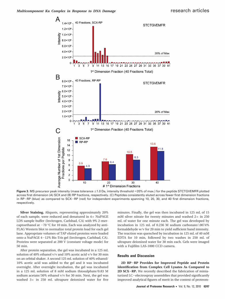
Figure 3. MS precursor peak intensity (mass tolerance (1.0 Da, intensity threshold )20% of max.) for the peptide STCTGVEMFR plottedacross first dimension (A) SCX and (B) RP fractions, respectively. (C) Peptides consistently eluted across fewer first dimension fractionsin RP-RP (blue) as compared to SCX-RP (red) for independent experiments spanning 10, 20, 30, and 40 first dimension fractions,respectively.
Multicomponent Ku Complex in Response to DNA Damage research articles
Journal of Proteome Research • Vol. 9, No. 12, 2010 6247

ics-based analyses of tyrosine signaling in embryonic stemcells.46 The primary challenge in this application was detectionof a rare post-translational modification (tyrosine phosphoryl-ation) in primary cells that exhibited relatively low levels ofsignaling activity. As an extension of these findings, we nextasked whether our 25 µm I.D. nanoflow columns could beintegrated within an automated, multidimension fractionationplatform for analysis of higher complexity biological extracts.We chose RP-RP as our target implementation based on recentreports that suggested the potential for improved performanceas compared to other multidimensional fractionation strate-gies.25 Figure 1 shows a schematic representation of our 2-Dnanoflow system (see experimental section). Briefly, our plat-form is based on a Waters NanoACQUITY UHPLC modified toaccept self-packed columns46 and reconfigured with an ad-ditional two-position, 6-port valve to enable a precolumneffluent split, in a vented column configuration.47,48 An on-board pump was used in isocratic mode to introduce samplesor first dimension eluents loaded from autosampler vials, whilea second UHPLC binary pump generated the solvent gradientfor LC-MS/MS in the usual manner. This configurationfacilitated rapid switching between RP and SCX columns forthe first dimension separation, without the need to preparelarge quantities of fresh first dimension solvents. Unattendedacquisition of RP/SCX-RP MS/MS data was facilitated by useof a computer-controlled electrospray positioning device (Digi-
tal PicoView, New Objective, Woburn, MA) that automaticallymoved the emitter tip beneath a wash station (see Figure 1)for sample loading in-between fractions. This eliminated build-up of salts or other residue on the outer surface of the emitter,that would otherwise degrade performance. Under theseconditions and for the sample types analyzed herein, wetypically achieved 2-3 months of continuous operation withno observable loss in first- or second-dimension columnperformance.
As an initial evaluation for analysis of complex mixtures, wechose a commercially prepared tryptic digest of E. coli lysate(see Experimental Section). First, we performed pilot studiesto determine first dimension salt (SCX) and organic (RP) eluentconcentrations that provided for uniform distribution of pep-tide identifications across a complete 2-D LC-MS/MS analysis(data not shown). Next, 200 ng of tryptic peptides derived fromE.coli were analyzed by either SCX-RP or RP-RP, usingidentical mass spectrometry acquisition methods. RP-RPprovided for a higher number of unique peptide (Figure 2a)and protein (Figure 2b) identifications across the full range offractionation conditions analyzed, as compared to SCX-RP(Supplementary Tables 2 and 3 provide a full list of peptideand protein identifications, Supporting Information). Consis-tent with these data, we also observed that RP-RP providedfor identification of more peptides per protein (Figure 2b).
Given the different retention mechanisms of reversed phaseand strong cation exchange resins, we next asked if peptidesidentified by either SCX-RP or RP-RP differed in their respec-tive physio-chemical properties. For example, we observed thatthe average length of peptides identified remained very con-sistent at 16 and 15 amino acid residues for SCX-RP andRP-RP, respectively, across all conditions analyzed (Figure 2b).Next we applied a permutation test51 to decipher any biaspresent in the amino acid content of peptides identified by eachfractionation technique. Figure 2c demonstrates that onlyhistidine was enriched in peptides identified with SCX-RP,while no statistically significant bias was observed for the aminoacid content of peptides identified by RP-RP. Data associatedwith the permutation test is provided in Supplementary Table4 (Supporting Information). Collectively the data above suggestthat RP-RP provided for an overall increase in peptideidentification, rather than improved performance for a par-ticular subclass of peptides that otherwise would go undetected.These results are consistent with previous work by Dowell etal.27 in which no significant differences in the physiochemicalproperties of proteins were observed across various multidi-mensional peptide fractionation techniques. To further eluci-date the performance discrepancy between RP-RP and SCX-RPwe next analyzed first dimension (SCX and RP) peak capacitiesfor the data in Figure 2. The use of peptide identification toverify the presence of a given peptide in consecutive firstdimension fractions was complicated by the stochastic natureof MS/MS. In an effort to mitigate this effect we used acombination of precursor mass tolerance ((1 Da) and seconddimension LC elution time ((1 min) to search for each peptidein first dimension fractions adjacent to that in which theoriginal identification occurred. Precursors within the masstolerance window, but below 20% relative abundance ascompared to the most intense instance of the identifiedpeptide, were not considered (see experimental section). Figure3a and b shows representative data for the peptide, STCT-GVEMFR dentified by both techniques, with 40 first dimensionfractions. Precursors putatively matching the identified peptide
Figure 4. Histogram plots of MS precursor intensities for peptidesdetected by both fractionation techniques (black bars) and forthose peptides identified uniquely by SCX-RP (A, red bars) andRP-RP (B, blue bars), respectively.
research articles Zhou et al.
6248 Journal of Proteome Research • Vol. 9, No. 12, 2010
spanned 7 first dimension fractions in SCX-RP, but eluted inonly 1 fraction in the RP-RP analysis. Accordingly, the maxi-mum precursor signal intensity observed for STCTGVEMFR wassignificantly higher for RP-RP as compared to SCX-RP. OverallRP-RP consistently demonstrated superior peak capacity asevidenced by the average number of first dimension fractionsspanned by all identified peptides (Figure 3c).
In separate experiments, we used a data analysis approachsimilar to that described above to explore the reproducibilityof RP-RP fractionation. From duplicate, 10 fraction RP-RPexperiments (as in Figure 2a, left column), and a stringentthreshold that required a peptide identification in both 2-DLC-MS/MS analyses to qualify as “reproduced,” we observedthat 78% of all peptides were found in the same first dimensionfraction across replicate analyses. As was noted above, thestochastic nature of MS/MS likely makes this result the lowerbound of reproducibility. In fact, the use of more permissivecriteria as described above (second dimension retention timewindow (1 min and precursor mass tolerance of (1 Da)demonstrated that potentially 95% of all identified peptidesappeared in the same fraction in replicate 2-D RP-RP analyses(data not shown).
We next asked whether the improved peak capacity of 2-DRP-RP (Figure 3a and b) would provide for identification ofpeptides over a wider dynamic range as compared to 2-DSCX-RP. To address this question we compared precursorintensities of peptides identified in common between SCX-RPand RP-RP as well as those sequences uniquely associated with
each fractionation technique. Figure 4 shows histogram plotsfor the precursor intensities of the commonly detected peptides(black bars) and those identified uniquely by SCX-RP (red bars)and RP-RP (blue bars), respectively. Consistent with the datain Figures 2 and 3, we observed that the distributions of unique-and commonly identified peptides overlapped in the case ofSCX-RP (Figure 4a) while those peptides uniquely identifiedby RP-RP (Figure 4b, blue bars) systematically clustered towardthe low end of the abundance range as compared to peptidesidentified by both techniques (Figure 4b, black bars).
Collectively our data suggest that RP-RP provides a repro-ducible platform for online peptide fractionation that outper-forms SCX-RP in terms of total number, and dynamic range,of peptide identifications, primarily due to increased peakcapacity in the first dimension.
2D Fractionation for Proteomic Characterization ofNuclear Multicomponent Ku Complexes. As described above,there is growing appreciation that control of cellular physiologyoccurs through a delicate balance of protein post-translationalmodification and protein-protein interactions. As a result,numerous targeted and large-scale studies have attempted todecipher the dynamics of protein complexes in the context ofnormal physiology, and sought to understand how disruptionof protein-protein interactions contributes to human disease.4,30
For example, the Ku proteins are involved in a number ofcellular functions, including telomere maintenance, transcrip-tional regulation, apoptosis and antigen-receptor gene rear-rangement. In particular the Ku proteins are essential compo-
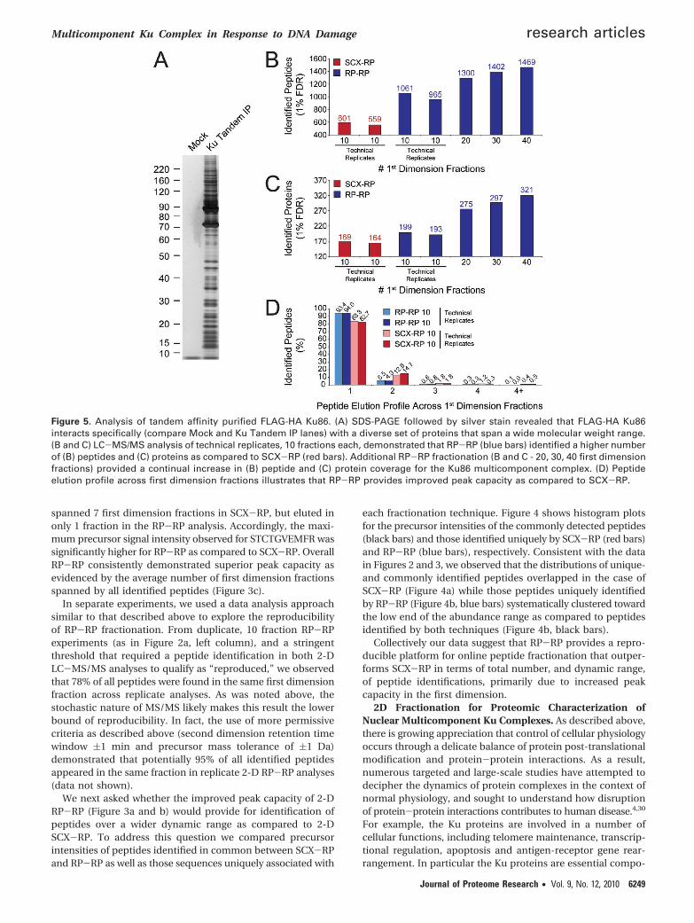
Figure 5. Analysis of tandem affinity purified FLAG-HA Ku86. (A) SDS-PAGE followed by silver stain revealed that FLAG-HA Ku86interacts specifically (compare Mock and Ku Tandem IP lanes) with a diverse set of proteins that span a wide molecular weight range.(B and C) LC-MS/MS analysis of technical replicates, 10 fractions each, demonstrated that RP-RP (blue bars) identified a higher numberof (B) peptides and (C) proteins as compared to SCX-RP (red bars). Additional RP-RP fractionation (B and C - 20, 30, 40 first dimensionfractions) provided a continual increase in (B) peptide and (C) protein coverage for the Ku86 multicomponent complex. (D) Peptideelution profile across first dimension fractions illustrates that RP-RP provides improved peak capacity as compared to SCX-RP.
Multicomponent Ku Complex in Response to DNA Damage research articles
Journal of Proteome Research • Vol. 9, No. 12, 2010 6249

nents of the response to DNA damage.52 Consistent with theinvolvement of Ku in multiple pathways, a recent massspectrometry-based study identified some 90 protein groupsin association with Ku, immunoaffinity purified from HEK 293whole cell lysate.45 Moreover, several reports have describedhow disruption of Ku protein complexes contributes to aber-rations in multiple cellular processes.53
Based on the apparent molecular complexity and functionaldiversity of Ku, we asked whether proteomics analysis basedon RP-RP fractionation would provide additional molecularresolution for the Ku complex. Toward this end we performedtandem affinity purification of FLAG-HA Ku86 from the nuclearfraction of FLAG-HA Ku86 HeLa S3 cells. As a negative control,TAP was performed in parallel from parental HeLa S3 cells.After elution of the complex with HA peptide, a small aliquotcorresponding to approximately 20% of the total eluate wasresolved by SDS-PAGE and visualized with silver stain. Figure5a shows that the Ku complex consists of a very large numberof proteins spanning a wide range of stoichiometry andmolecular weight. The absence of detected proteins in thecontrol lane suggested that the majority of protein bandsresolved by SDS-PAGE represented specific protein interactionswithin the Ku complex.
Next, we performed replicate LC-MS/MS analyses for bothSCX-RP and RP-RP, collecting 10 first dimension fractions foreach. Figure 5b and c show that, although each techniqueprovided reproducible fractionation, RP-RP provided for de-tection of significantly more unique peptides and proteins ascompared to SCX-RP (Supplementary Table 5 provides infor-mation for all peptide and protein identifications, SupportingInformation). Consistent with the data in Figures 2 and 3, weobserved that ∼94% of all peptides detected by RP-RP wereconstrained within a single, first dimension fraction (Figure 5d),while in the corresponding SCX-RP experiment, only ∼83%of identified peptides met this criteria. Given these data we nextasked whether further RP-RP fractionation depth wouldprovide for identification of additional proteins in the Kucomplex. In subsequent RP-RP experiments, equal aliquots ofKu complex were analyzed by acquisition of 20, 30, and 40 firstdimension fractions, respectively. Figure 5b and c show thatin fact we observed a continual increase in the number ofpeptide and protein identifications as a function of fraction-ation depth, again confirming the high peak capacity affordedby RP-RP fractionation.
In the context of these experiments, we observed manyproteins previously identified as partners of Ku 70/86, includ-ing: Werner syndrome associated protein (WRN) the poly(ADP-ribose) polymerase I (PARP-1), YY1, the RNA-dependentHelicase A/Nuclear DNA-dependent Helicase II/(RHA/NDH-II/DDX9), and several heterogeneous nuclear proteins(hnRNPs).45 Collectively, our results add significantly to therepertoire of known Ku complex members (see SupplementaryTable 5, Supporting Information).
As we intended to use iTRAQ-based quantification in thecourse of these studies, we next explored the potential forreporter ion scrambling due to coelution of peptides withsimilar mass-to-charge ratios. Using an approach similar to thatdescribed in Figure 3, we queried all MS scans preceding eachMS/MS event that yielded a peptide identification, and re-corded the number of other peptide precursors that appearedwithin a m/z range of -0.5 to +1.0 Da with respect to theidentified peptide. As before, coeluting species were scored aspotential interferents when their precursor intensity exceeded20% relative to that of the peptide of interest. By this relativelycrude measure, we observed potential contaminants associatedwith 12.7% of all peptides identified in the 10 fraction SCX-RPexperiment. In the corresponding 10 fraction RP-RP analysis,only 7.4% of identified peptides coeluted with a potentialinterfering species. Consistent with our observations for im-proved peak capacity associated with RP-RP, the potentialprecursor contamination rate dropped to 2.4% at the highestfractionation depth (data not shown).
Ku Multicomponent Protein Complex Is DynamicallyRemodeled in Response to γ Radiation. While Ku participatesin many cellular processes it is primarily associated with theDNA damage response.54 An emerging body of work hasrevealed that Ku along with several protein interaction partners,including SSRP1, WRN, and nucleolin, relocalize within thenucleus or are otherwise activated, via post-translationalmodification for example, in response to genotoxic stress.Moreover recent reports suggest that Ku/DNA-PK and SSRP155
may also play a role in sensitizing tumor cells to chemothera-peutic agents.55,56
Given these intriguing results and the underlying complexityof DNA repair pathways,53 we next asked whether RP-RPfractionation combined with stable isotope labeling would
Figure 6. The FLAG-HA Ku86 multicomponent complex is remod-eled in response to DNA damage. HeLa S3 cells that expressedFLAG-HA Ku86 were subjected to γ radiation, with complexespurified at 0, 0.5, 2, and 5 h after exposure. (A) Aliquots of elutedproteins were resolved by SDS-PAGE and visualized by silverstain; arrows indicate molecular weight regions at which subtlechanges in complex membership were observed. (B) Comparisonof proteins identified in TAP-purified FLAG-HA Ku86 complexesfrom gamma-treated HeLa S3 cells with those found in the nativecomplex (Figure 5, 40 frxn) revealed ∼80% reproducibility forRP-RP analysis. (C) Analysis of iTRAQ ratios for proteins identi-fied in the context of γ radiation indicated that protein member-ship in the complex decreased by ∼30% (relative to the nativecomplex) over a time course of 6 h after gamma irradiation.
research articles Zhou et al.
6250 Journal of Proteome Research • Vol. 9, No. 12, 2010

provide sufficient peak capacity to decipher the moleculardynamics of Ku protein complexes in response to DNA damage.Toward this end we used a model system in which HeLa S3cells that expressed FLAG-HA Ku86 were subjected to γradiation to induce DNA damage. Tandem affinity purificationwas performed at varying time intervals after treatment, and asmall aliquot of each HA eluate was resolved on SDS-PAGE.Comparison of the gel lanes (Figure 6a) revealed that changesin protein membership of the Ku complex under these condi-tions were subtle at best. Next, the remaining portions of eachTAP elution were digested with trypsin, labeled with iTRAQreagents, and analyzed by 40 fraction RP-RP MS/MS as inFigure 5.
ProteinPilot57 was used to facilitate analysis (e.g., identifica-tion and quantification) of all peptides and proteins. In totalwe detected and quantified 321 proteins within the Ku complexat time points, 0, 0.5, 2, and 5 h, relative to treatment with γradiation. We observed several proteins known to interact withKu including DNA-PKcs, WRN, NCL,45 and H2AX.58 Encourag-ingly, the overlap in protein identifications between this
experiment and our analysis of the basal complex (Figure 5,40 RP-RP fractions) was 82%, again illustrating the reproduc-ibility of TAP and RP-RP fractionation (Figure 6b). To betterunderstand global changes in Ku complex membership, wenext normalized all peptide ratios to those of FLAG-HA Ku asan internal control, and then plotted the fraction of proteinpartners whose membership in the complex was modulatedacross the time course analyzed. Figure 6c shows that proteinmembership in the complex generally decreased as a result ofDNA damage.
Given the large number of detected proteins, we next appliedhierarchical clustering to group proteins based on their coor-dinate response to γ radiation. Figure 7a shows that proteinmembers of the Ku complex clustered into 3 primary groups(see also Supplementary Table 6, Supporting Information);further analysis based on KEGG pathways revealed that groups2 and 3 were highly enriched for ribosomal proteins (Figure7b). Interestingly, several proteins reported to play a role inDNA repair, including DDX21, SSRP1, DNA-PKcs, along withseveral heterogeneous nuclear ribonucleoproteins,59,60 clus-
Figure 7. (A) Hierarchical clustering of iTRAQ ratios, relative to the zero time point (Figure 6A, left lane), partitioned the response ofFLAG-HA Ku86 associated proteins to gamma irradiation into three major groups. Functional analysis of each group based on KEGGPathways (B) revealed that groups 2 and 3 were highly enriched for ribosomal proteins, while multiple DNA repair pathways wereoverrepresented in group 1 (B - Top, and C). Several proteins previously demonstrated to interact with Ku (DNA-PK, SSRP1, WRN, andHNRNPC), along with novel complex members (DDX21 and RCC2) validated in this study (Figure 8) are indicated for group 1.
Multicomponent Ku Complex in Response to DNA Damage research articles
Journal of Proteome Research • Vol. 9, No. 12, 2010 6251
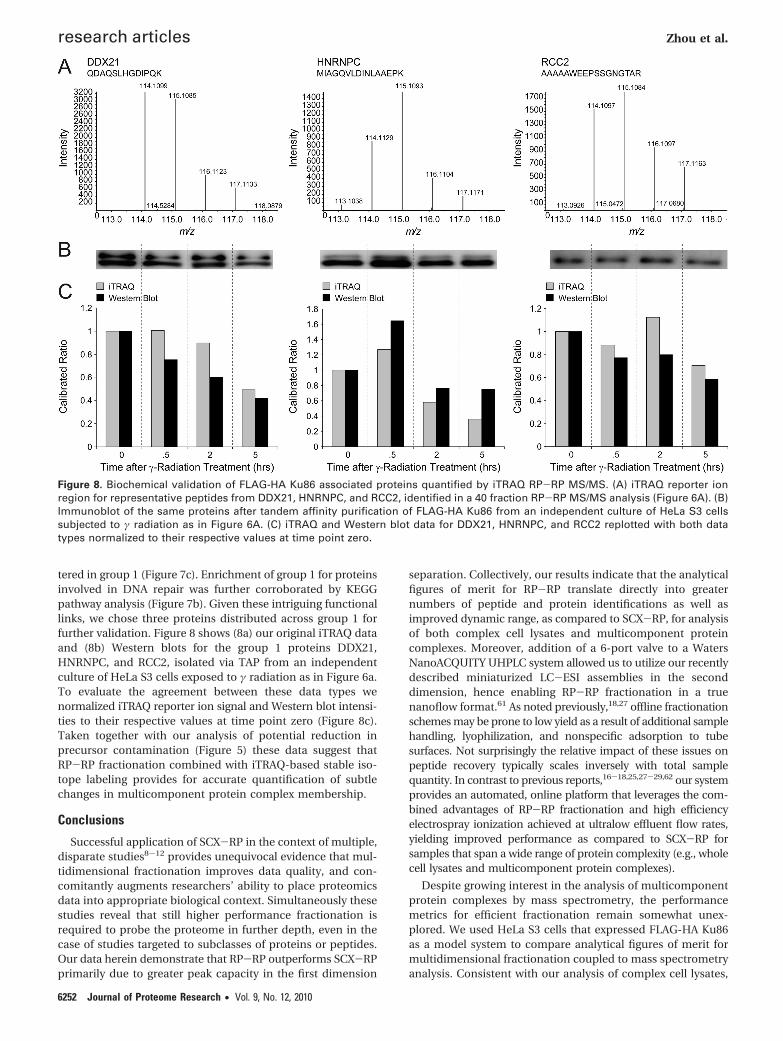
tered in group 1 (Figure 7c). Enrichment of group 1 for proteinsinvolved in DNA repair was further corroborated by KEGGpathway analysis (Figure 7b). Given these intriguing functionallinks, we chose three proteins distributed across group 1 forfurther validation. Figure 8 shows (8a) our original iTRAQ dataand (8b) Western blots for the group 1 proteins DDX21,HNRNPC, and RCC2, isolated via TAP from an independentculture of HeLa S3 cells exposed to γ radiation as in Figure 6a.To evaluate the agreement between these data types wenormalized iTRAQ reporter ion signal and Western blot intensi-ties to their respective values at time point zero (Figure 8c).Taken together with our analysis of potential reduction inprecursor contamination (Figure 5) these data suggest thatRP-RP fractionation combined with iTRAQ-based stable iso-tope labeling provides for accurate quantification of subtlechanges in multicomponent protein complex membership.
Conclusions
Successful application of SCX-RP in the context of multiple,disparate studies8-12 provides unequivocal evidence that mul-tidimensional fractionation improves data quality, and con-comitantly augments researchers’ ability to place proteomicsdata into appropriate biological context. Simultaneously thesestudies reveal that still higher performance fractionation isrequired to probe the proteome in further depth, even in thecase of studies targeted to subclasses of proteins or peptides.Our data herein demonstrate that RP-RP outperforms SCX-RPprimarily due to greater peak capacity in the first dimension
separation. Collectively, our results indicate that the analyticalfigures of merit for RP-RP translate directly into greaternumbers of peptide and protein identifications as well asimproved dynamic range, as compared to SCX-RP, for analysisof both complex cell lysates and multicomponent proteincomplexes. Moreover, addition of a 6-port valve to a WatersNanoACQUITY UHPLC system allowed us to utilize our recentlydescribed miniaturized LC-ESI assemblies in the seconddimension, hence enabling RP-RP fractionation in a truenanoflow format.61 As noted previously,18,27 offline fractionationschemes may be prone to low yield as a result of additional samplehandling, lyophilization, and nonspecific adsorption to tubesurfaces. Not surprisingly the relative impact of these issues onpeptide recovery typically scales inversely with total samplequantity. In contrast to previous reports,16-18,25,27-29,62 our systemprovides an automated, online platform that leverages the com-bined advantages of RP-RP fractionation and high efficiencyelectrospray ionization achieved at ultralow effluent flow rates,yielding improved performance as compared to SCX-RP forsamples that span a wide range of protein complexity (e.g., wholecell lysates and multicomponent protein complexes).
Despite growing interest in the analysis of multicomponentprotein complexes by mass spectrometry, the performancemetrics for efficient fractionation remain somewhat unex-plored. We used HeLa S3 cells that expressed FLAG-HA Ku86as a model system to compare analytical figures of merit formultidimensional fractionation coupled to mass spectrometryanalysis. Consistent with our analysis of complex cell lysates,
Figure 8. Biochemical validation of FLAG-HA Ku86 associated proteins quantified by iTRAQ RP-RP MS/MS. (A) iTRAQ reporter ionregion for representative peptides from DDX21, HNRNPC, and RCC2, identified in a 40 fraction RP-RP MS/MS analysis (Figure 6A). (B)Immunoblot of the same proteins after tandem affinity purification of FLAG-HA Ku86 from an independent culture of HeLa S3 cellssubjected to γ radiation as in Figure 6A. (C) iTRAQ and Western blot data for DDX21, HNRNPC, and RCC2 replotted with both datatypes normalized to their respective values at time point zero.
research articles Zhou et al.
6252 Journal of Proteome Research • Vol. 9, No. 12, 2010

we observed that RP-RP provided for identification of signifi-cantly more unique peptides and proteins as compared toSCX-RP, for an equivalent fractionation depth. Moreover, weobserved a continual increase in the number of proteinsidentified as a function of fractionation depth, suggesting thatefforts aimed at systematic analysis of discrete protein com-plexes will benefit from higher peak capacity separation strate-gies. Our data add some 175 putatively novel members to therepertoire of known proteins in the multicomponent Kucomplex.
To further explore the utility of RP-RP fractionation for theanalysis of protein complexes, we sought to characterizechanges in the Ku complex in response to DNA damageinduced by γ radiation. Although Ku86 and Ku70 are knownto play important roles in DNA repair, we observed only modestmodulation of the associated complex at the level of a silver-stained gel, over a time course of 5 h after exposure of cells toγ radiation. To gain additional insight, we utilized iTRAQ stableisotope labels in conjunction with RP-RP MS to decipher Kuprotein complex dynamics under these conditions. Hierarchicalclustering revealed coordinate participation of many proteinsknown to associate with Ku, including WRN,63 DNA-PK,55,64
and H2AX,58 in addition to a wide range of proteins notpreviously reported as members of the multicomponent Kucomplex (see Supplementary Table 6, Supporting Information).For example, our data significantly expand the number ofribosomal proteins that may participate in the Ku86 complex.When considered collectively with previous work,45 our resultsraise the intriguing possibility that Ku86 regulates translationof specific mRNAs via recruitment of ribosomal componentsto their site of synthesis. Consistent with this hypothesis Ku86and Ku70, among other proteins, have been reported to interactwith the 5′UTR sequence of p53. Indeed, this DNA sequenceis critical for the increased expression of p53 upon DNAdamage.65 We used Western blot to validate the response ofHNRNPC, DDX21, and RCC2 to γ radiation; the agreementbetween our immunoblot and proteomics data suggest thatmembership of many proteins within the Ku complex isdynamically modulated in response to DNA damage. Moreover,our ability to monitor modest ratios for members of the Kucomplex suggests that our online RP-RP fractionation platformprovides sufficient peak capacity to minimize deleteriousscrambling66,67 of iTRAQ reporter ions. In the specific cases ofRCC2,68 HNRNPC,59 and DDX21, their roles in DNA repair havebeen linked to different protein kinases, ATM/ATR,68 DNA-PKcs,69 and JNK,60 respectively. Collectively these observationshighlight the complex interplay between protein-proteininteractions and phosphorylation in regulation of cellularresponse to DNA damage.
Given our results above, along with recent work thathighlighted the complex nature of DNA repair pathways,68 itis not surprising to find that the molecular response to DNAdamage, or other genotoxic insult, is observed across sub-nuclear compartments. For example, relocalization of WRN,SSRP1 and Ku86 between the nucleolus and nucleoplasm hasbeen linked with different types of cellular stress.55 Manyproteins identified in our study, particularly those in group 1(Figure 7c), shuttle between different subnuclear compartmentsas a result of DNA damage. While the mechanistic impact ofprotein relocalization, for example DNA-PK, SSRP1, and DDX21in our work, or WRN,63 Spt1670 and TRF271 as describedelsewhere, remains unclear, our data provide an expanded setof proteins, putatively involved in DNA repair pathways, that
are viable candidates for further biochemical and functionalcharacterization. In this context, immunohistochemical-basedassays would provide orthogonal validation of protein-proteininteractions in addition to exquisite temporal and subcellularlocalization data. These assays, when combined with proteom-ics-based analysis of protein complexes involved in DNA repair,will provide a powerful means to decipher the molecularresponse to therapeutic strategies that rely on induction ofgenotoxic stress.
Acknowledgment. We thank Eric Smith for preparationof figures and valuable discussion. Generous support for thiswork was provided by the Dana-Farber Cancer Institute andthe National Institutes of Health, NHGRI (P50HG004233), andNINDS (P01NS047572).
Supporting Information Available: Supplementarytables. This material is available free of charge via the Internetat http://pubs.acs.org.
References(1) Pandey, A.; Mann, M. Proteomics to study genes and genomes.
Nature 2000, 405 (6788), 837–846.(2) Aebersold, R.; Mann, M. Mass spectrometry-based proteomics.
Nature 2003, 422 (6928), 198–207.(3) Hille, J. M.; Freed, A. L.; Watzig, H. Possibilities to improve
automation, speed and precision of proteome analysis: A com-parison of two-dimensional electrophoresis and alternatives.Electrophoresis 2001, 22 (19), 4035–4052.
(4) Kocher, T.; Superti-Furga, G. Mass spectrometry-based functionalproteomics: from molecular machines to protein networks. Nat.Methods 2007, 4 (10), 807–815.
(5) Sterner, J. L.; Johnston, M. V.; Nicol, G. R.; Ridge, D. P. Signalsuppression in electrospray ionization Fourier transform massspectrometry of multi-component samples. J. Mass Spectrom.2000, 35 (3), 385–391.
(6) Maltby, D.; Lynn, A.; Baker, P. Proteomics on a thermo finniganLTQ-FTICR mass spectrometer. Mol. Cell. Proteomics 2005, 4 (8),S442–S442.
(7) Second, T. P.; Blethrow, J. D.; Schwartz, J. C.; Merrihew, G. E.;MacCoss, M. J.; Swaney, D. L.; Russell, J. D.; Coon, J. J.; Zabrouskov,V. Dual-Pressure Linear Ion Trap Mass Spectrometer Improvingthe Analysis of Complex Protein Mixtures. Anal. Chem. 2009, 81(18), 7757–7765.
(8) Washburn, M. P.; Wolters, D.; Yates, J. R. Large-scale analysis ofthe yeast proteome by multidimensional protein identificationtechnology. Nat. Biotechnol. 2001, 19 (3), 242–247.
(9) Carlton, J. M.; Angiuoli, S. V.; Suh, B. B.; Kooij, T. W.; Pertea, M.;Silva, J. C.; Ermolaeva, M. D.; Allen, J. E.; Selengut, J. D.; Koo, H. L.;Peterson, J. D.; Pop, M.; Kosack, D. S.; Shumway, M. F.; Bidwell,S. L.; Shallom, S. J.; van Aken, S. E.; Riedmuller, S. B.; Feldblyum,T. V.; Cho, J. K.; Quackenbush, J.; Sedegah, M.; Shoaibi, A.;Cummings, L. M.; Florens, L.; Yates, J. R.; Raine, J. D.; Sinden, R. E.;Harris, M. A.; Cunningham, D. A.; Preiser, P. R.; Bergman, L. W.;Vaidya, A. B.; Van Lin, L. H.; Janse, C. J.; Waters, A. P.; Smith, H. O.;White, O. R.; Salzberg, S. L.; Venter, J. C.; Fraser, C. M.; Hoffman,S. L.; Gardner, M. J.; Carucci, D. J. Genome sequence andcomparative analysis of the model rodent malaria parasite Plas-modium yoelii yoelii. Nature 2002, 419 (6906), 512–519.
(10) Florens, L.; Washburn, M. P.; Raine, J. D.; Anthony, R. M.; Grainger,M.; Haynes, J. D.; Moch, J. K.; Muster, N.; Sacci, J. B.; Tabb, D. L.;Witney, A. A.; Wolters, D.; Wu, Y. M.; Gardner, M. J.; Holder, A. A.;Sinden, R. E.; Yates, J. R.; Carucci, D. J. A proteomic view of thePlasmodium falciparum life cycle. Nature 2002, 419 (6906), 520–526.
(11) Koller, A.; Washburn, M. P.; Lange, B. M.; Andon, N. L.; Deciu, C.;Haynes, P. A.; Hays, L.; Schieltz, D.; Ulaszek, R.; Wei, J.; Wolters,D.; Yates, J. R. Proteomic survey of metabolic pathways in rice.Proc. Natl. Acad. Sci. U.S.A. 2002, 99 (18), 11969–11974.
(12) Fournier, M. L.; Gilmore, J. M.; Martin-Brown, S. A.; Washburn,M. P. Multidimensional separations-based shotgun proteomics.Chem. Rev. 2007, 107 (8), 3654–3686.
(13) Mawuenyega, K. G.; Kaji, H.; Yamauchi, Y.; Shinkawa, T.; Saito,H.; Taoka, M.; Takahashi, N.; Isobe, T. Large-scale identificationof Caenorhabditis elegans proteins by multidimensional liquid
Multicomponent Ku Complex in Response to DNA Damage research articles
Journal of Proteome Research • Vol. 9, No. 12, 2010 6253

chromatography - Tandem mass spectrometry. J. Proteome Res.2003, 2 (1), 23–35.
(14) Taoka, M.; Yamauchi, Y.; Shinkawa, T.; Kaji, H.; Motohashi, W.;Nakayama, H.; Takahashi, N.; Isobe, T. Only a small subset of thehorizontally transferred chromosomal genes in Escherichia coliare translated into proteins. Mol. Cell. Proteomics 2004, 3 (8), 780–787.
(15) Nagano, K.; Taoka, M.; Yamauchi, Y.; Itagaki, C.; Shinkawa, T.;Nunomura, K.; Okamura, N.; Takahashi, N.; Izumi, T.; Isobe, T.Large-scale identification of proteins expressed in mouse embry-onic stem cells. Proteomics 2005, 5 (5), 1346–1361.
(16) Essader, A. S.; Cargile, B. J.; Bundy, J. L.; Stephenson, J. L. Acomparison of immobilized pH gradient isoelectric focusing andstrong-cation-exchange chromatography as a first dimension inshotgun proteomics. Proteomics 2005, 5 (1), 24–34.
(17) Cargile, B. J.; Talley, D. L.; Stephenson, J. L. Immobilized pHgradients as a first dimension in shotgun proteomics and analysisof the accuracy of pI predictability of peptides. Electrophoresis2004, 25 (6), 936–945.
(18) Slebos, R. J. C.; Brock, J. W. C.; Winters, N. F.; Stuart, S. R.; Martinez,M. A.; Li, M.; Chambers, M. C.; Zimmerman, L. J.; Ham, A. J.; Tabb,D. L.; Liebler, D. C. Evaluation of Strong Cation Exchange versusIsoelectric Focusing of Peptides for Multidimensional LiquidChromatography-Tandem Mass Spectrometry. J. Proteome Res.2008, 7 (12), 5286–5294.
(19) de Godoy, L. M. F.; Olsen, J. V.; Cox, J.; Nielsen, M. L.; Hubner,N. C.; Frohlich, F.; Walther, T. C.; Mann, M. Comprehensive mass-spectrometry-based proteome quantification of haploid versusdiploid yeast. Nature 2008, 455 (7217), 1251–U60.
(20) Picotti, P.; Bodenmiller, B.; Mueller, L. N.; Domon, B.; Aebersold,R.; Full Dynamic Range Proteome Analysis of, S. cerevisiae byTargeted Proteomics. Cell 2009, 138 (4), 795–806.
(21) Balgley, B. M.; Wang, W. J.; Song, T.; Fang, X. P.; Yang, L.; Lee,C. S. Evaluation of confidence and reproducibility in quantitativeproteomics performed by a capillary isoelectric focusing-basedproteomic platform coupled with a spectral counting approach.Electrophoresis 2008, 29 (14), 3047–3054.
(22) Fang, X. P.; Balgley, B. M.; Wang, W. J.; Park, D. M.; Lee, C. S.Comparison of multidimensional shotgun technologies targetingtissue proteomics. Electrophoresis 2009, 30 (23), 4063–4070.
(23) Wang, Y. J.; Rudnick, P. A.; Evans, E. L.; Li, J.; Zhuang, Z. P.; DeVoe,D. L.; Lee, C. S.; Balgley, B. M. Proteome analysis of microdissectedtumor tissue using a capillary isoelectric focusing-based multidi-mensional separation platform coupled with ESI-tandem MS. Anal.Chem. 2005, 77 (20), 6549–6556.
(24) Chen, J. Z.; Balgley, B. M.; DeVoe, D. L.; Lee, C. S. Capillaryisoelectric focusing-based multidimensional concentration/sepa-ration platform for proteome analysis. Anal. Chem. 2003, 75 (13),3145–3152.
(25) Gilar, M.; Olivova, P.; Daly, A. E.; Gebler, J. C. Two-dimensionalseparation of peptides using RP-RP-HPLC system with differentpH in first and second separation dimensions. J. Sep. Sci. 2005,28 (14), 1694–1703.
(26) Delmotte, N.; Lasaosa, M.; Tholey, A.; Heinzle, E.; Huber, C. G.Two-dimensional reversed-phase x ion-pair reversed-phase HPLC:an alternative approach to high-resolution peptide separation forshotgun proteome analysis. J. Proteome Res. 2007, 6 (11), 4363–4373.
(27) Dowell, J. A.; Frost, D. C.; Zhang, J.; Li, L. J. Comparison of two-dimensional fractionation techniques for shotgun proteomics.Anal. Chem. 2008, 80 (17), 6715–6723.
(28) Gilar, M.; Olivova, P.; Daly, A. E.; Gebler, J. C. Orthogonality ofseparation in two-dimensional liquid chromatography. Anal.Chem. 2005, 77 (19), 6426–6434.
(29) Nakamura, T.; Kuromitsu, J.; Oda, Y. Evaluation of comprehensivemultidimensional separations using reversed-phase, reversed-phase liquid chromatography/mass spectrometry for shotgunproteomics. J. Proteome Res. 2008, 7 (3), 1007–1011.
(30) Ewing, R. M.; Chu, P.; Elisma, F.; Li, H.; Taylor, P.; Climie, S.;McBroom-Cerajewski, L.; Robinson, M. D.; O’Connor, L.; Li, M.;Taylor, R.; Dharsee, M.; Ho, Y.; Heilbut, A.; Moore, L.; Zhang, S.;Ornatsky, O.; Bukhman, Y. V.; Ethier, M.; Sheng, Y.; Vasilescu, J.;Abu-Farha, M.; Lambert, J. P.; Duewel, H. S.; Stewart, I. I.; Kuehl,B.; Hogue, K.; Colwill, K.; Gladwish, K.; Muskat, B.; Kinach, R.;Adams, S. L.; Moran, M. F.; Morin, G. B.; Topaloglou, T.; Figeys,D. Large-scale mapping of human protein-protein interactions bymass spectrometry. Mol. Syst. Biol. 2007, 3, 17.
(31) Gavin, A. C.; Bosche, M.; Krause, R.; Grandi, P.; Marzioch, M.;Bauer, A.; Schultz, J.; Rick, J. M.; Michon, A. M.; Cruciat, C. M.;Remor, M.; Hofert, C.; Schelder, M.; Brajenovic, M.; Ruffner, H.;Merino, A.; Klein, K.; Hudak, M.; Dickson, D.; Rudi, T.; Gnau, V.;
Bauch, A.; Bastuck, S.; Huhse, B.; Leutwein, C.; Heurtier, M. A.;Copley, R. R.; Edelmann, A.; Querfurth, E.; Rybin, V.; Drewes, G.;Raida, M.; Bouwmeester, T.; Bork, P.; Seraphin, B.; Kuster, B.;Neubauer, G.; Superti-Furga, G. Functional organization of theyeast proteome by systematic analysis of protein complexes.Nature 2002, 415 (6868), 141–147.
(32) Bouwmeester, T.; Bauch, A.; Ruffner, H.; Angrand, P. O.; Bergamini,G.; Croughton, K.; Cruciat, C.; Eberhard, D.; Gagneur, J.; Ghidelli,S.; Hopf, C.; Huhse, B.; Mangano, R.; Michon, A. M.; Schirle, M.;Schlegl, J.; Schwab, M.; Stein, M. A.; Bauer, A.; Casari, G.; Drewes,G.; Gavin, A. C.; Jackson, D. B.; Joberty, G.; Neubauer, G.; Rick, J.;Kuster, B.; Superti-Furga, G. A physical and functional map of thehuman TNF-alpha NF-kappa B signal transduction pathway. Nat.Cell Biol. 2004, 6 (2), 97.
(33) Gavin, A. C.; Aloy, P.; Grandi, P.; Krause, R.; Boesche, M.; Marzioch,M.; Rau, C.; Jensen, L. J.; Bastuck, S.; Dumpelfeld, B.; Edelmann,A.; Heurtier, M. A.; Hoffman, V.; Hoefert, C.; Klein, K.; Hudak, M.;Michon, A. M.; Schelder, M.; Schirle, M.; Remor, M.; Rudi, T.;Hooper, S.; Bauer, A.; Bouwmeester, T.; Casari, G.; Drewes, G.;Neubauer, G.; Rick, J. M.; Kuster, B.; Bork, P.; Russell, R. B.; Superti-Furga, G. Proteome survey reveals modularity of the yeast cellmachinery. Nature 2006, 440 (7084), 631–636.
(34) Florens, L.; Carozza, M. J.; Swanson, S. K.; Fournier, M.; Coleman,M. K.; Workman, J. L.; Washburn, M. P. Analyzing chromatinremodeling complexes using shotgun proteomics and normalizedspectral abundance factors. Methods 2006, 40 (4), 303–311.
(35) Rolfs, A.; Hu, Y.; Ebert, L.; Hoffmann, D.; Zuo, D.; Ramachandran,N.; Raphael, J.; Kelley, F.; McCarron, S.; Jepson, D. A.; Shen, B.;Baqui, M. M. A.; Pearlberg, J.; Taycher, E.; DeLoughery, C.;Hoerlein, A.; Korn, B.; LaBaer, J. A Biomedically Enriched Collec-tion of 7000 Human ORF Clones. PLoS ONE 2008, 3 (1), e1528.
(36) Esposito, D.; Garvey, L. A.; Chakiath, C. S. Gateway Cloning forProtein Expression. In 2009, 31–54.
(37) Forstner, M.; Leder, L.; Mayr, L. M. Optimization of proteinexpression systems for modern drug discovery. Expert Rev. Pro-teomics 2007, 4 (1), 67–78.
(38) Ramachandran, N.; Raphael, J. V.; Hainsworth, E.; Demirkan, G.;Fuentes, M. G.; Rolfs, A.; Hu, Y.; LaBaer, J. Next-generation high-density self-assembling functional protein arrays. Nat Meth 2008,5 (6), 535–538.
(39) Park, J.; Hu, Y.; Murthy, T. V. S.; Vannberg, F.; Shen, B.; Rolfs, A.;Hutti, J. E.; Cantley, L. C.; LaBaer, J.; Harlow, E.; Brizuela, L.Building a human kinase gene repository: Bioinformatics, molec-ular cloning, and functional validation. Proc. Natl. Acad. Sci. U.S.A.2005, 102 (23), 8114–8119.
(40) Liang, S. F.; Yu, Y. B.; Yang, P. Y.; Gu, S.; Xue, Y.; Chen, X. Analysisof the protein complex associated with 14-3-3 epsilon by adeuterated-leucine labeling quantitative proteomics strategy. J. Chro-matogr., B 2009, 877 (7), 627–634.
(41) Yang, M. Z.; Wang, X. L.; Zhang, L.; Yu, C. Y.; Zhang, B. B.; Cole,W.; Cavey, G.; Davidson, P.; Gibson, G. Demonstration of theinteraction of transforming growth factor beta 2 and type Xcollagen using a modified tandem affinity purification tag. J. Chro-matogr., B 2008, 875 (2), 493–501.
(42) Van Leene, J.; Witters, E.; Inze, D.; De Jaeger, G. Boosting tandemaffinity purification of plant protein complexes. Trends Plant Sci.2008, 13 (10), 517–520.
(43) Kyriakakis, P.; Tipping, M.; Abed, L.; Veraksa, A. Tandem affinitypurification in Drosophila - The advantages of the GS-TAP system.Fly 2008, 2 (4), 229–235.
(44) Gregan, J.; Riedel, C. G.; Petronczki, M.; Cipak, L.; Rumpf, C.; Poser,I.; Buchholz, F.; Mechtler, K.; Nasmyth, K. Tandem affinitypurification of functional TAP-tagged proteins from human cells.Nat. Protoc. 2007, 2 (5), 1145–1151.
(45) Burckstummer, T.; Bennett, K. L.; Preradovic, A.; Schutze, G.;Hantschel, O.; Superti-Furga, G.; Bauch, A. An efficient tandemaffinity purification procedure for interaction proteomics in mam-malian cells. Nat. Methods 2006, 3 (12), 1013–1019.
(46) Ficarro, S. B.; Zhang, Y.; Lu, Y.; Moghimi, A. R.; Askenazi, M.; Hyatt,E.; Smith, E. D.; Boyer, L.; Schlaeger, T. M.; Luckey, C. J.; Marto,J. A. Improved Electrospray Ionization Efficiency Compensates forDiminished Chromatographic Resolution and Enables ProteomicsAnalysis of Tyrosine Signaling in Embryonic Stem Cells. Anal.Chem. 2009, 81 (9), 3440–3447.
(47) Licklider, L. J.; Thoreen, C. C.; Peng, J. M.; Gygi, S. P. Automationof nanoscale microcapillary liquid chromatography-tandem massspectromentry with a vented column. Anal. Chem. 2002, 74 (13),3076–3083.
(48) van der Heeft, E.; ten Hove, G. J.; Herberts, C. A.; Meiring, H. D.;van Els, C. A.; de Jong, A. P. A microcapillary column switchingHPLC-electrospray ionization MS system for the direct identifica-
research articles Zhou et al.
6254 Journal of Proteome Research • Vol. 9, No. 12, 2010

tion of peptides presented by major histocompatibility complexclass I molecules. Anal. Chem. 1998, 70 (18), 3742–3751.
(49) Askenazi, M.; Parikh, J. R.; Marto, J. A. mzAPI: a new strategy forefficiently sharing mass spectrometry data. Nat. Methods 2009, 6(4), 240–242.
(50) Parikh, J. R.; Askenazi, M.; Ficarro, S. B.; Cashorali, T.; Webber,J. T.; Blank, N. C.; Zhang, Y.; Marto, J. A. multiplierz: an extensibleAPI based desktop environment for proteomics data analysis. BMCBioinform. 2009, 10, 364.
(51) Ficarro, S. B.; Parikh, J. R.; Blank, N. C.; Marto, J. A. Niobium(V)oxide (Nb2O5): Application to phosphoproteomics. Anal. Chem.2008, 80 (12), 4606–4613.
(52) Martin, S. G.; Laroche, T.; Suka, N.; Grunstein, M.; Gasser, S. M.Relocalization of telomeric Ku and SIR proteins in response to DNAstrand breaks in yeast. Cell 1999, 97 (5), 621–633.
(53) Downs, J. A.; Jackson, S. P. A means to a DNA end: The many rolesof Ku. Nat. Rev. Mol. Cell Biol. 2004, 5 (5), 367–378.
(54) Helleday, T.; Petermann, E.; Lundin, C.; Hodgson, B.; Sharma, R. A.DNA repair pathways as targets for cancer therapy. Nat. Rev.Cancer 2008, 8 (3), 193–204.
(55) Dejmek, J.; Iglehart, J. D.; Lazaro, J. B. DNA-Dependent ProteinKinase (DNA-PK)-Dependent Cisplatin-Induced Loss of NucleolarFacilitator of Chromatin Transcription (FACT) and Regulation ofCisplatin Sensitivity by DNA-PK and FACT. Mol. Cancer Res. 2009,7 (4), 581–591.
(56) Bolderson, E.; Richard, D. J.; Zhou, B. B.; Khanna, K. K. Recentadvances in cancer therapy targeting proteins involved in DNAdouble-strand break repair. Clin. Cancer Res. 2009, 15 (20), 6314–20.
(57) Shilov, I. V.; Seymour, S. L.; Patel, A. A.; Loboda, A.; Tang, W. H.;Keating, S. P.; Hunter, C. L.; Nuwaysir, L. M.; Schaeffer, D. A. Theparagon algorithm, a next generation search engine that usessequence temperature values and feature probabilities to identifypeptides from tandem mass spectra. Mol. Cell. Proteomics 2007,6 (9), 1638–1655.
(58) Du, Y. C.; Gu, S.; Zhou, J. H.; Wang, T. Y.; Cai, H.; MacInnes, M. A.;Bradbury, E. M.; Chen, X. The dynamic alterations of H2AXcomplex during DNA repair detected by a proteomic approachreveal the critical roles of Ca2+/calmodulin in the ionizingradiation-induced cell cycle arrest. Mol. Cell. Proteomics 2006, 5(6), 1033–1044.
(59) Christian, K. J.; Lang, M. A.; Raffalli-Mathieu, F. Interaction ofheterogeneous nuclear ribonucleoprotein C1/C2 with a novel cis-regulatory element within p53 mRNA as a response to cytostaticdrug treatment. Mol. Pharmacol. 2008, 73 (5), 1558–1567.
(60) Mialon, A.; Thastrup, J.; Kallunki, T.; Mannermaa, L.; Westermarck,J.; Holmstrom, T. H. Identification of nucleolar effects in JNK-deficient cells. FEBS Lett. 2008, 582 (20), 3145–3151.
(61) Ficarro, S. B.; Zhang, Y.; Lu, Y.; Moghimi, A. R.; Askenazi, M.; Hyatt,E.; Smith, E. D.; Boyer, L.; Schlaeger, T. M.; Luckey, C. J.; Marto,J. A. Improved electrospray ionization efficiency compensates fordiminished chromatographic resolution and enables proteomicsanalysis of tyrosine signaling in embryonic stem cells. Anal. Chem.2009, 81 (9), 3440–3447.
(62) Dowell, J. A.; Heyden, W. V.; Li, L. Rat neuropeptidomics by LC-MS/MS and MALDI-FTMS: Enhanced dissection and extractiontechniques coupled with 2D RP-RP HPLC. J. Proteome Res. 2006,5 (12), 3368–3375.
(63) Sakamoto, S.; Nishikawa, K.; Heo, S. J.; Coto, M.; Furuichi, Y.;Shimamoto, A. Werner helicase relocates into nuclear foci inresponse to DNA damaging agents and co-localizes with RPA andRad51. Genes Cells 2001, 6 (5), 421–430.
(64) Shi, M. G.; Vivian, C. J.; Lee, K. J.; Ge, C. M.; Morotomi-Yano, K.;Manzl, C.; Bock, F.; Sato, S.; Tomomori-Sato, C.; Zhu, R. H.; Haug,J. S.; Swanson, S. K.; Washburn, M. P.; Chen, D. J.; Chen, B. P. C.;Villunger, A.; Florens, L.; Du, C. Y. DNA-PKcs-PIDDosome: ANuclear Caspase-2-Activating Complex with Role in G2/M Check-point Maintenance. Cell 2009, 136 (3), 508–520.
(65) Takagi, M.; Absalon, M. J.; McLure, K. G.; Kastan, M. B. Regulationof p53 translation and induction after DNA damage by ribosomalprotein L26 and nucleolin. Cell 2005, 123 (1), 49–63.
(66) Ow, S. Y.; Salim, M.; Noirel, J.; Evans, C.; Rehman, I.; Wright, P. C.iTRAQ Underestimation in Simple and Complex Mixtures: “TheGood, the Bad and the Ugly. J. Proteome Res. 2009, 8 (11), 5347–5355.
(67) Karp, N. A.; Huber, W.; Sadowski, P. G.; Charles, P. D.; Hester, S. V.;Lilley, K. S. Addressing accuracy and precision issues in iTRAQquantitation. Mol. Cell. Proteomics 2010.
(68) Matsuoka, S.; Ballif, B. A.; Smogorzewska, A.; McDonald, E. R.;Hurov, K. E.; Luo, J.; Bakalarski, C. E.; Zhao, Z. M.; Solimini, N.;Lerenthal, Y.; Shiloh, Y.; Gygi, S. P.; Elledge, S. J. ATM and ATRsubstrate analysis reveals extensive protein networks responsiveto DNA damage. Science 2007, 316 (5828), 1160–1166.
(69) Zhang, S. S.; Schlott, B.; Gorlach, M.; Grosse, F. DNA-dependentprotein kinase (DNA-PK) phosphorylates nuclear DNA helicase II/RNA helicase A and hnRNP proteins in an RNA-dependentmanner. Nucleic Acids Res. 2004, 32 (1), 1–10.
(70) Heo, K.; Kim, H. J.; Choi, S. H.; Choi, J. Y.; Kim, K. W.; Gu, J. F.;Lieber, M. R.; Yang, A. S.; An, W. J. FACT-mediated exchange ofhistone variant H2AX regulated by phosphorylation of H2AX andADP-ribosylation of Spt16. Mol. Cell 2008, 30 (1), 86–97.
(71) Buscemi, G.; Zannini, L.; Fontanella, E.; Lecis, D.; Lisanti, S.; Delia,D. The Shelterin Protein TRF2 Inhibits Chk2 Activity at Telomeresin the Absence of DNA Damage. Curr. Biol. 2009, 19 (10), 874–879.
PR1004696
Multicomponent Ku Complex in Response to DNA Damage research articles
Journal of Proteome Research • Vol. 9, No. 12, 2010 6255