oocyte formation by mitotically active germ cells purified
TRANSCRIPT
©20
12 N
atur
e A
mer
ica,
Inc.
All
righ
ts r
eser
ved.
A R T I C L E S
NATURE MEDICINE ADVANCE ONLINE PUBLICATION 1
Since the early 1950s, management of ovarian insufficiency and failure, including infertility caused by aging or insults, was restricted by the belief that the oocyte-containing pool of follicles set forth at birth is not amenable to renewal1. In 2004, studies with mice challenged the idea of a fixed ovarian reserve being endowed during the perinatal period2. Based on results from several approaches, it was concluded that the ovaries of adult mice possess rare female germline or oogonial stem cells (OSCs) that generate oocytes in a way analogous to the spermato-gonial stem cell support of sperm production in adult testes3. Although this previous study was criticized by many in the field4, several years later OSCs were successfully isolated from neonatal and adult mouse ovaries. The cells were shown to stably proliferate in vitro for months5,6 and spontaneously generate what seem to be immature oocytes in cul-ture6. Transplantation studies showed that GFP-expressing mouse OSCs injected into ovaries of chemotherapy-conditioned adult mice differ-entiate into mature eggs that are ovulated, fertilize and produce viable offspring5. These findings, along with other studies of mice7–9, have opened the possibility of using OSCs as agents for transplantation and targets for therapies designed to modulate ovarian function and fertil-ity4,10. Additionally, identification of dormant OSCs in atrophic ovaries of aged mice, which resume in vivo oogenesis when exposed to a young-adult (2 months of age) ovarian environment11, indicates that ovarian aging may be reversible10,12. However, any consideration of a possible clinical utility of these cells requires firm evidence that comparable oocyte-producing cells exist in ovaries of reproductive-age women.
As a first step toward this goal, we critically assessed a recently reported protocol to obtain OSCs from adult mouse ovaries5. This approach relies on the immunological detection of a putative cell-surface variant of Ddx4 (DEAD box polypeptide 4, also commonly
referred to as mouse vasa homolog or Mvh), an evolutionarily con-served germ-cell-specific RNA helicase13–15, coupled with cell sorting using magnetic beads. Two issues surfaced that we felt might pre-clude application of this protocol to isolate candidate OSCs from adult human ovaries. First, Ddx4 is widely considered to be a cytoplasmic protein and, thus, its reported detection on the surface of mouse OSCs is at odds with the results from prior studies10,16. Second, even if a cell-surface variant of Ddx4 is expressed by mouse OSCs, immuno-magnetic sorting is a relatively crude approach to cell isolation that often results in the enrichment of a desired cell type in fractions that are contaminated with non-targeted cells carried over during the col-umn washing and flushing steps. Magnetic bead sorting also does not distinguish between viable and damaged or dead cells and does not allow for simultaneous assessment of other cellular features, such as yield, size or co-expression of additional markers. Our objectives were to initially test with mice whether or not OSCs possess an externally exposed epitope of Ddx4 and, if so, to use this property for valida-tion of a fluorescence-activated cell sorting (FACS)-based protocol to purify viable OSCs from the dispersed ovaries of adult mice. We next determined whether this technology could also be used to purify an equivalent population of candidate OSCs from the ovarian cortical tissue of healthy reproductive-age women. Once we isolated mouse and human OSCs by FACS, we then tested their growth properties and functionality using both in vitro and in vivo approaches.
RESULTSValidation of a FACS-based protocol for OSC isolationThe antibody to Ddx4 used previously to isolate mouse OSCs by immunomagnetic sorting is a rabbit polyclonal antibody against the
1Vincent Center for Reproductive Biology, Massachusetts General Hospital Vincent Department of Obstetrics and Gynecology, Massachusetts General Hospital, Boston, Massachusetts, USA. 2Department of Obstetrics, Gynecology and Reproductive Biology, Harvard Medical School, Boston, Massachusetts, USA. 3Department of Obstetrics and Gynecology, Saitama Medical Center, Saitama Medical University, Saitama, Japan. 4These authors contributed equally to this work. Correspondence should be addressed to J.L.T. ([email protected]).
Received 26 August 2011; accepted 11 January 2012; published online 26 February 2012; doi:10.1038/nm.2669
Oocyte formation by mitotically active germ cells purified from ovaries of reproductive-age womenYvonne A R White1,2,4, Dori C Woods1,2,4, Yasushi Takai3, Osamu Ishihara3, Hiroyuki Seki3 & Jonathan L Tilly1,2
Germline stem cells that produce oocytes in vitro and fertilization-competent eggs in vivo have been identified in and isolated from adult mouse ovaries. Here we describe and validate a fluorescence-activated cell sorting-based protocol that can be used with adult mouse ovaries and human ovarian cortical tissue to purify rare mitotically active cells that have a gene expression profile that is consistent with primitive germ cells. Once established in vitro, these cells can be expanded for months and can spontaneously generate 35- to 50-mm oocytes, as determined by morphology, gene expression and haploid (1n) status. Injection of the human germline cells, engineered to stably express GFP, into human ovarian cortical biopsies leads to formation of follicles containing GFP-positive oocytes 1–2 weeks after xenotransplantation into immunodeficient female mice. Thus, ovaries of reproductive-age women, similar to adult mice, possess rare mitotically active germ cells that can be propagated in vitro as well as generate oocytes in vitro and in vivo.
©20
12 N
atur
e A
mer
ica,
Inc.
All
righ
ts r
eser
ved.
A R T I C L E S
2 ADVANCE ONLINE PUBLICATION NATURE MEDICINE
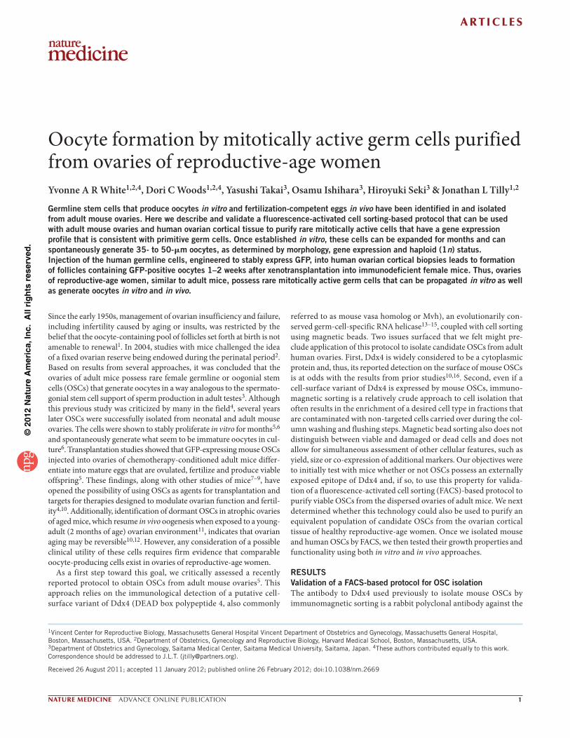
C terminus of the protein (hereafter called the COOH antibody)5. We obtained this antibody, along with a goat polyclonal antibody against the N terminus of Ddx4 (hereafter called the NH2 antibody), for our comparative studies. An immunofluorescence analysis of ovaries from young-adult mice using either of the two antibodies showed an iden-tical pattern of Ddx4 expression that was restricted to the oocytes (Fig. 1a). We then used each antibody for immunomagnetic sorting of dispersed ovary tissue from young-adult mice5. We obtained no cells in the bead fraction when we used the NH2 antibody; however, we observed 5- to 8- m cells bound to the magnetic beads when we used the COOH antibody (Fig. 1b). An analysis of the cells isolated with the COOH antibody revealed a germline gene expression pattern consistent with that previously reported for mouse OSCs isolated using immunomagnetic sorting5 (Supplementary Fig. 1). Although we always detected the isolated oocytes in the non-immunoreactive wash fraction using the COOH antibody when assessed in parallel (Fig. 1b), a more thorough marker analysis of the Ddx4-positive viable cell fraction obtained by immunomagnetic sorting revealed several oocyte-specific mRNAs, including mRNA of the Nobox (new-born ovary homeobox), Zp3 (zona pellucida glycoprotein 3) and Gdf9 (growth differentiation factor 9) genes (Supplementary Fig. 1). These findings indicate that although mouse oocytes do not show evidence of cell-surface expression of Ddx4 when analyzed as individual
entities (Fig. 1b), mouse oocytes are nonetheless a contaminating cell type after immunomagnetic sorting of OSCs from dispersed mouse ovary tissue. This outcome probably reflects either a non-specific phys-ical carryover of small oocytes with OSCs during column washing and flushing or a reactivity of cytoplasmic Ddx4 in plasma-membrane–compromised (damaged) oocytes with the COOH antibody.
Therefore, we next tested the reactivity of each of the two antibod-ies with dispersed mouse ovarian cells by FACS. In agreement with the results from the magnetic bead sorting, we only obtained viable Ddx4-positive cells when we used the COOH antibody (Fig. 1c). However, if we fixed and permeabilized the ovarian cells before FACS, we also obtained Ddx4-positive cells using the NH2 antibody (Fig. 1c). Furthermore, if we fixed, permeabilized and re-sorted the viable Ddx4-positive cells that we isolated by FACS using the COOH antibody, the same cell population was also recognized by the NH2 antibody (Fig. 1d). As a final means to confirm the validity of this OSC isolation method (Fig. 1e), we assessed the fractions of cells obtained at each step of the protocol by gene expression analysis using the following markers for germ cells: Prdm1 (PR domain containing 1 with ZNF domain, which has the human gene ortholog PRDM1, also commonly referred to as BLIMP1), Dppa3 (developmental pluripotency-associated 3, with the human gene ortholog DPPA3, also commonly referred to as STELLA), Ifitm3 (interferon induced
a
c
d
NH2
NH2 NH2
NH2 NH2
Size exclusion (µm)fFiltration
Ovary <70 >70 <35 >35 +ve –ve
FiltrationDdx4sorted
fractionsNoRT
NH2
COOH
COOH Live
Ddx4neg neg Ddx4
NH2Permeabilized
neg Ddx4
Live
Live
Anti-goat-488
Anti-goat-488
Anti-goat-488
Ant
i-rab
bit-
AP
C
Ant
i-rab
bit-
AP
C
Ant
i-rab
bit-
AP
C PermeabilizedPermeabilizedLive
CO
OH
bFraction 1
NH
2
Fraction 2 Fraction 3
e
FACS sort
COOH
FACSre-sort
Live Fixed and permeabilized
Ovarian somatic cellsDdx4 external andinternalDdx4 internal
No cells
Live Fixed and permeabilized
101
101
102
102
103
103
104
104
105
105
Single oocytes
Dispersed ovary
Figure 1 FACS-based protocol for OSC isolation. (a) Immunofluorescence analysis of Ddx4 expression (green; with a blue DAPI counterstain) in young adult mouse ovaries using antibodies against the N (NH2) or the C (COOH) terminus of the protein (scale bars, 50 m). (b) Immunomagnetic sorting of dispersed mouse ovaries or isolated oocytes using antibodies against the N or C terminus of Ddx4. Fraction 1, cells plus beads before separation; fraction 2, wash fraction (non-immunoreactive); fraction 3, bead fraction (Ddx4-positive cells, which are highlighted by white arrows) (scale bars, 50 m). (c) FACS plots from the analysis of live or permeabilized cells from dispersed mouse ovaries using antibodies against the N or C terminus of Ddx4. Viable Ddx4-positive cells were only detected with the COOH antibody (red dashed box, reacted with anti-rabbit-APC), whereas permeabilization enabled the isolation of Ddx4-positive cells using the NH2 antibody (blue dashed box, reacted with anti-goat-488). (d) Permeabilization of viable Ddx4-positive cells obtained with the COOH antibody (red dashed boxes) enabled re-isolation of the same cells by FACS using the NH2 antibody (blue dashed box), as shown by the shift in fluorescence (the red to blue color indicates the shift from APC-positive live cells to 488-positive fixed and permeabilized cells). (e) Schematic of FACS protocols used to validate the Ddx4 COOH antibody for the isolation of viable OSCs. (f) Expression analysis of germline markers (Prdm1, Dppa3, Ifitm3, Tert, Ddx4 and Dazl) and oocyte markers (Nobox, Zp3 and Gdf9) in each cell fraction produced during the ovarian dispersion process to obtain cells for FACS-based isolation of OSCs. +ve, Ddx4-positive viable cell fraction after FACS; −ve, Ddx4-negative viable cell fraction after FACS; no RT, PCR of RNA sample without reverse transcription; -actin, sample loading control. Samples were resolved through agarose gels and stained with ethidium bromide to visualize DNA bands, with the resultant images inverted for photography and display purposes.
©20
12 N
atur
e A
mer
ica,
Inc.
All
righ
ts r
eser
ved.
A R T I C L E S
NATURE MEDICINE ADVANCE ONLINE PUBLICATION 3
transmembrane protein 3, also commonly referred to as Fragilis, with the human gene ortholog IFITM3), Tert (telomerase reverse tran-scriptase, with the human gene ortholog TERT), Ddx4 (with the human gene ortholog DDX4) and Dazl (deleted in azoospermia-like, with the human gene ortholog DAZL). For oocytes, we used the markers Nobox, Zp3 and Gdf9. We minced and enzymatically digested adult mouse ovarian tissue and passed it through a 70- m filter to remove large tissue clumps and then through a 35- m filter to obtain the final fraction of cells for FACS. Every fraction of cells in each step of the protocol, with the exception of the Ddx4-positive viable cell frac-tion obtained by FACS, expressed all germline and oocyte markers (Fig. 1f). Although FACS-sorted Ddx4-positive cells expressed all germline markers, we detected no oocyte markers in these cells (Fig. 1f). Thus, unlike the oocyte contamination observed when we isolated OSCs by immunomagnetic sorting using the Ddx4 COOH antibody (Supplementary Fig. 1), use of this antibody with FACS provided a strategy to obtain OSCs free of oocytes.
FACS isolation of candidate OSCs from adult human ovariesUsing the COOH antibody with FACS, we consistently isolated viable DDX4-positive cells of 5–8 m in diameter from human ovarian cortical tissue of all subjects analyzed (n = 6), with a percent yield (1.7% 0.6% (mean s.e.m.) DDX4-positive as compared to the total viable cells sorted) that was comparable to the yield of OSCs from young-adult mouse ovaries that we processed in parallel (1.5% 0.2% Ddx4-positive compared to the total viable cells sorted; n = 15). The percent yield is the incidence of Ddx4-positive cells in the final pool of viable single cells sorted by FACS, which represents only a fraction of the total number of cells present in the ovaries before processing. To estimate the incidence of OSCs per ovary, we used young-adult mice to determine the genomic DNA content per ovary (1,774.44 g
426.15 g (mean s.e.m.); n = 10) and divided this value into the genomic DNA content per fraction of viable cells sorted per ovary (16.41 g 4.01 g; n = 10). Assuming the genomic DNA content per cell in these mice is equivalent, this allowed us to determine how much of the total ovarian cell pool was represented by the total viable
sorted cell fraction obtained after processing. Using this correction factor, we estimated the incidence of OSCs per ovary in the mice to be 0.014% 0.002% (0.00926 × (1.5% 0.2%)). The OSC yield varied across multiple replicates, but we consistently obtained between 250 to slightly over 1,000 viable Ddx4-positive cells from each young-adult mouse ovary after FACS of dispersates that we initially prepared from a pool of four young-adult mouse ovaries.
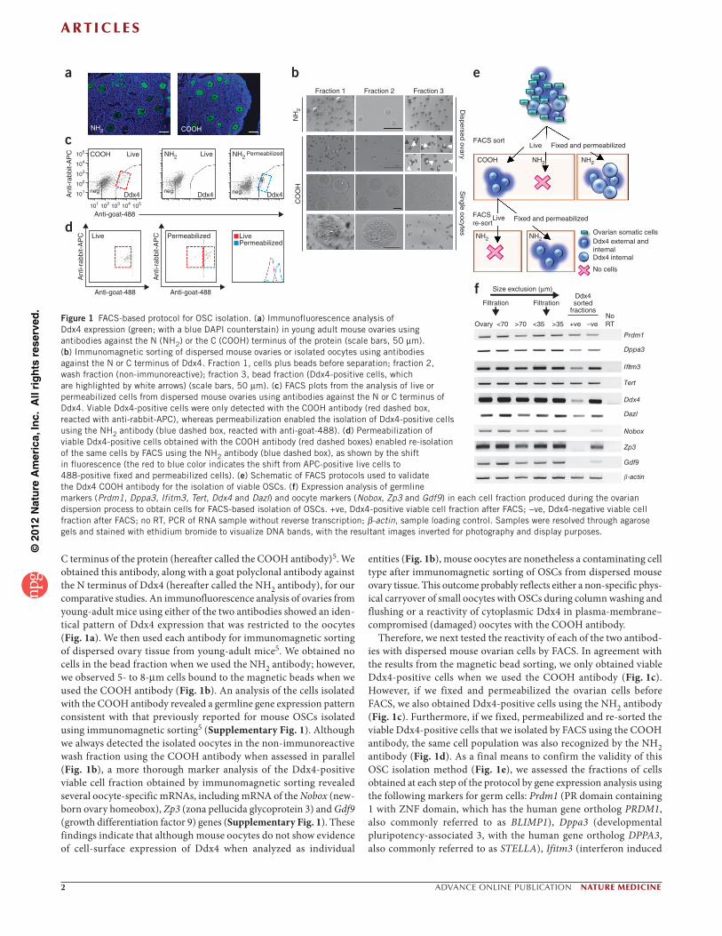
Analyses of freshly isolated Ddx4- and DDX4-positive cells from mouse and human ovaries, respectively, (Fig. 2a,b) revealed similar sizes and morphology between the mouse and human cells (Fig. 2c,d), as well as matched gene expression profiles rich in markers for early germ cells17–19 (the markers Prdm1 and PRDM1, Dppa3 and DPPA3, Ifitm3 and IFITM3, and Tert and TERT for mice and humans, respec-tively; Fig. 2e). These findings agree with the previously reported morphology and gene expression profile of mouse OSCs5,6. To further define characteristic features of Ddx4-positive cells obtained from adult ovaries using FACS, we first performed an in vivo teratoma formation assay. We felt this was a crucial step, as a recent study reported the isolation of stem cells positive for Pou5f1 (POU domain class 5 transcription factor 1) from adult mouse ovaries that pos-sess the teratoma-forming capacity of embryonic stem cells (ESCs) and induced pluripotent stem cells20. All (100%) of the nonobese diabetic (NOD)-severe combined immunodeficient (SCID) mice we transplanted with 1 × 10⁵ mouse ESCs that we used as positive controls (n = 3) developed teratomas within 3 weeks of transplanta-tion; however, we observed no teratomas in NOD-SCID mice that we transplanted in parallel with 1 × 105 Ddx4-positive cells freshly isolated from adult mouse ovaries (n = 3), even at 24 weeks after transplantation (Fig. 2f–h). Thus, although OSCs express numerous stem cell and primitive germ cell markers5,6 (Figs. 1f and 2e), these cells are distinct from the other types of pluripotent stem cells that have previously been described.
We next engineered FACS-purified mouse OSCs to express GFP using retroviral transduction after we established the cells as actively dividing germ-cell-only cultures in vitro (see below). We then used these cells to test the ability of mouse OSCs to generate oocytes after
Figure 2 Isolation of OSCs from adult mouse and human ovaries. (a,b) Representative histological appearance of adult ovarian tissue used for the isolation of human (a) and mouse (b) OSCs. Scale bars, 100 m. (c,d) Morphology of viable cells isolated by FACS based on cell-surface expression of DDX4 (human, left) or Ddx4 (mouse, right). Scale bars, 10 m; insets show a twofold higher magnification of individual OSCs. (e) Gene expression profile of the starting ovarian material and freshly isolated OSCs showing an assessment of three different subjects as examples for the human ovarian tissue analyses. Samples were resolved through agarose gels and stained with ethidium bromide to visualize DNA bands, with the resultant images inverted for photography and display purposes. (f,g) Teratoma formation assay showing an absence of tumors in NOD-SCID mice 24 weeks after receiving injections of freshly isolated mouse OSCs (f) compared with development of tumors in mice 3 weeks after the injection of an equivalent number of mouse ESCs (g; with the tumor highlighted by a black-dashed oval). (h) Examples of cells from all three germ layers in a representative ESC-derived teratoma, with a neural rosette highlighted at the same magnification in the inset of the left panel (scale bars, 100 m).
a
c
e
dg
h
b f
Mouse Human ovary Human OSCs
1 2 3 1 2 3Ovary OSCsNoRT
NoRT
Prdm1/PRDM1
Dppa3/DPPA3
Ifitm3/IFITM3
Tert/TERT
Ectoderm Endoderm Mesoderm-actin

©20
12 N
atur
e A
mer
ica,
Inc.
All
righ
ts r
eser
ved.
A R T I C L E S
4 ADVANCE ONLINE PUBLICATION NATURE MEDICINE
transplantation into the ovaries of adult female mice. To ensure that the outcomes we obtained were reflective of the stable integration of the transplanted cells into the ovaries and also that the outcomes were not complicated by pre-transplantation–induced damage to the gonads, we injected GFP-expressing mouse OSCs into ovaries of non-chemotherapy–conditioned wild-type recipient mice at 2 months of age, and we maintained the mice for 5–6 months before analysis. At between 7 and 8 months of age, we induced the transplanted mice to ovulate using exogenous gonadotropins, after which we collected their ovaries and any oocytes released into the oviducts. We readily detected developing follicles containing GFP-positive oocytes, along with host follicles containing GFP-negative oocytes, in ovaries of all female mice that received GFP-expressing mouse OSCs that were initially purified by FACS (Fig. 3a,b).
After oviductal flushing, we observed complexes containing expanded cumulus cells surrounding centrally located oocytes both lacking and expressing GFP. Mixing these complexes with sperm from wild-type male mice resulted in fertilization and the development of preimplantation embryos, with those embryos that were derived from fertilized GFP-positive eggs retaining GFP expression through the hatching blastocyst stage (Fig. 3c–e). From the five adult wild-type female mice transplanted with GFP-expressing OSCs 5–6 months before evaluation, we retrieved a total of 31 cumulus-oocyte com-plexes from the oviducts, 23 of which successfully fertilized to produce embryos. The presence of cumulus cells around each oocyte made it impossible for us to accurately determine the number of GFP-negative as compared to GFP-positive oocytes ovulated. However, an evaluation of the 23 embryos produced after in vitro fertilization (IVF) revealed that eight were GFP positive, with all five mice tested releasing at least one egg at ovulation that fertilized to produce a GFP-positive embryo. These findings indicate that mouse OSCs purified by FACS using the DDX4 COOH antibody, like their previously reported counterparts isolated from mouse ovaries by immunomagnetic sorting using the same antibody5, generate functional oocytes in vivo. However, our data also show that chemotherapy conditioning before transplanta-tion is not, as previously reported5, required for OSCs to engraft and generate functional oocytes in adult mouse ovary tissue.
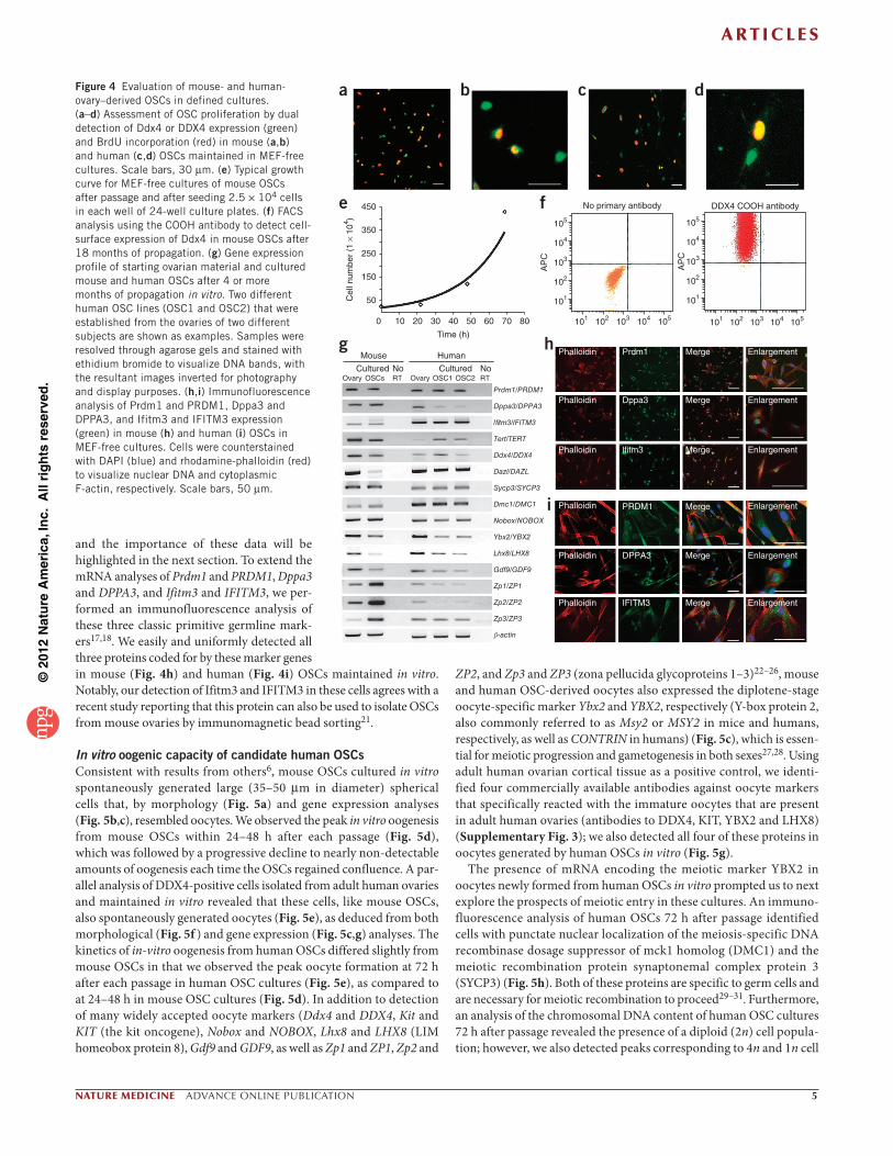
In vitro characterization of candidate human OSCsUsing parameters described previously for the in vitro propagation of mouse OSCs5, we placed adult mouse-ovary–derived Ddx4- positive cells and adult human-ovary–derived DDX4-positive cells into defined cultures with mitotically inactive mouse embryonic fibro-blasts (MEFs) as feeders. We could establish freshly isolated OSCs as clonal cell lines, and the colony formation efficiency for human OSCs not seeded onto MEFs ranged from 0.18–0.40%. We could not per-form an accurate assessment of colony formation efficiency by OSCs using MEFs as initial feeders, the latter of which greatly facilitate the establishment of mouse and human OSCs in vitro. After 10–12 weeks (mouse) or 4–8 weeks (human) in culture, actively dividing germ-cell colonies became readily apparent (Supplementary Fig. 2). Once the OSCs were established and proliferating, the cells could be reestab-lished as germ-cell-only cultures in the absence of MEFs without loss of proliferative potential. A dual analysis of Ddx4 or DDX4 expression and bromodeoxyuridine (BrdU) incorporation in MEF-free cultures of mouse or human OSCs, respectively, revealed large numbers of cells double positive for Ddx4 (mouse) or DDX4 (human) and BrdU (Fig. 4a–d). These data confirmed that the adult mouse- and human-ovary-derived OSCs were actively dividing. At this stage, the mouse cells required passage at confluence every 4–5 d, with the cultures being split 1:6 or 1:8 (with an estimated doubling time of 14 h; Fig. 4e). The rate of mouse OSC proliferation was approximately two-fold to threefold higher than that of the human OSCs maintained in parallel, the latter of which required passage at confluence every 7 d, with cultures being split at either 1:3 or 1:4.
Cell-surface expression of Ddx4 was detectable on the surface of more than 95% of the OSCs after 18 months of propagation (Fig. 4f). The remaining cells not positive for cell-surface expression of Ddx4 after FACS using the COOH antibody were large (up to 35 m in diameter, with any cells >35 m excluded prior to FACS by filtra-tion) spherical cells spontaneously produced by OSCs during culture, which showed cytoplasmic expression of Ddx4 and will be described in detail in the next section. A gene expression analysis of the cultured cells confirmed maintenance of early germline markers (Fig. 4g). We also detected several oocyte-specific markers in these cultures,
Figure 3 Mouse OSCs generate functional eggs after intraovarian transplantation. (a,b) Examples of growing follicles containing GFP-negative (a) and GFP-positive (b) (brown; with a blue hematoxylin counterstain) oocytes in ovaries of wild-type mice injected with GFP-expressing OSCs 5–6 months before analysis. (c) Examples of ovulated GFP-negative eggs (in cumulus-oocyte complexes) and the resultant embryos (embryos at the two-cell (2-cell), four-cell (4-cell), and blastocyst (B) stages are shown as examples) generated by IVF after induced ovulation of wild-type female mice that received intraovarian transplantation of GFP-expressing OSCs 5–6 months before analysis. Scale bars, 30 m. (d,e) Examples of GFP-positive eggs (in cumulus-oocyte complexes) obtained from the oviducts after induced ovulation of wild-type female mice that received intraovarian transplantation of GFP-expressing OSCs 5–6 months before analysis. These eggs were fertilized in vitro using wild-type sperm, resulting in two-cell embryos that progressed through preimplantation development (examples of GFP-positive embryos at the two-cell, four-cell, eight-cell (8-cell), compacted morula (CM), expanded morula (EM), blastocyst and hatching blastocyst (HB) stages are shown) to form hatching blastocysts 5–6 d after fertilization. Scale bars, 30 m.
a
c
d
e
bGFP-negative oocytes GFP-positive oocytes
Egg
Egg
Egg Egg
Egg 2-cell
2-cell
2-cell 2-cell 8-cell CM
2-cell 4-cell
4-cell
B
HBBEM
©20
12 N
atur
e A
mer
ica,
Inc.
All
righ
ts r
eser
ved.
A R T I C L E S
NATURE MEDICINE ADVANCE ONLINE PUBLICATION 5
and the importance of these data will be highlighted in the next section. To extend the mRNA analyses of Prdm1 and PRDM1, Dppa3 and DPPA3, and Ifitm3 and IFITM3, we per-formed an immunofluorescence analysis of these three classic primitive germline mark-ers17,18. We easily and uniformly detected all three proteins coded for by these marker genes in mouse (Fig. 4h) and human (Fig. 4i) OSCs maintained in vitro. Notably, our detection of Ifitm3 and IFITM3 in these cells agrees with a recent study reporting that this protein can also be used to isolate OSCs from mouse ovaries by immunomagnetic bead sorting21.
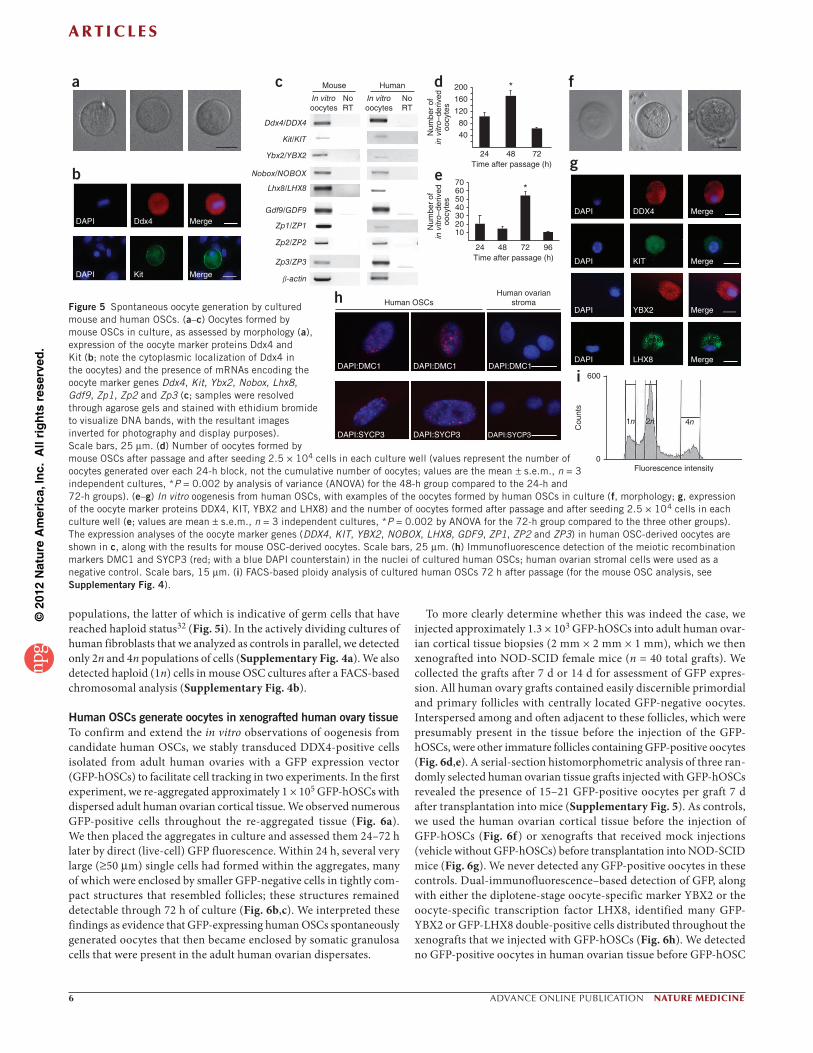
In vitro oogenic capacity of candidate human OSCsConsistent with results from others6, mouse OSCs cultured in vitro spontaneously generated large (35–50 m in diameter) spherical cells that, by morphology (Fig. 5a) and gene expression analyses (Fig. 5b,c), resembled oocytes. We observed the peak in vitro oogenesis from mouse OSCs within 24–48 h after each passage (Fig. 5d), which was followed by a progressive decline to nearly non-detectable amounts of oogenesis each time the OSCs regained confluence. A par-allel analysis of DDX4-positive cells isolated from adult human ovaries and maintained in vitro revealed that these cells, like mouse OSCs, also spontaneously generated oocytes (Fig. 5e), as deduced from both morphological (Fig. 5f ) and gene expression (Fig. 5c,g) analyses. The kinetics of in-vitro oogenesis from human OSCs differed slightly from mouse OSCs in that we observed the peak oocyte formation at 72 h after each passage in human OSC cultures (Fig. 5e), as compared to at 24–48 h in mouse OSC cultures (Fig. 5d). In addition to detection of many widely accepted oocyte markers (Ddx4 and DDX4, Kit and KIT (the kit oncogene), Nobox and NOBOX, Lhx8 and LHX8 (LIM homeobox protein 8), Gdf9 and GDF9, as well as Zp1 and ZP1, Zp2 and
ZP2, and Zp3 and ZP3 (zona pellucida glycoproteins 1–3)22–26, mouse and human OSC-derived oocytes also expressed the diplotene-stage oocyte-specific marker Ybx2 and YBX2, respectively (Y-box protein 2, also commonly referred to as Msy2 or MSY2 in mice and humans, respectively, as well as CONTRIN in humans) (Fig. 5c), which is essen-tial for meiotic progression and gametogenesis in both sexes27,28. Using adult human ovarian cortical tissue as a positive control, we identi-fied four commercially available antibodies against oocyte markers that specifically reacted with the immature oocytes that are present in adult human ovaries (antibodies to DDX4, KIT, YBX2 and LHX8) (Supplementary Fig. 3); we also detected all four of these proteins in oocytes generated by human OSCs in vitro (Fig. 5g).
The presence of mRNA encoding the meiotic marker YBX2 in oocytes newly formed from human OSCs in vitro prompted us to next explore the prospects of meiotic entry in these cultures. An immuno-fluorescence analysis of human OSCs 72 h after passage identified cells with punctate nuclear localization of the meiosis-specific DNA recombinase dosage suppressor of mck1 homolog (DMC1) and the meiotic recombination protein synaptonemal complex protein 3 (SYCP3) (Fig. 5h). Both of these proteins are specific to germ cells and are necessary for meiotic recombination to proceed29–31. Furthermore, an analysis of the chromosomal DNA content of human OSC cultures 72 h after passage revealed the presence of a diploid (2n) cell popula-tion; however, we also detected peaks corresponding to 4n and 1n cell
a
e 450 No primary antibody DDX4 COOH antibody
105
105
104
104
103
103
102
102
101
105
104
103
102
101
101 105104103102101
Cel
l num
ber
(1 ×
104 )
AP
C
AP
C
f
h
i
g
b c d
350
250
150
50
0 10 20 30 40
Time (h)
50 60 70
Mouse
Ovary OvaryOSCs OSC2OSC1RT RT
HumanCulturedCultured No No
80
Phalloidin
Phalloidin
Phalloidin
Phalloidin
Phalloidin
Phalloidin
Prdm1
Dppa3
l!tm3
PRDM1
DPPA3
IFITM3
Merge
Merge
Merge
Merge
Merge
Merge
Enlargement
Enlargement
Enlargement
Enlargement
Enlargement
Enlargement
Figure 4 Evaluation of mouse- and human-ovary–derived OSCs in defined cultures. (a–d) Assessment of OSC proliferation by dual detection of Ddx4 or DDX4 expression (green) and BrdU incorporation (red) in mouse (a,b) and human (c,d) OSCs maintained in MEF-free cultures. Scale bars, 30 m. (e) Typical growth curve for MEF-free cultures of mouse OSCs after passage and after seeding 2.5 × 10⁴ cells in each well of 24-well culture plates. (f) FACS analysis using the COOH antibody to detect cell-surface expression of Ddx4 in mouse OSCs after 18 months of propagation. (g) Gene expression profile of starting ovarian material and cultured mouse and human OSCs after 4 or more months of propagation in vitro. Two different human OSC lines (OSC1 and OSC2) that were established from the ovaries of two different subjects are shown as examples. Samples were resolved through agarose gels and stained with ethidium bromide to visualize DNA bands, with the resultant images inverted for photography and display purposes. (h,i) Immunofluorescence analysis of Prdm1 and PRDM1, Dppa3 and DPPA3, and Ifitm3 and IFITM3 expression (green) in mouse (h) and human (i) OSCs in MEF-free cultures. Cells were counterstained with DAPI (blue) and rhodamine-phalloidin (red) to visualize nuclear DNA and cytoplasmic F-actin, respectively. Scale bars, 50 m.
©20
12 N
atur
e A
mer
ica,
Inc.
All
righ
ts r
eser
ved.
A R T I C L E S
6 ADVANCE ONLINE PUBLICATION NATURE MEDICINE
populations, the latter of which is indicative of germ cells that have reached haploid status32 (Fig. 5i). In the actively dividing cultures of human fibroblasts that we analyzed as controls in parallel, we detected only 2n and 4n populations of cells (Supplementary Fig. 4a). We also detected haploid (1n) cells in mouse OSC cultures after a FACS-based chromosomal analysis (Supplementary Fig. 4b).
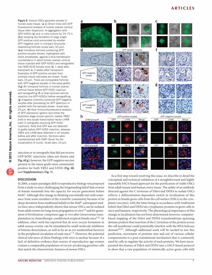
Human OSCs generate oocytes in xenografted human ovary tissueTo confirm and extend the in vitro observations of oogenesis from candidate human OSCs, we stably transduced DDX4-positive cells isolated from adult human ovaries with a GFP expression vector (GFP-hOSCs) to facilitate cell tracking in two experiments. In the first experiment, we re-aggregated approximately 1 × 10⁵ GFP-hOSCs with dispersed adult human ovarian cortical tissue. We observed numerous GFP-positive cells throughout the re-aggregated tissue (Fig. 6a). We then placed the aggregates in culture and assessed them 24–72 h later by direct (live-cell) GFP fluorescence. Within 24 h, several very large ( 50 m) single cells had formed within the aggregates, many of which were enclosed by smaller GFP-negative cells in tightly com-pact structures that resembled follicles; these structures remained detectable through 72 h of culture (Fig. 6b,c). We interpreted these findings as evidence that GFP-expressing human OSCs spontaneously generated oocytes that then became enclosed by somatic granulosa cells that were present in the adult human ovarian dispersates.
To more clearly determine whether this was indeed the case, we injected approximately 1.3 × 10³ GFP-hOSCs into adult human ovar-ian cortical tissue biopsies (2 mm × 2 mm × 1 mm), which we then xenografted into NOD-SCID female mice (n = 40 total grafts). We collected the grafts after 7 d or 14 d for assessment of GFP expres-sion. All human ovary grafts contained easily discernible primordial and primary follicles with centrally located GFP-negative oocytes. Interspersed among and often adjacent to these follicles, which were presumably present in the tissue before the injection of the GFP-hOSCs, were other immature follicles containing GFP-positive oocytes (Fig. 6d,e). A serial-section histomorphometric analysis of three ran-domly selected human ovarian tissue grafts injected with GFP-hOSCs revealed the presence of 15–21 GFP-positive oocytes per graft 7 d after transplantation into mice (Supplementary Fig. 5). As controls, we used the human ovarian cortical tissue before the injection of GFP-hOSCs (Fig. 6f ) or xenografts that received mock injections (vehicle without GFP-hOSCs) before transplantation into NOD-SCID mice (Fig. 6g). We never detected any GFP-positive oocytes in these controls. Dual-immunofluorescence–based detection of GFP, along with either the diplotene-stage oocyte-specific marker YBX2 or the oocyte-specific transcription factor LHX8, identified many GFP-YBX2 or GFP-LHX8 double-positive cells distributed throughout the xenografts that we injected with GFP-hOSCs (Fig. 6h). We detected no GFP-positive oocytes in human ovarian tissue before GFP-hOSC
DAPI
a
b
f
g
c d
e
DAPI
DAPI
DAPI
DAPI
DAPI
Ddx4DDX4
YBX2
LHX8
Kit
Mouse Human
In vitrooocytes
NoRT
Ddx4/DDX4
Kit/KIT
Ybx2/YBX2
Nobox/NOBOX
Lhx8/LHX8
Gdf9/GDF9
Zp1/ZP1
Zp2/ZP2
Zp3/ZP3
-actin
NoRT
In vitrooocytes
KIT
MergeMerge
Merge
Merge
Merge
Merge
200 *1601208040
24 48
70*
72
24 48 72 96
60
Time after passage (h)
Time after passage (h)
Num
ber
ofin
vitr
o–de
rived
oocy
tes
Num
ber
ofin
vitr
o–de
rived
oocy
tes 50
40302010
i 600
0
1n 2n 4n
Fluorescence intensity
Cou
nts
h
DAPI:SYCP3
DAPI:DMC1 DAPI:DMC1
Human OSCsHuman ovarian
stroma
DAPI:DMC1
DAPI:SYCP3 DAPI:SYCP3
Figure 5 Spontaneous oocyte generation by cultured mouse and human OSCs. (a–c) Oocytes formed by mouse OSCs in culture, as assessed by morphology (a), expression of the oocyte marker proteins Ddx4 and Kit (b; note the cytoplasmic localization of Ddx4 in the oocytes) and the presence of mRNAs encoding the oocyte marker genes Ddx4, Kit, Ybx2, Nobox, Lhx8, Gdf9, Zp1, Zp2 and Zp3 (c; samples were resolved through agarose gels and stained with ethidium bromide to visualize DNA bands, with the resultant images inverted for photography and display purposes). Scale bars, 25 m. (d) Number of oocytes formed by mouse OSCs after passage and after seeding 2.5 × 10⁴ cells in each culture well (values represent the number of oocytes generated over each 24-h block, not the cumulative number of oocytes; values are the mean s.e.m., n = 3 independent cultures, *P = 0.002 by analysis of variance (ANOVA) for the 48-h group compared to the 24-h and 72-h groups). (e–g) In vitro oogenesis from human OSCs, with examples of the oocytes formed by human OSCs in culture (f, morphology; g, expression of the oocyte marker proteins DDX4, KIT, YBX2 and LHX8) and the number of oocytes formed after passage and after seeding 2.5 × 10⁴ cells in each culture well (e; values are mean s.e.m., n = 3 independent cultures, *P = 0.002 by ANOVA for the 72-h group compared to the three other groups). The expression analyses of the oocyte marker genes (DDX4, KIT, YBX2, NOBOX, LHX8, GDF9, ZP1, ZP2 and ZP3) in human OSC-derived oocytes are shown in c, along with the results for mouse OSC-derived oocytes. Scale bars, 25 m. (h) Immunofluorescence detection of the meiotic recombination markers DMC1 and SYCP3 (red; with a blue DAPI counterstain) in the nuclei of cultured human OSCs; human ovarian stromal cells were used as a negative control. Scale bars, 15 m. (i) FACS-based ploidy analysis of cultured human OSCs 72 h after passage (for the mouse OSC analysis, see Supplementary Fig. 4).
©20
12 N
atur
e A
mer
ica,
Inc.
All
righ
ts r
eser
ved.
A R T I C L E S
NATURE MEDICINE ADVANCE ONLINE PUBLICATION 7
injection or in xenografts that did not receive GFP-hOSC injections (data not shown and Fig. 6f,g); however, the GFP-negative oocytes present in the tissue grafts were consistently positive for both YBX2 and LHX8 (Fig. 6h and Supplementary Fig. 3).
DISCUSSIONIn 2004, a major paradigm shift in reproductive biology was proposed from a study in mice challenging the longstanding belief that ovaries of female mammals lose the capacity for oocyte generation before birth2. Although this change in thinking was initially met with resist-ance from some members of the scientific community because of its sharp deviation from traditional beliefs in the field4, subsequent stud-ies have since independently shown that mouse OSCs can be isolated from adult ovaries for long-term propagation in vitro5,6 and for gener-ation of fertilization-competent eggs in vivo after intraovarian trans-plantation in chemotherapy-conditioned recipient female mice5,21. In addition, other work has reported that de novo oocyte formation in adult mouse ovaries can be stimulated by small-molecule inhibitors of histone deacetylases, as well as by an as-yet unidentified factor(s) in the peripheral circulation of male mice7–9. However, the potential clinical relevance of these findings with mice is unclear because of a lack of definitive evidence that ovaries of reproductive-age women contain a comparable population of oocyte-producing germline cells that match the characteristic features of mouse OSCs.
As a first step toward resolving this issue, we describe in detail the conceptual and technical validation of a straightforward and highly repeatable FACS-based approach for the purification of viable OSCs from adult mouse and human ovary tissue. The utility of an antibody directed against the C terminus of Ddx4 and DDX4 to isolate OSCs reflects a differentiation-dependent switch in localization of this protein in female germ cells from the cell surface (OSCs) to the cyto-plasm (oocytes), with the latter being in accordance with traditional beliefs that Ddx4 and DDX4 are cytoplasmic proteins in germ cells in mice and humans, respectively. The physiological importance of this change in localization has not been determined; however, computer-based mapping of the Ddx4 and DDX4 transmembrane-spanning domain predicts that insertion of the C terminus of the protein across the cell membrane could potentially interfere with the RNA helicase domain10,33. Although additional work will be needed to test this prediction, movement of proteins into and out of various cellular compartments is a post-translational mechanism that is commonly used by cells to regulate the activity of such proteins. We have incor-porated this feature of Ddx4 and DDX4 into a FACS-based protocol to show that a rare population of mitotically active germ cells with
a
d f
ge
h
b c
GFP-positive oocytes
GFP-positive oocytesGFP
GFP
GFP
GFP
GFP-negativeoocyte
GFP-negativeoocyte
GFP-negative oocytesLHX8 pre-graft
YBX2 pre-graft
LHX8
YBX2 YBX2
LHX8Merge
Merge
Merge
Merge
Figure 6 Human OSCs generate oocytes in human ovary tissue. (a–c) Direct (live-cell) GFP fluorescence analysis of human ovarian cortical tissue after dispersion, re-aggregation with GFP-hOSCs (a) and in vitro culture for 24–72 h (b,c) showing the formation of large single GFP-positive cells surrounded by smaller GFP-negative cells in compact structures resembling follicles (scale bars, 50 m). (d,e) Immature follicles containing GFP- positive oocytes (brown, highlighted with black arrowheads, against a blue hematoxylin counterstain) in adult human ovarian cortical tissue injected with GFP-hOSCs and xenografted into NOD-SCID female mice (d, 1 week after transplant; e, 2 weeks after transplant). Examples of GFP-positive oocytes from multiple tissue replicates are shown. Scale bars, 25 m. There are comparable follicles with GFP-negative oocytes in the same grafts. (f,g) All immature follicles in human ovarian cortical tissue before GFP-hOSC injection and xenografting (f) or that received vehicle injection (no GFP-hOSCs) before xenografting (g) (negative controls) contained GFP-negative oocytes after processing for GFP detection in parallel with the samples shown. Scale bars, 25 m. (h) Dual immunofluorescence analysis of GFP expression (green) and either the diplotene-stage oocyte-specific marker YBX2 (red) or the oocyte transcription factor LHX8 (red) in xenografts receiving GFP-hOSC injections. Note that GFP was not detected in grafts before GFP-hOSC injection, whereas YBX2 and LHX8 were detected in all oocytes before and after injection. Sections were counterstained with DAPI (blue) for the visualization of nuclei. Scale bars, 25 m.
©20
12 N
atur
e A
mer
ica,
Inc.
All
righ
ts r
eser
ved.
A R T I C L E S
8 ADVANCE ONLINE PUBLICATION NATURE MEDICINE
gene expression profiles and growth characteristics that are remark-ably similar to mouse OSCs can be reliably isolated from ovaries of healthy young women and propagated long-term in vitro.
Our successful purification of what seems to be, by all criteria tested, the human equivalent of mouse OSCs was facilitated by two main factors: nearly 3 years of work in our laboratory to test and refine the protocol based on the use of antibodies to Ddx4 and DDX4 for isola-tion of OSCs using FACS rather than immunomagnetic sorting, as was reported initially5, and rare access to entire ovarian cortical tissues that had been vitrified and cryopreserved after the removal of both ovaries from women in their twenties and early thirties. Recent stud-ies of mouse OSCs by others have showed the ability of these cells to generate immature oocytes in vitro6, as well as oocytes that mature into developmentally competent eggs in vivo5. Regarding the latter find-ing, it is notable that prior studies of mammalian germline stem cell transplantation using spermatogonial stem cells or OSCs have been performed with chemotherapy-conditioned hosts as recipients5,21,34,35, presumably to open niches for effective donor-cell engraftment. Our observations show that for OSCs, adult female mice do not require chemotherapy conditioning before transplantation for these cells to effectively engraft in the ovaries and function long term ( 5 months), at least as measured by their ability to generate oocytes that mature in follicles that yield fertilization-competent eggs after ovulation. For ethical and legal reasons, as well as technical feasibility limitations related to a lack of validated protocols for efficiently maturing human primordial follicles to the antral stage in vitro for the isolation of metaphase-II oocytes, our current endpoint analysis of the equivalent cells in humans could not be as comprehensive. Nonetheless, we have established a consistent and close parallelism between human-ovary–derived DDX4-positive cells and mouse OSCs in terms of strategy of purification, yield from adult ovary tissue, morphology, primitive germline gene expression profile, in vitro growth properties, mitotic activity, meiotic activity and the ability to form oocytes in defined cultures in vitro and in injected ovary tissue in vivo.
Therefore, based on the multiple experimental lines of evidence reported herein, we feel it is reasonable to conclude that the rare cells with cell-surface expression of DDX4 that are present in the ovaries of reproductive-age women represent adult human OSCs. In addition to opening a new research field in human reproductive biology that was inconceivable less than 10 years ago, clear evidence for the existence of these cells in women may offer new opportunities to expand on and enhance current fertility-preservation strategies. For example, with assisted reproductive technologies involving cryopreservation of ovar-ian cortical tissue already in development for females with cancer36,37, isolation and expansion of OSCs from this tissue before or after cryo-preservation might be useful for new fertility applications. In fact, we found that these cells can be consistently obtained from cryopreserved and thawed human ovarian tissue samples, and that these cells per se can be cryopreserved and thawed months later with minimal loss for successful establishment in vitro. In addition, the availability of a detailed protocol for the purification of these newly discovered cells from human ovary tissue provides us and others with a much more physiologically relevant in-vitro model system from which to study human female germ cell development compared to the ESC-derived or induced pluripotent stem cell-derived germline cells that are currently used as models for human female gametogenesis38–42.
METHODSMethods and any associated references are available in the online version of the paper at http://www.nature.com/naturemedicine/.
Note: Supplementary information is available on the Nature Medicine website.
ACKNOWLEDGMENTSThe authors thank L. Prickett-Rice and K. Folz-Donahue of the Harvard Stem Cell Institute Flow Cytometry Core Facility and J. Groeneweg for expert technical assistance. We also thank J.R. Mann and K.J. MacLaughlin for the provision of TgOG2 transgenic mice. This work was supported by a Method to Extend Research in Time (MERIT) Award from the US National Institute on Aging (NIH R37-AG012279), the Henry and Vivian Rosenberg Philanthropic Fund, the Sea Breeze Foundation and Vincent Memorial Hospital Research Funds. This work was conducted while D.C.W. was supported in part by a Ruth L. Kirschstein National Research Service Award (NIH F32-AG034809).
AUTHOR CONTRIBUTIONSY.A.R.W., D.C.W. and J.L.T. designed the experiments, analyzed the data and wrote the manuscript. Y.A.R.W. and D.C.W. conducted the experiments. Y.T., O.I. and H.S. collected, cryopreserved and provided human ovarian cortical tissue. J.L.T. directed the project. All authors reviewed and approved the final manuscript for submission.
COMPETING FINANCIAL INTERESTSThe authors declare competing financial interests: details accompany the full-text HTML version of the paper at http://www.nature.com/naturemedicine/.
Published online at http://www.nature.com/naturemedicine/. Reprints and permissions information is available online at http://www.nature.com/reprints/index.html.
1. Zuckerman, S. The number of oocytes in the mature ovary. Recent Prog. Horm. Res. 6, 63–108 (1951).
2. Johnson, J., Canning, J., Kaneko, T., Pru, J.K. & Tilly, J.L. Germline stem cells and follicular renewal in the postnatal mammalian ovary. Nature 428, 145–150 (2004).
3. Brinster, R.L. Male germline stem cells: from mice to men. Science 316, 404–405 (2007).
4. Tilly, J.L., Niikura, Y. & Rueda, B.R. The current status of evidence for and against postnatal oogenesis in mammals: a case of ovarian optimism versus pessimism? Biol. Reprod. 80, 2–12 (2009).
5. Zou, K. et al. Production of offspring from a germline stem cell line derived from neonatal ovaries. Nat. Cell Biol. 11, 631–636 (2009).
6. Pacchiarotti, J. et al. Differentiation potential of germ line stem cells derived from the postnatal mouse ovary. Differentiation 79, 159–170 (2010).
7. Johnson, J. et al. Oocyte generation in adult mammalian ovaries by putative germ cells in bone marrow and peripheral blood. Cell 122, 303–315 (2005).
8. Wang, N. & Tilly, J.L. Epigenetic status determines germ cell meiotic commitment in embryonic and postnatal mammalian gonads. Cell Cycle 9, 339–349 (2010).
9. Niikura, Y., Niikura, T., Wang, N., Satirapod, C. & Tilly, J.L. Systemic signals in aged males exert potent rejuvenating effects on the ovarian follicle reserve in mammalian females. Aging 2, 999–1003 (2010).
10. Tilly, J.L. & Telfer, E.E. Purification of germline stem cells from adult mammalian ovaries: a step closer towards control of the female biological clock? Mol. Hum. Reprod. 15, 393–398 (2009).
11. Niikura, Y., Niikura, T. & Tilly, J.L. Aged mouse ovaries possess rare premeiotic germ cells that can generate oocytes following transplantation into a young host environment. Aging 1, 971–978 (2009).
12. Massasa, E., Costa, X.S. & Taylor, H.S. Failure of the stem cell niche rather than loss of oocyte stem cells in the aging ovary. Aging 2, 1–2 (2010).
13. Fujiwara, Y. et al. Isolation of a DEAD-family protein gene that encodes a murine homolog of Drosophila vasa and its specific expression in germ cell lineage. Proc. Natl. Acad. Sci. USA 91, 12258–12262 (1994).
14. Castrillon, D.H., Quade, B.J., Wang, T.Y., Quigley, C. & Crum, C.P. The human VASA gene is specifically expressed in the germ lineage. Proc. Natl. Acad. Sci. USA 97, 9585–9590 (2000).
15. Noce, T., Okamoto-Ito, S. & Tsunekawa, N. Vasa homolog genes in mammalian germ cell development. Cell Struct. Funct. 26, 131–136 (2001).
16. Normile, D. Reproductive biology. Study suggests a renewable source of eggs and stirs more controversy. Science 324, 320 (2009).
17. Saitou, M., Barton, S.C. & Surani, M.A. A molecular programme for the specification of germ cell fate in mice. Nature 418, 293–300 (2002).
18. Ohinata, Y. et al. Blimp1 is a critical determinant of the germ cell lineage in mice. Nature 436, 207–213 (2005).
19. Dolci, S. et al. Stem cell factor activates telomerase in mouse mitotic spermatogonia and in primordial germ cells. J. Cell Sci. 115, 1643–1649 (2002).
20. Gong, S.P. et al. Embryonic stem cell-like cells established by culture of adult ovarian cells in mice. Fertil. Steril. 93, 2594–2601 (2010).
21. Zou, K., Hou, L., Sun, K., Xie, W. & Wu, J. Improved efficiency of female germline stem cell purification using Fragilis-based magnetic bead sorting. Stem Cells Dev. 20, 2197–2204 (2011).
©20
12 N
atur
e A
mer
ica,
Inc.
All
righ
ts r
eser
ved.
A R T I C L E S
NATURE MEDICINE ADVANCE ONLINE PUBLICATION 9
22. Suzumori, N., Yan, C., Matzuk, M.M. & Rajkovic, A. Nobox is a homeobox-encoding gene preferentially expressed in primordial and growing oocytes. Mech. Dev. 111, 137–141 (2002).
23. Rajkovic, A., Pangas, S.A., Ballow, D., Suzumori, N. & Matzuk, M.M. NOBOX deficiency disrupts early folliculogenesis and oocyte-specific gene expression. Science 305, 1157–1159 (2004).
24. Pangas, S.A. et al. Oogenesis requires germ cell-specific transcriptional regulators Sohlh1 and Lhx8. Proc. Natl. Acad. Sci. USA 103, 8090–8095 (2006).
25. Elvin, J.A., Clark, A.T., Wang, P., Wolfman, N.M. & Matzuk, M.M. Paracrine actions of growth differentiation factor-9 in the mammalian ovary. Mol. Endocrinol. 13, 1035–1048 (1999).
26. Zheng, P. & Dean, J. Oocyte-specific genes affect folliculogenesis, fertilization, and early development. Semin. Reprod. Med. 25, 243–251 (2007).
27. Gu, W. et al. Mammalian male and female germ cells express a germ cell-specific Y-Box protein, MSY2. Biol. Reprod. 59, 1266–1274 (1998).
28. Yang, J. et al. Absence of the DNA-/RNA-binding protein MSY2 results in male and female infertility. Proc. Natl. Acad. Sci. USA 102, 5755–5760 (2005).
29. Page, S.L. & Hawley, R.S. The genetics and molecular biology of the synaptonemal complex. Annu. Rev. Cell Dev. Biol. 20, 525–558 (2004).
30. Yuan, L. et al. Female germ cell aneuploidy and embryo death in mice lacking the meiosis-specific protein SCP3. Science 296, 1115–1118 (2002).
31. Kagawa, W. & Kurumizaka, H. From meiosis to postmeiotic events: uncovering the molecular roles of the meiosis-specific recombinase Dmc1. FEBS J. 277, 590–598 (2010).
32. West, F.D., Mumaw, J.L., Gallegos-Cardenas, A., Young, A. & Stice, S.L. Human haploid cells differentiated from meiotic competent clonal germ cell lines that originated from embryonic stem cells. Stem Cells Dev. 20, 1079–1088 (2011).
33. Abban, G. & Johnson, J. Stem cell support of oogenesis in the human. Hum. Reprod. 24, 2974–2978 (2009).
34. Brinster, R.L. & Zimmermann, J.W. Spermatogenesis following male germ-cell transplantation. Proc. Natl. Acad. Sci. USA 91, 11298–11302 (1994).
35. Brinster, C.J. et al. Restoration of fertility by germ cell transplantation requires effective recipient preparation. Biol. Reprod. 69, 412–420 (2003).
36. Oktay, K. & Karlikaya, G. Ovarian function after transplantation of frozen, banked autologous ovarian tissue. N. Engl. J. Med. 342, 1919 (2000).
37. Sönmezer, M. & Oktay, K. Orthotopic and heterotopic ovarian tissue transplantation. Best Pract. Res. Clin. Obstet. Gynaecol. 24, 113–126 (2010).
38. Hübner, K. et al. Derivation of oocytes from mouse embryonic stem cells. Science 300, 1251–1256 (2003).
39. Ko, K. & Schöler, H.R. Embryonic stem cells as a potential source of gametes. Semin. Reprod. Med. 24, 322–329 (2006).
40. Nicholas, C.R., Haston, K.M., Grewall, A.K., Longacre, T.A. & Reijo Pera, R.A. Transplantation directs oocyte maturation from embryonic stem cells and provides a therapeutic strategy for female infertility. Hum. Mol. Genet. 18, 4376–4389 (2009).
41. Kee, K., Angeles, V.T., Flores, M., Nguyen, H.N. & Reijo Pera, R.A. Human DAZL, DAZ and BOULE genes modulate primordial germ-cell and haploid gamete formation. Nature 462, 222–225 (2009).
42. Nicholas, C.R., Chavez, S.L., Baker, V.L. & Reijo Pera, R.A. Instructing an embryonic stem cell-derived oocyte fate: lessons from endogenous oogenesis. Endocr. Rev. 30, 264–283 (2009).
©20
12 N
atur
e A
mer
ica,
Inc.
All
righ
ts r
eser
ved.
NATURE MEDICINE doi:10.1038/nm.2669
ONLINE METHODSMice. Wild-type C57BL/6 and NOD-SCID mice were from Charles River Laboratories and Jackson Laboratories, respectively. Transgenic mice with germline-specific GFP expression (TgOG2)43–45 were obtained, bred and genotyped as described7,46. All animal protocols were reviewed and approved by the institutional care and use committee of Massachusetts General Hospital.
Human samples. After obtaining written informed consent, ovaries were surgi-cally removed from six female subjects between 22 and 33 years of age with gen-der identity disorder who were receiving sex reassignment at Saitama Medical Center. The outer cortical layer of each ovary was carefully removed, vitrified and cryopreserved47. For our experiments, the cryopreserved tissue was thawed using the Cryotissue Thawing Kit (KITAZATO BioPharma). All experiments with human tissue were conducted without subject identifiers under a proto-col reviewed and approved by the institutional review board of Massachusetts General Hospital (protocol number 2008P001640).
DDX4 antibodies. Polyclonal antibodies against the C (ab13840; Abcam) or N (AF2030; R&D Systems) terminus of DDX4 were from Abcam or R&D Systems, respectively.
Immunomagnetic sorting. Dissociated ovarian cells or isolated oocytes were processed by immunomagnetic sorting using streptavidin-conjugated microbeads (Miltenyi) or secondary-antibody–conjugated Dynabeads (Invitrogen), as described5.
Flow cytometry. Antibody-labeled ovarian cell suspensions were subjected to FACS using a BD Biosciences FACSAria II cytometer (Harvard Stem Cell Institute) gated against negative controls (unstained cells and cell fractions processed without primary antibody).
Teratoma formation. Mouse ESCs (mESC v6.5) or freshly isolated mouse OSCs were injected subcutaneously near the rear haunch of NOD-SCID female mice, which were then monitored weekly for up to 6 months after injection for tumor formation.
OSC cultures. Mouse and human OSCs were established on mitotically inactivated immortalized MEFs and propagated in vitro, essentially as described5. To assess OSC proliferation, MEF-free OSC cultures were treated with 10 M BrdU (Sigma-Aldrich) for 48 h for dual-immunofluorescence–based detection of BrdU incorporation and Ddx4 or DDX4 expression, essentially as described5.
Gene expression. The presence of each indicated mRNA was assessed by con-ventional RT-PCR using the primers listed in Supplementary Tables 1 (mouse) and 2 (human).
Immunoanalyses. Fixed ovarian tissues, OSCs or oocytes were assessed by stand-ard immunohistochemical or immunocytochemical methods using antibodies that recognize Ddx4 and DDX4 (C terminus: ab13840, Abcam; N terminus: AF2030, R&D Systems), Kit and KIT (sc1494, Santa Cruz Biotechnology), Prdm1 and PRDM1 (ab81961, Abcam), Dppa3 and DPPA3 (ab19878, Abcam), Ifitm3 and IFITM3 (mouse: ab15592, human: ab74699, Abcam), DMC1 (ab96613, Abcam), SYCP3 (NB300-232, NOVUS Biologicals), YBX2 (ab33164, Abcam) or LHX8 (ab41519, Abcam).
Ploidy analysis. A chromosomal analysis of mouse and human OSCs was per-formed using a BD Biosciences FACSAria II cytometer, essentially as described32. Human fetal kidney fibroblasts (KEK 293, Invitrogen) were used as control somatic cells.
Generation of GFP-expressing OSCs. One microgram of pBabe-Gfp vector DNA (Addgene plasmid repository #10668) was transfected (Lipofectamine, Invitrogen) into the Platinum-A retroviral packaging cell line (Cell Biolabs). Transduction of OSCs was performed using fresh viral supernatant in the pres-ence of polybrene (Sigma-Aldrich). Mouse and human OSCs with expression of GFP were purified by FACS after an initial 1 week of propagation and then were expanded for 2 additional weeks before a second round of FACS purification to obtain the cells used for the experiments.
Transplantation of mouse OSCs. GFP-expressing mouse OSCs (1 × 10⁴) were injected directly into each ovary of wild-type mice at 2 months of age. Between 7 and 8 months of age, the mice were subjected to an induced ovulation protocol. Cumulus-oocyte complexes were collected from the oviducts and assessed by direct fluorescence microscopy for GFP expression. For IVF, cumulus-oocyte complexes were mixed with wild-type sperm for 4–5 h, washed and transferred to potassium simplex optimized medium with amino acids (Irvine Scientific). Light and fluorescence microscopic examinations were performed every 24 h for a total of 144 h to monitor embryo development48. Ovarian tissue harvested at the time of oocyte collection was processed for immunohistochemical detec-tion of GFP, as detailed46.
Human ovarian tissue re-aggregation. Dissociated human ovarian cortex was incubated with 35 g ml–1 phytohemaglutannin (PHA; Sigma), along with 1 × 10⁵ GFP-expressing human OSCs for 10 min at 37 °C. The cell mix was pelleted to create the tissue aggregate, which was then placed onto a Millicell 0.4- m culture plate insert (Millipore) in OSC culture medium. Aggregates were incubated at 37 °C in 5% CO2 and 95% air, and live-cell GFP imaging was performed 24, 48 and 72 h later.
Intraovarian OSC injection and xenografting. Human ovarian cortical tissue injected with approximately 1.3 × 10³ GFP-expressing human OSCs was grafted into NOD-SCID female mice49,50. Xenografts were removed 7 d or 14 d after transplantation and were processed for immunohistochemical detection of GFP expression46. For the dual marker analysis, we performed immunofluorescence-based detection of GFP and either YBX2 or LHX8 (see above).
Data analyses. All experiments were independently replicated at least three times. Quantitative data (means s.e.m. of combined results) were analyzed by one-way ANOVA using Prism software, whereas the qualitative images presented are representative of outcomes obtained in replicate experiments.
Additional methods. Detailed methodology is described in the Supplementary Methods.
43. Yeom, Y.I. et al. Germline regulatory element of Oct-4 specific for the totipotent cycle of embryonal cells. Development 122, 881–894 (1996).
44. Yoshimizu, T. et al. Germline-specific expression of the Oct-4/green fluorescent protein (GFP) transgene in mice. Dev. Growth Differ. 41, 675–684 (1999).
45. Szabó, P.E. et al. Allele specific expression of imprinted genes in mouse migratory primordial germ cells. Mech. Dev. 115, 157–160 (2002).
46. Lee, H.-J. et al. Bone marrow transplantation generates immature oocytes and rescues long-term fertility in a preclinical mouse model of chemotherapy-induced premature ovarian failure. J. Clin. Oncol. 25, 3198–3204 (2007).
47. Kagawa, N., Silber, S. & Kuwayama, M. Successful vitrification of bovine and human ovarian tissue. Reprod. Biomed. Online 18, 568–577 (2009).
48. Selesniemi, K., Lee, H.-J., Muhlhauser, A. & Tilly, J.L. Prevention of maternal aging-associated oocyte aneuploidy and meiotic spindle defects in mice by dietary and genetic strategies. Proc. Natl. Acad. Sci. USA 108, 12319–12324 (2011).
49. Weissman, A. et al. Preliminary experience with subcutaneous human ovarian cortex transplantation in the NOD-SCID mouse. Biol. Reprod. 60, 1462–1467 (1999).
50. Matikainen, T. et al. Aromatic hydrocarbon receptor-driven Bax gene expression is required for premature ovarian failure caused by biohazardous environmental chemicals. Nat. Genet. 28, 355–360 (2001).