open access textbook of general surgery - vula · barium enema may be useful when planning surgery...
TRANSCRIPT

OPEN ACCESS TEXTBOOK OF GENERAL SURGERY DIVERTICULAR DISEASE OF THE COLON B Baigrie C McGuire EPIDEMIOLOGY AND INCIDENCE Diverticular disease has traditionally been a disease of western society, and is related to a diet deficient in fiber. It is one of the most common conditions in the developed world, but only a minority of patients will ever experience complications. Once considered very rare in sub- Saharan Africa, its rising incidence mirrors the widespread adoption of a western lifestyle. The incidence increases with age, affecting almost one third of Caucasians by the age of 50 and two- thirds by the age of 80. In the Caucasian population the sigmoid is most commonly affected, whereas right sided disease is more common amongst Asians. Previously unknown in black Africans, a few case series have emerged, such as 20 cases in five years in black South Africans and a recent series of 31 cases in 5 years in Kampala, Uganda. The distribution and pattern follows Caucasian patterns of sigmoid and left sided involvement. DEFINITIONS · A diverticulum is a sac-like
protrusion of the colonic wall. · Diverticulosis merely describes the
presence of diverticula. · Diverticulitis refers to inflammation
of diverticula. PATHOPHYSIOLOGY Anatomically there are two types of diverticulae: · True diverticulae, which are sac-
like protrusions consisting of all the layers of the colonic wall
· False or pseudo-diverticulae which are sac-like protrusions consisting of mucosa and submucosa only.
Diverticular disease of the colon refers to pseudo-diverticulae. A single true
diverticulum is sometimes found in the caecum and is a congenital abnormality. Along the length of most of the colon, the outer longitudinal muscle layer is divided into three bands called the taenia coli. One is adjacent to the attachment of the meso-colon, and two are on the anti-mesenteric side. Diverticulae occur at four well defined weak points in the bowel wall where the blood vessels penetrate the muscle layer. They occur on either side of the mesenteric taenia and on the mesenteric side of the anti-mesenteric taeniae (Figure 1). The taenia diffuse into a circumferential layer at the level of the upper rectum, hence the rarity of diverticulosis below the recto sigmoid junction. Diverticulae can occur in the appendix but is exceedingly rare.
Figure 1. Weak points of colonic wall where blood vessels enter. (Sourced from Jones DJ. Diverticular disease. In: Jones DJ, Irving MH, editors. ABC of colorectal diseases. Grat Britain: BMJ publishing group; 1993. p. 47.)

Figure 2. Diverticulitis of the appendix (Sourced from Dr R. Baigrie) An important predisposing factor for diverticular disease is a low fiber diet. Diverticuiae are thought to arise as a pulsion phenomenon secondary to raised intraluminal pressure, which weakens the bowel wall. Patients with low fiber diets have dense, sometimes large stools, which alters colonic motility and produces increased intraluminal pressures leading to the herniation of the mucosa and submucosa through the weak points. This occurs most commonly in the sigmoid colon, and in about half of patients with diverticular disease, it is confined to the sigmoid colon. Muscular hypertrophy, spasm, hyper segmentation and irregular contractions are purported to represent peristaltic attempts to propel the stools in a constipated patient. However, only about half the patients report constipation, possibly implying other aetiological factors. Diverticular disease is not a premalignant condition, but its high incidence results in its frequent co-existence with colon cancer. CLINICAL PRESENTATION Diverticular disease can present in four ways: · Asymptomatic · Symptomatic · Diverticular bleed
· Diverticulitis and its complications In the absence of inflammation (diverticulitis), the term diverticulosis is preferred. Asymptomatic diverticulosis Most people with diverticular disease are asymptomatic, unaware of their condition unless detected incidentally at colonoscopic screening or laparotomy. Symptomatic diverticulosis These patients present with attacks of left iliac fossa pain, which is at times colicky in nature, bloating, flatulence and altered bowel habit. Their symptoms often disappear after defaecation or passage of flatulence. They often present clinically with tenderness in the left iliac fossa with no clinical or laboratory signs of inflammation. The clinical picture of- diverticulosis often overlaps with that of irritable bowel syndrome (IBS) which can co-exist with asymptomatic diverticulosis, leading to diagnostic confusion. Surgery is rarely indicated and a high fiber diet is recommended. However, contrary to popular belief, there is no reason to avoid tomato and other pips for fear of them sticking in a diverticulum. Barium enema has generally been superseded by colonoscopy, but the latter may be technically very difficult and is associated with a small risk of colon rupture.

Figure 3. Barium enema demonstrating multiple sigmoid diverticulae (Sourced from Fry RD, Mahmoud J, Maron DJ, et al. Colon and Rectum. In: Townsend CM, Beachamp RD, Evers BM, Mattox KL, editors. Sabiston textbook of surgery. 18th ed. Canada: Elsevier Inc.; 2008 p. 1364) Increasingly, abdominal CT is used instead of, or to supplement colonoscopy. In some situations, barium enema may be useful when planning surgery as it provides the clearest indication of the extent and severity of disease. DIVERTICULITIS In diverticulitis there is extramural pericolic inflammation and localised peritonitis which sometimes progresses to perforation and abscess formation. The process begins with obstruction of the diverticulum leading to increased intraluminal pressure, inflammation and perforation. This is often caused by a faecolith. (Figure 4)
Figure 4. Obstruction of a diverticulum by a faecolith. (Sourced from Dr R. Baigrie) Abscesses can involve adjacent structures, resulting in fistulae of which colovesical fistulae are the most common. Severe or recurrent inflammation results in fibrosis and ultimately stricturing. Although this is rarely sufficient to cause large bowel obstruction unless associated with
constipation, it can be difficult to distinguish from a malignant stricture. The literature reports a widely varied lifetime risk of acute diverticulitis of between 0.1 — 20 percent. In our experience, the incidence of patients requiring hospitalisation for diverticulitis is less than one percent. |\/lost patients diagnosed with diverticulitis by primary health practitioners have IBS, resulting in a high incidence of inappropriate antibiotic use. Diverticulitis most commonly occurs in the sigmoid colon. These patients usually present with left iliac fossa pain, fever, bloating and leucocytosis. Alteration of bowel habit is usual and constipation or loose stools may result. While urinary symptoms (which are caused by the proximity of the bladder to the inflamed sigmoid colon) may occur, rectal bleeding is rare. Diverticulitis of an isolated diverticulum of the caecum is clinically indistinguishable from appendicitis. The best investigation is CT scan with or without contrast depending on the patient's condition. Barium enema should be used with caution because of the risk of increasing colonic pressure and causing complications. It is also very uncomfortable in the acute setting and clearing out the barium afterwards is problematic. Left sided colonoscopy has similar disadvantages but may be useful if a co-existent malignancy is suspected. All patients should have a diagnostic colonoscopy some time after the acute episode has settled.
Figure 5. Diverticulae as seen during colonoscopy. (Sourced from Dr R. Baigrie)
Diverticulitis may be complicated or uncomplicated. Uncomplicated diverticulitis occurs when an inflammed or perforated diverticulum is walled off by omentum or adjacent structures resulting in localised inflammation. Most patients require hospitalisation and should be placed on antibiotics such as ciprofloxin and metronidazole to cover gram negative bacilli and anaerobes. This commonly results in rapid clinical improvement within 48 — 72 hrs. Antibiotics can be administered orally when tolerated, and continued for 7 — 10 days. Several weeks after the symptoms have subsided, investigations should be conducted to evaluate the extent of diverticulosis and to exclude cancer. Colonoscopy is preferred. Following successful conservative therapy for a first attack of diverticulitis, approximately one-third of patients will remain asymptomatic, one-third will have episodic cramps, and one-third will suffer a second episode of diverticulitis. Historically, the prognosis has been thought to become worse with second and subsequent attacks, resulting in the recommendation of elective surgery after two or three episodes. However, more recent data suggest that patients with recurrent diverticulitis do not have higher morbidity and mortality, and repeated attacks may get increasingly less severe and eventually cease spontaneously. Level l data and grade A recommendations are not available for surgery in this setting, and the decision for elective colectomy should be made on a case to case basis. Complicated diverticulitis occurs in the following circumstances: · The perforation leads to formation
of a localised abscess · Fistula formation to adjacent
organs, usually the bladder · Uncontained perforation leading to
generalised peritonitis
· Colonic stricture causing bowel obstruction
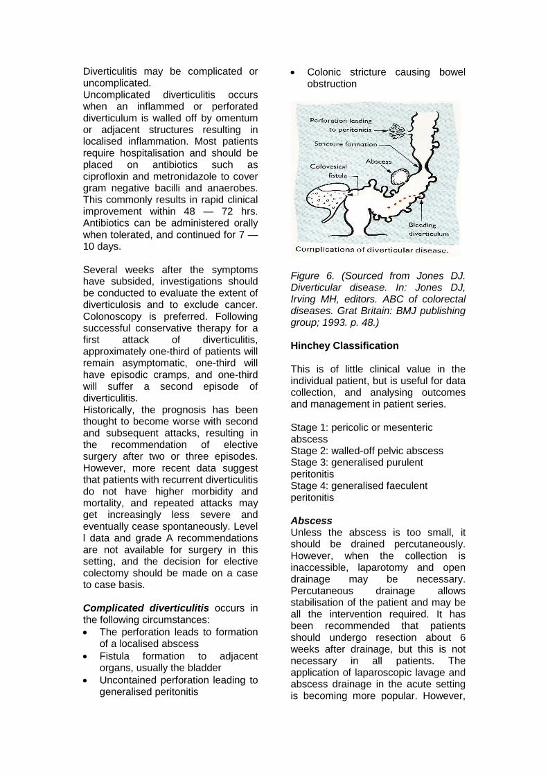
Figure 6. (Sourced from Jones DJ. Diverticular disease. In: Jones DJ, Irving MH, editors. ABC of colorectal diseases. Grat Britain: BMJ publishing group; 1993. p. 48.) Hinchey Classification This is of little clinical value in the individual patient, but is useful for data collection, and analysing outcomes and management in patient series. Stage 1: pericolic or mesenteric abscess Stage 2: walled-off pelvic abscess Stage 3: generalised purulent peritonitis Stage 4: generalised faeculent peritonitis Abscess Unless the abscess is too small, it should be drained percutaneously. However, when the collection is inaccessible, laparotomy and open drainage may be necessary. Percutaneous drainage allows stabilisation of the patient and may be all the intervention required. It has been recommended that patients should undergo resection about 6 weeks after drainage, but this is not necessary in all patients. The application of laparoscopic lavage and abscess drainage in the acute setting is becoming more popular. However,
the presence of frank faecal material is widely regarded as an indication to convert to open surgery. Fistulae These result from an abscess eroding into an adjacent organ. The bladder is most commonly involved and patients present with pyuria, faecaluria, pneumaturia and recurrent urinary tract infections (Figure 7 and 8). Colonoscopy will usually exclude cancer (figure 9) which is also a cause of a colovesical fistula, although the procedure may not be possible as a result of fibrotic distortion. The fistula is excised along with the diseased segment of colon and primary anastomosis is usually appropriate. A large bladder opening may require suture closure, but small openings should be left alone and a bladder catheter left in situ for about 10 days or until a cystogram demonstrates absence of a leak. Omentum should be interposed between the colon and bladder at the time of surgery. Fistulae also occur to the vagina, uterus, small bowel and skin.
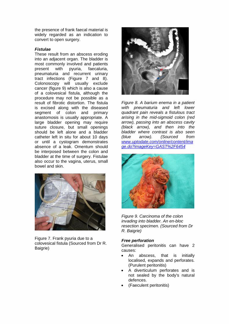
Figure 7. Frank pyuria due to a colovesical fistula (Sourced from Dr R. Baigrie)
Figure 8. A barium enema in a patient with pneumaturia and left lower quadrant pain reveals a fistulous tract arising in the mid-sigmoid colon (red arrow), passing into an abscess cavity (black arrow), and then into the bladder where contrast is also seen (blue arrow). (Sourced from www.uptodate.com/online/content/image.do?imageKey=GAST%2F6454
Figure 9. Carcinoma of the colon invading into bladder. An en-bloc resection specimen. (Sourced from Dr R. Baigrie) Free perforation Generalised peritonitis can have 2 causes: · An abscess, that is initially
localised, expands and perforates. (Purulent peritonitis)
· A diverticulum perforates and is not sealed by the body's natural defences.
· (Faeculent peritonitis)

Colonic perforation is associated with a high mortality rate. Management includes intravenous fluid resuscitation, analgesia and antibiotics. In the case of generalised peritonitis, an urgent laparotomy or laparoscopy is required and it is often not possible to determine whether the perforation is due to cancer or diverticulitis. A Hartmann's procedure is the safest operation, but in the hands of an experienced surgeon resection and primary anastomosis is appropriate in highly selected patients. Colonic obstruction Can occur under 2 circumstances: · ln the acute setting, small bowel
obstruction occurs when small bowel adheres to the infectious process. it is managed with nasogastric drainage if necessary, antibiotics and percutaneous drainage of any abscesses.
· Chronic narrowing of the sigmoid colon results from muscular hypertrophy of the bowel wall or a fibrotic stricture. These rarely cause complete obstruction but can present a diagnostic problem if a contrast study reveals a sigmoid stricture in an area containing numerous diverticulae. A cancer of the sigmoid colon needs to be considered in this scenario.
Diverticular haemorrhage Bleeding results from eccentric rupture of a damaged blood vessel into the lumen, as it passes through the wall of a diverticulum in the submucosal plane.
Figure 10. Blood vessel in a diverticulum separated from the bowel lumen by only mucosa. (Sourced from www.uptodate.com/online/content/image.do?imageKey=GAST%2F6275)
Figure11. Transilluminated diverticulae seen during on-table colonoscopy, demonstrating their thin wall and obvious vasculature. (Sourced from Dr R. Baigrie) Over time, the vessel is exposed to stercoral injury along its luminal aspect, leading to thinning of the media over the dome, or at the neck, of the diverticulum. These changes predispose to rupture. Inflammation and diverticulitis do not predispose to bleeding. Diverticular bleed usually presents with brisk rectal bleeding and is the commonest cause of life threatening lower GI haemorrhage. Approximately 3-5 percent of patients with diverticulosis will present with significant GI blood loss. Bleeding is uncommon under the age of 50, and is rare in patients presenting with acute diverticulitis. It is usually abrupt in onset and painless. Patients may have mild lower abdominal cramps and rectal urgency, followed by the passage of red blood or clots. Bleeding stops spontaneously in 75% of patients. Although 90% of diverticula are in the left colon, bleeding is from the right colon (proximal to the splenic flexure) in at least 50% of patients. This argues strongly against empirical left hemicolectomy or segmental sigmoid colectomy in patients with uncontrolled haemorrhage without an identified source. Initial management is the same as any other gastro-intestinal bleed and consists of resuscitation, localisation of bleeding and

management of the cause. The best method of diagnosis and treatment is colonoscopy after rapid bowel preparation. A bleeding vessel can sometimes be controlled by adrenaline injection, heater probe or other method, but successful endoscopic control is rare. If the source of bleeding cannot be identified, the investigation of choice is CT angiography, improved resolution has resulted in this non-invasive, quicker and simpler investigation replacing mesenteric angiography as the first choice investigation.
Figure 12. CT angiography identifying the site of active diverticular bleeding (Sourced from Dr Derek Solomon)
Figure 13. Bleeding identified during mesenteric angiography (Sourced from Dr Derek Solomon) lt is more sensitive. Successful mesenteric angiography requires a rate of bleeding that is at least 1 ml/min, and in this setting arterial embolisation has become accepted as a safe and effective therapy. This has success rates as high as 70-90 percent. The theoretical risk of
ischaemia seldom results in infarction of the colonic segment, although patients may experience pain, fever and tachycardia, and require very close observation. Technetium-99m tagged red blood cell scan, although more sensitive (bleeding rate 0.1-O.5ml/min) is less reliable and does not allow the therapeutic option. Because the bleeding is arterial in origin, it usually stops abruptly and investigations are invariably non-diagnostic in the stable patient. The challenge is to facilitate emergency endoscopy or angiography in the acutely bleeding patient while continuing resuscitation. Surgical intervention is reserved for failure of medical, angiographic, or endoscopic therapies. lntra-operative colonoscopy may assist in localisation of bleeding. Segmental colonic resection is indicated if the site of bleeding is known, but rebleeding is reported in 15 percent of patients in the first year after angiographic directed resection and 40 percent after negative angiography. Because the right colon is the commonest site of origin in patients with persistent bleeding, a subtotal colectomy with ileorectal anastomosis may be required if angiography or endoscopy does not identify a definite bleeding site. The mortality and morbidity of a subtotal colectomy is significant, especially as these patients are usually elderly with other co-morbidities. However, it stands to reason that if a patient who presents with a second episode of bleeding has a greater than 50% chance of further bleeding, then consideration should be given to elective surgical resection after two episodes of significant self-limited diverticular haemorrhage.
This work is licensed under a Creative Commons Attribution 3.0 Unported License.