optical and biological considerations for confocal microscopy
TRANSCRIPT

Optical and Biological Considerations for Confocal Microscopy
Steven Ruzin Ph.D.CNR Biological Imaging FacilityUniversity of California, Berkeley

Specimen
Excitation filter
Dichroic mirror
A B
Objective
Barrier filter
Arc light source
E p i f l u o r e s c e n c e M i c r o s c o p e

Diffraction and Out-Of-Focus light
d
d = Airy disk (1 Airy unit)
Out-of-focus information

Marvin Minsky’s 1957 Confocal Microscope
Specimen d
Pinhole diameter = d; excludes diffraction rings
Source pinhole

Pinhole Diameter and Confocal slice thickness

Sarastro 1989 Confocal Microscope Design
d
xy
Objective
Scanner
Laser
Lens
Object plane
Detector 2
Dichroic Beam Splitter 1
Focus motor/ “Stepper Motor”
Dichroic Beam Splitter 2
Detector 1Pinhole
d

Ch 1 (32 PMTs)
Ch 2
Ch 3
pinholesDichroics
Sample Plane
Laser
TransmittedLight detector
diffraction grating
Zeiss LSM Meta; Leica SP2, Olympus FV1000
Contemporary Laser scanning Confocal Microscope Design

Fluorescence Emission Properties
DAPI SYTO 11
Stokes Shift

Discriminating Fluorescent Probes
A-488 A-594 A-488 A-546 A-568 A-594
Molecular Probes’ Alexa Dyes
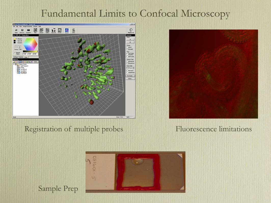
Fundamental Limits to Confocal Microscopy
Registration of multiple probes Fluorescence limitations
Sample Prep
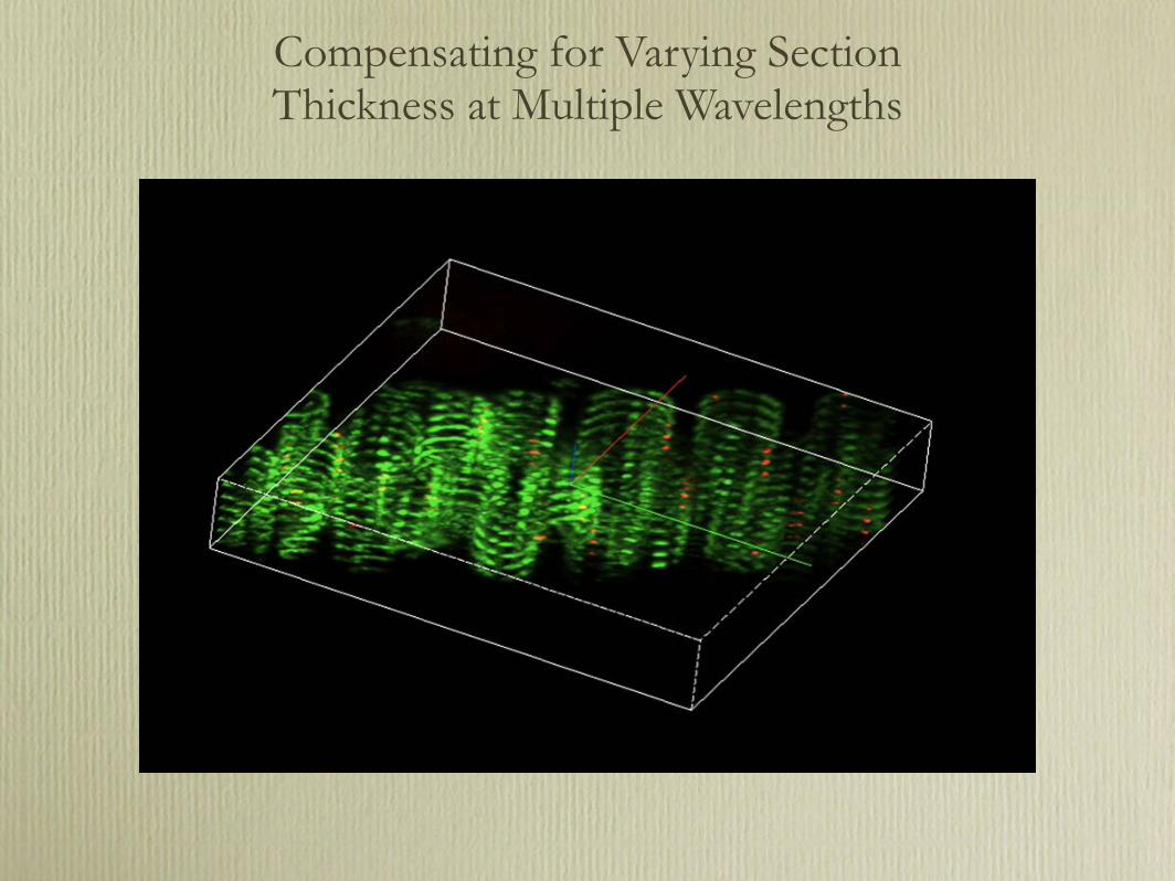
Compensating for Varying Section Thickness at Multiple Wavelengths

Adjusting the Optical Slice Thickness
Barley Aleurone protoplast
Adjust pinholes to normalize optical slice thickness

Pinhole Adjustment yields:• Sections in-register
• Normalized confocal slice thickness
C. elegans Z. mays

The problem of simultaneous UV/Vis excitation
UVVis
Collimator adjustment for separate laser fibers
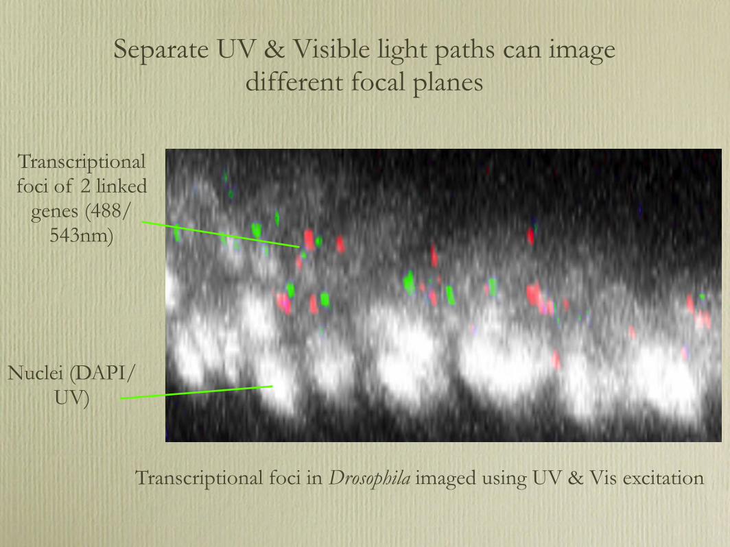
Transcriptional foci in Drosophila imaged using UV & Vis excitation
Nuclei (DAPI/ UV)
Transcriptional foci of 2 linked
genes (488/543nm)
Separate UV & Visible light paths can image different focal planes

UV/Vis Ex misalignment

UV-excited and Vis-excited probes imaged to the same plane
BacteriaDAPI and Autofluorescence
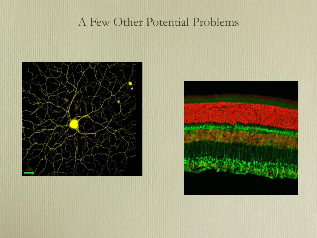
A Few Other Potential Problems

Standard coverslip designations
#1 = 130–170µm#1.5 = 160–190µm#2 = 170–250µm
Variation in Coverslip Thickness

Objectives are most efficient when the correct coverslip is used
Barley
Mismatched coverslip thickness results in decreased energy, reduced depth resolution, and axial shift in focus

Autofluorescence
Z. mays
Insect leg

Autofluorescence
Ex 488Em 505–550
Ex543Em LP560
??Paraffin-embedded
Ex 488, 543Em LP560
Aldehyde-inducedLung, H&E
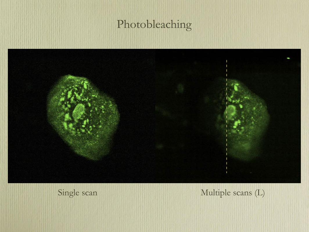
Photobleaching
Single scan Multiple scans (L)
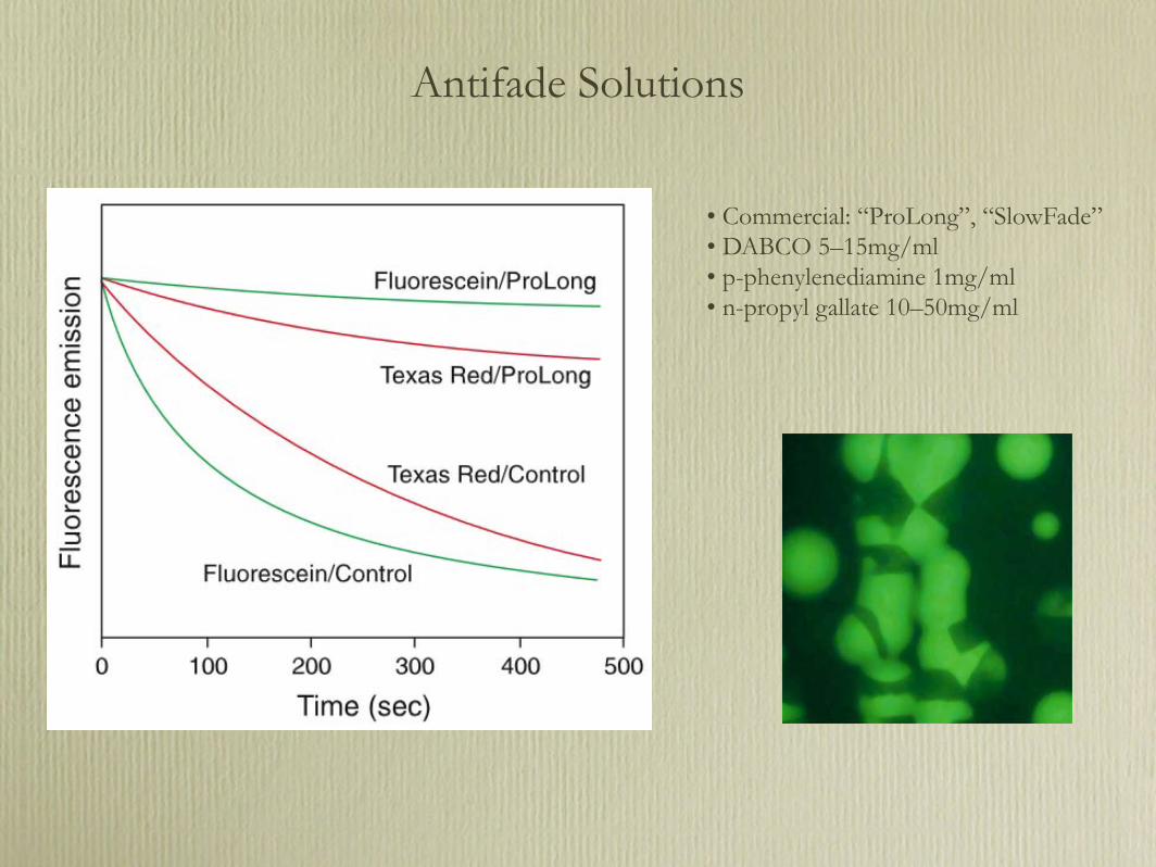
Antifade Solutions
• Commercial: “ProLong”, “SlowFade”• DABCO 5–15mg/ml• p-phenylenediamine 1mg/ml• n-propyl gallate 10–50mg/ml

Traditional Fluorescent Probes
HOOC CH2CH2
HOOC
COOH
OHO
CH2CH2 COOH
O
BCECF
Mg2
C
OCH3OC20H39
CH3
I II
IV III
VHH
OOC O
H
NN
Chlorophyll
NH2
H2NC
C
NH2
NH2NH
DAPI
OCH3C
O O
O
O
CO CH3O
FDA
H2N
NH2
H3C
CH3CH3
N
I-
N
PI
+ +
I-
H2N NH2
COOCH3
Rhodamine 123
CH3OOC CH2OOC CH2CH2
HOOC
COOH
OHO
CH2CH2 COOCH23COOCH3
O
BCECF-AM

Contemporary Fluorescent Probes: Alexa Dyes
Alexa 488O
C
NH2H2N O
SO3SO3
OH
O
O
- -
+
-

Environmental Sensitivity: pH

Sample Preparation
• Mountant contains glycerol and antifade solution• Seal coverslip edges well• Remove “old” immersion oil before re-viewing• Refreezing is not recommended
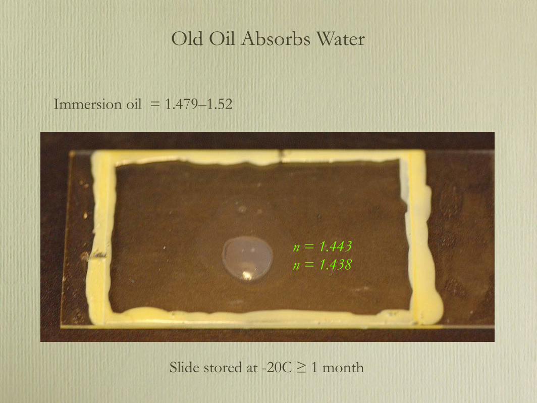
Old Oil Absorbs Water
n = 1.443n = 1.438
Immersion oil = 1.479–1.52
Slide stored at -20C ≥ 1 month

Sampling Techniques

Nyquist Sampling
128x128, 4x Average 512x512, 4x Average
Rate ≥ 2f(highest)

Averaging pixel intensity can increase signal-to-noise
Pixel Averaging

The Color Lookup Table“Let me just take a look at it.”

Imaging System Assigns Colors Based on Assumptions

Thank you
Giardia