optical biometer al-scan - ocusoft.deocusoft.de/ulib/ndk/alscan/al_scan_6p_2_k.pdf · the al-scan...
TRANSCRIPT
AL-Scan Optical Biometer
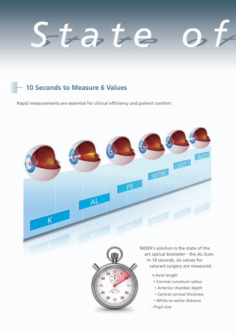
S t a t e o f 10 Seconds to Measure 6 Values
Rapid measurements are essential for clinical efficiency and patient comfort.
NIDEK’s solution is the state of the art optical biometer - the AL-Scan. In 10 seconds, six values for cataract surgery are measured:
• Axial length
• Corneal curvature radius
• Anterior chamber depth
• Central corneal thickness
• White-to-white distance
• Pupil size
T h e A r tf t h e A r tt h e A r t
Y direction
Z direction X direction
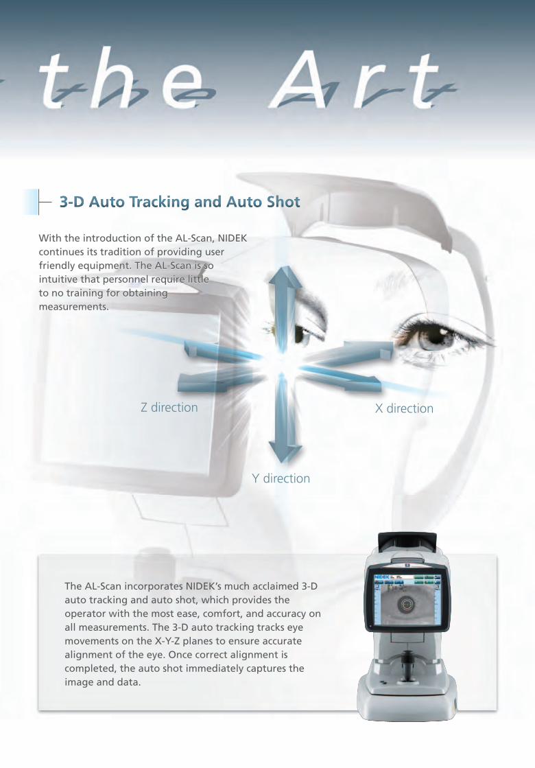
3-D Auto Tracking and Auto Shot
With the introduction of the AL-Scan, NIDEK With the introduction of the AL-Scan, NIDEK continues its tradition of providing user continues its tradition of providing user friendly equipment. The AL-Scan is so friendly equipment. The AL-Scan is so intuitive intuitive that personnel require little that personnel require little to no training for obtaining to no training for obtaining measurements.measurements.
With the introduction of the AL-Scan, NIDEK continues its tradition of providing user friendly equipment. The AL-Scan is so intuitive that personnel require little to no training for obtaining measurements.
The AL-Scan incorporates NIDEK’s much acclaimed 3-D auto tracking and auto shot, which provides the operator with the most ease, comfort, and accuracy on all measurements. The 3-D auto tracking tracks eye movements on the X-Y-Z planes to ensure accurate alignment of the eye. Once correct alignment is completed, the auto shot immediately captures the image and data.
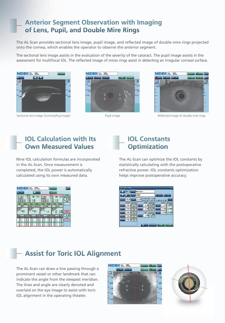
Anterior Segment Observation with Imaging of Lens, Pupil, and Double Mire Rings
The AL-Scan provides sectional lens image, pupil image, and reflected image of double mire rings projected onto the cornea, which enables the operator to observe the anterior segment.
The sectional lens image assists in the evaluation of the severity of the cataract. The pupil image assists in the assessment for multifocal IOL. The reflected image of mires rings assist in detecting an irregular corneal surface.
Sectional lens image (Scheimpflug image) Pupil image Reflected image of double mire rings
IOL Calculation with Its Own Measured Values
Nine IOL calculation formulas are incorporated in the AL-Scan. Once measurement is completed, the IOL power is automatically calculated using its own measured data.
IOL Constants Optimization
The AL-Scan can optimize the IOL constants by statistically calculating with the postoperative refractive power. IOL constants optimization helps improve postoperative accuracy.
Assist for Toric IOL Alignment
The AL-Scan can draw a line passing through a prominent vessel or other landmark that can indicate the angle from the steepest meridian. The lines and angle are clearly denoted and overlaid on the eye image to assist with toric IOL alignment in the operating theater.
110°110°
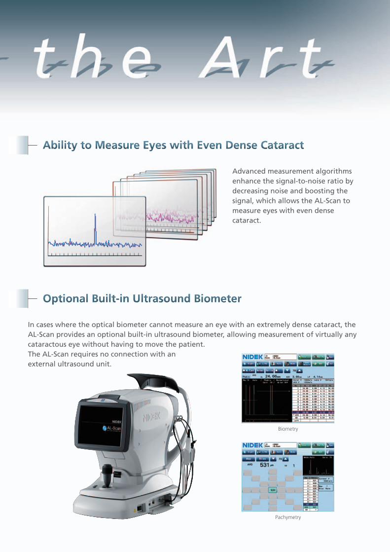
t h e A r tAbility to Measure Eyes with Even Dense Cataract
Advanced measurement algorithms enhance the signal-to-noise ratio by decreasing noise and boosting the signal, which allows the AL-Scan to measure eyes with even dense cataract.
Optional Built-in Ultrasound Biometer
In cases where the optical biometer cannot measure an eye with an extremely dense cataract, the AL-Scan provides an optional built-in ultrasound biometer, allowing measurement of virtually any cataractous eye without having to move the patient. The AL-Scan requires no connection with an external ultrasound unit.
Biometry
Pachymetry
HEAD OFFICE34-14 Maehama, HiroishiGamagori, Aichi 443-0038, JapanTelephone : +81-533-67-6611Facsimile : +81-533-67-6610URL : http://www.nidek.co.jp
[Manufacturer ]
TOKYO OFFICE(International Div.)3F Sumitomo Fudosan Hongo Bldg., 3-22-5 Hongo, Bunkyo-ku, Tokyo113-0033, JapanTelephone : +81-3-5844-2641Facsimile : +81-3-5844-2642URL : http://www.nidek.com
NIDEK INC.47651 Westinghouse DriveFremont, CA 94539, U.S.A.Telephone : +1-510-226-5700 : +1-800-223-9044 (US only)Facsimile : +1-510-226-5750URL : http://usa.nidek.com
NIDEK TECHNOLOGIES SrlVia dell'Artigianato, 6 / A35020 Albignasego (Padova), ItalyTelephone : +39 049 8629200 / 8626399Facsimile : +39 049 8626824URL : http://www.nidektechnologies.it
NIDEK S.A.Europarc13, rue Auguste Perret94042 Creteil, FranceTelephone : +33-1-49 80 97 97Facsimile : +33-1-49 80 32 08URL : http://www.nidek.fr
CNIDEK 2012 Printed in Japan AL-Scan 2
Specifications and design are subject to change without notice.
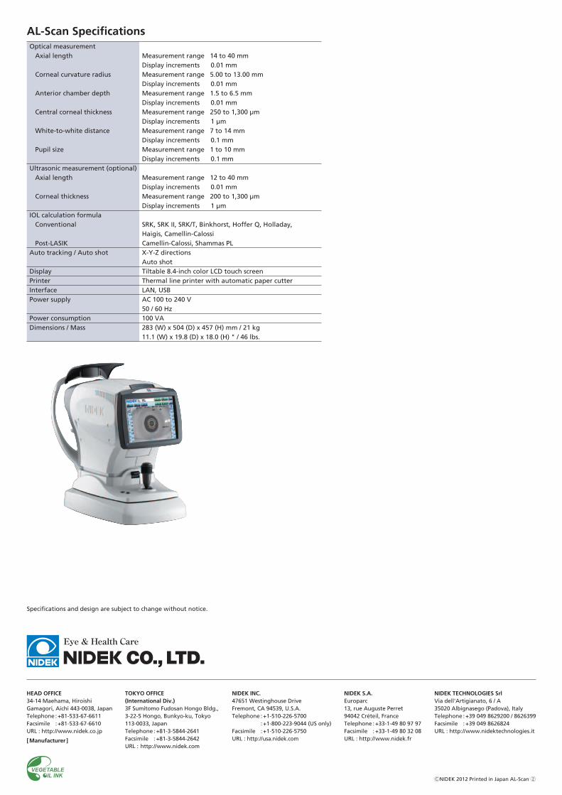
AL-Scan Specifications
Measurement range 14 to 40 mmDisplay increments 0.01 mmMeasurement range 5.00 to 13.00 mmDisplay increments 0.01 mmMeasurement range 1.5 to 6.5 mmDisplay increments 0.01 mmMeasurement range 250 to 1,300 µmDisplay increments 1 µmMeasurement range 7 to 14 mmDisplay increments 0.1 mmMeasurement range 1 to 10 mmDisplay increments 0.1 mm
Measurement range 12 to 40 mmDisplay increments 0.01 mmMeasurement range 200 to 1,300 µmDisplay increments 1 µm
SRK, SRK II, SRK/T, Binkhorst, Hoffer Q, Holladay, Haigis, Camellin-CalossiCamellin-Calossi, Shammas PLX-Y-Z directionsAuto shotTiltable 8.4-inch color LCD touch screenThermal line printer with automatic paper cutterLAN, USBAC 100 to 240 V50 / 60 Hz100 VA283 (W) x 504 (D) x 457 (H) mm / 21 kg11.1 (W) x 19.8 (D) x 18.0 (H) " / 46 lbs.
Optical measurementAxial length
Corneal curvature radius
Anterior chamber depth
Central corneal thickness
White-to-white distance
Pupil size
Ultrasonic measurement (optional)Axial length
Corneal thickness
IOL calculation formulaConventional
Post-LASIKAuto tracking / Auto shot
DisplayPrinterInterfacePower supply
Power consumptionDimensions / Mass