orbital trauma david m. yousem, m.d., m.b.a. johns hopkins medical institution
DESCRIPTION
N.A. The following is not an indication for surgical correction of orbital Fx 1. A. Double vision 2. B. Enophthalmos 3. C. Greater than 50% floor involvement 4. D. Exophthalmos 5. E. None of the aboveTRANSCRIPT
Orbital Trauma
David M. Yousem, M.D., M.B.A.Johns Hopkins Medical Institution
0% 0% 0% 0% 0%
1 2 3 4 5
N.A. What constrains a retinal detachment?
1. A. Ciliary body2. B. Hyaloid vessels3. C. Ora Serrata4. D. Zonular ligaments5. E. Orbital septum
0% 0% 0% 0% 0%
1 2 3 4 5
N.A. The following is not an indication for surgical correction of
orbital Fx
1. A. Double vision2. B. Enophthalmos3. C. Greater than 50%
floor involvement4. D. Exophthalmos5. E. None of the above
• Describe injuries to globe (bulbar)• List indications for acute globe
intervention• Describe retrobulbar injuries including
fractures (intraconal/conal/extraconal)• Discuss controversies re: fracture
intervention
Orbital Trauma Goals and Objectives
Orbital Trauma : Background
• Trauma to eye = 3% of ED visits• 4.5% of all orbital pathology is from
trauma• 40% of monocular blindness in US is
from trauma• Some findings require acute
treatment
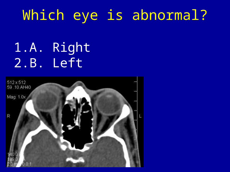
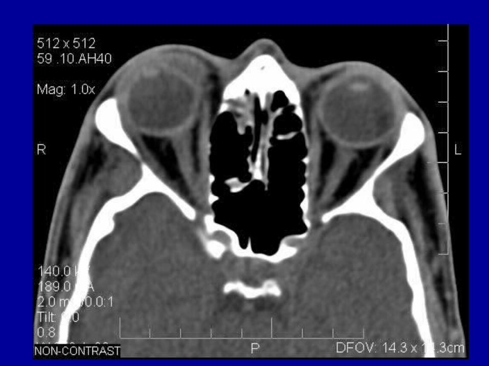
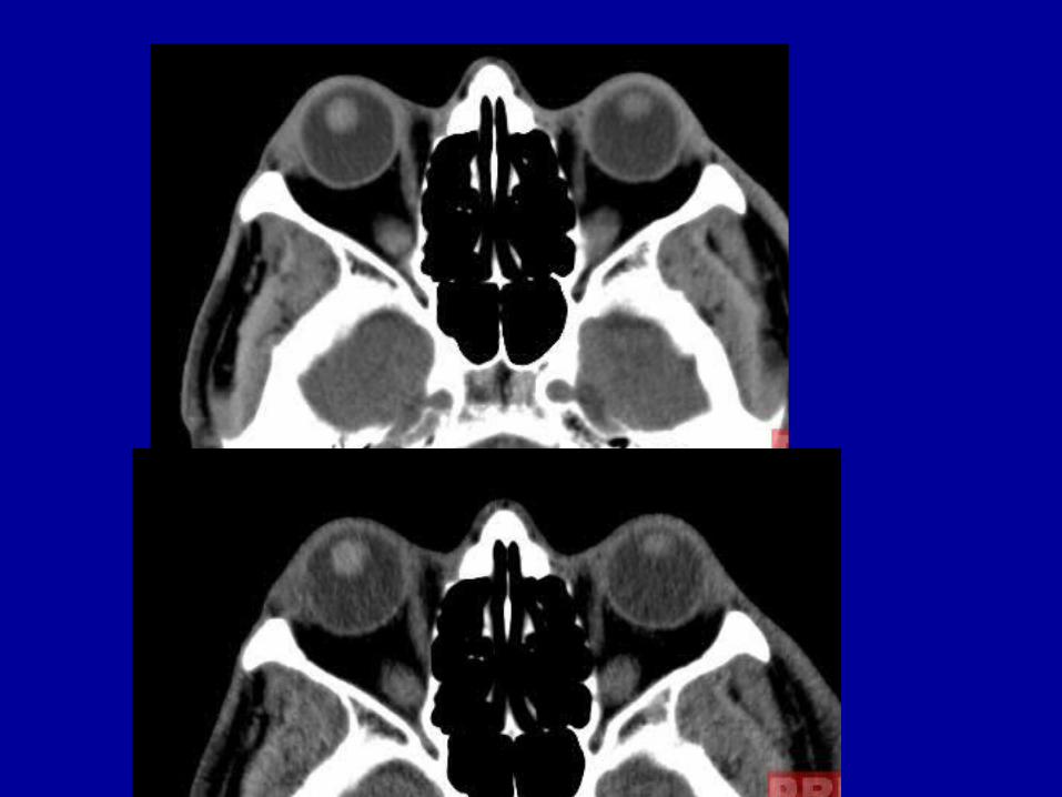
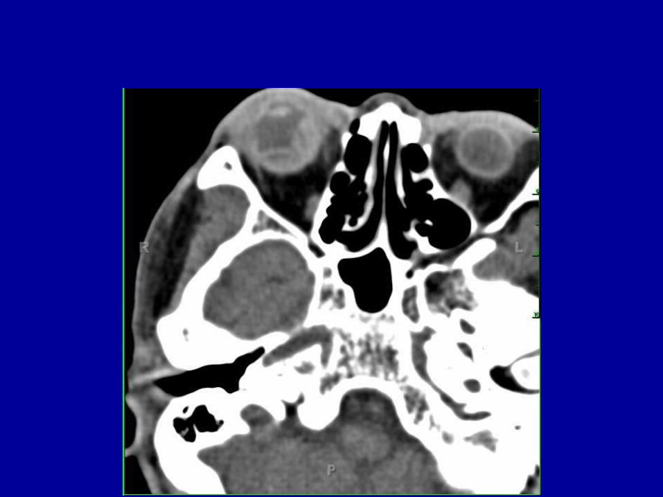
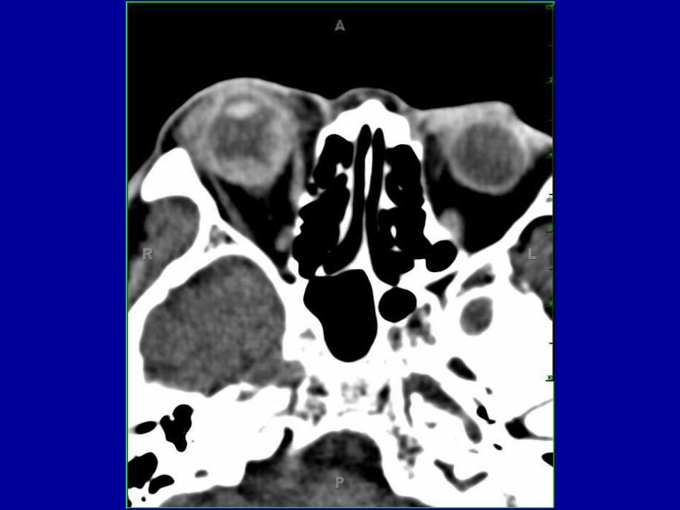
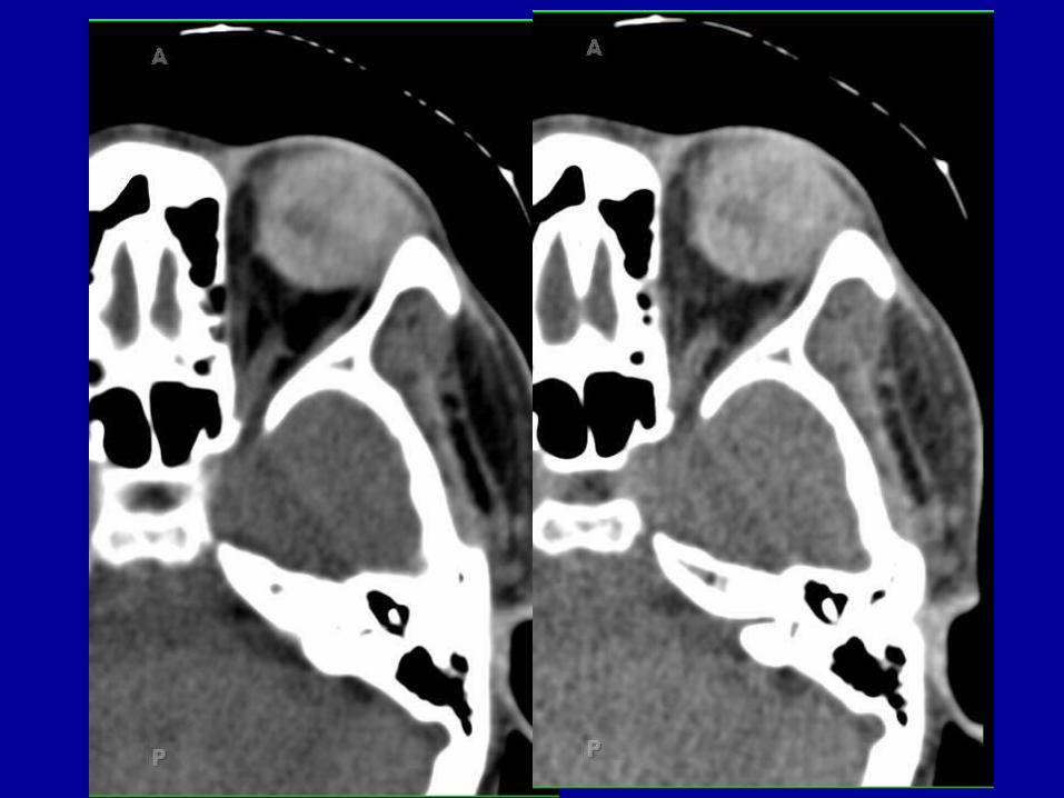
Which eye is abnormal?
1. A. Right2. B. Left
Ocular Blood Locations:
• Anterior chamber: anterior hyphema• Posterior chamber: posterior
hyphema• Vitreous: vitreous hemorrhage• Choroidal detachment• Retinal detachment
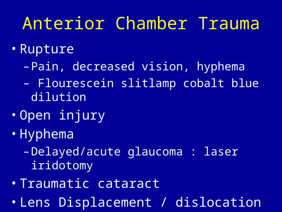
Anterior Chamber Trauma
• Rupture– Pain, decreased vision, hyphema– Flourescein slitlamp cobalt blue dilution
• Open injury• Hyphema
– Delayed/acute glaucoma : laser iridotomy• Traumatic cataract• Lens Displacement / dislocation
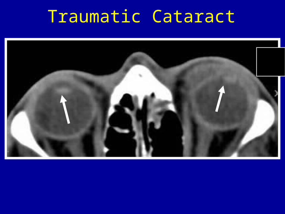
Traumatic Cataract
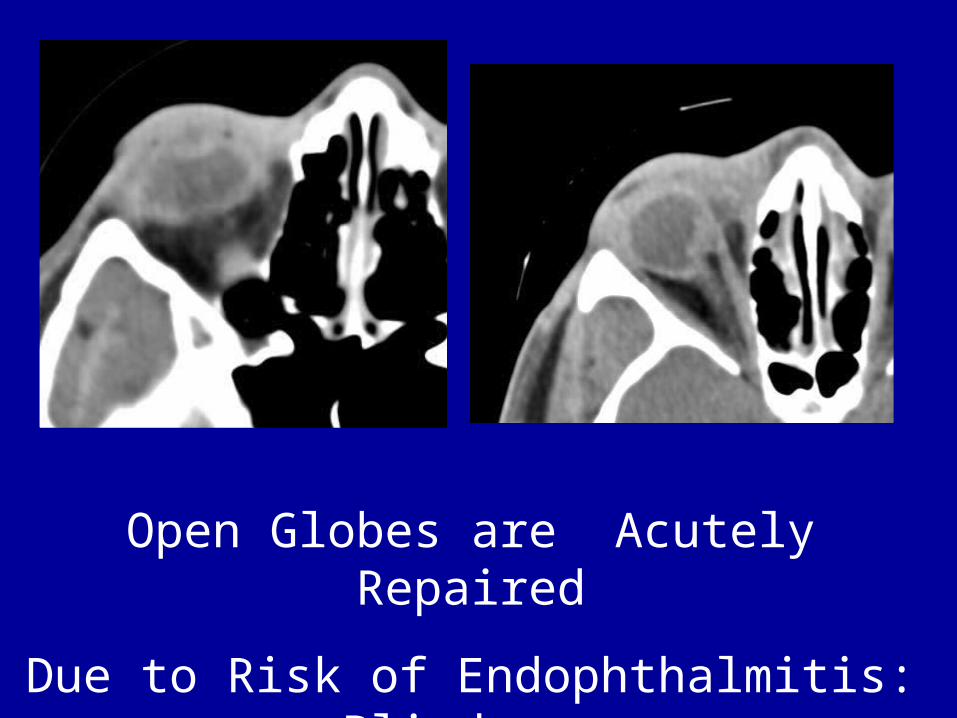
Open Globes are Acutely Repaired
Due to Risk of Endophthalmitis: Blindness
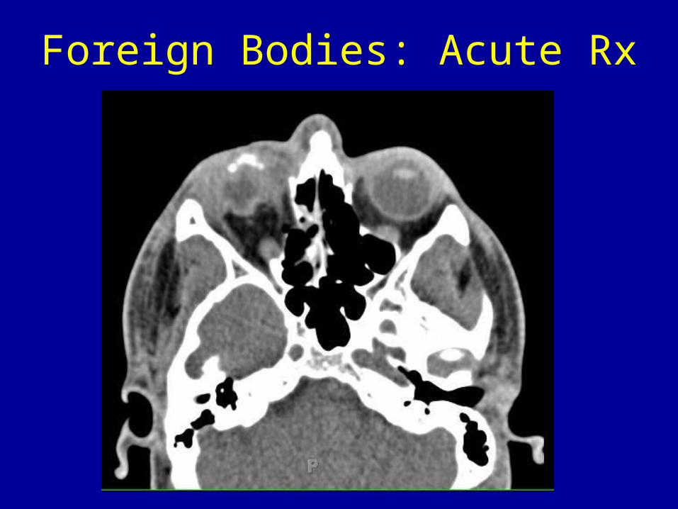
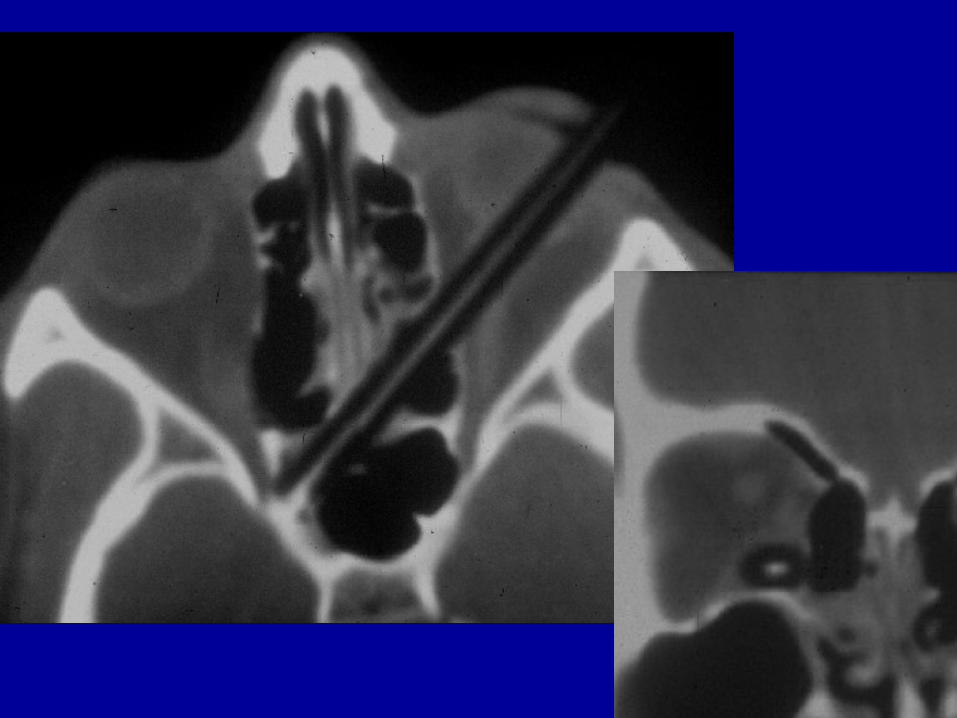
Foreign Bodies: Acute Rx
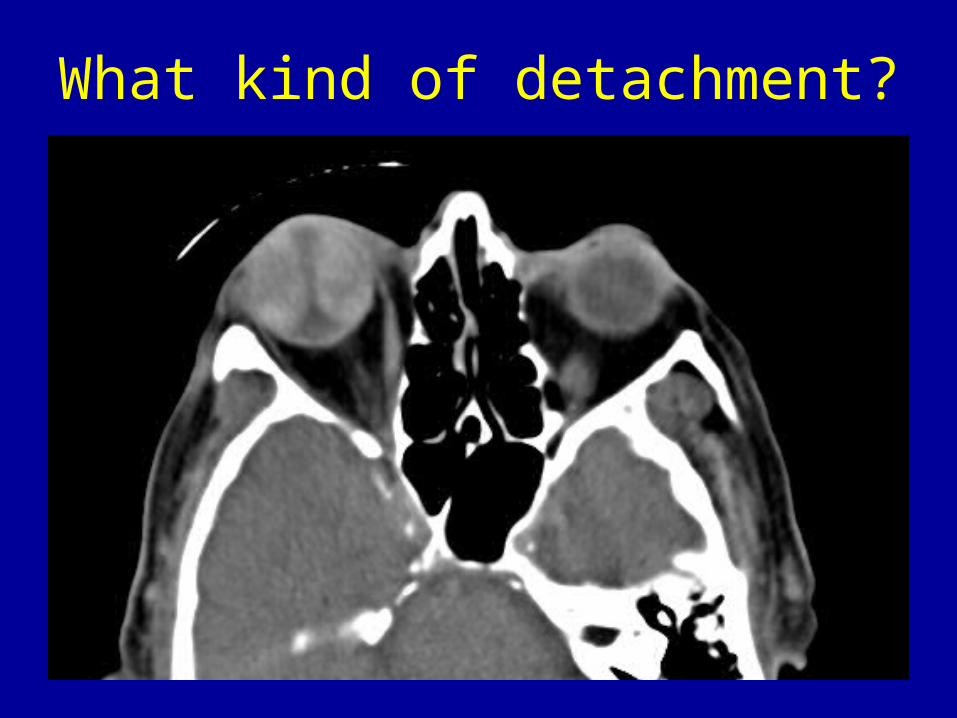
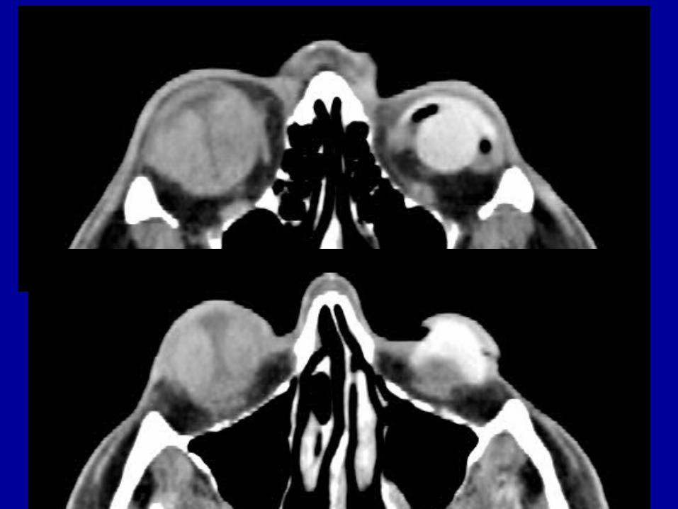
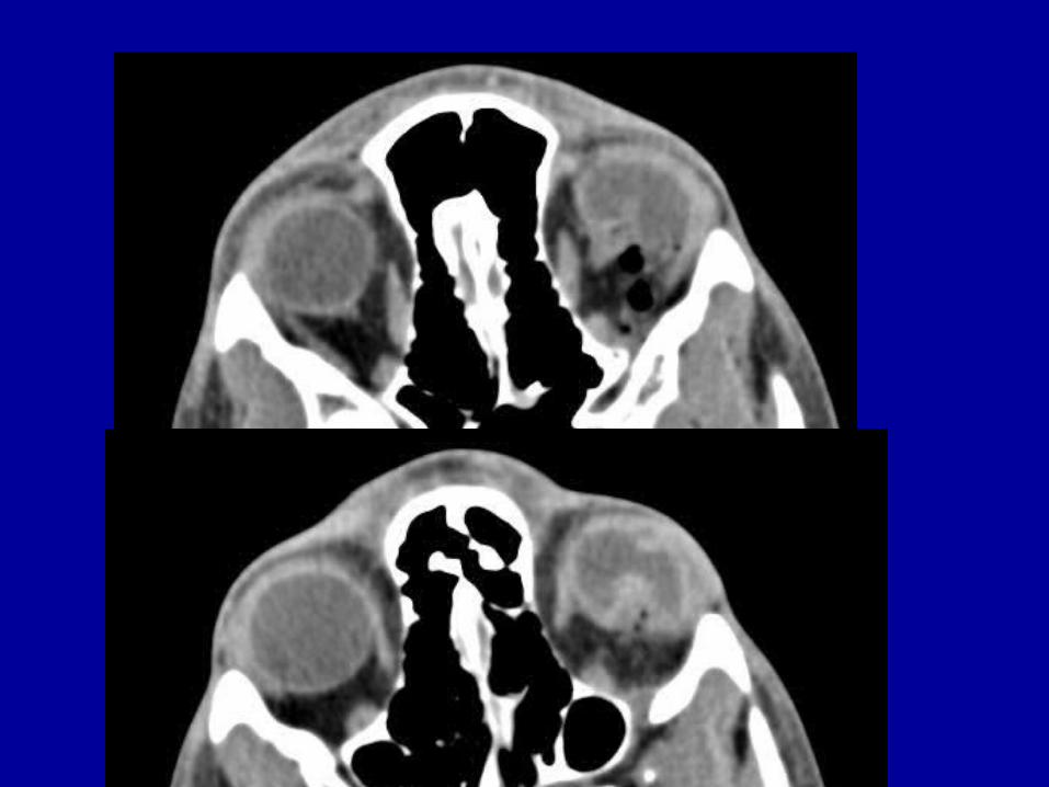
What kind of detachment?
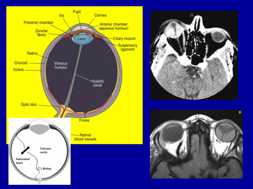
Ocular Membranes
• Retinal detachment– NAT!
• Choroidal detachment• Subhyaloid detachment• Puncture
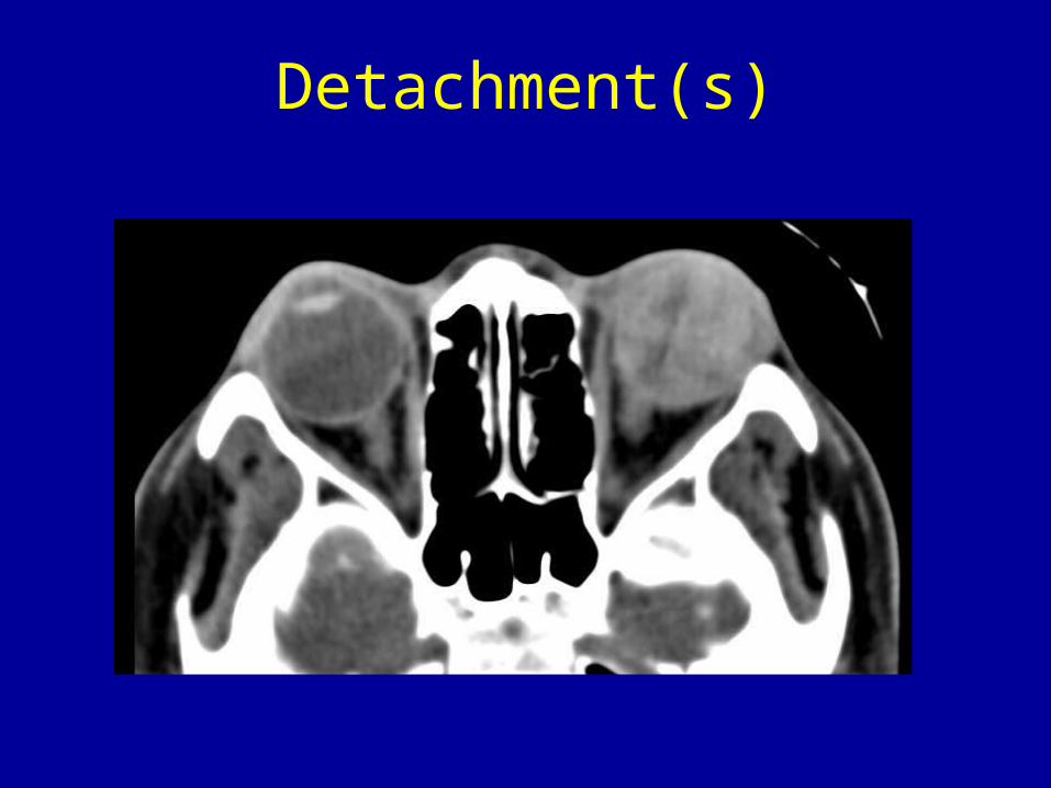
Detachment(s)
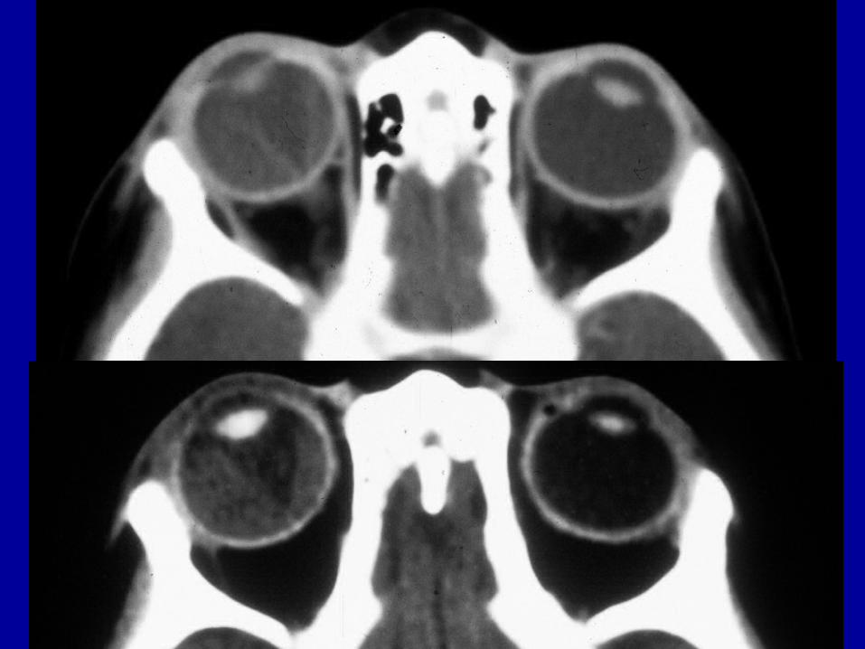
Vitreous Chamber
• Classic rupture• Ocular hypotony• Hemorrhage• Puncture• Late effect: Phthisis Bulbi
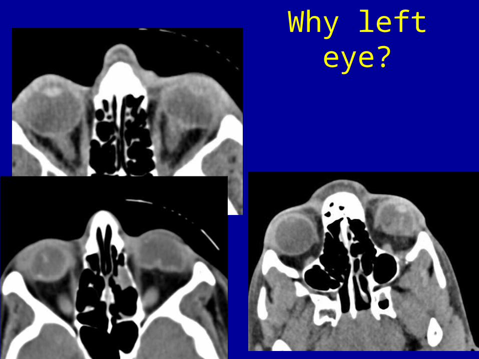
Why left eye?
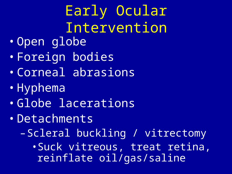
Early Ocular Intervention• Open globe• Foreign bodies• Corneal abrasions• Hyphema• Globe lacerations• Detachments
– Scleral buckling / vitrectomy• Suck vitreous, treat retina, reinflate
oil/gas/saline
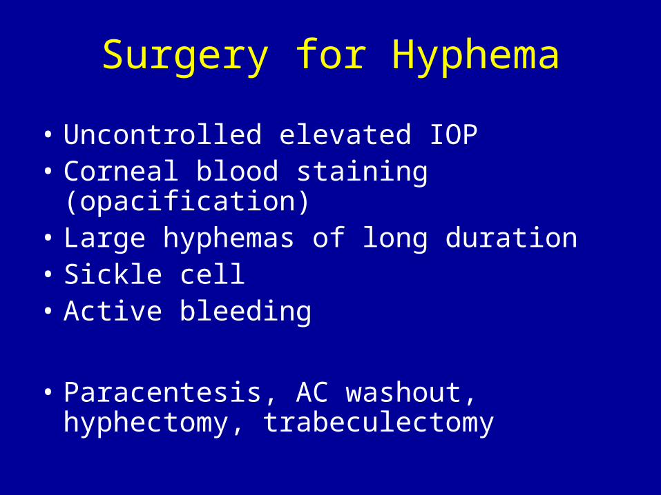
Surgery for Hyphema
• Uncontrolled elevated IOP• Corneal blood staining (opacification)• Large hyphemas of long duration • Sickle cell• Active bleeding
• Paracentesis, AC washout, hyphectomy, trabeculectomy
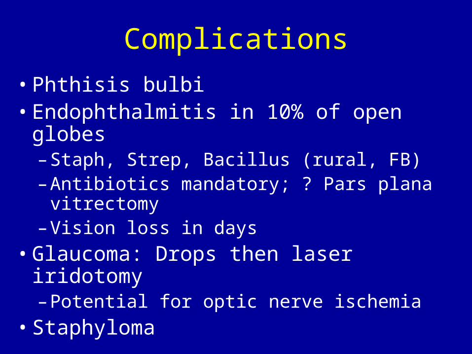
Complications
• Phthisis bulbi• Endophthalmitis in 10% of open globes
– Staph, Strep, Bacillus (rural, FB)– Antibiotics mandatory; ? Pars plana
vitrectomy– Vision loss in days
• Glaucoma: Drops then laser iridotomy– Potential for optic nerve ischemia
• Staphyloma
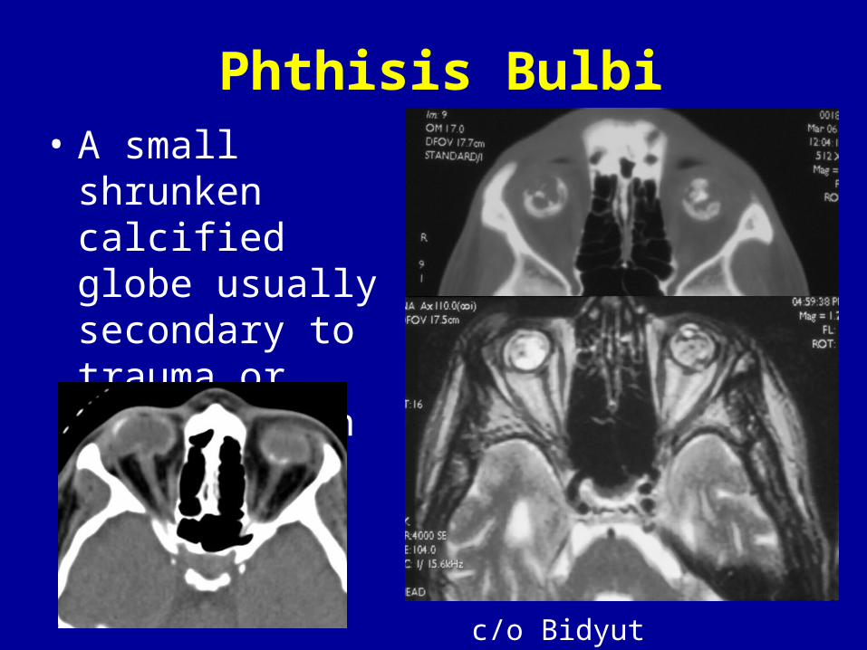
Phthisis Bulbi• A small shrunken
calcified globe usually secondary to trauma or inflammation
c/o Bidyut Pramanik
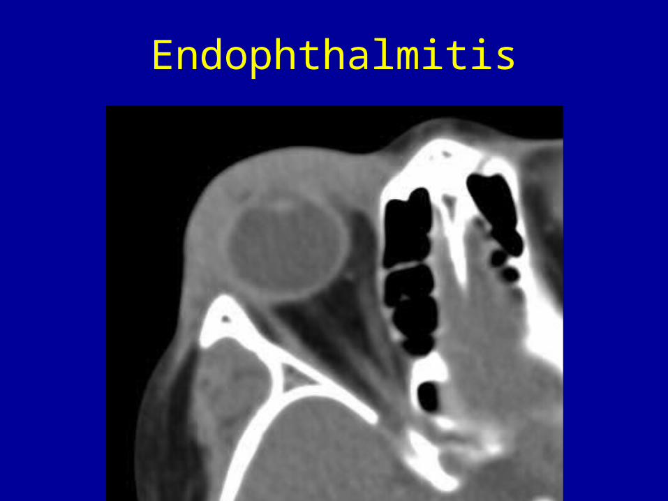
Endophthalmitis

Staphyloma• Acquired defects in the
sclera or cornea• Posterior staphyloma is
associated with increasing globe size
• Usually on the temporal side of optic nerve
• Outward bulging with uveoscleral thinning
• Anterior staphyloma is seen with RA
c/o Bidyut Pramanik

Enucleation
• Blind painful eye• Endophthalmitis (esp open globe)• Phthisis bulbi• Severe traumatic rupture• Unsightly eye• Glaucoma
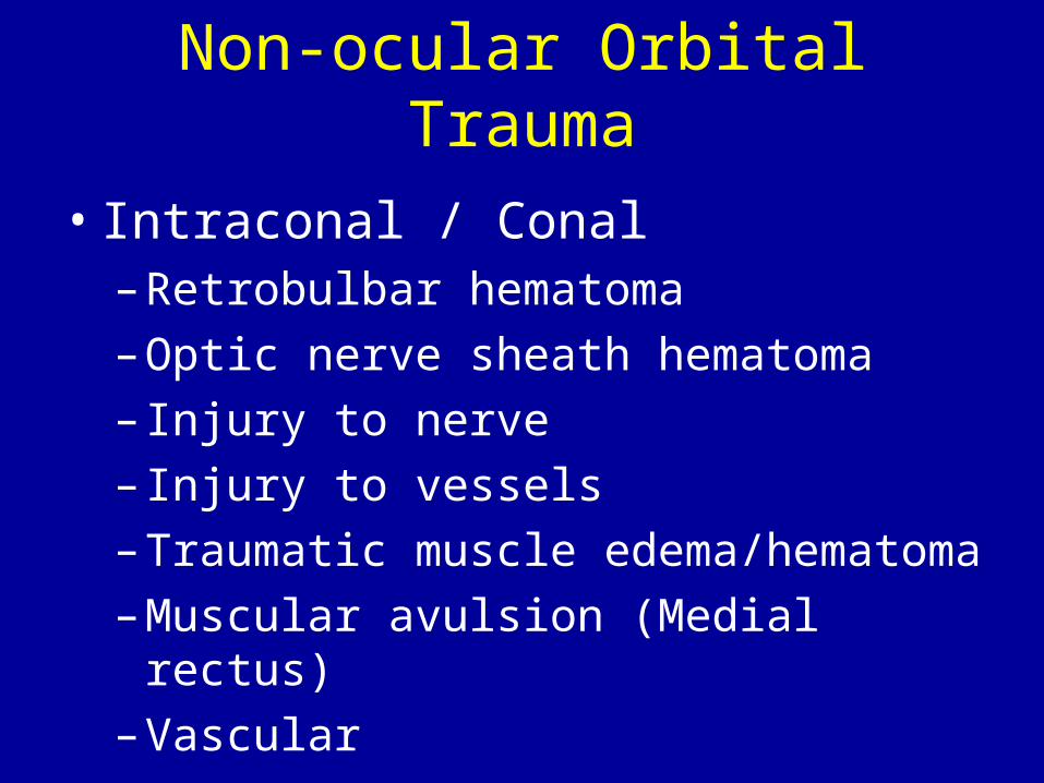
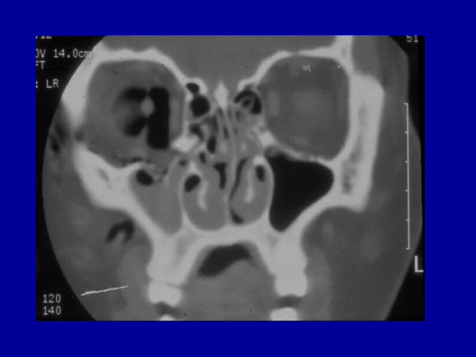
Non-ocular Orbital Trauma
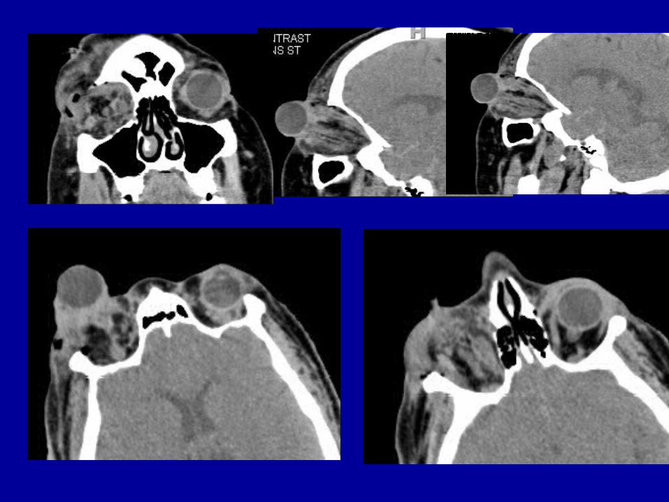
• Intraconal / Conal– Retrobulbar hematoma– Optic nerve sheath hematoma– Injury to nerve– Injury to vessels– Traumatic muscle edema/hematoma– Muscular avulsion (Medial rectus)– Vascular
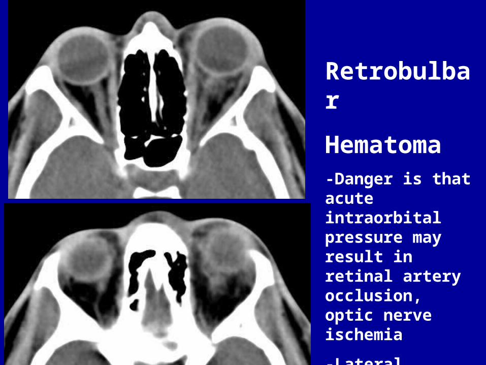
Retrobulbar
Hematoma-Danger is that acute intraorbital pressure may result in retinal artery occlusion, optic nerve ischemia
-Lateral canthotomy decompression
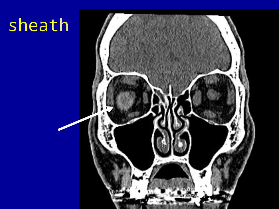
sheath
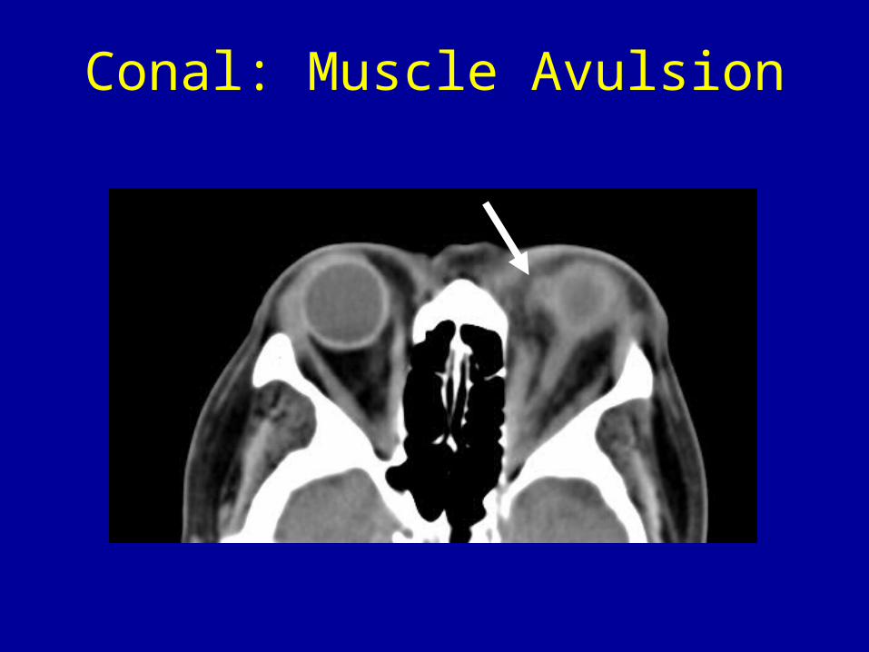
Conal: Muscle Avulsion
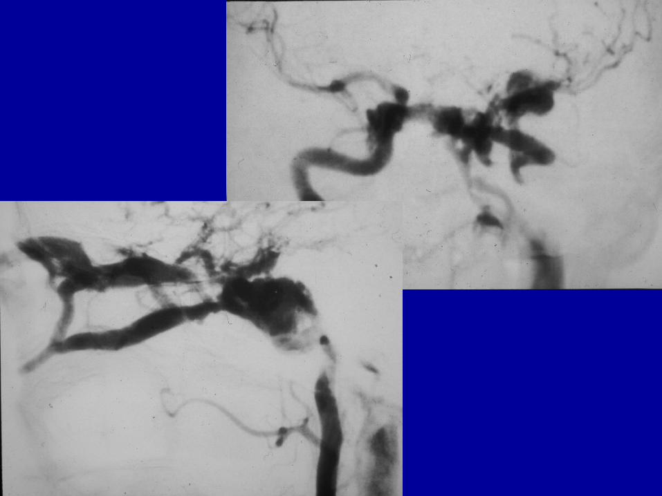
Orbital Trauma Vascular
• Carotid-cavernous fistula• Pseudoaneurysm• Varicosities

Carotid Cavernous Fistula
• May result in EOM enlargement due to venous engorgement
• All EOMs involved• Superior Ophthalmic Vein is dilated• Usually unilateral

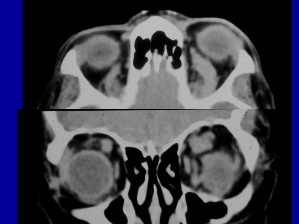
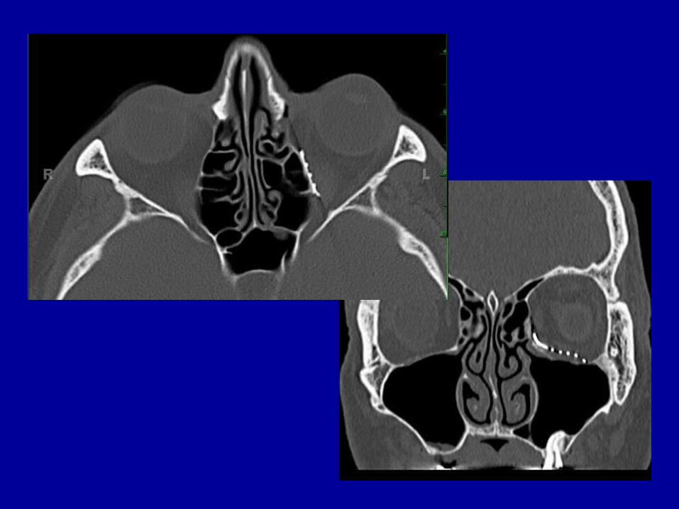
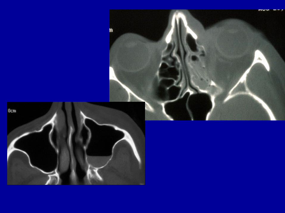
Extraconal: Orbital Fractures
• Orbital rim• Orbital floor• Medial orbital wall: lamina papyracea• Lateral orbital wall• Superior wall
– Globe injuries occur in 10-25% of patients with orbital fractures
Indications for Surgery for Orbital Fractures
• Enophthalmos > 2 mm (> 50% of floor)• Hypoglobus (downward displaced globe)• Diplopia
– Edema, heme, n. palsy, direct trauma• Increase in orbital volume > 1 cc
– Correlates with enophthalmos• Limited mobility (entrapment of EOM)• Compressive optic neuropathy
Kontio R, Lindquist C. OMFC 2009: 21: 209-220
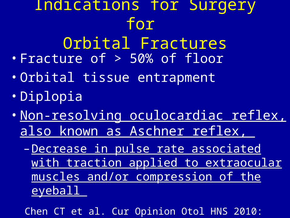
Indications for Surgery for Orbital Fractures
• Fracture of > 50% of floor• Orbital tissue entrapment• Diplopia• Non-resolving oculocardiac reflex, also
known as Aschner reflex, – Decrease in pulse rate associated with
traction applied to extraocular muscles and/or compression of the eyeball
Chen CT et al. Cur Opinion Otol HNS 2010: 18: 311-6
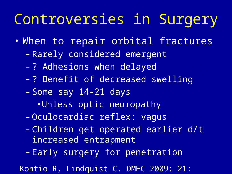
Controversies in Surgery• When to repair orbital fractures
– Rarely considered emergent– ? Adhesions when delayed– ? Benefit of decreased swelling– Some say 14-21 days
• Unless optic neuropathy– Oculocardiac reflex: vagus– Children get operated earlier d/t increased
entrapment– Early surgery for penetration
Kontio R, Lindquist C. OMFC 2009: 21: 209-220

Controversies in Surgery
• What to repair with– Must be rigid to contain orbital contents– Restore form and volume– Contourable
• Autogenous grafts (iliac bone)– ? Too rigid, difficult to place
• Alloplasts (non/resorbable)– Many varieties
• Titanium mesh, MedporKontio R, Lindquist C. OMFC 2009: 21: 209-220
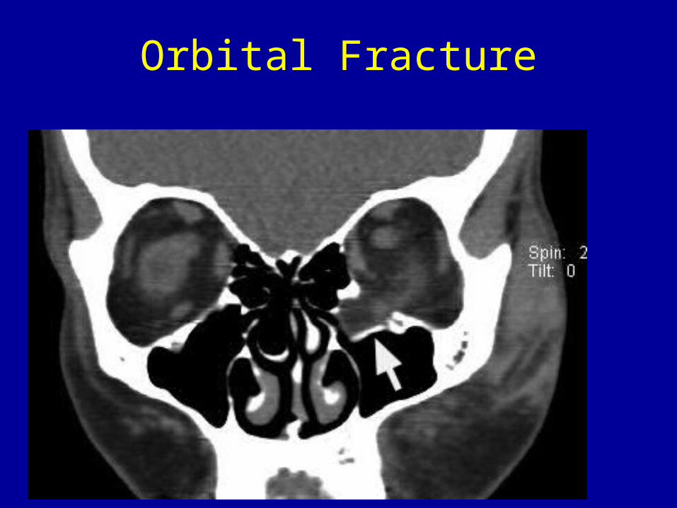
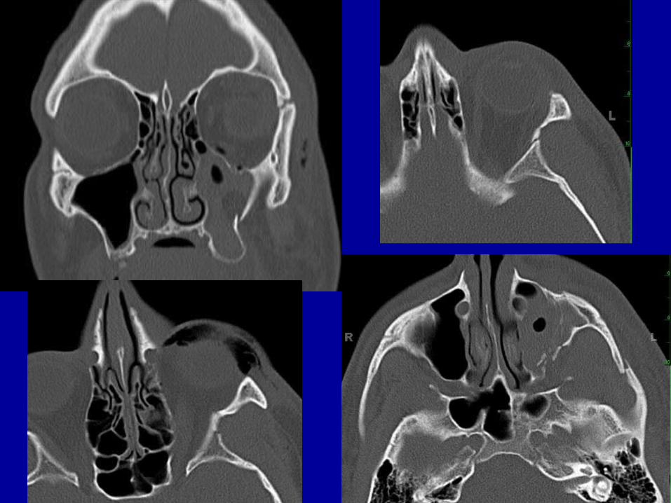
Orbital Fracture
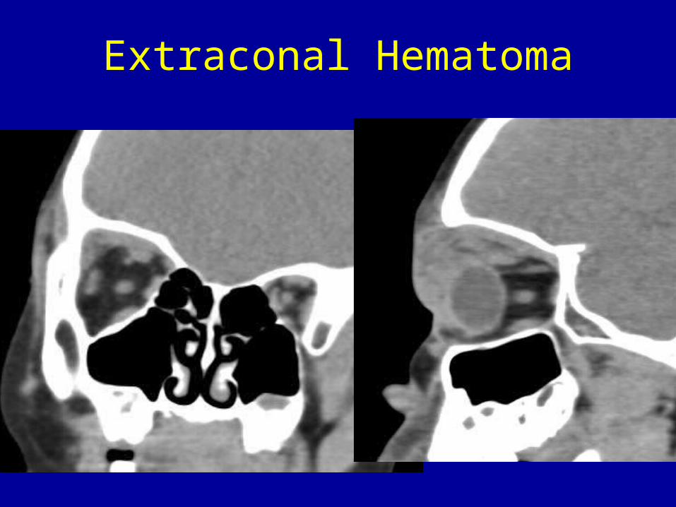
Extraconal Hematoma
Conclusions
• A common indication in ED practice• Ocular, non-ocular findings often
equally important• Some fractures should be treated
acutely• Long term sequelae