orexin-neuromodulated cerebellar circuit controls ... · orexin-neuromodulated cerebellar circuit...
TRANSCRIPT
Orexin-neuromodulated cerebellar circuit controlsredistribution of arterial blood flows for defensebehavior in rabbitsNaoko Nisimarua,b, Chetan Mittala,c,1, Yoshinori Shiraia,d, Thongchai Sooksawatee,2, Prabu Anandaraja,c,1,Tsutomu Hashikawaa, Soichi Nagaoa, Akiko Arataa,f, Takeshi Sakuraig, Miyuki Yamamotoh, and Masao Itoa,3
aRIKEN Brain Science Institute, 2-1 Hirosawa, Wako, Saitama 351-0198, Japan; bDepartment of Physiology, Faculty of Medicine, Oita University,1-1 Idaigaoka,Hasama,Yufu, Oita 879-5593, Japan; cIndian Institute of Technology, Kharagpur 721-302, India; dDepartment of Neuroplasticity, Shinshu University GraduateSchool of Medicine, 3-1-1 Asahi, Matsumoto, Nagano 390-8621, Japan; eDepartment of Physiology, Faculty of Pharmaceutical Science, ChulalongkornUniversity, Bangkok 10330, Thailand; fDepartment of Physiology, Hyogo College of Medicine, 1-1 Mukogawa, Nishinomiya, Hyogo 663-8501, Japan;gDepartment of Molecular Neuroscience and Integrative Physiology, Faculty of Medicine, Kanazawa University, Kanazawa, Ishikawa 920-8640, Japan;and hComprehensive Human Studies, University of Tsukuba, 1-1 Tennodai, Tsukuba, Ibaraki 305-8575, Japan
This contribution is part of the special series of Inaugural Articles by members of the National Academy of Sciences elected in 2007.
Contributed by Masao Ito, July 9, 2013 (sent for review April 27, 2012)
We investigated a unique microzone of the cerebellum located infolium-p (fp) of rabbit flocculus. In fp, Purkinje cells were potentlyexcited by stimulation of the hypothalamus or mesencephalic peri-aqueductal gray, which induced defense reactions. Using multipleneuroscience techniques, we determined that this excitation wasmediated via beaded axons of orexinergic hypothalamic neuronspassing collaterals through the mesencephalic periaqueductalgray. Axonal tracing studies using DiI and biotinylated dextranamine evidenced the projection of fp Purkinje cells to the ventro-lateral corner of the ipsilateral parabrachial nucleus (PBN). Be-cause, in defense reactions, arterial blood flow has been knownto redistribute from visceral organs to active muscles, we hypoth-esized that, via PBN, fp adaptively controls arterial blood flowredistribution under orexin-mediated neuromodulation that couldoccur in defense behavior. This hypothesis was supported by ourfinding that climbing fiber signals to fp Purkinje cells were elicitedby stimulation of the aortic nerve, a high arterial blood pressure,or a high potassium concentration in muscles, all implying errors inthe control of arterial blood flow. We further examined the arte-rial blood flow redistribution elicited by electric foot shock stimuliin awake, behaving rabbits. We found that systemic administra-tion of an orexin antagonist attenuated the redistribution and thatlesioning of fp caused an imbalance in the redistribution betweenactive muscles and visceral organs. Lesioning of fp also diminishedfoot shock-induced increases in the mean arterial blood pressure.These results collectively support the hypothesis that the fp micro-complex adaptively controls defense reactions under orexin-mediatedneuromodulation.
somatosympathetic | vestibulosympathetic | OX-1R antagonist |bicuculline | baroreceptor
The cerebellar cortex consists of numerous microzones, eachextending 10 mm2 or so (ref. 1; for review, see ref. 2). Each
microzone receives three distinct types of input, that is, mossyfibers, climbing fibers, and beaded fibers. Beaded fibers containcertain amines or neuropeptides (for review, see refs. 3 and 4)and distribute diffusely; supposedly, they determine the generalactivity or the mode of operation of their target neurons, that is,neuromodulation, unlike input/output-specific transmission inmossy fibers and climbing fibers (5). Several microzones havebeen analyzed with regard to their circuit mechanisms and spe-cific reflex functions (not only somatic; some are autonomic)(6, 7), but the actual roles of beaded fibers remain largely unknown.In this study, we focused on a particular microzone located in theflocculus [folium-p (fp)] (8, 9), which receives beaded fiberscontaining orexins (hypocretins) (10). Orexins consist of A and Bisopeptides containing 33 and 28 amino acids, respectively, andhave been implicated in sleep and feeding (11–13).
A functionally unique feature of fp is that a large portion ofPurkinje cells in it are excited by stimulation of the classic de-fense areas in the hypothalamus and mesencephalic periaqu-eductal gray (PAG) (14). Electrical or chemical stimulation ofthe defense areas induces complex motor activities for “fight orflight” and associated cardiovascular responses such as rapidincrease of blood pressure (BP) (15–17). In natural behavingconditions, harmful stimuli activate the defense areas via theamygdala (for review, see ref. 18). Involvement of orexins in thecardiovascular defense reactions has been suggested becausethe genetically induced orexin deficiency in mice leads to atten-uation of the transient increase of blood pressure evoked fromdefense areas (19, 20).We analyzed neuronal circuit connections to and from fp using
axonal transport tracers. Importantly, we found that Pukinje cellsin fp project their axons to the ventrolateral corner of the ipsi-lateral parabrachial nucleus (PBN), a major cardiovascular centerin the brainstem (21, 22). It was previously shown that PBNreceives Purkinje cell axons from the anterior vermis (21) as wellas the middle (6) and lateralmost regions (7) of the nodulus-uvula. These observations suggest that PBN acts as a functionalinterface between the cerebellum and supraspinal cardiovascularcenters (21). In defense reactions, arterial blood flow is redis-tributed from visceral organs and resting muscles to active muscles(17) by the action of the sympathetic nervous system (23). Subtlecontrol of this redistribution is needed to maintain cardiovascularhomeostasis while fulfilling the high demand for arterial bloodsupply to muscles actively involved in defense reactions.On the above-introduced backgrounds, we attempted to define
the role of orexins in fp function and the role of fp in the controlof arterial blood flow. We used rabbits because their flocculushas discernible regular folial divisions (8, 9), which is not the casein rats or mice.
A preliminary report of this work was delivered by N.N. and M.I. at the 35th InternationalCongress of Physiological Sciences, March 31–April 5, 2005, San Diego, CA, abstract 512.
Author contributions: N.N., A.A., M.Y., and M.I. designed research; N.N., C.M., Y.S.,T. Sooksawate, P.A., T.H., S.N., A.A., M.Y., and M.I. performed research; T. Sakurai con-tributed new reagents/analytic tools; N.N., C.M., Y.S., T. Sooksawate, A.A., M.Y., and M.I.analyzed data; and N.N., T. Sakurai, and M.I. wrote the paper.
The authors declare no conflict of interest.
Freely available online through the PNAS open access option.1On leave from: Indian Institute of Technology, Kharagpur 721-302, India.2On leave from: Chulalongkorn University, Bangkok 10330, Thailand.3To whom correspondence should be addressed. E-mail: [email protected].
This article contains supporting information online at www.pnas.org/lookup/suppl/doi:10.1073/pnas.1312804110/-/DCSupplemental.
14124–14131 | PNAS | August 27, 2013 | vol. 110 | no. 35 www.pnas.org/cgi/doi/10.1073/pnas.1312804110

ResultsOrexin-Immunopositive Beaded Fibers in Rabbit Cerebellum. In rab-bits, the flocculus lies in the rostroventrolateral corner of thecerebellum (Fig. 1A) and consists of six folia, fm, f1, f2, f3, f4,and fp (8. 9) (Fig. 1B). In coronal sections, a cut face of fp can beidentified by its flattened triangular appearance, with the peakpointing ventrolaterally (Fig. 1C). As examined in three com-plete sets of serial coronal sections (60 μm thick) of the leftflocculus, the Purkinje cell layer in fp maximally expands dor-soventrally by 3.0 ± 0.1 mm (mean ± SE of mean, throughoutthis article) and anteroposteriorly by 3.1 ± 0.2 mm, the total areabeing 7.2 ± 0.9 mm2.Immunohistochemical data on orexins are abundant in the
literature for mice and rats but are scarce for rabbits; therefore,we collected our own data for confirmation. Within the cere-bellum, we observed orexin-immunopositive fibers located al-most exclusively in the flocculus, with a rare detection of suchfibers in the vermis, as similarly observed in rats (10). A fluo-rescence microscopy image shows the beaded appearance ofthese fibers, which lie adjacent to Purkinje cells and ascend to themolecular layer, some running parallel to the Purkinje cell layer(Fig. 2 A and B). In one flocculus, we counted 46 such fibers inthe molecular layer, 74 in the Purkinje cell layer, and 71 in thegranular layer. As summarized for three rabbits (Table S1), thedensity of orexin-immunopositive fibers in cross-sectional areasof fp is 2.77 times as high as that in all other (non-fp) folia ofthe flocculus. The common presence of orexin-immunopositivefibers may justify our classification of fp as part of the rabbitflocculus whereas Tan et al. (9) deemed fp as part of the ventralparaflocculus.
Origin of Orexin-Immunopositive Fibers. Within the rabbit hypo-thalamus, orexin-immunopositive neurons distribute around thefornix over the perifornical nucleus, dorsomedial hypothalamus,and lateral hypothalamic area (Fig. S1A). As similarly observedin rats (10), orexin-immunopositive axons of these cells innervatea number of structures in the brainstem and densely fill PAG(Fig. S1C). Orexin B-conjugated saporin (SAP, a ribosome-inactivating protein) has been used to degenerate neurons thathad orexin receptor-2 (OX-2R), including orexin-immunopositiveneurons; however, SAP actually degenerated not only theseneurons, but also most of the other neurons in the posterior
hypothalamus (24). Therefore, we injected SAP into the hypo-thalamus as a nonspecific lesioning agent. In two rabbits in whichSAP-induced lesions covered the hypothalamus defense areabilaterally (except for some surviving orexin-immunopositive neu-rons) (Fig. S1B), orexin-immunopositive fibers in PAG largely di-minished (Fig. S1D); this finding confirms that these fibers arebranches of orexin-immunopositive fibers of hypothalamic origin.Abnormally low basal BP was reported to prevail in genetically
orexin-deficient mice (19), but abnormally low basal BP was notthe case in our two rabbits systemically injected with orexinreceptor-1 (OX-1R) antagonists; mean BP were 92 and 105 mmHgagainst the control values 98 and 108 mmHg, respectively. In threeSAP-treated rabbits, the average of the mean BP was also normal:92 ± 5 mmHg against 91 ± 3 mmHg in 20 control rabbits. Dif-ferent neural mechanisms appear to underlie the maintenance ofbasal BP and the transient BP increase in defense reactions, atleast in rabbits.
Involvement of Orexins in Defense Reactions. Under general anes-thesia, the hypothalamus was explored with a bipolar needleelectrode for stimulation (SE1; Fig. 1). At certain depths alonga dorsoventral track, a train of 100-Hz bursts (8-50 pulses; eachpulse, 100–500 μA; 0.3 ms wide) effectively induced a transientincrease in BP by 20–100 mmHg (Fig. 3A, Fig. S2A). On theaverage of mean BP curves (Fig. 3I), the mean increase is 49 ±4 mmHg (n = 20) and the mean latency is 0.70 ± 0.03 s (n = 19).Four lines of evidence support the view that this transient BPincrease is mediated by orexinergic hypothalamic neurons. First,when optimal stimulation sites for this effect were mapped invarious experiments (shown in Figs. 3–5 and 7), they covered theregion designated as the hypothalamic defense area (Fig. S2B)where orexin-immunopositive neurons distributed (Fig. S1A).Second, systemic administration (7 mg/kg) of an OX-1R antag-onist, SB-334867 (25) or SB-408124 (26), markedly attenuatedthese BP increases (Fig. 3 C, G, and J). Third, in all of the fourrabbits tested 2–6 wk after SAP injection, orexin-immunopositiveneurons had largely disappeared from the hypothalamus (Fig.S1B; compare with Fig. S1A), and, accordingly, hypothalamusstimulation (even as strong as 500 μA) failed to increase meanBP (5 ± 3 mmHg). Note that, in these experiments, the locationof the stimulating electrode was controlled stereotaxically to beclose to the fornix and later confirmed histologically (for exam-ple, * in Fig. S1B). Fourth, prepro-orexin–null knockout mice(19) and orexinergic neuron-ablated transgenic mice (20) havebeen reported to exhibit an attenuated BP increase during hypo-thalamus stimulation.Stimulation within or at its border of PAG also induced BP
increase as mapped in Fig. S2D. As would be expected, this ef-fect was blocked by OX-1R antagonists (Fig. 3 E andG). We alsonoted that hypothalamus/PAG stimulation evoked an increasein the rate of integrated electromyograph discharges in nuchalmuscles with a latency of ∼0.1 s (Fig. 3 B and F). Importantly,OX-1R antagonists did not affect these motor responses (Figs. 3D and H), which suggests that orexins are not involved in motorcomponents of defense reactions (see Discussion).
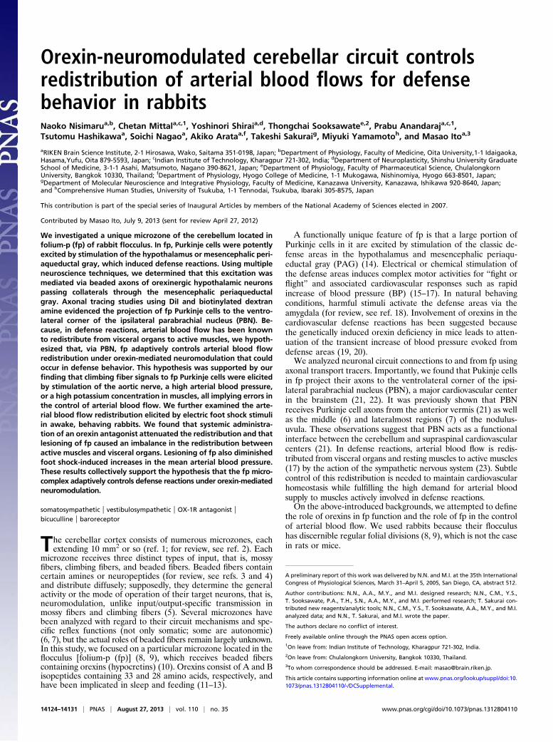
Fig. 1. Folium-p of rabbit flocculus. (A) Left side view of rabbit brain. ME,glass microelectrode; SE1, stimulating needle electrode inserted in the hy-pothalamus; SE2, another electrode in PAG. (B) Enlarged left side view of theflocculus showing its folial divisions. fp is exposed by retracting parts of theneighboring f4 and fv. Red spots, positions of the last microelectrode tracksin 21 experiments. Vertical lines 1–5 are drawn at 400, 800, 1,200, 1,600, and2,000 μm from the caudal pole of fp. (C) Coronal section of the left flocculusat the level 4 in B. LN, lateral nucleus.
Fig. 2. Orexinergic axons in flocculus. (A and B) Beaded orexin-immuno-positive fibers double-stained for calbindin (red) and orexins (green). ML,molecular layer; PC, Purkinje cell.
Nisimaru et al. PNAS | August 27, 2013 | vol. 110 | no. 35 | 14125
NEU
ROSC
IENCE
INAUGURA
LART
ICLE

Orexinergic Excitation of Purkinje Cells. When a glass microelec-trode was inserted through the dorsal paraflocculus, it reachedipsilateral fp, neighboring f4, or the ventralmost folium of theventral paraflocculus (fv), at depths of ∼7 mm (Fig. 1C). Sam-pled Purkinje cells spontaneously discharged simple spikes (Fig.4 A and B) at 10–90 spikes per s (mean 40.3 ± 4.0 spikes per s,n = 28) and complex spikes at 1–7 spikes per s (mean 3.8 ± 0.3spikes per s, n =19). Hypothalamus/PAG stimulation on eitherthe left or right side frequently induced a transient increase inthe rate of simple spike discharges, as shown in Fig. 4A. Inperistimulus time histograms (PSTHs), this excitation starts uponstimulation and lasts for a fraction of 1 s to 3–5 s (Fig. 4C). Theresponses occurring within 1 s, with statistical significance in thepermutation test (shaded in Fig. 4 C–E), were taken as the pri-mary effects of hypothalamus/PAG stimulation.Among the Purkinje cells sampled in fp and its neighboring
folia (fv, f4), 38–40% were excited by hypothalamus stimulationand 27–32% were excited by PAG stimulation (Table S2). Whenplotted on standardized coronal sections, the vast majority ofthese excited Purkinje cells were located in fp (Fig. 5 A and C).The remaining small fraction of excited Purkinje cells protrudedinto the neighboring f4 (Fig. 5 A, 2; A, 3; C, 2; and C, 3). Thehypothalamic origin of the hypothalamus/PAG stimulation-evokedexcitation was reflected in the effects of SAP-induced lesions ofthe hypothalamus; in the lesioned rabbits, hypothalamus stimu-lation excited Purkinje cells at a significantly lower rate (5%) thanin control (38%) or sham-operated (32%) rabbits [Wilcoxon–Mann–Whitney (WMW) test, P < 0.01] (Table S2).
When applied iontophoretically at 50 nA through a seven-barreled pipette to individual Purkinje cells (SI Materials and
Methods, Microiontophoresis), OX-1R antagonists effectivelydepressed the hypothalamus/PAG stimulation-evoked excitation(Fig. 4F). Between before and during iontophoresis, the differ-ence in the histograms was significant in 12 of the 16 cells ex-amined [Wilcoxon signed-rank test for matched samples (WSM)during a 3-s poststimulation period, P = 0.0001–0.020]. In thismeasurement, data obtained using either SB-334867 (n = 11) orSB408124 (n = 5) and by either hypothalamus (n = 9) or PAGstimulation (n = 7) were pooled together because these factorsmade no significant difference in the data obtained. In the ab-sence of hypothalamic/PAG stimulation, iontophoresis of anOX-1R antagonist at 50 nA caused a gradual decrease in the rateof spontaneous simple spike discharges by 40.2 ± 11.6% (n = 5;P = 0.0009 to <0.0001) on average at 20–29 s (Fig. 4G, blue). Incontrast, iontophoresis of orexin A at 50 nA rapidly increasedthe rate of simple spike discharges from Purkinje cells (Fig. 4G,red). The average increase observed at 20–29 s after the onset ofiontophoresis was 40.4 ± 6.6% [n = 5; Wilcoxon test for singlesamples (WSS) P = 0.02–0.006]. These observations indicate thatin fp Purkinje cells, orexins are spontaneously released to providea background excitation and also mediate the hypothalamus/PAGstimulation-evoked excitation. Because the ionotophoresis of 6-cyano-7-nitroquinoxaline-2, 3-dione (CNQX), an alpha-amino-3-hydroxy- 5-methyl-4-isoxazole propionate (AMPA) receptor an-tagonist did not affect hypothalamus/PAG-stimulation-inducedexcitation (Fig. S3, n = 4), it is unlikely that Purkinje cells wereexcited by glutamate that might be coreleased with orexins.
Nonorexinergic Inhibition of Purkinje Cells. The hypothalamus/PAGstimulation at sites optimal for induction of the rapid BP rise
Fig. 3. Defense reactions evoked from the hypo-thalamus. (A–D) Stimulation of hypothalamus. Up-ward arrows indicate the moments of application ofa train of 100-Hz pulses lasting 0.5 s. (A) Chart re-cord of BP. (B) Integrated EMG recorded from nu-chal muscles. (C and D) Similar to A and B, butrecorded 10 min after i.v. injection of SB-334867.(E–H) Similar to A and D, but with stimulation ofPAG. (I) Twenty times-averaged mean BP curve. ▴,the start of increase. (J) Plots changes in peakamplitudes of hypothalamus stimulation-evoked BPincreases (red) and short-latency discharges in nu-chal muscles (green) after i.v. injection of an OX-1Rantagonist.
Fig. 4. Recording from Purkinje cells. (A) Upperrecord, BP. Lower record, simple and complex spikedischarges. Dots indicate complex spikes. The lefthypothalamus was stimulated (horizontal bar). (B)Similar to A, but for another Purkinje cell. Speci-mens of simple and complex spikes are shown in anexpanded time scale. (C–E) Examples of PSTHs forsimple spike responses to hypothalamus/PAG stim-ulation. Ordinates, simple spike discharge frequencyrelative to the average frequency during 5-s pres-timulation periods. Deviations of the PSTHs fromthat level occurring during 1-s poststimulationperiods are shaded. Squares indicate the period ofthe stimulation. (F) Averaged PSTHs showing exci-tation before (red) and during (blue) continuousapplication of OX-1R antagonists at 100 nA (n = 16).A vertically hatched band indicates the 3-s periodfor statistical comparison between the two groupsof plots. (G) Effects of iontophoresis of orexin-A(red) or OX-1R antagonists (blue) on spontaneous simple spike discharges. An obliquely hatched band indicates iontophoresis (n = 5 for each of the red andblue plots). (H) Similar to F, but with the effect of continuous iontophoresis of bicuculline on hypothalamus/PAG-induced inhibition (n = 5).
14126 | www.pnas.org/cgi/doi/10.1073/pnas.1312804110 Nisimaru et al.
(Fig. S2) induced not only excitation in a population of Purkinjecells in and near fp (see above) but also inhibition in anotheroverlapping population of them (Fig. 4 B and D). The inhibitedand excited Purkinje cells were mixed side by side (Fig. 5 B andD), and, in some Purkinje cells, excitation and inhibition appearedin succession (Fig. 4E), indicating that both excitation and in-hibition converged to these Purkinje cells.However, the inhibition should have nonhypothalamic origins
because SAP lesions in the hypothalamus did not impair theinhibition (Table S2); for example, left hypothalamus stimulationin SAP-treated rabbits inhibited 56% of the Purkinje cells ex-amined compared with 46% in control or 51% in sham-operatedrabbits. Note also that iontophoresis of OX-1R antagonists didnot affect the inhibition (Fig. 4F; see the inhibition phase at 0–1 s).Therefore, it is likely that the inhibition arises from nonorexinergicneurons, which might be located outside of the hypothalamus butextending their axons through the hypothalamus. Because bicu-culline applied at 10–20 nA effectively attenuated the inhibition onaverage by 37 ± 4% (n = 5; WSM, P = 0.01576–0.00042) (Fig. 4H),the inhibition should be mediated by GABA, at least in part.Note that PAG stimulation evoked inhibition at a considerably
higher rate (68–74%) than hypothalamus stimulation (45–46%)(Table S2). Note also that Purkinje cells inhibited by PAGstimulation extended laterally into fv (Fig. 5 D, 3). It may be thatthe inhibition evoked from hypothalamus and that evoked fromPAG are mediated by a different pathway(s), at least in part. Aquestion may arise how the nonorexinergic inhibition in fp
Purkinje cells contributes to defense reactions. However, we leavethis question open until we determine origins of the inhibition.
Input–Output Connections of fp. For axonal tracing, we first usedDiI for its special advantage that it could be applied precisely tofp in formol-fixed postmortem cerebella under direct vision (27).In seven such brain specimens, we observed DiI-labeled axons offp Purkinje cells to form a bundle that seemingly extended to theipsilateral PBN, displaying an axon terminal-like varicose patternthere (Fig. 6 A and B). These fibers could be axons of fp Purkinjecells projecting to PBN, but the possibility cannot be excludedthat these are in part retrogradely labeled afferent fibers to fp. Infact, some PBN neurons were retrogradely labeled (see Fig. 6Cand below). We then adopted anterogradely transported bio-tinylated dextran amine (BDA) (28) and injected stereotaxicallyto the fp. In the case of such injection shown in Fig. 6 D–F, weobserved axon terminal-like structures (F) in the ventrolateralcorner of the ipsilateral PBN (E). The injected BDA extended tof1–f4 folia (D); however, because no evidence was obtained inprevious studies that Purkinje cells in these non-fp folia projectto PBN, it would be reasonable to assume that the BDA-labeledaxon terminal-like structures in PBN originated from fp Purkinjecells (Discussion).Those neurons retrogradely labeled by DiI from fp (Fig. 6C)
are a possible source of mossy fibers projecting to fp. Wecounted 177 retrogradely labeled neurons in the PBN of sevenrabbits, among which 126 lay in the lateralmost part of the PBNand the remaining 51 in the medial subnucleus. In one rabbit,WGA-HRP injected into fp also retrogradely labeled neurons inthe pedunculopontine nucleus (PPN) (Fig. S4 E and F). A fewdispersed hypothalamus neurons were also labeled (Fig. S4 Cand D) in the manner reported previously by Haines et al. (1984)(29). They could be orexinergic neurons. DiI injected into fp alsolabeled retrogradely neurons in the contralateral inferior olivarynucleus (ION). In four rabbits, 113 DiI-labeled neurons werelocated in the caudolateral portion of the principal olive (Fig. 6G and H). WGA-HRP (SI Materials and Methods, HRP Labeling)injected into fp also labeled neurons in this ION area (Fig. S4B).These ION neurons are presumed to be the origin of climbingfibers projecting to fp.
Climbing Fiber Signals. We explored the peripheral sources ofclimbing fiber signals to fp Purkinje cells and found that a sig-nificant (permutation test, P = 0.00033–0.048) transient increasein the rate of climbing fiber discharges occurred under the fol-lowing three conditions: (i) electric pulse stimulation of the aorticnerve; a significant effect occurred in four of the six Purkinjecells examined, with latencies of 0.06–0.14 s (Fig. 7A); (ii)electric hypothalamic stimulation that caused a rapid BP in-crease; a significant effect occurred in 10 of the 21 Purkinje cellsexamined, with latencies of 0.5–2.0 s (Fig. 7B); and (iii) quickone-shot injections of 40 mM KCl solution (4 mL at a time) tohindlimb muscles via left iliac artery; a significant effect occurredwithin a few seconds after the onset of the injection in 10 of the20 Purkinje cells tested (Fig. 7C). Implications of these threetypes of stimulus are later discussed (Discussion). When hypo-thalamus/PAG stimulation-excited Purkinje cells were selected,significant increases in the climbing fiber discharge rate wereobserved in 2/2, 8/11, and 6/11 cells tested under the above-mentioned three conditions. The overall frequency of occurrencewas 16 of the 24 excited Purkinje cells (67%). That similarlycalculated for hypothalamus/PAG stimulation-inhibited Purkinjecells was 8/23 (35%), half of that for excited cells.
Arterial Blood Flow and BP in Behaving Rabbits. Electric foot shockstimuli (FS) evoke defense reactions in a freely moving animal(30). We adopted 2–4 mA stimuli, lasting 1, 3, or 30 s, as FS.These stimuli evoked quick turning-around locomotion of therabbit within the cage. Trials were repeated once every 10 minfour to seven times in one session to obtain averaged responses
Fig. 5. Location of hypothalamus/PAG stimulation-excited or -inhibitedPurkinje cells. The 2–5, coronal sections of a standard flocculus specimendrawn at four levels indicated in Fig. 1B. The sites recording from Purkinjecells were determined on serial coronal sections and then replotted on themost closely positioned specimen section. (A) Hypothalamus stimulation-excited Purkinje cells (in red). (B) Hypothalamus stimulation-inhibited Pur-kinje cells (in blue). (C and D) Similar to A and B, but with PAG stimulation.Data were obtained from 7 rabbits with hypothalamus stimulation and 13rabbits with PAG stimulation.
Nisimaru et al. PNAS | August 27, 2013 | vol. 110 | no. 35 | 14127
NEU
ROSC
IENCE
INAUGURA
LART
ICLE

to FS. In each rabbit, a session was repeated several times atintervals of 6–7 d.In the first set of experiments, probes for arterial blood flow
were attached to the right and/or left femoral artery that suppliesleg muscles, or one to the celiac artery that supplies major vis-ceral organs. In rabbits in the control state (Fig. 8A, curves a and b),each FS regularly induced transient increases in the femoral arterialflow (FAF) and simultaneous decreases in the visceral arterialflow (VAF), representing the redistribution of arterial blood flowfrom visceral organs to active muscles. We measured the averageincrease of FAF or decrease of VAF during the 30 s of FSstimulation relative to their prestimulation magnitude; FS in-creased FAF by 75 ± 12% (n = 7; n in this section means thenumber of rabbits used) and decreased VAF by 53 ± 7% (n = 8).Fig. 8A also shows that i.v. one-shot injection of an OX-1R an-tagonist (7 mg/kg) decreased FS-induced changes in both FAFand VAF (curves c and d). These decreases were relatively short-lasting and diminished in 20 min. As compared just before andwithin 15 min after the injection, the FS-induced FAF increaseswere depressed by 57 ± 9% (n = 5; t test, P = 0.008), and theFS-induced VAF decreases were depressed by 54 ± 12% (n = 4;P = 0.034).After these measurements, kainate solution (0.1%, 1 μL at
a time) was injected to fps bilaterally under general anesthesia.When measured more than 5 d later, the amount of 30-s FS-induced FAF enhancement was 2.33 ± 0.29 (n = 3) times largerthan that before the kainate treatment (t test, P = 0.037). Incontrast, the amount of FS-induced VAF reduction was smaller,0.39 ± 0.12 (n = 3) times that before the kainate treatment
(t test, P = 0.045). It was also noted (Fig. 8B, curves e and f) thatthe time course of 30-s FS-evoked VAF decrease became sig-nificantly faster; half-decay time was reduced from 20 to 24 s to6 to 15 s (by 45 ± 9%, n = 3; t test P = 0.028) after the kainatetreatment. The half decay time for FAF increase also becameshorter from 60 to 100 s to 24 to 58 s, by 59 ± 15% (n = 3)on average, but this shortening is not statistically significant(P = 0.108).In the second set of experiments, mean BP was measured at
the right femoral artery. In the control state, 30-s FS regularlycaused a modest increase of mean BP (20 ± 3 mmHg, n = 7).During 30-s FS, the increased BP began to decrease and, afterthe cessation of FS, further decreased to the prestimulation levelor slightly below that (Fig. 8C, curve g). FS for 1 or 3 s inducedchanges in mean BP somewhat smaller in amplitude but similarin time course to those induced by 30-s FS (see below). Theserabbits then received bilateral kainite treatment, and FS-inducedchanges in the mean BP were measured 6 d thereafter. In onerabbit, 30-s FS caused a 16-mmHg decrease instead of the 16-mmHg increase observed before the kainite treatment (Fig. 8C,curve h). In another rabbit, 30-s FS caused a 10-mmHg increaseof the mean BP, but this increase was smaller than the 25-mmHgincrease observed before kainate treatment. A third rabbit wastested with 1-s FS, which caused a 15-mmHg increase in themean BP before but a 10-mmHg decrease after kainate treat-ment. These observations consistently suggest that the fp nor-mally acts to maintain BP at a modestly enhanced level duringdefense reactions.
Fig. 6. DiI- and BDA-labeled outputs and inputs of fp. (A) Coronal sections of cerebellum and medulla through right fp, into which DiI was injected. DiI-containing fibers were traced through three consecutive sections (100 μm thick) and superposed as drawn in red. LN, lateral cerebellar nucleus; NV. Mes.,mesencephalic trigeminal nucleus; SCP, superior cerebellar peduncle. (B and C) Photomicrographs for two small areas (b and c) enclosed in A showing varicouspattern of the labeled axons (B) and retrogradely labeled neurons (C), respectively. (D) A section of the brainstem of another rabbit, relatively rostral cor-responding to the level 5 in Fig. 4. BDA covered fp but extended also to the other floccular folia. (E) A 45-μm-thick section cut at 810 μm caudal to D, showingtissues around SCP in a larger scale. Wavy short lines indicate BDA-labeled axons. LPB, lateral PBN; MPB, medial PBN; mcp, middle cerebellar peduncle. (F) Aphotomicrograph of the part enclosed by a rectangle in E showing axon terminal-like structure labeled by BDA. (G) A section of the ventrolateral medulla atthe caudal level of the principal olive (PO) of a third rabbit. Only the side contralateral to the fp injected with DiI is shown. (H) A photomicrograph for the areaenclosed by a square in (G) showing retrogradely labeled IO neurons.
14128 | www.pnas.org/cgi/doi/10.1073/pnas.1312804110 Nisimaru et al.

DiscussionNeuronal Circuit for Cardiovascular Defense Reactions. On the basisof previous reports from many authors and the present data, Fig.9 delineates the neuronal circuit for cardiovascular defensereactions. As the cardiovascular defense reactions are typicallyevoked by an electric foot shock (Fig. 8), they should primarilyinvolve a somatosympathetic reflex (SSR) (31), which is evokedby stimulation of the skin, joints, or muscles; after these reactionsare mediated by a segmental circuit (SGC), they act upon thesympathetic preganglionic neurons located in the intermedio-lateral nucleus (IML) of the spinal cord. The activated IML maythen generate spatiotemporal patterns of arterial blood flowamong muscles and visceral organs (23, 32). A supraspinalpathway (SSP) involving spinal and trigeminal sensory nuclei(SVN) (33) and sympathetic premotor neurons in the rostroven-trolateral nucleus (RVL) superposes on the SSR pathway. An-other pathway mediated by PBN neurons further parallels SSP;PBN neurons receive excitatory inputs from various sensorypathways including PPN (34) and in turn project to RVL (35).According to a general finding in vestibular/cerebellar nuclei (2),we assume that various inputs to PBN are derived from collat-erals of mossy fibers (Fig. 9). RVL forms a complex neuronalcircuit with other cardiovascular brainstem nuclei (for review,see ref. 36), including the nucleus tractus solitarius (NTS). NTS
receives baroreceptor signals and in turn activates the caudalventrolateral medulla (CVL), which inhibits RVL, forming thebaroreflex pathway. Eventually, NTS also projects to vagal car-dioinhibitory (VCI) neurons, adding to the baroreceptor reflex(BRR). The paraventricular nucleus of the hypothalamus mayalso contribute to cardiovascular defense reactions via two-wayconnections with NTS, but these connections are left out fromFig. 9 for simplicity.In this study, DiI injected into fp under direct vision labeled
a bundle of axons that course between fp and the ventrolateralcorner of PBN (Fig. 6 A and B). This bundle, at least in part,would contain axons of fp Purkinje cells directly projecting toPBN. BDA stereotaxically injected into fp labeled axon terminal-like structures in the ventrolateral corner of PBN (Fig. 6 D–F),supporting the view that fp Purkinje cells project directly to theventrolateral corner of PBN. Even though the injected BDAspread to f1–f4 (Fig. 6D), previous studies revealed no Purkinjecell projection from these non-fp folia to PBN (9, 37, 38).Therefore, it is likely that the cells projecting to PBN were fpPurkinje cells, presumably for control of cardiovascular defensereactions. Note that a previous study also revealed a directprojection of fp Purkinje cells to vestibular nuclei [see figures 2and 3 of Tan et al. (9)]. Therefore, fp Purkinje cells could also beinvolved in some vestibular function. This finding is in agreementwith that reported for the lateral nodulus–uvula area, from whichPurkinje cells project to not only PBN but also vestibular nuclei(7). Lesioning of this area impaired cardiovascular adjustment tochanges in head position and posture, suggesting that Purkinjecells in this area contribute to the control of vestibulosympa-thetic reflexes (39). One may suppose that fp is involved in notonly SSRs but also a certain vestibulo-autonomic function duringdefense reactions; this function, however, has yet to be specified.How is the circuit in Fig. 9 coupled with motor activities such as
quick locomotion in defense behavior? One way is feedback fromactive muscles via mechanoreceptors or K+ release. However, inthis study, we recognized that a hypothalamus/PAG stimulationevoked short latency activity in nuchal muscles (Fig. 3 B and F),which suggests the presence of a feedforward central commandthat drives motor systems (40). This central command was notaffected by systemic administration of OX-1R antagonists (Fig. 3Dand H). We may suppose that a nonorexinergic central commandcomes from the mesencephalic locomotor region, but presently wehave little knowledge about its origin or relation to the feedbackfrom active muscles via mechanoreceptors or K+ release.
Orexinergic Neuromodulation. We have shown that orexin-immu-nopositive fibers originate from the classic defense area in thehypothalamus, extend branches through PAG, and reach theflocculus with the highest density in fp. Two lines of evidenceindicate that these fibers mediate the hypothalamus/PAG stim-ulation-induced excitation of fp Purkinje cells. First, OX-1Rantagonists effectively depressed this excitation (Fig. 4F). Sec-ond, SAP-induced lesions of hypothalamic neurons markedly de-creased the frequency of occurrence of the excitation among fp
Fig. 7. Sources of climbing fiber signals. Complex spikes were recorded fromfp Purkinje cells and distinguished from simple spikes by their character-istic configurations. (A) Raster diagram (a) and corresponding PSTH (b) forcomplex spike discharges evoked from an fp Purkinje cell. ▾, moment ofstimulation of the right aortic nerve with electric single pulses. Ordinate in b,discharge rate of complex spikes/s. (B) PSTH of complex spikes evoked byhypothalamic stimulation (time at 0 s) that induced transient mean BP in-crease. (C) Similar to B, but for one-shot injection of 40 mM KCl solution intoleft iliac artery at time 0 s.
Fig. 8. Electric foot shock-evoked changes of arte-rial blood flow and blood pressure. (A) Redis-tribution of arterial blood flow. Curves a andb indicate control FAF and VAF, and the superposedcurves c and d indicate FAF and VAF following sys-temic administration of OX-1R antagonist. Eachcurve indicates an average of four trials in the samesession. (B) Changes in arterial blood flow recordedin the same rabbit as that whose data are shown inA before (curves a and b, same data as in A) and5 d after kainite injection into bilateral fp regions(curves e and f, average of six trials). (C ) Changes inmean BP (MBP) evoked by foot-shock stimulation.Curve g, before kainite lesioning; curve h, after kainate lesioning. (D) Difference between the curves g and h. Horizontal bars indicate the period of footshock stimulation.
Nisimaru et al. PNAS | August 27, 2013 | vol. 110 | no. 35 | 14129
NEU
ROSC
IENCE
INAUGURA
LART
ICLE

Purkinje cells (Table S2). Therefore, despite their disperseddiffuse distribution in fp (Fig. 2), the orexin-immunopositivefibers prove to exert a substantial excitatory action on Purkinjecells. Peculiarly, there has been no literature confirming a sig-nificant presence of OX-1 or OX-2 mRNA in the cerebellum ofrodents (41). Nevertheless, the absence of mRNA in cell somatamay not exclude the possibility that proteins of OX-1 or OX-2exist in peripheral dendrites.Orexin-immunopositive fibers are sparsely distributed and form
diffuse connections to their target neurons not only in fp but also inbroad areas of the brainstem, including PBN (42). This fact impliesthat orexins released from these fibers do not provide input/output-specific information, but they govern the general activity of theirtarget neurons and thereby switch the operationalmode of the targetneuronal circuit in the manner of neuromodulation (43). In Fig. 9,the dashed line labeled fb represents a return projection from thePBN to the hypothalamic region where orexinergic neurons dis-tribute (44). This projection may feed back orexin-evoked activi-ties in the medulla and cerebellum to hypothalamic orexinergicneurons. Orexins act on G protein-coupled receptors (45) andexcite target neurons (46) by inducing membrane depolarizationvia a decrease in membrane conductance for potassium ions (47),and possibly also via Ca2+ entry through transient receptor potentialcation (TRPC) channels (48).Innately fearful or fear-conditioned stimuli activate hypothalamic
orexinergic neurons via the amygdala (18). Activated orexinergicneurons would switch the cardiovascular defense system to an alertmode from other modes. Peripheral stimuli would then sensitivelyevoke defense reactions. In a nonalert mode, in which orexinergicneurons are inactive, the same peripheral stimuli may not evokedefense reactions. This conjecture is supported by the presentfinding that systemic administration of OX-1R antagonists de-pressed the FS-evoked defense reactions (Fig. 8A). The presentstudy provides a prototype of the selectionmechanism for behavioralrepertoires via neuropeptidergic or aminergic neuromodulation.Each peptide or aminemay associatewith a set of structures throughthe spinal cord, brainstem, and cerebellum, which jointly expressa specific behavior.Orexinergic fibers impinge also fm and f1–f4, less densely than
in fp but still significantly (Table S1). From these floccular folia,Purkinje cells project axons to vestibular nuclear neurons me-diating the vestibuloocular reflex (37, 38), but roles of orexins inthis reflex has not been clarified. Orexin A excites lateral ves-tibular nuclear neurons and improves vestibular-related motorbehavior in rats (49), but the relevance of these neurons to theflocculus remains unclear.
Cerebellar Control of Cardiovascular Defense Reactions. We foundthat Purkinje cells in fp receive climbing fiber signals via theaortic nerve (Fig. 7A), which arise from baroreceptors that sense
an increase in BP. Climbing fiber signals were also induced bythe hypothalamic stimulation (Fig. 7B). Because the latencies ofthese signals (0.5–2.0 s) match those of the hypothalamus stim-ulation-evoked BP increase (0.5–1.0 s, see Fig. 3I), it is likely thatclimbing fiber signals are evoked as a consequence of the tran-sient BP increase. During the FS-induced redistribution of ar-terial blood flow, BP increased modestly (by 20 mmHg; seeResults), and therefore the hypothalamus/PAG stimulation-evoked relatively large BP increase (by 49 mmHg, see Results)should imply errors in cardiovascular control. Intraarterial in-jection of a high-potassium solution into muscles (Fig. 7B)should also represent errors because it mimics the failure ofsufficient removal of potassium secreted from contracting musclefibers by an increased arterial blood flow. Potassium accumu-lated in the interstitial space of active muscles stimulates groupIII and IV afferents (50, 51). Activities of these afferents willeventually reach the inferior olive as error signals. To summa-rize, fp appears to adaptively control arterial blood flow to avoidan excessive rise of BP and accumulation of potassium in activemuscles during defense reactions. This interpretation is consis-tent with the general view that a cerebellar circuit learns by re-ferring to climbing fiber error signals (2).In Fig. 9, fp is connected to the neuronal circuits for cardio-
vascular defense reactions (SSP and SSR; see above) via PBN.Comparison of FS-induced responses before and after fplesioning (Fig. 8 B, C, and D) revealed twofold roles of fp indefense reactions. First, after fp lesioning, FS-induced FAFincreases became significantly larger whereas FS-induced VAFdecreases became significantly smaller and shorter-lasting. Ap-parently, fp normally functions for effectively balancing arterialblood flow in magnitude and time course between active musclesand visceral organs/resting muscles. This balancing function mustbe mediated by the sympathetic nervous system that regulatesvasodilatation and vasoconstriction. Second, fp lesioning im-paired FS-induced modest BP increases, which either decreasedor were converted to decreases (Fig. 8D). Because BP is a func-tion of vascular resistance and cardiac output, fp may regulateBP in defense reactions by adjusting vaso-dilatation/-constrictionamong arteries to achieve appropriate total vascular resistance.The role of fp in defense reactions can thus be interpreted con-sistently with the cerebellar adaptive control system model (2).
Materials and MethodsAnimals.Male adult albino rabbits (2.4–3.1 kg) were obtained from Japan SLCInc. and were housed in an environment-controlled, 12-h-light/12-h-darkroom, receiving water and food ad libitum. In addition, male young (1.0 kg)albino rabbits were similarly obtained, and they were used only for DiItracing. We followed the Guidelines for the Care and Use of LaboratoryAnimals of RIKEN. The Wako Animal Experiment Committee of RIKEN ap-proved the plan of our experiments.
Standard Surgery. A rabbit was anesthetized by an i.v. injection of urethane(600 mg/kg body weight) and α-chloralose (60 mg/kg body weight), artifi-cially respired, and mounted on a stereotaxic frame in a prone position. Inacute experiments, BP was monitored through a heparin-filled polyethylenetube inserted into the abdominal aorta through the right femoral artery andconnected to a pressure gauge (MP-3, AP-600G, Nihon Kohden). After eachexperiment, the rabbit was deeply anesthetized and perfused transcardiallywith 1 L of PBS mixed with 10,000 units of heparin and then with 1.5 L of ice-cold 4% paraformaldehyde in PBS.
Lesioning. Under urethane-α-chloralose–induced anesthesia, 490 ng of SAP(Advanced Targeting Systems) in 0.5 μL of PBS (Otsuka) or 980 ng of SAP in2 μL of PBS was injected at one time to the hypothalamus using a Hamiltonsyringe, bilaterally or unilaterally. In sham-operated rabbits, 1 μL of PBSsolution was bilaterally injected to the hypothalamus. Kainate solution(0.1%, 1 μL at a time) was injected stereotaxically into bilateral fp regions.Later histological examinations revealed the loss of Purkinje cells over largeparts of fp (Fig. S5).
Recording from Purkinje Cells. Glass microelectrodes were filled with a solu-tion containing 2 M NaCl and Fast Green FCF dye (electric resistance, 3–5
Fig. 9. Neuronal circuit diagram for cerebellar control of defense reactions.AMY, amygdala; BR, baroreceptor; CF, climbing fiber; fb, feedback pathway;GR, granule cell; HTH, hypothalamus; MF, mossy fber; PF, parallel fiber.Other abbreviations are defined in the text.
14130 | www.pnas.org/cgi/doi/10.1073/pnas.1312804110 Nisimaru et al.

Mohm). Unit spikes of Purkinje cells conventionally recorded were analyzedwith a MAP System (Plexon Inc.) using MAP control software (RASPUTIN). Toanalyze PSTHs, we adopted nonparametric statistical methods (WMW, WSS,WSM). The permutation test of two independent data sets was also used (SIMaterials and Methods, Statistical Analysis).
Iontophoretic Drug Application. Drugs were applied to Purkinje cells ionto-phoretically through seven-barreled micropipettes (SI Materials and Meth-ods, Microiontophoresis). Each barrel was filled with one of the followingsolutions: 20 mM bicuculline methochloride in 0.165 M NaCl (pH 4.0), 4 mMCNQX in 0.154 M NaCl (pH 7.0), 0.3 mM orexin-A (pH 7.0), or 0.5 mMSB334867/SB408124 in 0.165 M NaCl (0.1% DMSO) (pH 6.0). All of the drugswere purchased from Tocris or Sigma.
Axonal Transport Tracers. Tetramethylrhodamine and biotin conjugated Dextran(10,000 MW, Lysin fixable, D-3312) and a vector (VECTASTAIN ABC Kit, Standard,PK-4000) were purchased fromMolecular Probes. The 0.02–0.1 μL of 5% solutionat a time was injected to fp bilaterally (28). One week later, the brain tissueswere cut to thin sections (45 μm thick).
1,1-Dioctadecyl-3,3,3′,3′-tetramethyl-indocarbocyanine perchloride (DiI)was purchased from Invitrogen Molecular Probes TH. Under direct vision ofa formalin-fixed cerebellum, a DiI crystal was placed on fp and pushed intothe cortex using a needle, or 50% ethanol solution with dissolved DiI wasinjected into fp (27). More than 4 mo later, the brain tissues were cut intoserial sections (100 μm).
Behavioral Tests. To monitor arterial blood flow, an ultrasonic pulse Dopplerflow meter (VF-1, PD-20/10, Christal Biotech) was used. To monitor BP byradiotelemetry (Primetech; TA11PA-D-70), a probewas chronically embeddedin the abdominal cavity and connected to the abdominal aorta. Defensereactions were tested in a cage (size 48 × 48 × 45 (height) cm). The top of thebox was open. The floor was a grid that was connected to a shock generator(scrambler, SGS-003DX; Muromachi).
Details of the methods used, figures of a confirmatory nature, and nu-merical data are shown in SI Materials and Methods.
ACKNOWLEDGMENTS. We thank Mr. Masahiro Ito, Ms. Mari Ito-Shiga,Dr. Mari Anzai, and Ms. Moeko Kudo for technical assistance. We aregrateful for the continuous support given by RIKEN Brain Science Institute.
1. Oscarsson O (1979) Functional units of the cerebellum-sagittal zones and microzones.Trends Neurosci 2:144–145.
2. Ito M (2006) Cerebellar circuitry as a neuronal machine. Prog Neurobiol 78(3-5):272–303.
3. King JS, Cummings SL, Bishop GA (1992) Peptides in cerebellar circuits. Prog Neurobiol39(4):423–442.
4. Ito M (2009) Functional roles of neuropeptides in cerebellar circuits. Neuroscience162(3):666–672.
5. Schweighofer N, Doya K, Kuroda S (2004) Cerebellar aminergic neuromodulation:Towards a functional understanding. Brain Res Brain Res Rev 44(2-3):103–116.
6. Paton JFR, Spyer KM (1990) Brain stem regions mediating the cardiovascular responseselicited from the posterior cerebellar cortex in the rabbit. J Physiol 427:533–552.
7. Sadakane K, Kondo M, Nisimaru N (2000) Direct projection from the cardiovascularcontrol region of the cerebellar cortex, the lateral nodulus-uvula, to the brainstem inrabbits. Neurosci Res 36(1):15–26.
8. Nagao S, Ito M, Karachot L (1985) Eye field in the cerebellar flocculus of pigmentedrabbits determined with local electrical stimulation. Neurosci Res 3(1):39–51.
9. Tan J, Epema AH, Voogd J (1995) Zonal organization of the flocculovestibular nucleusprojection in the rabbit: A combined axonal tracing and acetylcholinesterase histo-chemical study. J Comp Neurol 356(1):51–71.
10. Nambu T, et al. (1999) Distribution of orexin neurons in the adult rat brain. Brain Res827(1-2):243–260.
11. de Lecea L, et al. (1998) The hypocretins: Hypothalamus-specific peptides with neu-roexcitatory activity. Proc Natl Acad Sci USA 95(1):322–327.
12. Sakurai T, et al. (1998) Orexins and orexin receptors: A family of hypothalamic neu-ropeptides and G protein-coupled receptors that regulate feeding behavior. Cell92(4):573–585.
13. Chemelli RM, et al. (1999) Narcolepsy in orexin knockout mice: Molecular genetics ofsleep regulation. Cell 98(4):437–451.
14. Nisimaru N, Ito M (2005) 35th International Congress of Physiological Sciences, March31–April 5, 2005: Abstracts (Federation of American Societies for Experimental Bi-ology, Bethesda, MD).
15. Silveira MCL, Graeff FG (1992) Defense reaction elicited by microinjection of kainicacid into the medial hypothalamus of the rat: Antagonism by a GABAA receptoragonist. Behav Neural Biol 57(3):226–232.
16. Markgraf CG, et al. (1991) Hypothalamic, midbrain and bulbar areas involved in thedefense reaction in rabbits. Physiol Behav 49(3):493–500.
17. Adams DB, Baccelli G, Mancia G, Zanchetti A (1969) Cardiovascular changes duringnaturally elicited fighting behavior in the cat. Am J Physiol 216(5):1226–1235.
18. Sakurai T (2007) The neural circuit of orexin (hypocretin): Maintaining sleep andwakefulness. Nat Rev Neurosci 8(3):171–181.
19. Kayaba Y, et al. (2003) Attenuated defense response and low basal blood pressure inorexin knockout mice. Am J Physiol Regul Integr Comp Physiol 285(3):R581–R593.
20. Zhang W, Sakurai T, Fukuda Y, Kuwaki T (2006) Orexin neuron-mediated skeletalmuscle vasodilation and shift of baroreflex during defense response in mice. Am JPhysiol Regul Integr Comp Physiol 290(6):R1654–R1663.
21. Supple WF, Jr., Kapp BS (1994) Anatomical and physiological relationships betweenthe anterior cerebellar vermis and the pontine parabrachial nucleus in the rabbit.Brain Res Bull 33(5):561–574.
22. Chamberlin NL, Saper CB (1992) Topographic organization of cardiovascular re-sponses to electrical and glutamate microstimulation of the parabrachial nucleus inthe rat. J Comp Neurol 326(2):245–262.
23. Koba S, Xing J, Sinoway LI, Li J (2007) Differential sympathetic outflow elicited byactive muscle in rats. Am J Physiol Heart Circ Physiol 293(4):H2335–H2343.
24. Gerashchenko D, Chou TC, Blanco-Centurion CA, Saper CB, Shiromani PJ (2004) Effectsof lesions of the histaminergic tuberomammillary nucleus on spontaneous sleep inrats. Sleep 27(7):1275–1281.
25. Smart D, et al. (2001) SB-334867-A: The first selective orexin-1 receptor antagonist. BrJ Pharmacol 132(6):1179–1182.
26. Langmead CJ, et al. (2004) Characterisation of the binding of [3H]-SB-674042, a novelnonpeptide antagonist, to the human orexin-1 receptor. Br J Pharmacol 141(2):340–346.
27. Honig MG, Hume RI (1989) Dil and diO: Versatile fluorescent dyes for neuronal la-belling and pathway tracing. Trends Neurosci 12(9):333–335, 340–341.
28. Veenman CL, Reiner A, Honig MG (1992) Biotinylated dextran amine as an anterogradetracer for single- and double-labeling studies. J Neurosci Methods 41(3):239–254.
29. Haines DE, Dietrichs E, Sowa TE (1984) Hypothalamo-cerebellar and cerebello-hypothalamic pathways: A review and hypothesis concerning cerebellar circuitswhich may influence autonomic centers affective behavior. Brain Behav Evol 24(4):198–220.
30. Passerin AM, et al. (2000) Role of locus coeruleus in foot shock-evoked Fos expressionin rat brain. Neuroscience 101(4):1071–1082.
31. Sato A, Schmidt RF (1973) Somatosympathetic reflexes: Afferent fibers, centralpathways, discharge characteristics. Physiol Rev 53(4):916–947.
32. Kerman IA, Yates BJ (1999) Patterning of somatosympathetic reflexes. Am J Physiol277(3 Pt 2):R716–R724.
33. Cechetto DF, Standaert DG, Saper CB (1985) Spinal and trigeminal dorsal horn pro-jections to the parabrachial nucleus in the rat. J Comp Neurol 240(2):153–160.
34. Plowey ED, Kramer JM, Beatty JA, Waldrop TG (2002) In vivo electrophysiologicalresponses of pedunculopontine neurons to static muscle contraction. Am J PhysiolRegul Integr Comp Physiol 283(5):R1008–R1019.
35. Miura M, Takayama K (1991) Circulatory and respiratory responses to glutamate stim-ulation of the lateral parabrachial nucleus of the cat. J Auton Nerv Syst 32(2):121–133.
36. Guyenet PG (2006) The sympathetic control of blood pressure. Nat Rev Neurosci 7(5):335–346.
37. Yamamoto M (1978) Localization of rabbit’s flocculus Purkinje cells projecting to thecerebellar lateral nucleus and the nucleus prepositus hypoglossi investigated by meansof the horseradish peroxidase retrograde axonal transport. Neurosci Lett 7(2-3):197–202.
38. De Zeeuw CI, Wylie DR, DiGiorgi PL, Simpson JI (1994) Projections of individual Pur-kinje cells of identified zones in the flocculus to the vestibular and cerebellar nuclei inthe rabbit. J Comp Neurol 349(3):428–447.
39. Nisimaru N, Okahara K, Yanai S (1998) Cerebellar control of the cardiovascular re-sponses during postural changes in conscious rabbits. Neurosci Res 32(3):267–271.
40. Tsuchimochi H, Hayes SG, McCord JL, Kaufman MP (2009) Both central command andexercise pressor reflex activate cardiac sympathetic nerve activity in decerebrate cats.Am J Physiol Heart Circ Physiol 296(4):H1157–H1163.
41. Marcus JN, et al. (2001) Differential expression of orexin receptors 1 and 2 in the ratbrain. J Comp Neurol 435(1):6–25.
42. Peyron C, et al. (1998) Neurons containing hypocretin (orexin) project to multipleneuronal systems. J Neurosci 18(23):9996–10015.
43. Marder E, Thirumalai V (2002) Cellular, synaptic and network effects of neuro-modulation. Neural Netw 15(4-6):479–493.
44. Bester H, Besson JM, Bernard JF (1997) Organization of efferent projections from theparabrachial area to the hypothalamus: A Phaseolus vulgaris-leucoagglutinin study inthe rat. J Comp Neurol 383(3):245–281.
45. Zhu Y, et al. (2003) Orexin receptor type-1 couples exclusively to pertussis toxin-insensitive G-proteins, while orexin receptor type-2 couples to both pertussis toxin-sensitive and -insensitive G-proteins. J Pharmacol Sci 92(3):259–266.
46. Eriksson KS, Sergeeva O, Brown RE, Haas HL (2001) Orexin/hypocretin excites the his-taminergic neurons of the tuberomammillary nucleus. J Neurosci 21(23):9273–9279.
47. Murai Y, Akaike T (2005) Orexins cause depolarization via nonselective cationic andK+ channels in isolated locus coeruleus neurons. Neurosci Res 51(1):55–65.
48. Larsson KP, et al. (2005) Orexin-A-induced Ca2+ entry: Evidence for involvement oftrpc channels and protein kinase C regulation. J Biol Chem 280(3):1771–1781.
49. Zhang J, et al. (2011) A role for orexin in central vestibular motor control. Neuron69(4):793–804.
50. Rybicki KJ, Waldrop TG, Kaufman MP (1985) Increasing gracilis muscle interstitial po-tassium concentrations stimulate group III and IV afferents. J Appl Physiol 58(3):936–941.
51. Kumazawa T, Mizumura K (1977) Thin-fibre receptors responding to mechanical, chem-ical, and thermal stimulation in the skeletal muscle of the dog. J Physiol 273(1):179–194.
Nisimaru et al. PNAS | August 27, 2013 | vol. 110 | no. 35 | 14131
NEU
ROSC
IENCE
INAUGURA
LART
ICLE