overlapping distributions of orexin/hypocretin- and...
TRANSCRIPT

Overlapping Distributions of Orexin/Hypocretin- and Dopamine-�-
Hydroxylase Immunoreactive Fibers inRat Brain Regions Mediating Arousal,
Motivation, and Stress
BRIAN A. BALDO,1* ROGER A. DANIEL,1 CRAIG W. BERRIDGE,1,2
AND ANN E. KELLEY1,2
1Department of Psychiatry, University of Wisconsin-Madison Medical School,Madison, Wisconsin 53719
2Department of Psychology, University of Wisconsin-Madison, Madison, Wisconsin 53719
ABSTRACTA double-label immunohistochemical study was carried out to investigate overlap between
dopamine-�-hydroxylase (D�H) -immunopositive projections and the projections of hypothalamicneurons containing the arousal- and feeding-related peptide, orexin/hypocretin (HCRT), in ratbrain. Numerous intermingled HCRT-immunopositive and D�H-immunopositive fibers wereseen in a ventrally situated corridor extending from the hypothalamus to deep layers of theinfralimbic cortex. Both fiber types avoided the nucleus accumbens core, caudate putamen, andthe globus pallidus. In the diencephalon, overlap was observed in several hypothalamic areas,including the perifornical, dorsomedial, and paraventricular nuclei, as well as in the paraven-tricular thalamic nucleus. Intermingled HCRT-containing and D�H-containing fibers extendedfrom the hypothalamus into areas within the medial and central amygdala, terminating at themedial border of the lateral subdivision of the central nucleus of the amygdala. Dense overlapbetween the two fiber types was also observed in the periaqueductal gray, particularly in thevicinity of the dorsal raphe, as well as (to a lesser extent) in the ventral tegmental area, theretrorubral field, and the pedunculopontine tegmental nucleus. Hypocretin-containing cell bod-ies, located in the perifornical and lateral hypothalamus, were embedded within a dense plexusof D�H-immunopositive fibers and boutons, with numerous cases of apparent contacts of D�H-containing boutons onto HCRT-immunopositive soma and dendrites. HCRT-containing fiberswere observed amid the noradrenergic cells of the locus coeruleus, and in the vicinity of the A1,A2, and A5 cell groups. Hence, the projections of these two arousal-related systems, originatingin distinctly different parts of the brain, jointly target several forebrain regions and brainstemmonoaminergic nuclei involved in regulating core motivational processes. J. Comp. Neurol. 464:220–237, 2003. © 2003 Wiley-Liss, Inc.
Indexing terms: norepinephrine; hypothalamus; locus coeruleus; extended amygdala; behavioral
state
It is well-established that central noradrenergic sys-tems participate in the neural control of arousal, atten-tion, and behavioral state (Foote et al., 1980; Foote andMorrison, 1987). For example, electrophysiological studieshave demonstrated that discharge rates of locus coeruleusneurons, the principal source of norepinephrine in theforebrain, are positively related to electroencephalo-graphic (EEG) and behavioral indices of arousal bothacross the sleep–wake cycle and within the waking state(Hobson et al., 1975; Foote et al., 1980; Aston-Jones andBloom, 1981a). Moreover, pharmacologic manipulations
Grant sponsor: National Institute on Drug Abuse (A.E.K., C.W.B.);Grant number: DA-09311; Grant number: DA-04788; Grant number: DA-10681; Grant number: DA-00389; Grant sponsor: National Institute ofMental Health (B.A.B., C.W.B.); Grant number: MH-12626; Grant number:MH-62359.
*Correspondence to: Brian A. Baldo, Department of Psychiatry, Univer-sity of Wisconsin-Madison Medical School, 6001 Research Park Blvd.,Madison, WI 53719. E-mail: [email protected]
Received 31 January 2002; Revised 13 March 2003; Accepted 9 April 2003DOI 10.1002/cne.10783Published online the week of July 28, 2003 in Wiley InterScience (www.
interscience.wiley.com).
THE JOURNAL OF COMPARATIVE NEUROLOGY 464:220–237 (2003)
© 2003 WILEY-LISS, INC.
Abbreviations
12 hypoglossal nucleus3V third ventricle7n facial nerveac anterior commissureaca anterior commissure, anterior partAcbC nucleus accumbens coreAcbSh nucleus accumbens shellACo anterior cortical amygdaloid nucleusAH anterior hypothalamic areaAOP anterior olfactory nucleus, posterior partAP area postremaAq aqueductArc arcuate nucleusAVDM anteroventral thalamic nucleus, dorsomedial partBIC nucleus of the brachium of the inferior colliculusBLA basolateral amygdaloid nucleus, anterior partBLP basolateral amygdaloid nucleus, posterior partBLV basolateral amygdaloid nucleus, ventral partBMA basomedial amygdaloid nucleus, anterior partBMP basomedial amygdaloid nucleus, posterior partBSTIA bed nucleus of the stria terminalis, intra-amygdaloid divi-
sionBSTL bed nucleus of the stria terminalis, lateral divisionBSTM bed nucleus of the stria terminalis, medial divisionBSTMA bed nucleus of the stria terminalis, medial division, ante-
rior partCeC central amygdaloid nucleus, capsular partCeL central amygdaloid nucleus, lateral divisionCeM central amygdaloid nucleus, medial divisionCl claustrumCM central medial thalamic nucleuscp cerebral peduncleCPu caudate/putamenD3V dorsal third ventricleDEn dorsal endopiriform nucleusDLG dorsal lateral geniculate nucleusDMD dorsomedial nucleus of the hypothalamus, dorsal partDP dorsal peduncular cortexDpMe deep mesencephalic nucleusDR dorsal rapheD�H dopamine � hydroxylaseec external capsulef fornixGi gigantocellular reticular nucleusGP globus pallidusHCRT orexin/hypocretinHDB nucleus of the horizontal limb of the diagonal bandic internal capsuleicp inferior cerebellar peduncleIL infralimbic cortexInfCol inferior colliculusIO inferior oliveIPAC interstitial nucleus of the posterior limb of the anterior
commissureIPACL IPAC, lateral partIRt intermediate reticular nucleusKF Kolliker-Fuse nucleusLaDL lateral amygdaloid nucleus, dorsolateral partLaVL lateral amygdaloid nucleus, ventrolateral partLaVM lateral amygdaloid nucleus, ventromedial partLC locus coeruleusLGP lateral globus pallidusLH lateral hypothalamic areaLHb lateral habenular nucleusLPGi lateral paragigantocellular nucleusLPO lateral preoptic areaLRt lateral reticular nucleusLSD lateral septal nucleus, dorsal partLSI lateral septal nucleus, intermediate partLV lateral ventriclemcp middle cerebellar peduncleMCPO magnocellular preoptic nucleusMD mediodorsal thalamic nucleusMdD medullary reticular nucleus, dorsal part
Me5 mesencephalic trigeminal nucleusMeAD medial amygdaloid nucleus, anterodorsal partMePV medial amygdaloid nucleus, posteroventral partMG medial geniculate nucleusMHb medial habenular nucleusml medial lemniscusMM medial mamillary nucleusMo5 motor trigeminal nucleusmp mamillary peduncleMPA medial preoptic areaMS medial septummt mamillothalamic tractMTu medial tuberal nucleusMVePC medial vestibular nucleus, parvicellular partopt optic tractox optic chiasmPa paraventricular nucleus of the hypothalamusPCRt parvicellular reticular nucleusPF parafascicular thalamic nucleusPH posterior hypothalamic areaPLd paralambdoid septal nucleusPMV premamillary nucleus, ventral partPn pontine reticular nucleusPnC pontine reticular nucleus, caudal partPnV pontine reticular nucleus, ventral partPPTg pedunculopontine tegmental nucleusPrC precommissural nucleusPrL prelimbic cortexPr5 principal sensory trigeminal nucleusPSTh parasubthalamic nucleusPT parataenial thalamic nucleusPV paraventricular thalamic nucleusPVA paraventricular thalamic nucleus, anterior partPVP paraventricular thalamic nucleus, posterior partpy pyramidal tractRe reuniens thalamic nucleusRh rhomboid thalamic nucleusRRF retrorubral fields5 sensory root of the trigeminal nerveSCh suprachiasmatic nucleusscp superior cerebellar peduncleSFi septofimbrial nucleusSHi septohippocampal nucleusSI substantia innominataSIB substantia innominata, basal partSID substantia innominata, dorsal partSIV substantia innominata, ventral partsm stria medullaris of the thalamusSNC substantia nigra pars compactaSNR substantia nigra pars reticulataSO supraoptic nucleusSol nucleus of the solitary tractSOR supraoptic nucleus, retrochiasmatic partSp5 spinal trigeminal nucleussp5 spinal trigeminal tractSp5C spinal trigeminal nucleus, caudal partSp5I spinal trigeminal nucleus, interpolar partSpVe spinal vestibular nucleusSPO superior paraolivary nucleusst stria terminalisSTh subthalamic nucleusSubB subbrachial nucleusSubCD subcoeruleus nucleus, dorsal partSubCV subcoeruleus nucleus, ventral partSuM supramammillary nucleusSupCol superior colliculusVLG ventral lateral geniculate nucleusVMH ventromedial nucleus of the hypothalamusVO ventral orbital cortexVP ventral pallidumVPM ventral posteromedial thalamic nucleusVRe ventral reuniens thalamic nucleusVTA ventral tegmental areaZI zona incerta
221HYPOCRETIN/NOREPINEPHRINE OVERLAP IN RAT BRAIN

that increase firing rates of locus coeruleus neurons, orthat stimulate postsynaptic noradrenergic receptors, in-crease cortical EEG patterns and behavioral concomitantsof alert waking (Berridge and Foote, 1991, 1996; Berridgeand O’Neill, 2001). In addition to arousal-related changesin the tonic activity of locus coeruleus cells, there arephasic changes, observed during alert waking states, in-duced by the presentation of salient stimuli (Foote et al.,1980; Aston-Jones and Bloom, 1981b). Thus, the extensivenoradrenergic projection originating in the locus coeruleusparticipates in neural events underlying both the transi-tion between sleeping and waking and, within waking, theprocessing of salient sensory information.
It was recently discovered that the hypothalamic pep-tide orexin/hypocretin (HCRT) also plays a crucial role inthe central regulation of behavioral state. Initially, HCRTwas thought to be primarily a feeding-related peptide(Sakurai et al., 1998). However, there is considerable ev-idence supporting a prominent role of HCRT in arousal-related processes. For example, the genetic basis for ca-nine narcolepsy is a mutation that inactivates thehypocretin-2 receptor subtype (Lin et al., 1999). Similarly,human narcoleptics have abnormally low levels of HCRTin their cerebrospinal fluid (Nishino et al., 2000) and re-duced numbers of hypothalamic HCRT-containing neu-rons (Thannickal et al., 2000). Ex vivo studies in ratsshowed that the activity of HCRT-containing neurons, asindexed by expression of the immediate early gene productFos is positively correlated with behavioral state, suchthat a greater number of HCRT-containing cells expressFos in association with wakefulness (Estabrooke et al.,2001). Moreover, intraventricular infusion of HCRT in-creases cortical EEG indices of arousal and wakefulness inrats (Piper et al., 2000; Espana et al., 2001).
Given the similar state-modulatory actions of the nor-adrenergic and hypocretin systems, it is possible thatthese two systems act within a common set of neuralcircuits to achieve these actions. In support of this hypoth-esis, it has been shown both norepinephrine and HCRTincrease time spent awake when injected into a circum-scribed region of the basal forebrain extending from theanterior portion of the medial septum and nucleus accum-bens shell to the posterior aspect of the medial preopticarea (Berridge et al., 1996; Berridge and Foote, 1996,Espana et al., 2001). In addition, the locus coeruleus re-ceives a dense HCRT innervation (Peyron et al., 1998;Cutler et al., 1999; Hagan et al., 1999; Horvath et al.,1999) and HCRT acts in the vicinity of this brainstemstructure to increase firing rates of locus coeruleus neu-rons (Hagan et al., 1999; Horvath et al., 1999; Bourgin etal., 2000). Hence, there is anatomic and physiological ev-idence that many of the arousal-related effects of the locuscoeruleus–noradrenergic and HCRT systems are, at leastin part, mediated by actions within a common set of ter-minal fields. Moreover, these two neurotransmitter sys-tems display some degree of reciprocal connectivity in thatthe HCRT system innervates the locus coeruleus and mod-ulates locus coeruleus–noradrenergic neurotransmission.Currently, the extent to which HCRT-synthesizing neu-rons receive noradrenergic innervation remains unknown.
Noradrenergic and HCRT projections target a widerange of brain regions associated with state-dependentphysiological, affective, motivational, and cognitive pro-cesses. For example, as determined in separate single-label studies of these two systems, structures such as the
central amygdala and bed nucleus of the stria terminalis,which have extensive connections to autonomic or endo-crine effector systems and are activated by stressful envi-ronmental challenges, receive considerable noradrenergicand HCRT input. Moreover, several brain regions thatmediate appetitive and reward-related processes, includ-ing the lateral hypothalamus and ventral tegmental area,contain noradrenergic and HCRT fibers, and, in the case ofthe lateral hypothalamus, HCRT-containing cell bodies(Fuxe et al., 1968; Swanson and Hartman, 1975; Asan,1993; Peyron et al., 1998; Cutler et al., 1999).
Despite extensive information regarding the distribu-tion of these two modulatory neurotransmitter systems,the extent to which they interact and regulate commonsubstrates within their terminal fields remains unknown.It is often difficult to overlay and match fiber distributionsfrom two different single-label studies, because the ana-tomic sections presented in each of the studies frequentlyare not directly comparable. This, in turn, can limit thedetailed assessment of how closely axonal projections oftwo different systems co-mingle and jointly respect thesame boundaries within complex structures, an importantconsideration given the anatomic heterogeneity of the re-gions targeted by the norepinephrine and HCRT systems.
Thus, the present study was undertaken to provide adetailed, systematic description of overlap between thedistributions of noradrenergic and HCRT-containing ax-ons in the rat brain, using a double-labeling technique.Our emphasis was on well-established forebrain sub-strates of arousal and state-dependent cognitive and af-fective processes. Hypocretin-containing and D�H-containing fibers were immunohistochemically labeledand stained distinguishable colors with separate peroxi-dase reactions. In this manner, HCRT-containing andD�H-containing axons could be directly examined in thesame tissue sections. Issues of particular interest were (1)whether HCRT-containing and D�H-containing fibersjointly respect the boundaries of previously identified com-partments of anatomically heterogeneous structures, (2)whether D�H-containing fibers in the hypothalamus arelocated in the vicinity of HCRT-containing cell bodies, and(3) whether the structures innervated by both fiber typesare related either anatomically or by virtue of a commonset of behavioral/physiological processes that they regu-late.
MATERIALS AND METHODS
Subjects
Male Sprague-Dawley rats (Harlan, Madison, WI), eachweighing 280–300 g upon arrival, were used in this study.Rats were group-housed in acrylic cages with cob beddingand maintained in a temperature and light controlledenvironment (12 hour light/dark cycle, lights on at 7:00AM). Standard rat chow and slightly acidified tap waterwas available ad libitum. All facilities and procedureswere in accordance with the guidelines regarding animaluse and care put forth by the National Institutes of Healthof the United States, were supervised and approved by theInstitutional Animal Care and Use Committee of the Uni-versity of Wisconsin, and were accredited by the Associa-tion for the Assessment and Accreditation of LaboratoryAnimal Care.
222 B.A. BALDO ET AL.

Histology and Immunohistochemistry
Animals (n � 4) were deeply anesthetized with sodiumpentobarbital (70 mg/kg, i.p.) and perfused transcardiallywith heparinized saline (1 unit of heparin/ml 0.9% saline;heparin was obtained from SoloPak Laboratories, ElkGrove Village, IL), followed immediately by 500 ml of 4%paraformaldehyde in 0.01 M phosphate buffer. The brainswere post-fixed in the paraformaldehyde solution over-night and subsequently taken through graded sucrose so-lutions (10%–20%–30% sucrose in 0.01 M phosphate-buffered saline [PBS], pH 7.3) at 4°C until the brains sankin the 30% sucrose solution (96–120 hours).
The brains were then frozen and 40-�m coronal sectionswere taken through the brain on a cryostat microtome.Every third section was placed into an individual wellcontaining 0.1 M PBS with 0.1% sodium azide (pH 7.3)and stored at 5°C for at least 24 hours.
For immunohistochemical processing, sections werewashed four times (10 minutes each time) in 0.01 M PBS.Endogenous peroxidase activity was inhibited by incubat-ing slides in a quenching solution containing 10% metha-nol and 0.75% hydrogen peroxide in 0.01 M PBS for 10minutes at room temperature. Sections were then washedagain and incubated for 48 hours at 4°C with primaryrabbit anti-rat prepro-hypocretin antibody #2050 (1:2,000,a generous gift of Dr. L. de Lecea; see Peyron et al., 1998)in an antibody dilution buffer (0.1% Triton X-100, 0.1%casein, and 0.1% sodium azide in 0.1 M PBS, pH 7.2).
After incubation, tissue was rinsed with 0.01 M PBS,and then exposed to a goat anti-rabbit biotinylated sec-ondary antibody (Vector Laboratories, Burlingame, CA)for 2 hours. Tissue was rinsed with 0.01 M PBS, exposedto an avidin-biotin-peroxidase complex (ABC complex,Vector Laboratories) for 1 hour, rinsed again with 0.01 MPBS, and stained with diaminobenzidine (DAB, VectorLaboratories) to yield a brown precipitate.
Sections were then washed in 0.01 M PBS and exposedto a monoclonal mouse anti-rat D�H primary antibody(1:1,500, Chemicon International, Inc., Temecula, CA) di-luted in the aforementioned antibody dilution buffer. Sec-tions were exposed to the anti-D�H antibody for 48 hoursat 5°C. After incubation in the anti-D�H primary anti-body, sections were rinsed with 0.01 M PBS, and takenthrough all steps described above (except that a horseanti-mouse secondary antibody was used), omitting thequench step. Tissue was exposed to the Vector SG chroma-gen (Vector Laboratories), which stained D�H-containingneurons and processes a blue–gray color. Sections weremounted on Fisher Superfrost Plus slides (Fisher Scien-tific, Pittsburgh, PA), air-dried for 24 hours, takenthrough graded alcohols (95–100%), cleared in xylene for 5days, and cover-slipped with Permount (Fisher Scientific)mounting medium.
Light microscopy and image analysis
Brightfield images in Figures 2, 3, and 5 were capturedwith a SPOT Diagnostics Inc. CCD camera coupled to aLeica DMR light microscope, using SPOT software version2.2 on a PC-based microcomputer. Images in Figure 4were captured with a Zeiss AxioCam HRC CCD cameramounted on a Zeiss AxioScope 2 light microscope, inter-faced with a PC-based microcomputer. Images were im-ported into Adobe Photoshop, where contrast, sharpness,and color balance were modified slightly to match the
actual stained tissue. Under brightfield microscopy,hypocretin-containing cell bodies and processes, whichwere stained with DAB, appeared brown, whereas D�H-containing processes (stained with the Vector SG chroma-gen) appeared blue–gray.
RESULTS
Cortex
The majority of the neocortex contained very light over-lap between HCRT-containing and D�H-containing axons.A moderate-to-dense network of D�H-containing fiberswas observed throughout the majority of the cortex, sim-ilar to that previously described (for review, see Foote etal., 1983). These fibers were thin and branching, andcontained small, evenly spaced boutons. Scattered ran-domly throughout these D�H-containing fibers were a fewthick, varicose HCRT-containing axons.
One notable exception to this pattern was a region ex-tending from the ventral border of anterior infralimbiccortex, through the dorsal peduncular cortex, and into thetenia tecta (Fig. 1A). This area contained a dense collec-tion of thick, varicose HCRT-immunopositive fibers inter-mingled with numerous D�H-containing axons. This dualinnervation was located in the deep layers of cortex adja-cent to the ventral portion of the corpus callosum andspread ventrolaterally beneath the tip of the corpus callo-sum to fill an area between the corpus callosum and themedial border of the anterior commissure.
Septal regions
Lateral septum. The lateral septum contained consid-erable overlap between HCRT- and D�H-immunopositivefibers. There was a collection of long, thin D�H-containingfibers with a few small varicosities coursing dorsally fromthe region of the olfactory tubercle though the lateralseptum. These were likely fibers of passage ascendingtoward targets in the cortex or hippocampus, as describedpreviously (see Swanson and Hartman, 1975). Dispersedamong these axons were thin, branching D�H-containingfibers with numerous large boutons (Fig. 1C–E). Of inter-est, there was a very dense, discrete patch of these thin,varicose processes located in the central part of the lateralseptum, approximately midway between midline and thelateral ventricle (Fig. 1E). Intermingled with the D�H-immunopositive elements throughout the lateral septumwere numerous short, thick, HCRT-containing fibers withmany large boutons. There was a patch of particularlydense HCRT innervation abutting the corpus callosumand the dorsal portion of the lateral ventricle (Fig. 1D,E).
At more posterior levels in which the fornix and thedecussation of the anterior commissure were visible, theHCRT and D�H innervation of dorsal portions of the lat-eral septum was considerably less dense. Nevertheless,there were numerous thick, varicose HCRT-immunopositivefibers observed in the dorsolateral sectors of the septum,particularly in the zone directly adjacent to the lateralventricle (Fig. 1F). In addition, the ventral sector of thelateral septum contained a moderately dense overlap ofthin D�H-containing processes and thick, HCRT-immunopositive axons (Fig. 1F).
Medial septum/diagonal band. The entire antero-posterior extent of the diagonal band and the medial sep-tum contained moderate to high densities of overlapping
223HYPOCRETIN/NOREPINEPHRINE OVERLAP IN RAT BRAIN

HCRT-containing and D�H-containing fibers. This dualHCRT/ D�H innervation mainly skirted the nucleus ac-cumbens shell, thereby delineating the medial border of
this structure, although a few fibers crossed into the nu-cleus accumbens shell (Fig. 1B,C). The density of HCRT-containing fibers was particularly heavy in the dorsome-
Fig. 1. A–U: Chartings representing anatomic distributions andrelative densities of orexin/hypocretin (HCRT) -immunopositive anddopamine-�-hydroxylase (D�H) -immunopositive fibers in the ratbrain. HCRT-containing axonal processes are shown as orange-colored representations of axonal fibers, and areas containing D�H-containing processes are mapped out with a solid blue color. For bothneuromodulator systems, regional fiber densities are depicted as
largely absent, light, moderate, or heavy, as indicated in the key.HCRT-containing cell bodies are indicated by small red circles andD�H-containing cell bodies by small blue squares; each circle orsquare represents approximately four cells. Coronal brain sectionsand labels were adapted from the atlas of Paxinos and Watson (1998).Numbers indicate distance in millimeters from bregma. For abbrevi-ations, see list.
224 B.A. BALDO ET AL.

dial portions of the diagonal band and medial septum andsomewhat lighter in the ventrolateral sector of the diago-nal band. At the level of the decussation of the anteriorcommissure, moderate to heavy distributions of thin D�H-containing fibers with large boutons intermingled withthick, varicose HCRT-containing fibers were observed inthe most ventromedial portion of the medial septum, im-mediately ventral to the fornix (Fig. 1F).
Basal ganglia
Caudate putamen and globus pallidus. The caudateputamen was mostly devoid of HCRT- or D�H-containing
fibers. Two exceptions were the anterior caudate putamenand the ventrolateral striatum. The anterior caudate pu-tamen contained a low density of long, varicose HCRT-containing fibers (Fig. 1B). The ventrolateral sector of thestriatum contained a moderately dense patch of HCRT-containing fibers. This innervation, consisting of thick,branching axons with numerous large boutons, appearedto be associated with a collection of HCRT-immunopositive fibers located immediately lateral to thestriatum within the claustrum and deep layers of sur-rounding cortex (Fig. 1C). These fibers spread mediallyacross and underneath the ventral tip of the corpus callo-
Figure 1 (Continued)
225HYPOCRETIN/NOREPINEPHRINE OVERLAP IN RAT BRAIN

sum to innervate the striatum. Apart from these two ar-eas, only a very few randomly scattered HCRT- or D�H-containing fibers were noted in the caudate putamen.Similarly to the caudate putamen, the globus palliduscontained few, sparsely distributed HCRT- or D�H-containing fibers (Fig. 1G,H).
Nucleus accumbens and ventral pallidum. The mostanterior sectors of the nucleus accumbens contained veryfew HCRT- or D�H-immunopositive fibers. Proceedingcaudally, the HCRT innervation grew progressivelydenser in two areas: the most medial aspects of the shell,and the region of the nucleus accumbens directly ventralto the lateral ventricle (Fig. 1B,C). In the medial shell,HCRT-containing fibers were interspersed with D�H-immunopositive axons; the density of both fiber types waslight in the middle of the anteroposterior extent of theaccumbens and became progressively heavier as one pro-ceeded caudally. The intermingled HCRT and D�H inner-vation of the medial shell appeared to represent a contin-uation of the dense innervation of the medial septum/diagonal band area by these two fiber types (Figs. 1B,C,2B).
The density of both HCRT-immunopositive and D�H-immunopositive fibers increased dramatically in the cau-dal nucleus accumbens, and particularly in the transitionzone between the nucleus accumbens and the bed nucleusof the stria terminalis. Within the caudal shell, there weremany thin, branching D�H-containing fibers with large
boutons; these fibers were especially dense in two bandsfollowing the medial boundaries of the accumbens shelland ventral pallidum (Fig. 2E; see also Zaborszky andCullinan, 1996; Berridge et al., 1997; Delfs et al., 1998). Inthe transition zone between the nucleus accumbens andthe bed nucleus of the stria terminalis, the D�H-immunopositive fibers were most dense in a crescent-shaped zone outlining the medial edge of the anteriorcommissure and thinned somewhat, although still re-maining quite dense, medially toward and within the ven-tral pallidum (Fig. 1E). The D�H immunoreactivity wasintermingled with thick- and thin-gauge HCRT-containing fibers with many large boutons. This dual in-nervation was largely absent from the nucleus accumbenscore, caudate putamen, and interstitial nucleus of theposterior limb of the anterior commissure; these regionspresented as a homogeneous area containing a few, ran-domly dispersed fibers (Figs. 1C–E, 2A).
Bed nucleus of the stria terminalis andsubstantia innominata
The bed nucleus of the stria terminalis contained amongthe highest levels of HCRT/D�H overlap of any structurein the brain. A dense network of highly arborized D�H-immunoreactive fibers with many large boutons was ob-served in the bed nucleus of the stria terminalis, with thehighest concentrations of fibers located in the ventral por-tion of this nucleus (Figs. 1F,G, 2C). Indeed, the D�H
Figure 1 (Continued)
226 B.A. BALDO ET AL.
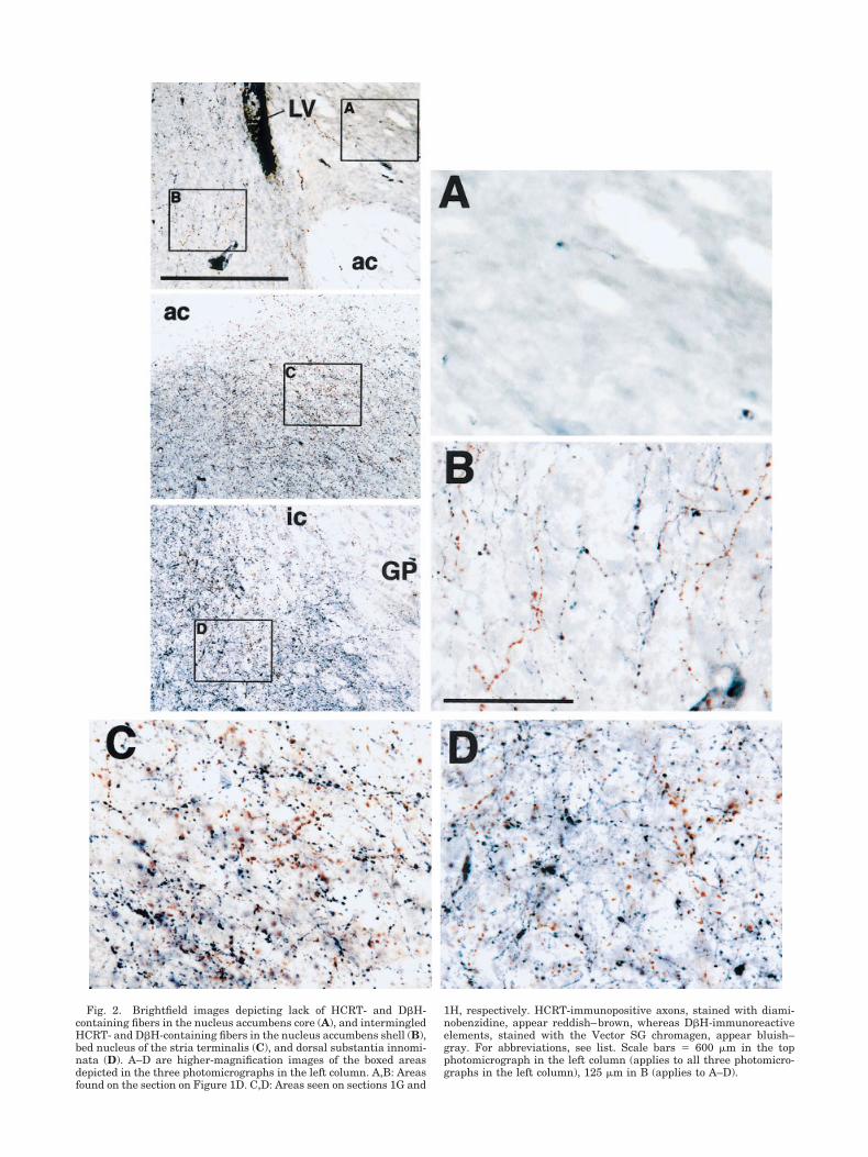
Fig. 2. Brightfield images depicting lack of HCRT- and D�H-containing fibers in the nucleus accumbens core (A), and intermingledHCRT- and D�H-containing fibers in the nucleus accumbens shell (B),bed nucleus of the stria terminalis (C), and dorsal substantia innomi-nata (D). A–D are higher-magnification images of the boxed areasdepicted in the three photomicrographs in the left column. A,B: Areasfound on the section on Figure 1D. C,D: Areas seen on sections 1G and
1H, respectively. HCRT-immunopositive axons, stained with diami-nobenzidine, appear reddish–brown, whereas D�H-immunoreactiveelements, stained with the Vector SG chromagen, appear bluish–gray. For abbreviations, see list. Scale bars � 600 �m in the topphotomicrograph in the left column (applies to all three photomicro-graphs in the left column), 125 �m in B (applies to A–D).

staining in this region was sufficiently intense as to beplainly apparent to the naked eye, and appeared in themicroscope as a dense plexus of immunoreactive boutons.Dorsal to the anterior commissure, the D�H innervationthinned somewhat, although still remaining quite dense.Distributed throughout the bed nucleus of the stria termi-nalis, and intermingled with D�H-containing elementsboth dorsal and ventral to the anterior commissure, werenumerous varicose, branching HCRT-immunoreactiveprocesses. At more anterior levels, these fibers were par-ticularly heavily concentrated in the dorsal regions of thebed nucleus of the stria terminalis, in a triangle-shapedterritory bounded by the anterior commissure and theinternal capsule (Fig. 1F); the axons in this subregionappeared to be contiguous with the HCRT innervation ofthe most caudal aspect of the nucleus accumbens. Therewere also considerable numbers of HCRT-containing fi-bers distributed throughout the dense patch of D�H im-munoreactivity in the ventral bed nucleus of the striaterminalis (Figs. 1F,G, 2C); these axons were typicallythick with many large boutons.
The dense D�H innervation of the ventral bed nucleusof the stria terminalis appeared to be contiguous with aheavy patch of immunopositive fibers and boutons visibleat more posterior levels in the ventral part and sections ofthe basal part of the substantia innominata (Fig. 1F,G; seealso Chang, 1989). Within the substantia innominata,there was considerable overlap of these D�H fibers withthick, varicose HCRT-containing axons, particularly inthe zone abutting the medial border of the globus pallidus,where the anterior portion of the internal capsule wasvisible (Figs. 1G,H, 2D). This dual innervation dropped offsharply within the globus pallidus, thereby distinctly de-fining the globus pallidus/substantia innominata bound-ary.
Starting in the dense patch of D�H-immunoreactiveboutons in the substantia innominata and continuing dor-sally along the medial perimeter of the globus pallidus andanterior portion of the internal capsule, many long, thinD�H-containing fibers, likely fibers of passage, were ob-served. Intermingled with these axons were long, thin,varicose HCRT-containing fibers oriented in parallel tothe D�H-containing axons.
In the basal substantia innominata, the dual HCRT/D�H innervation was less dense. A light distribution ofHCRT-containing fibers was noted in this region, inter-mixed with a moderate density of D�H-immunopositiveelements.
Hypothalamus
Both HCRT-immunoreactive and D�H-innunoreactiveaxons were present in abundance in the medial and lat-eral preoptic areas (Fig. 1F–H). The HCRT and D�H in-nervation of these regions appeared to be contiguous with,albeit somewhat less dense than, the innervation of thebed nucleus of the stria terminalis. This dual innervationextended ventrally to the base of the brain, although thedensity of HCRT-containing fibers was somewhat lighterin the suprachiasmatic nucleus relative to adjacent re-gions of the medial preoptic area. The supraoptic nucleuswas heavily innervated by D�H-immunopositive fibersbut contained only a few scattered HCRT-containing ax-ons (Fig. 1H).
Hypocretin-containing cell bodies were observed in thehypothalamus, concentrated mainly in the perifornical
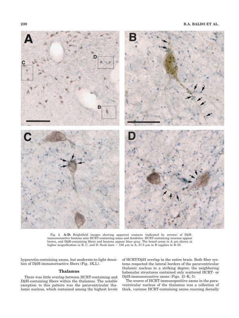
and lateral hypothalamus, with a few cells scattered an-teromedially in the paraventricular nucleus, and ventro-laterally in the medial tuberal area (Fig. 1I–L; see alsoPeyron et al., 1998; Sakurai et al., 1998). The hypocretin-containing cell bodies in the perifornical area were embed-ded within a dense collection of D�H-containing fiberslikely originating in the segment of the principal adrener-gic bundle that passes through the zona incerta, fromwhere it sends a branch laterally to innervate the para-ventricular nucleus and ventral aspect of the dorsomedialnucleus (Swanson and Hartman, 1975). Accordingly, wenoted smooth, thin D�H-containing fibers, collected intodiscrete fascicular bundles (consistent with fibers of pas-sage), in the vicinity of the zona incerta and internalcapsule. Emanating from these bundles was a denseswath of D�H-immunopositive fibers and boutons span-ning the hypothalamus from the zona incerta, through thehypocretin cell body-rich perifornical area and medial as-pect of the lateral hypothalamus, to the paraventricularnucleus (and at more posterior levels, the dorsal portion ofthe dorsomedial nucleus; Fig. 1J,K). At more caudal lev-els, this network of D�H immunoreactivity extended ven-trally into the ventromedial hypothalamic area and themedial tuberal nucleus. Within these regions, the D�H-containing fibers were thin, highly arborized, and con-tained many large boutons, features indicative of terminalfields. In many cases, there was the impression of synapticcontacts between hypocretin cell bodies and D�H-containing fibers and boutons (Fig. 4A–D).
There was considerable overlap between hypocretin-containing and D�H-containing fibers in a hypothalamiczone located between the paraventricular nucleus and theperifornical area. The paraventricular nucleus itself (asmentioned above) contained a very dense plexus of D�H-immunoreactive boutons; the staining in this region wasso intense that it could be seen with the naked eye (Figs.1H,I, 5A). Thick, varicose hypocretin-containing axonsoriginating in the perifornical area were also seen in theparaventricular nucleus intermingled with the D�H-containing boutons; these hypocretin fibers were mostheavily concentrated in the dorsomedial portion of thisstructure, in a region directly above the third ventricle,although there were also considerable numbers of fibersdistributed throughout the rest of the nucleus (Figs. 1H,I,5A). At more posterior levels, considerable overlap ofhypocretin-containing and D�H-containing fibers wasnoted in the dorsomedial hypothalamic nucleus, especiallyin the lateral portion of this nucleus (Fig. 1J,K); this dualinnervation appeared to be contiguous with the denseconcentration of hypocretin- and D�H-immunopositive fi-bers in the perifornical area.
Extending laterally from the fornix, moderate to heavyconcentrations of intermingled hypocretin-containing andD�H-containing fibers were observed in an area extendingthrough the lateral hypothalamus and traversing the zonebetween the internal capsule and optic tract toward tar-gets in the amygdala (Fig. 1I,J). Many of the hypocretin-containing fibers in this region, particularly those directlyadjacent to the optic tract, were smooth, thin, and orientedin a parallel manner, suggestive of fibers of passage. TheD�H-immunoreactive fibers in this hypothalamus/amygdala transition zone were varicose and arborized,compatible with terminal field morphology. Ventral hypo-thalamic structures, such as the ventromedial nucleusand the arcuate nucleus, contained moderate densities of
228 B.A. BALDO ET AL.

Fig. 3. Brightfield images depicting diverse HCRT- and D�H-containing fiber distributions and morphologies in the medial (top left,A) and lateral (middle left, B) subdivisions of the central amygdaloidnucleus and in the basolateral complex (bottom left, C). A–C: Higher-magnification images of the boxed areas in the three photomicro-graphs in the left column represent areas found within the amygdalaon the section shown in Figure 1J. HCRT-immunopositive axons,
stained with diaminobenzidine, appear reddish–brown, whereasD�H-immunoreactive elements, stained with the Vector SG chroma-gen, appear bluish–gray. Arrows in the top and middle photomicro-graphs of the left column point to the same histologic feature. Forabbreviations, see list. Scale bars � 600 �m in the top photomicro-graph in the left column (applies to all three photomicrographs in thatcolumn), 125 �m in A (applies to A–C).
hypocretin-containing axons, but moderate-to-light densi-ties of D�H-immunoreactive fibers (Fig. 1K,L).
Thalamus
There was little overlap between HCRT-containing andD�H-containing fibers within the thalamus. The notableexception to this pattern was the paraventricular tha-lamic nucleus, which contained among the highest levels
of HCRT/D�H overlap in the entire brain. Both fiber sys-tems respected the lateral borders of the paraventricularthalamic nucleus to a striking degree; the neighboringhabenular structures contained only scattered HCRT- orD�H-immunoreactive axons (Figs. 1I–K, 5).
The source of HCRT-immunopositive axons in the para-ventricular nucleus of the thalamus was a collection ofthick, varicose HCRT-containing axons coursing dorsally
Fig. 4. A–D: Brightfield images showing apparent contacts (indicated by arrows) of D�H-immunoreactive boutons onto HCRT-containing soma and dendrites. HCRT-containing neurons appearbrown, and D�H-containing fibers and boutons appear blue–gray. The boxed areas in A are shown athigher magnification in B, C, and D. Scale bars � 150 �m in A, 37.5 �m in B (applies to B–D).
230 B.A. BALDO ET AL.

Fig. 5. Brightfield images depicting HCRT- and D�H-containing fi-bers in the paraventricular nucleus of the hypothalamus (top left, A), theparaventricular thalamic nucleus (middle left, B), and the posteriorventral periaqueductal gray, in the vicinity of the dorsal raphe (bottomleft, C). A–C: Higher-magnification images of the boxed areas indicatedin the three photomicrographs in the left column depict areas found on
the sections shown in Figure 1I, 1K, and 1R, respectively. HCRT-immunopositive axons, stained with diaminobenzidine, appear reddish–brown, whereas D�H-immunoreactive elements, stained with the VectorSG chromagen, appear bluish–gray. For abbreviations, see list. Scalebars � 600 �m in the top photomicrograph in the left column (applies toall the photomicrographs in that column), 125 �m in A (applies to A–C).

along the midline, spanning the diencephalon from thedorsal portion of the paraventricular nucleus of the hypo-thalamus up to the base of the dorsal third ventricle (Fig.1J,K). A segment of this projection branched off to followthe ventral contour of the centromedial thalamic nucleus,whereas the remainder of the fibers continued dorsally tofill, at more anterior levels, the parataenial nucleus (Fig.1H), and further caudally, the paraventricular thalamicnucleus (Figs. 1I–K, 5B). The HCRT-containing fiberswere interdigitated with the two heavy patches of D�Himmunoreactivity within the paraventricular thalamicnucleus. These D�H-containing fibers were highly ar-borized and varicose, giving the impression at lower mag-nifications of homogeneous, densely packed immunoreac-tive boutons concentrated in bilateral oval-shaped patcheswithin the paraventricular thalamic nucleus (Fig. 5B). Atmore posterior levels, the dual HCRT/D�H innervation ofthe paraventricular nucleus extended ventrolaterally intothe region of the mediodorsal nucleus (Fig. 1L).
The remainder of the thalamus contained very fewhypocretin-containing fibers, and a thin distribution ofvery fine D�H-containing fibers. The dorsomedial part ofthe anteroventral thalamic nucleus contained a denseplexus of D�H-immunopositive axons (Fig. 1I); however,HCRT-containing fibers were largely absent from this re-gion.
Amygdala
The distributions and morphologies of D�H- and HCRT-containing fibers varied considerably among the subre-gions of the amygdala. A moderately dense network ofintermingled HCRT- and D�H-immunoreactive fibers wasobserved extending laterally from the hypothalamus, tra-versing the hypothalamus/amygdala transition zone, andarching over the optic tract to innervate the amygdaloiddivision of the bed nucleus of the stria terminalis, themedial subdivision of the central nucleus (Fig. 1I,J), and,to a lesser extent, the medial amygdaloid nucleus. Boththe HCRT-containing and the D�H-containing fibers inthese regions were arborized and contained numerousboutons, resembling fibers seen, for example, in the bednucleus of the stria terminalis and the dorsal substantiainnominata. This heavy dual innervation ended at themedial border of the lateral subdivision of the centralnucleus, which contained only a few, scattered D�H- orHCRT-containing fibers, and was thereby similar in ap-pearance to the overlying amygdalostriatal transitionzone and the caudate putamen (Figs. 1I–K, 3A,B).
The basolateral amygdaloid complex contained a mod-erate density of D�H-immunopositive fibers; however,there were few HCRT-containing fibers within this struc-ture (Figs. 1J,K, 3C). Unlike the fibers in the central andmedial amygdaloid nuclei, the D�H-immunopositive ax-ons in the basolateral complex were long, thin, and con-tained very small boutons. These fibers resembled thoseseen throughout the cortex. Scattered amid the D�H-containing fibers in the basolateral complex were a fewthick, varicose HCRT-immunopositive axons.
Hippocampus
The hippocampus contained almost no overlap betweenhypocretin-containing and D�H-containing fibers (Fig. 1I–P). Only a very few, randomly scattered hypocretin-containing axons were observed in the hippocampus. Incontrast, D�H-containing fibers were present throughout
the entire hippocampus; these axons were observed to bemost heavily concentrated in the dentate gyrus and, moreposteriorly, adjacent to fields CA1–CA3 (see also Swansonand Hartman, 1975).
Midbrain, pons, and medulla
The periaqueductal gray contained substantial overlapbetween HCRT-containing and D�H-containing fibers(Fig. 1N–P). Thick, varicose HCRT-immunopositive axonswere observed surrounding the cerebral aqueduct and fill-ing the medial aspects of the ventral and lateral quad-rants of the gray matter. These HCRT-containing fiberswere intermingled with thin, varicose D�H-immunoreactiveaxons. The dorsal regions of the periaqueductal gray con-tained fewer HCRT-containing and D�H -containing fi-bers. Extending laterally from the periaqueductal graywas a strip of HCRT-immunopositive axons, which tra-versed the midbrain and extended into the medial aspectof the medial geniculate complex (Fig. 1O). Within thisregion, the HCRT-containing axons were intermingledwith thin, varicose, highly arborized D�H-immunopositivefibers.
At more caudal levels, the intense HCRT innervation ofthe midline and ventrolateral portions of the periaqueduc-tal gray spread more laterally. Within these sectors, par-ticularly at more caudal levels, the thick, varicose HCRT-immunoreactive axons were intermingled with amoderate-to-heavy density of branching D�H-containingfibers with numerous prominent boutons. This overlapwas particularly apparent in the vicinity of the dorsalraphe (Figs. 1Q,R, 5C). The D�H axons and terminals inthese areas were associated with bilateral D�H-containing fiber bundles localized just outside the ventro-lateral border of the periaqueductal gray. Interspersedthrough these bundles was a moderate density of HCRTinnervation, part of a strip of HCRT-containing fibersextending ventrally and laterally from the periaqueductalgray. Similar to the pattern seen at more rostral levels,the density of HCRT and D�H immunoreactivity thinnedsomewhat in dorsal sectors of the periaqueductal gray.
Moderate overlap between HCRT-containing and D�H-containing fibers was also observed in the ventral tegmen-tal area (Fig. 1N–P). This region contained a moderateconcentration of thick, varicose HCRT-containing fibersintermingled with highly varicose and arborized D�H-immunopositive axons. The HCRT innervation of the ven-tral tegmental area continued laterally along the ventralcontour of the medial lemniscus, partially extending intothe medial lemniscus itself, as well as the paranigralnucleus, the parabrachial pigmented nucleus, and the dor-sal portion of the substantia nigra pars compacta, butavoiding the substantia nigra pars reticulata (Fig. 1O,P).Thin, varicose D�H-immunopositive axons overlapped theHCRT innervation along the ventral contour of the mediallemniscus but penetrated further ventrally into the sub-stantia nigra pars compacta than did the HCRT-containing fibers. A dense collection of D�H-immunoreactive axons was also observed arching alongthe dorsal contour of the medial lemniscus; there wererelatively few HCRT-containing axons within this region.Moderate overlap between the two fiber types was alsoseen more caudally in the vicinity of the retrorubral fieldand pedunculopontine tegmental nucleus (Fig. 1Q).
In accordance with previous reports (Peyron et al., 1998;Cutler et al., 1999; Hagan et al., 1999; Horvath et al.,
232 B.A. BALDO ET AL.

1999), a dense concentration of HCRT-immunopositivefibers was observed intermingled with the D�H-immunopositive noradrenergic cell bodies of the locus co-eruleus. These axons were branching and varicose andwere distributed throughout both the cell body-containingand dendritic regions of the locus coeruleus (Fig. 1S).Varicose HCRT-immunopositive axons were also noted inthe dorsal aspect of the pontine central gray, along theboundary of the fourth ventricle. These axons were inter-mingled among the D�H-containing elements (both den-drites and varicose axons) extending from the locus coer-uleus. Considerable overlap between the two fiber typeswas also noted in the parabrachial area (Fig. 1S) and in astrip extending through the subcoerulear region along thetrajectory of D�H-containing cell bodies, extending be-tween the locus coeruleus and the A5 noradrenergicgroup. There was a moderate-to-high density of HCRT-containing axons in this strip just lateral the D�H-containing soma, in the dendritic zones of these neurons(particularly in the vicinity of A5). Nevertheless, only afew clear instances of actual contacts onto D�H-containing dendrites or cell bodies were noted.
There was a moderate-to-dense concentration of HCRT-immunopositive fibers in the nucleus of the solitary tract,where the A2 noradrenergic neurons are found (Fig.1T,U). The densest concentrations of these axons werelocated just dorsally and laterally to the A2 noradrenergiccells, with scattered fibers extending into the A2 region,and, in a few cases, making apparent contacts onto nor-adrenergic neurons. The A1 noradrenergic area containedonly a few, randomly scattered HCRT-containing fibers(Fig. 1T,U).
DISCUSSION
Summary of results
Substantial overlap between HCRT- and D�H-containing projections was noted in the following zones:(1) a ventrally situated corridor extending rostrally fromthe HCRT-containing neurons in the perifornical/lateralhypothalamus through the anteroposterior extent of theforebrain (including medial hypothalamus, preoptic area,substantia innominata, bed nucleus of the stria termina-lis, medial and lateral septal regions, and medial nucleusaccumbens shell), penetrating into a circumscribed area inthe deep layers of infralimbic and dorsal peduncular cor-tex; (2) a ventrally situated corridor extending caudallyfrom the perifornical and lateral hypothalamus into mid-brain structures such as the ventral tegmental area, ret-rorubral field, and pedunculopontine tegmental nucleus;(3) ventral portions of the central gray, particularly in thevicinity of the dorsal raphe, and, further caudally, in themesencephalic gray and locus coeruleus; (4) a region ex-tending laterally from the perifornical hypothalamus, overthe optic tract, and into the intra-amygdaloid division ofthe bed nucleus of the stria terminalis and medial divisionof the central amygdaloid nucleus, ending abruptly at themedial border of the lateral division of this nucleus; (5) avery distinct area in the dorsal diencephalon including theparataenial, paraventricular, and mediodorsal thalamicnuclei. Except as noted above in (1), the majority of cortexcontained a moderately dense network of long, thin,branching D�H-containing fibers, which were similar inmorphology to those seen in the hippocampus and baso-
lateral amygdala. Intermixed with these cortical fiberswas a low density of randomly scattered, varicose HCRT-containing axons. Basal ganglia structures such as thecaudate putamen, nucleus accumbens core, and globuspallidus were devoid of both fiber types. Within HCRT cellbody-rich hypothalamic regions, a dense plexus of D�H-immunoreactive fibers and boutons was observed, withmany cases of apparent contacts of D�H-containing bou-tons onto HCRT soma and dendrites.
Although densely intermingled HCRT- and D�H-immunoreactive fibers were observed in numerous brainstructures (see above), the degree to which these processescontacted identical postsynaptic targets was not assessed;indeed, a general limitation of the light microscopy tech-niques used here is that they provide suggestive, ratherthan conclusive, evidence of synaptic contacts. Regardless,the present work was designed to provide a broad perspec-tive and descriptive catalog of the neuroanatomic sitesthat might support functional interactions betweenHCRT- and norepinephrine-containing terminal fields.These data can serve as a framework to guide both elec-tron microscopy studies, wherein more direct evidence ofsynaptic contacts can be obtained, as well as functionalstudies of HCRT/norepinephrine interactions.
Our general hypothesis was that D�H-immunoreactiveelements represented components of the norepinephrinesystem. Norepinephrine, however, is also a substrate forthe synthesis of epinephrine by means of the actions ofphenylethanolamine-N-methyl-transferase (PNMT) in ad-renergic neurons. Thus, the previously reported presenceof PNMT immunoreactivity in structures like the para-ventricular thalamic nucleus, perifornical hypothalamus,and medial amygdala (Hokfelt et al., 1984; Astier et al.,1987) raises the possibility that the D�H-containing pro-cesses in those structures represent adrenergic, ratherthan noradrenergic, fibers. Presently, the functional roleof epinephrine in the central nervous system is not well-characterized, although it is likely that this amine exertssome of the same effects as norepinephrine, consideringthat its affinity for noradrenergic receptors is close to thatof norepinephrine itself (Hoffman and Lefkowitz, 1995).Nevertheless, in general, D�H-containing projections inthe brain vastly outnumber PNMT-containing projections(Swanson and Hartman, 1975; Hokfelt et al., 1984), indi-cating that the majority of D�H-immunoreactive fibersobserved in the present study represented noradrenergicprocesses. More specifically, previous work examining themedial amygdala, stained separately in adjacent sectionsfor D�H and PNMT, revealed greater numbers of D�H-containing fibers than PNMT-immunopositive processes(Fallon and Ciofi, 1992; Asan, 1993). This finding indicatesthat the majority of D�H-containing elements in theamygdala are noradrenergic, and supports the principlethat “. . .levels of the earlier enzymes in the sequence ofcatecholamine synthesis are thought to be quite low incatecholaminergic fibers and terminals” (Fallon and Ciofi,1992).
HCRT/D�H overlap and theextended amygdala
The present findings provide support for the notion ofcontiguity and common modulation among several basalforebrain structures comprising the extended amygdala(for an extensive review of extended amygdala anatomy,see Alheid et al., 1995). For example, intermingled D�H-
233HYPOCRETIN/NOREPINEPHRINE OVERLAP IN RAT BRAIN

containing and HCRT-containing fibers were seenthroughout the bed nucleus of the stria terminalis. Theinnervation of the lateral bed nucleus of the stria termi-nalis appeared contiguous with a dense collection of bothfiber types in an area of the substantia innominata corre-sponding to the central and medial divisions of the sub-lenticular extended amygdala (Alheid et al., 1995). Pro-ceeding caudally, moderate concentrations of both fibertypes were noted in the medial subdivision of the centralamygdaloid nucleus. However, this dual innervation ter-minated at the medial border of the lateral subdivision ofthe central amygdaloid nucleus.
This distinction between the medial and lateral subdi-visions of the central nucleus is interesting in terms ofongoing debates regarding the classification and charac-terization of the various amygdaloid subregions. It hasbeen argued that portions of the central and medial amyg-daloid nuclei can be viewed as a specialized region of thestriatum. For example, in situ hybridization for glutamicacid decarboxylase mRNA reveals an unbroken swath ofneurons extending from the posterior caudate, throughthe amygdalostriatal transition zone, and into the centraland medial divisions of the amygdala (Swanson andPetrovich, 1998). This observation is in general agreementwith the present finding that the caudate putamen, amyg-dalostriatal transition zone, and the lateral subdivision ofthe central nucleus presented as a homogeneous regiondevoid of either HCRT- or D�H-containing fibers. Othershave proposed that the projections, intrinsic connectivity,and histochemical features of central amygdala subdivi-sions recapitulate features of core-shell-ventral pallidumorganization, with the lateral and medial subdivisions ofthe central nucleus nuclei displaying, respectively, “core-like” and “pallidum-like’” histochemical features and cir-cuitry (Cassell et al., 1999). Again, this model is consis-tent, in a general sense, with the present observation thatthe medial subdivision of the central nucleus, like portionsof the ventral pallidum, contains intermingled HCRT- andD�H-containing fibers, whereas the lateral subdivision,like the nucleus accumbens core, is devoid of either fibertype. Hence, the present data reveal further similaritiesbetween portions of the central amygdaloid nucleus andstriatum, with regard to distributions of overlappingHCRT and D�H projections to these areas.
The basolateral amygdaloid complex displayed a strik-ingly different pattern of innervation relative to the cen-tral amygdala subregions described above. Dopamine-�-hydroxylase immunopositive axons in the basolateralnucleus resembled the long, thin, branching D�H-containing fibers in the cortex, but differed from the short,varicose fibers and terminal boutons seen in the medialportions of the amygdala (see also Fallon and Ciofi, 1992;Asan, 1993, 1997). Moreover, the relative densities ofHCRT-containing vs. D�H-containing fibers in the baso-lateral nucleus of the amygdala (i.e., a moderate-to-heavydensity of D�H-containing fibers but only a few, scatteredHCRT-immunopositive axons) was very similar to the ra-tio of these two fiber types seen throughout the cortex butdiffered markedly from the ratio seen in the medial sub-division of the central amygdala, where both fiber typeswere present in abundance. Thus, the present results lendfurther support to the idea that the basolateral amygdalais structurally more similar to the cortex than it is to thecentral and medial divisions of the amygdala (McDonald,1992; Alheid et al., 1995; Swanson and Petrovich, 1998).
Functional considerations
Perhaps the most straightforward implication of thepresent data is that hypocretin and norepinephrine mayinteract functionally within the brain regions containingoverlapping projections from both systems. A comparisonof the distributions of hypocretin-1 receptor mRNA,hypocretin-2 receptor mRNA, and mRNA for �-adrenergicand �-adrenergic receptors reveals that some combinationof hypocretin and norepinephrine receptor subtypes ispresent in the main areas we identified as receiving over-lapping HCRT and D�H input (Asanuma et al., 1991;Talley et al., 1996; Rosin et al., 1996; Trivedi et al., 1998;Marcus et al., 2001), supporting the idea of functionalinteractions between these two neuromodulators. Thequestion then arises as to whether there is a unified set ofphysiological or behavioral processes subject to dualhypocretin/norepinephrine regulation that can be deducedon the basis of anatomic considerations. A review of theestablished functional roles of the areas most heavily in-nervated by the hypocretin and norepinephrine systemsindicates that these structures are involved in the regu-lation of arousal or behavioral state, autonomic functionand stress, and appetitively motivated behavior.
A compelling framework for hypocretin/norepinephrineinteractions in the control of behavioral state comes fromstudies of EEG and behavioral measures of arousal afterremote-controlled administration of either noradrenergicagonists or HCRT into the basal forebrain of sleeping rats.Infusions of either the �-adrenergic receptor agonist iso-proterenol (Berridge et al., 1996), the monoamine-releaseramphetamine (Berridge et al., 1999), or HCRT peptides(Espana et al., 2001) into a region encompassing the me-dial septum, caudomedial nucleus accumbens shell, andthe medial preoptic area elicited low-voltage cortical EEGpatterns indicative of waking, along with increased motoractivity. Examination of chartings from these studies re-veals an excellent concordance between the sites mostsensitive to the arousal-related effects of HCRT and nor-epinephrine, and the areas identified in the present workas containing moderate-to-dense concentrations of inter-mingled HCRT-containing and D�H-containing axons, es-pecially the medial septal region and the medial preopticarea. It would be of considerable interest, therefore, todetermine, in future studies, whether the combined ad-ministration of HCRT and norepinephrine into these sitesinteracts in an additive or superadditive manner in thecontrol of behavioral state.
In addition to HCRT-norepinephrine interactionswithin arousal-related forebrain regions, overlapping pro-jections from these two systems were noted within severalmonoaminergic nuclei within the midbrain and pons. Forexample, in agreement with prior studies, a dense concen-tration of HCRT-containing processes was observedwithin the locus coeruleus (Peyron et al., 1998; Cutler etal., 1999; Hagan et al., 1999; Horvath et al., 1999). We alsonoted considerable HCRT/ D�H overlap in the dorsal ra-phe and ventral tegmental area. Thus, HCRT and norad-renergic projections may interact to coordinate arousal-related dopaminergic, serotonergic, and noradrenergicinput to the basal forebrain and cortex.
Because we observed that HCRT-containing cell bodiesin the perifornical and lateral hypothalamus receive sub-stantial D�H innervation (including many cases of appar-ent contacts of D�H-immunopositive boutons onto HCRT-
234 B.A. BALDO ET AL.

containing soma), the question arose as to whether thesetwo systems interact reciprocally at the cell-body level.Thus, HCRT input to the A1 and A2 noradrenergic cellgroups, major sources of perifornical/lateral hypothalamicnorepinephrine (Woulfe et al., 1990; Aston-Jones et al.,1995), was examined. Although numerous HCRT-containing fibers were observed within the nucleus of thesolitary tract, site of the A2 noradrenergic cell group, mostof these HCRT fibers were in proximity to but not directlycontacting D�H-containing dendrites or soma (although afew examples of apparent contacts were noted). Only scat-tered HCRT fibers were seen in the vicinity of the A1region. These observations support the notion that HCRTmay modulate neuronal activity in the A2 cell group byinfluencing local circuitry in the vicinity of the noradren-ergic cell bodies, but do not form direct synapses ontothese cells.
Currently, there is a strong emphasis on the roles ofhypocretin and norepinephrine in arousal and sleep–wakeregulation. However, an examination of the distribution ofoverlapping HCRT and D�H innervation indicates thatthese neuromodulators are well-situated to mediate auto-nomic and endocrine functions as well. For example, thebed nucleus of the stria terminalis, which contains amongthe densest concentrations of intermingled HCRT-containing and D�H-containing processes in the entirebrain, projects to medullary centers such as the nucleus ofthe solitary tract and the parabrachial area to modulateautonomic processes (Schwaber et al., 1982; Sofroniew,1983; Grove, 1988; Moga et al., 1989; Loewy, 1991). More-over, there is substantial HCRT/D�H overlap in portionsof the extended amygdala, which projects to the paraven-tricular nucleus of the hypothalamus, a well-establishedcontrol center for hypothalamic–pituitary–adrenal axisregulation. The paraventricular nucleus itself containsnumerous, intermingled HCRT-containing and D�H-containing fibers. As a group, these brain structures areinvolved in centrally mediated autonomic and endocrineresponses associated with high arousal states, includingstress (e.g., Henke, 1984; Gray, 1993; Pacak et al., 1995;Koob, 1999), raising the possibility that joint hypocretin/norepinephrine actions in the bed nucleus of the striaterminalis and related areas of the extended amygdalaproduce a stressful or aversive behavioral state. This hy-pothesis is supported by the observation that central in-fusions of hypocretin enhance stress-related behaviorslike grooming, face-washing, and nonspecific chewing, be-yond a simple proportional increase resulting from thearousal-enhancing actions of this peptide (Espana et al.,2002).
It is important to note that a role for norepinephrine/hypocretin interactions in the control of appetitive orreward-related processes cannot be ruled out. For exam-ple, substantial HCRT/D�H overlap was seen in brainregions involved in feeding, such as the lateral hypothal-amus and caudomedial nucleus accumbens shell. We alsoobserved overlap within the ventral tegmental area. In-deed, HCRT-containing cell bodies are confined to thelateral/perifornical hypothalamus suggesting a funda-mental involvement, at least in a modulatory sense, inbrain reward processes. Thus, taken together, our obser-vations support a potential role for HCRT/adrenergic in-teractions in neural processes associated with both appet-itive and aversive states.
One of the most circumscribed areas of HCRT/D�Hoverlap was found in the paraventricular thalamic nu-cleus. This region of the thalamus receives inputs from thesuprachiasmatic nucleus (Watts et al., 1987) and projectsto the infralimbic cortex, nucleus accumbens, lateral sep-tum, bed nucleus of the stria terminalis, and amygdala(Moga et al., 1995; Bubser and Deutch, 1998), areas thatthemselves receive abundant overlapping HCRT-containing and D�H-containing projections. Based on itsconnectivity, it has been proposed that the paraventricu-lar thalamic nucleus is well-situated to modulate stress-related dopaminergic activity in the forebrain (Bubser andDeutch, 1999). Interestingly, the paraventricular tha-lamic nucleus also contains among the highest levels ofmelatonin receptor binding in the brain (Weaver et al.,1989; Williams et al., 1991). On the basis of these ana-tomic findings, it could be hypothesized that this region ofthe thalamus represents a control site where circadianinformation is received and then relayed to basal fore-brain regions that mediate adaptive behavioral state-related and motivational responses to environmental chal-lenges. Inputs from the hypocretin and norepinephrinesystems (and possibly the epinephrine system as well—see Hokfelt et al., 1984 and Astier et al., 1987) to theparaventricular nucleus are positioned to influence thesecircadian inputs, thereby potentially representing ameans for these neuromodulators to override circadiancontrol over basal forebrain sites relevant to motivationand arousal.
CONCLUSIONS
Although they originate in distinctly different parts ofthe brain, the hypocretin and norepinephrine systemsboth target structures implicated in an array of state-dependent physiological, cognitive, and affective pro-cesses, including those associated with appetitive andaversive motivational states. Within these regions,HCRT- and D�H-containing fibers are closely intermin-gled and, in several areas, respect precisely the sameboundaries. There are also numerous D�H-containing fi-bers and boutons in close apposition to HCRT-containingcell bodies, in a manner suggestive of synaptic contacts.Conversely, HCRT-containing axons contact the norad-renergic cells of the locus coeruleus and are positioned toinfluence neural circuitry in the vicinity of the A2 and A5noradrenergic cell groups. Thus, the present results pro-vide anatomic evidence supporting coordinatedhypocretin/norepinephrine actions within the brain.
ACKNOWLEDGMENTS
The authors thank Mrs. Carol Dizack for her artisticskill and assistance with the fiber chartings. We alsothank Dr. Charles F. Landry for advice on the immuno-histochemical procedures and for the use of his SPOT CCDimaging system, Dr. Steve Gammie for helpful insightsregarding our photographic images and for the use of hisAxioCam imaging system, Mr. Ken Sadeghian for techni-cal assistance, and Dr. Luis de Lecea for the generous giftof the anti–prepro-hypocretin antibody.
235HYPOCRETIN/NOREPINEPHRINE OVERLAP IN RAT BRAIN

LITERATURE CITED
Alheid GF, de Olmos, JS, Beltramino CA. 1995. Amygdala and extendedamygdala. In: Paxinos G, editor. The rat nervous system. San Diego:Academic Press, Inc. p 495–578.
Asan E. 1993. Comparative single and double immunolabeling with anti-sera against catecholamine biosynthetic enzymes: criteria for the iden-tification of dopaminergic, noradrenergic and adrenergic structures inselected rat brain areas. Histochemistry 99:427–442.
Asan E. 1997. Ultrastructural features of tyrosine-hydroxylase-immunoreactive afferents and their targets in the rat amygdala. CellTissue Res 288:449–469.
Asanuma M, Ogawa N, Mizukawa K, Haba K, Hirata H, Mori A. 1991.Distribution of the beta-2 adrenergic receptor messenger RNA in therat brain by in situ hybridization histochemistry: effects of chronicreserpine treatment. Neurochem Res 16:1253–1256.
Astier B, Kitahama K, Denoroy L, Jouvet M, Renaud B. 1987. Immuno-histochemical evidence for the adrenergic medullary longitudinal bun-dle as a major ascending pathway to the hypothalamus. Neurosci Lett78:241–246.
Aston-Jones G, Bloom FE. 1981a. Activity of norepinephrine-containinglocus coeruleus neurons in behaving rats anticipates fluctuations in thesleep-waking cycle. J Neurosci 1:876–886.
Aston-Jones G, Bloom FE. 1981b. Nonrepinephrine-containing locus coer-uleus neurons in behaving rats exhibit pronounced responses to non-noxious environmental stimuli. J Neurosci 1:887–900.
Aston-Jones G, Shipley MT, Grzanna R. 1995. The locus coeruleus, A5 andA7 noradrenergic cell groups. In: Paxinos G, editor. The rat nervoussystem. San Diego: Academic Press. p 183–205.
Berridge CW, Foote SL. 1991. Effects of locus coeruleus activation onelectroencephalographic activity in neocortex and hippocampus. J Neu-rosci 11:3135–3145.
Berridge CW, Foote SL. 1996. Enhancement of behavioral and electroen-cephalographic indices of waking following stimulation of noradrener-gic beta-receptors within the medial septal region of the basal fore-brain. J Neurosci 16:6999–7009.
Berridge CW, O’Neill J. 2001. Differential sensitivity to the wake-promoting actions of norepinephrine within the medial preoptic areaand the substantia innominata. Behav Neurosci 115:165–174.
Berridge CW, Bolen SJ, Manley MS, Foote SL. 1996. Modulation of fore-brain electroencephalographic activity in halothane-anesthetized ratvia actions of noradrenergic beta-receptors within the medial septalregion. J Neurosci 16:7010–7020.
Berridge CW, Stratford TL, Foote SL, Kelley AE. 1997. Distribution ofdopamine beta-hydroxylase-like immunoreactive fibers within theshell subregion of the nucleus accumbens. Synapse 27:230–241.
Berridge CW, O’Neil J, Wifler K. 1999. Amphetamine acts within themedial basal forebrain to initiate and maintain alert waking. Neuro-science 93:885–896.
Bourgin P, Huitron-Resendiz S, Spier AD, Fabre V, Morte B, Criado JR,Sutcliffe JG, Henriksen SJ, de Lecea L. 2000. Hypocretin-1 modulatesrapid eye movement sleep through activation of locus coeruleus neu-rons. J Neurosci 20:7760–7765.
Bubser M, Deutch AY. 1998. Thalamic paraventricular nucleus neuronscollateralize to innervate the prefrontal cortex and nucleus accumbens.Brain Res 787:304–310.
Bubser M, Deutch AY. 1999. Stress induces Fos expression in neurons ofthe thalamic paraventricular nucleus that innervate limbic forebrainsites. Synapse 32:13–22.
Cassell MD, Freedman LJ, Shi C. 1999. The intrinsic organization of thecentral extended amygdala. Ann N Y Acad Sci 877:217–241.
Chang HT. 1989. Noradrenergic innervation of the substantia innominata:a light and electron microscopic analysis of dopamine beta-hydroxylaseimmunoreactive elements in the rat. Exp Neurol 104:101–112.
Cutler DJ, Morris R, Sheridhar V, Wattam TA, Holmes S, Patel S, Arch JR,Wilson S, Buckingham RE, Evans ML, Leslie RA, Williams G. 1999.Differential distribution of orexin-A and orexin-B immunoreactivity inthe rat brain and spinal cord. Peptides 20:1455–1470.
Delfs JM, Zhu Y, Druhan JP, Aston-Jones GS. 1998. Origin of noradren-ergic afferents to the shell subregion of the nucleus accumbens: antero-grade and retrograde tract-tracing studies in the rat. Brain Res 806:127–140.
Espana RA, Baldo BA, Kelley AE, Berridge CW. 2001. Wake-promotingand sleep-suppressing actions of hypocretin (orexin): basal forebrainsites of action. Neuroscience 106:699–715.
Espana RA, Plahn S, Berridge CW. 2002. Circadian-dependent andcircadian-independent behavioral actions of hypocretin/orexin. BrainRes 943:224–236.
Estabrooke IV, McCarthy MT, Ko E, Chou TC, Chemelli RM, YanagisawaM, Saper CB, Scammell TE. 2001. Fos expression in orexin neuronsvaries with behavioral state. J Neurosci 21:1656–1662.
Fallon JH, Ciofi P. 1992. Distribution of monamines within the amygdala.In: Aggleton J, editor. The amygdala: neurobiological aspects of emo-tion, memory, and mental dysfunction. New York: Wiley-Liss Inc. p97–114.
Foote SL, Morrison JH. 1987. Extrathalamic modulation of cortical func-tion. Annu Rev Neurosci 10:67–95.
Foote SL, Aston-Jones G, Bloom FE. 1980. Impulse activity of locus coer-uleus neurons in awake rats and monkeys is a function of sensorystimulation and arousal. Proc Natl Acad Sci U S A 77:3033–3037.
Foote SL, Bloom FE, Aston-Jones G. 1983. Nucleus locus ceruleus: newevidence of anatomical and physiological specificity. Physiol Rev 63:844–914.
Fuxe K, Hamberger B, Hokfelt T. 1968. Distribution of noradrenalinenerve terminals in cortical areas of the rat. Brain Res 8:125–131.
Gray TS. 1993. Amygdaloid CRF pathways. Role in autonomic, neuroen-docrine, and behavioral responses to stress. Ann N Y Acad Sci 697:53–60.
Grove EA. 1988. Efferent connections of the substantia innominata in therat. J Comp Neurol 277:347–364.
Hagan JJ, Leslie RA, Patel S, Evans ML, Wattam TA, Holmes S, BenhamCD, Taylor SG, Routledge C, Hemmati P, Munton RP, Ashmeade TE,Shah AS, Hatcher JP, Hatcher PD, Jones DN, Smith MI, Piper DC,Hunter AJ, Porter RA, Upton N. 1999. Orexin A activates locus coer-uleus cell firing and increases arousal in the rat. Proc Natl Acad Sci US A 96:10911–110916.
Henke PG. 1984. The bed nucleus of the stria terminalis and immobiliza-tion-stress: unit activity, escape behaviour, and gastric pathology inrats. Behav Brain Res 11:35–45.
Hobson JA, McCarley RW, Wyzinski PW. 1975. Sleep-cycle oscillation:reciprocal discharge by two brainstem neuronal groups. Science 189:55–58.
Hoffman BB, Lefkowitz RJ. 1995. Catecholamines, sympathomimeticdrugs. In: Hardman JG, Limbird LE, Molinoff PB, Ruddon RW, GilmanAG, editors. Goodman & Gilman’s; the pharmacological basis of ther-apeutics. New York: McGraw-Hill. p 199–248.
Hokfelt T, Johansson O, Goldstein M. 1984. Central catecholamine neu-rons as revealed by immunohistochemistry with special reference toadrenaline neurons. In: Bjorklund A, Hokfelt T, editors. Classicaltransmitters in the CNS. New York: Elsevier. p 157–276.
Horvath TL, Peyron C, Diano S, Ivanov A, Aston-Jones G, Kilduff TS, vanDen Pol AN. 1999. Hypocretin (orexin) activation and synaptic inner-vation of the locus coeruleus noradrenergic system. J Comp Neurol415:145–159.
Koob GF. 1999. Corticotropin-releasing factor, norepinephrine, and stress.Biol Psychiatry 46:1167–1180.
Lin L, Faraco J, Li R, Kadotani H, Rogers W, Lin X, Qiu X, de Jong PJ,Nishino S, Mignot E. 1999. The sleep disorder canine narcolepsy iscaused by a mutation in the hypocretin (orexin) receptor 2 gene. Cell98:365–376.
Loewy AD. 1991. Forebrain nuclei involved in autonomic control. ProgBrain Res 87:253–268.
Marcus JN, Aschkenasi CJ, Lee CE, Chemelli RM, Saper CB, YanagisawaM, Elmquist JK. 2001. Differential expression of orexin receptors 1 and2 in the rat brain. J Comp Neurol 435:6–25.
McDonald AJ. 1992. Cell types and intrinsic connections of the amygdala.In: Aggleton JP, editor. The amygdala: neurobiological aspects of emo-tion, memory, and mental dysfunction. New York: Wiley-Liss Inc. p67–96.
Moga MM, Saper CB, Gray TS. 1989. Bed nucleus of the stria terminalis:cytoarchitecture, immunohistochemistry, and projection to the para-brachial nucleus in the rat. J Comp Neurol 283:315–332.
Moga MM, Weis RP, Moore RY. 1995. Efferent projections of the paraven-tricular thalamic nucleus in the rat. J Comp Neurol 359:221–238.
Nishino S, Ripley B, Overeem S, Lammers GJ, Mignot E. 2000. Hypocretin(orexin) deficiency in human narcolepsy. Lancet 355:39–40.
Pacak K, McCarty R, Palkovits M, Kopin IJ, Goldstein DS. 1995. Effects ofimmobilization on in vivo release of norepinephrine in the bed nucleusof the stria terminalis in conscious rats. Brain Res 688:242–246.
236 B.A. BALDO ET AL.

Paxinos G, Watson C. 1998. The rat brain in stereotaxic coordinates. SanDiego: Academic Press.
Peyron C, Tighe DK, van den Pol AN, de Lecea L, Heller HC, Sutcliffe JG,Kilduff TS. 1998. Neurons containing hypocretin (orexin) project tomultiple neuronal systems. J Neurosci 18:9996–10015.
Piper DC, Upton N, Smith MI, Hunter AJ. 2000. The novel brain neuropep-tide, orexin-A, modulates the sleep-wake cycle of rats. Eur J Neurosci12:726–730.
Rosin DL, Talley EM, Lee A, Stornetta RL, Gaylinn BD, Guyenet PG,Lynch KR. 1996. Distribution of alpha 2C-adrenergic receptor-likeimmunoreactivity in the rat central nervous system. J Comp Neurol372:135–165.
Sakurai T, Amemiya A, Ishii M, Matsuzaki I, Chemelli RM, Tanaka H,Williams SC, Richarson JA, Kozlowski GP, Wilson S, Arch JR, Buck-ingham RE, Haynes AC, Carr SA, Annan RS, McNulty DE, Liu WS,Terrett JA, Elshourbagy NA, Bergsma DJ, Yanagisawa M. 1998. Orex-ins and orexin receptors: a family of hypothalamic neuropeptides and Gprotein-coupled receptors that regulate feeding behavior. Cell 92:573–585.
Schwaber JS, Kapp BS, Higgins GA, Rapp PR. 1982. Amygdaloid and basalforebrain direct connections with the nucleus of the solitary tract andthe dorsal motor nucleus. J Neurosci 2:1424–1438.
Sofroniew MV. 1983. Direct reciprocal connections between the bed nu-cleus of the stria terminalis and dorsomedial medulla oblongata: evi-dence from immunohistochemical detection of tracer proteins. J CompNeurol 213:399–405.
Swanson LW, Hartman BK. 1975. The central adrenergic system. Animmunofluorescence study of the location of cell bodies and their effer-
ent connections in the rat utilizing dopamine-beta-hydroxylase as amarker. J Comp Neurol 163:467–505.
Swanson LW, Petrovich GD. 1998. What is the amygdala? Trends Neurosci21:323–331.
Talley EM, Rosin DL, Lee A, Guyenet PG, Lynch KR. 1996. Distribution ofalpha 2A-adrenergic receptor-like immunoreactivity in the rat centralnervous system. J Comp Neurol 372:111–134.
Thannickal TC, Moore RY, Nienhuis R, Ramanathan L, Gulyani S, AldrichM, Cornford M, Siegel JM. 2000. Reduced number of hypocretin neu-rons in human narcolepsy. Neuron 27:469–474.
Trivedi P, Yu H, MacNeil DJ, Van der Ploeg LH, Guan XM. 1998. Distri-bution of orexin receptor mRNA in the rat brain. FEBS Lett 438:71–75.
Watts AG, Swanson LW, Sanchez-Watts G. 1987. Efferent projections ofthe suprachiasmatic nucleus: I. Studies using anterograde transport ofPhaseolus vulgaris leucoagglutinin in the rat. J Comp Neurol 258:204–229.
Weaver DR, Rivkees SA, Reppert SM. 1989. Localization and character-ization of melatonin receptors in rodent brain by in vitro autoradiog-raphy. J Neurosci 9:2581–2590.
Williams LM, Martinoli MG, Titchener LT, Pelletier G. 1991. The ontogenyof central melatonin binding sites in the rat. Endocrinology 128:2083–2090.
Woulfe JM, Flumerfelt BA, Hrycyshyn AW. 1990. Efferent connections ofthe A1 noradrenergic cell group: a DBH immunohistochemical andPHA-L anterograde tracing study. Exp Neurol 109:308–322.
Zaborszky L, Cullinan WE. 1996. Direct catecholaminergic-cholinergic in-teractions in the basal forebrain. I. Dopamine-beta-hydroxylase- andtyrosine hydroxylase input to cholinergic neurons. J Comp Neurol374:535–554.
237HYPOCRETIN/NOREPINEPHRINE OVERLAP IN RAT BRAIN