orexin receptor 2 expression in the posterior … · orexin receptor 2 expression in the posterior...
TRANSCRIPT
Orexin receptor 2 expression in the posteriorhypothalamus rescues sleepiness in narcoleptic miceTakatoshi Mochizukia, Elda Arrigonia, Jacob N. Marcusb,c, Erika L. Clarka, Mihoko Yamamotoa, Michael Honerd,Edilio Borronid, Bradford B. Lowellb, Joel K. Elmquistb,e, and Thomas E. Scammella,1
aDepartment of Neurology and bDivision of Endocrinology, Beth Israel Deaconess Medical Center, Harvard Medical School, Boston, MA 02215; cMerckResearch Laboratories, West Point, PA 19486; dF. Hoffmann-La Roche, CH-4070 Basel, Switzerland; and eDepartments of Internal Medicine and Pharmacology,Division of Hypothalamic Research, University of Texas Southwestern Medical Center, Dallas, TX 75390
Edited by Richard D. Palmiter, University of Washington, Seattle, WA, and approved February 4, 2011 (received for review August 20, 2010)
Narcolepsy is caused by a loss of orexin/hypocretin signaling, re-sulting in chronic sleepiness, fragmented non-rapid eye movementsleep, and cataplexy. To identify the neuronal circuits underlyingnarcolepsy, we produced a mouse model in which a loxP-flankedgene cassette disrupts production of the orexin receptor type 2(OX2R; also known as HCRTR2), but normal OX2R expression canbe restored by Cre recombinase. Mice lacking OX2R signaling hadpoor maintenance of wakefulness indicative of sleepiness and frag-mented sleep and lacked any electrophysiological response toorexin-A in the wake-promoting neurons of the tuberomammillarynucleus. These defects were completely recovered by crossing themwith mice that express Cre in the female germline, thus globallydeleting the transcription-disrupter cassette. Then, by using anadeno-associated viral vector coding for Cre recombinase, we foundthat focal restoration of OX2R in neurons of the tuberomammillarynucleus and adjacent parts of the posterior hypothalamus com-pletely rescued the sleepiness of these mice, but their fragmentedsleep was unimproved. These observations demonstrate that thetuberomammillary region plays an essential role in the wake-promoting effects of orexins, but orexins must stabilize sleepthrough other targets.
histamine | Cre-loxP | G protein-coupled receptors | arousal
Narcolepsy is caused by a selective loss of the hypothalamicneurons producing the orexin (i.e., hypocretin) neuropep-
tides and is one of the most common causes of chronic sleepiness(1). In humans and mice, loss of orexin signaling results in un-stable sleep/wake states, with poor maintenance of wakefulness,fragmented sleep, and intrusions into wakefulness of elements ofrapid eye movement (REM) sleep, including brief episodes ofparalysis known as cataplexy. The orexin neuropeptides stronglyexcite many brain regions that regulate sleep/wake behavior,yet the key pathways through which orexins stabilize wakefulnessand sleep remain unknown.Orexins act through two receptors, OX1R and OX2R (also
known as HCRTR1 and HCRTR2), and the OX2R seems to playa critical role in the maintenance of wakefulness. Mice constitu-tively lacking OX2R are unable to maintain long bouts of wake-fulness and can fall asleep rapidly (2). In addition, an OX2Rantagonist strongly promotes sleep, whereas an OX1R antagonisthas no effect (3).Although it is clear that OX2R signaling is necessary for the
normal maintenance of wakefulness, the anatomic sites throughwhich this occurs remain unknown. OX2R is expressed in manywake-promoting brain regions, including the histaminergic neu-rons of the tuberomammillary nucleus (TMN), other mono-aminergic regions, cholinergic systems, and forebrain regions,including the thalamus and cortex (4). Several researchers havehypothesized that the TMN is a key site because orexin-A excitesthe TMN neurons and infusion of orexin-A near this regionpromotes wakefulness (5–7). However, this perspective is con-troversial as optogenetic activation of the orexin neurons pro-motes arousal in mice lacking histamine (8), and mice lacking
both OX1R and histamine H1 receptors have normal sleep/wakebehavior (9).The main goal of the present study was to determine the key
pathways through which OX2R signaling promotes wakefulness.We hypothesized that orexins stabilize wake through the TMNand adjacent regions. In addition, we predicted that if narco-leptic mice could produce longer periods of wakefulness, theywould develop stronger homeostatic sleep drive and their frag-mented sleep would become more consolidated. To define thesepathways, we produced mice with a loxP-flanked transcription-disrupter (TD) gene cassette that prevents expression of func-tional OX2R, but OX2R signaling in these mice can be reac-tivated by Cre recombinase. The advantage of this reactivationapproach is that OX2R expression can occur only in neurons thatwould normally express the receptor because the OX2R gene isdriven by the native promoter. We then induced local expressionof OX2R by microinjecting an adeno-associated viral vector(AAV) coding for Cre recombinase (AAV-Cre) to determinewhether focal rescue of orexin signaling in and around the TMNimproves the sleepiness and fragmented sleep of narcolepsy.
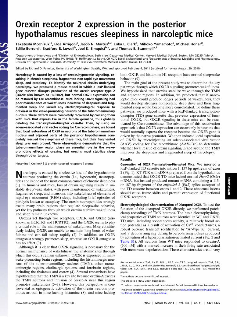
ResultsGeneration of OX2R Transcription-Disrupted Mice. We inserted aloxP-flanked TD cassette into intron 1, 137 bp upstream of exon2 (Fig. 1). RT-PCR with cDNA prepared from the hypothalamusdemonstrated that OX2R TD mice lacked normal Hcrtr2 (Ox2r)mRNA, but instead produced mutant mRNA containing a 115-or 187-bp fragment of the engrailed 2 (En2) splice acceptor ofthe TD cassette between exons 1 and 2. These abnormal insertsshould cause frame shifts and prevent expression of functionalOX2R receptors.
Electrophysiological Characterization of Disrupted OX2R. To test thefunction of the disrupted OX2R directly, we performed patch-clamp recordings of TMN neurons. The basic electrophysiolog-ical properties of TMN neurons were identical in WT and OX2RTD mice, including spontaneous activity, a relatively broad ac-tion potential as a result of activation of Ca2+ conductances, arobust outward transient rectification by “A”-type K+ current,and a depolarizing sag during hyperpolarizing pulses producedby activation of a hyperpolarization-activated current (Fig. 2 andTable S1). All neurons from WT mice responded to orexin-A(300 nM) with a marked increase in their firing rate associatedwith membrane depolarization. These characteristics are all very
Author contributions: T.M., J.N.M., B.B.L., J.K.E., and T.E.S. designed research; T.M., E.A.,J.N.M., E.L.C., M.Y., and M.H. performed research; E.B. contributed new reagents/analytictools; T.M., E.A., M.H., and T.E.S. analyzed data; and T.M., E.A., and T.E.S. wrote thepaper.
The authors declare no conflict of interest.
This article is a PNAS Direct Submission.1To whom correspondence should be addressed. E-mail: [email protected].
This article contains supporting information online at www.pnas.org/lookup/suppl/doi:10.1073/pnas.1012456108/-/DCSupplemental.
www.pnas.org/cgi/doi/10.1073/pnas.1012456108 PNAS | March 15, 2011 | vol. 108 | no. 11 | 4471–4476
NEU
ROSC
IENCE
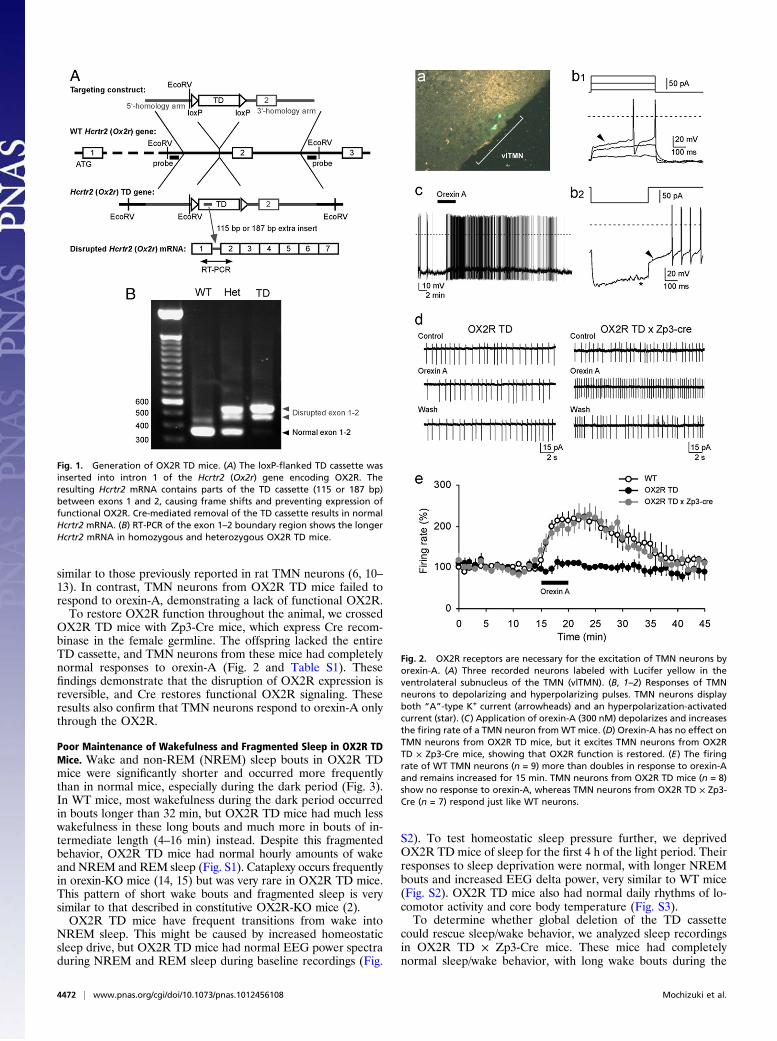
similar to those previously reported in rat TMN neurons (6, 10–13). In contrast, TMN neurons from OX2R TD mice failed torespond to orexin-A, demonstrating a lack of functional OX2R.To restore OX2R function throughout the animal, we crossed
OX2R TD mice with Zp3-Cre mice, which express Cre recom-binase in the female germline. The offspring lacked the entireTD cassette, and TMN neurons from these mice had completelynormal responses to orexin-A (Fig. 2 and Table S1). Thesefindings demonstrate that the disruption of OX2R expression isreversible, and Cre restores functional OX2R signaling. Theseresults also confirm that TMN neurons respond to orexin-A onlythrough the OX2R.
Poor Maintenance of Wakefulness and Fragmented Sleep in OX2R TDMice. Wake and non-REM (NREM) sleep bouts in OX2R TDmice were significantly shorter and occurred more frequentlythan in normal mice, especially during the dark period (Fig. 3).In WT mice, most wakefulness during the dark period occurredin bouts longer than 32 min, but OX2R TD mice had much lesswakefulness in these long bouts and much more in bouts of in-termediate length (4–16 min) instead. Despite this fragmentedbehavior, OX2R TD mice had normal hourly amounts of wakeand NREM and REM sleep (Fig. S1). Cataplexy occurs frequentlyin orexin-KO mice (14, 15) but was very rare in OX2R TD mice.This pattern of short wake bouts and fragmented sleep is verysimilar to that described in constitutive OX2R-KO mice (2).OX2R TD mice have frequent transitions from wake into
NREM sleep. This might be caused by increased homeostaticsleep drive, but OX2R TD mice had normal EEG power spectraduring NREM and REM sleep during baseline recordings (Fig.
S2). To test homeostatic sleep pressure further, we deprivedOX2R TD mice of sleep for the first 4 h of the light period. Theirresponses to sleep deprivation were normal, with longer NREMbouts and increased EEG delta power, very similar to WT mice(Fig. S2). OX2R TD mice also had normal daily rhythms of lo-comotor activity and core body temperature (Fig. S3).To determine whether global deletion of the TD cassette
could rescue sleep/wake behavior, we analyzed sleep recordingsin OX2R TD × Zp3-Cre mice. These mice had completelynormal sleep/wake behavior, with long wake bouts during the
Fig. 1. Generation of OX2R TD mice. (A) The loxP-flanked TD cassette wasinserted into intron 1 of the Hcrtr2 (Ox2r) gene encoding OX2R. Theresulting Hcrtr2 mRNA contains parts of the TD cassette (115 or 187 bp)between exons 1 and 2, causing frame shifts and preventing expression offunctional OX2R. Cre-mediated removal of the TD cassette results in normalHcrtr2 mRNA. (B) RT-PCR of the exon 1–2 boundary region shows the longerHcrtr2 mRNA in homozygous and heterozygous OX2R TD mice.
Fig. 2. OX2R receptors are necessary for the excitation of TMN neurons byorexin-A. (A) Three recorded neurons labeled with Lucifer yellow in theventrolateral subnucleus of the TMN (vlTMN). (B, 1–2) Responses of TMNneurons to depolarizing and hyperpolarizing pulses. TMN neurons displayboth “A”-type K+ current (arrowheads) and an hyperpolarization-activatedcurrent (star). (C) Application of orexin-A (300 nM) depolarizes and increasesthe firing rate of a TMN neuron from WT mice. (D) Orexin-A has no effect onTMN neurons from OX2R TD mice, but it excites TMN neurons from OX2RTD × Zp3-Cre mice, showing that OX2R function is restored. (E) The firingrate of WT TMN neurons (n = 9) more than doubles in response to orexin-Aand remains increased for 15 min. TMN neurons from OX2R TD mice (n = 8)show no response to orexin-A, whereas TMN neurons from OX2R TD × Zp3-Cre (n = 7) respond just like WT neurons.
4472 | www.pnas.org/cgi/doi/10.1073/pnas.1012456108 Mochizuki et al.
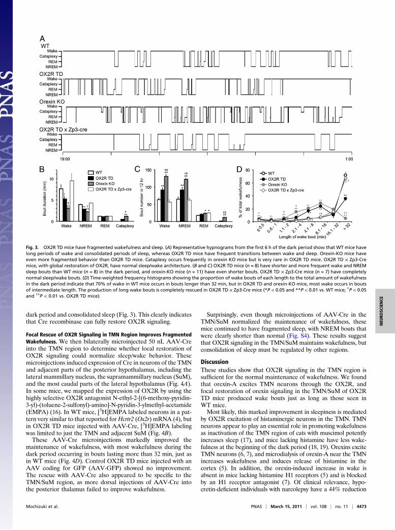
dark period and consolidated sleep (Fig. 3). This clearly indicatesthat Cre recombinase can fully restore OX2R signaling.
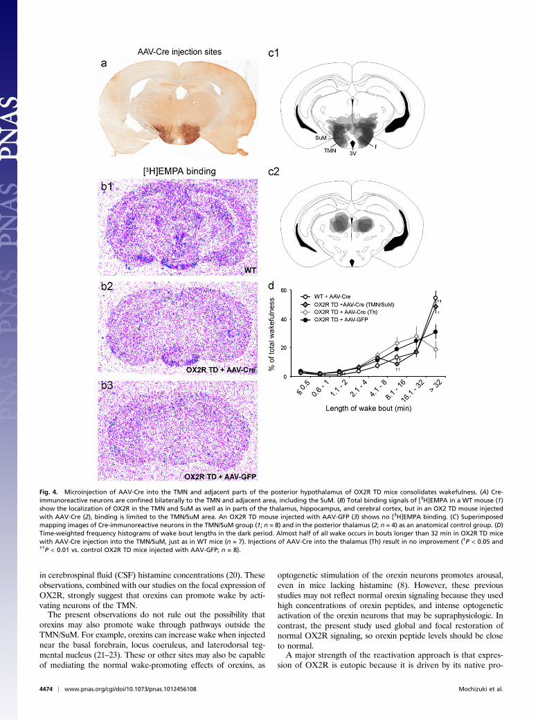
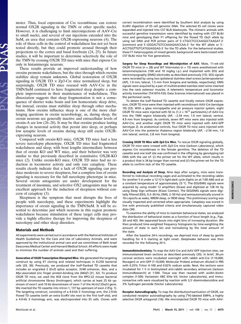
Focal Rescue of OX2R Signaling in TMN Region Improves FragmentedWakefulness. We then bilaterally microinjected 50 nL AAV-Creinto the TMN region to determine whether local restoration ofOX2R signaling could normalize sleep/wake behavior. Thesemicroinjections induced expression of Cre in neurons of the TMNand adjacent parts of the posterior hypothalamus, including thelateral mammillary nucleus, the supramammillary nucleus (SuM),and the most caudal parts of the lateral hypothalamus (Fig. 4A).In some mice, we mapped the expression of OX2R by using thehighly selective OX2R antagonist N-ethyl-2-[(6-methoxy-pyridin-3-yl)-(toluene-2-sulfonyl)-amino]-N-pyridin-3-ylmethyl-acetamide(EMPA) (16). In WT mice, [3H]EMPA labeled neurons in a pat-tern very similar to that reported forHcrtr2 (Ox2r) mRNA (4), butin OX2R TD mice injected with AAV-Cre, [3H]EMPA labelingwas limited to just the TMN and adjacent SuM (Fig. 4B).These AAV-Cre microinjections markedly improved the
maintenance of wakefulness, with most wakefulness during thedark period occurring in bouts lasting more than 32 min, just asin WT mice (Fig. 4D). Control OX2R TD mice injected with anAAV coding for GFP (AAV-GFP) showed no improvement.The rescue with AAV-Cre also appeared to be specific to theTMN/SuM region, as more dorsal injections of AAV-Cre intothe posterior thalamus failed to improve wakefulness.
Surprisingly, even though microinjections of AAV-Cre in theTMN/SuM normalized the maintenance of wakefulness, thesemice continued to have fragmented sleep, with NREM bouts thatwere clearly shorter than normal (Fig. S4). These results suggestthat OX2R signaling in the TMN/SuM maintains wakefulness, butconsolidation of sleep must be regulated by other regions.
DiscussionThese studies show that OX2R signaling in the TMN region issufficient for the normal maintenance of wakefulness. We foundthat orexin-A excites TMN neurons through the OX2R, andfocal restoration of orexin signaling in the TMN/SuM of OX2RTD mice produced wake bouts just as long as those seen inWT mice.Most likely, this marked improvement in sleepiness is mediated
by OX2R excitation of histaminergic neurons in the TMN. TMNneurons appear to play an essential role in promoting wakefulnessas inactivation of the TMN region of cats with muscimol potentlyincreases sleep (17), and mice lacking histamine have less wake-fulness at the beginning of the dark period (18, 19). Orexins exciteTMN neurons (6, 7), and microdialysis of orexin-A near the TMNincreases wakefulness and induces release of histamine in thecortex (5). In addition, the orexin-induced increase in wake isabsent in mice lacking histamine H1 receptors (5) and is blockedby an H1 receptor antagonist (7). Of clinical relevance, hypo-cretin-deficient individuals with narcolepsy have a 44% reduction
Fig. 3. OX2R TD mice have fragmented wakefulness and sleep. (A) Representative hypnograms from the first 6 h of the dark period show that WT mice havelong periods of wake and consolidated periods of sleep, whereas OX2R TD mice have frequent transitions between wake and sleep. Orexin-KO mice haveeven more fragmented behavior than OX2R TD mice. Cataplexy occurs frequently in orexin KO mice but is very rare in OX2R TD mice. OX2R TD × Zp3-Cremice, with global restoration of OX2R, have normal sleep/wake architecture. (B and C) OX2R TD mice (n = 8) have shorter and more frequent wake and NREMsleep bouts than WT mice (n = 8) in the dark period, and orexin-KO mice (n = 11) have even shorter bouts. OX2R TD × Zp3-Cre mice (n = 7) have completelynormal sleep/wake bouts. (D) Time-weighted frequency histograms showing the proportion of wake bouts of each length to the total amount of wakefulnessin the dark period indicate that 70% of wake in WT mice occurs in bouts longer than 32 min, but in OX2R TD and orexin-KO mice, most wake occurs in boutsof intermediate length. The production of long wake bouts is completely rescued in OX2R TD × Zp3-Cre mice (*P < 0.05 and **P < 0.01 vs. WT mice; †P < 0.05and ††P < 0.01 vs. OX2R TD mice).
Mochizuki et al. PNAS | March 15, 2011 | vol. 108 | no. 11 | 4473
NEU
ROSC
IENCE
in cerebrospinal fluid (CSF) histamine concentrations (20). Theseobservations, combined with our studies on the focal expression ofOX2R, strongly suggest that orexins can promote wake by acti-vating neurons of the TMN.The present observations do not rule out the possibility that
orexins may also promote wake through pathways outside theTMN/SuM. For example, orexins can increase wake when injectednear the basal forebrain, locus coeruleus, and laterodorsal teg-mental nucleus (21–23). These or other sites may also be capableof mediating the normal wake-promoting effects of orexins, as
optogenetic stimulation of the orexin neurons promotes arousal,even in mice lacking histamine (8). However, these previousstudies may not reflect normal orexin signaling because they usedhigh concentrations of orexin peptides, and intense optogeneticactivation of the orexin neurons that may be supraphysiologic. Incontrast, the present study used global and focal restoration ofnormal OX2R signaling, so orexin peptide levels should be closeto normal.A major strength of the reactivation approach is that expres-
sion of OX2R is eutopic because it is driven by its native pro-
Fig. 4. Microinjection of AAV-Cre into the TMN and adjacent parts of the posterior hypothalamus of OX2R TD mice consolidates wakefulness. (A) Cre-immunoreactive neurons are confined bilaterally to the TMN and adjacent area, including the SuM. (B) Total binding signals of [3H]EMPA in a WT mouse (1)show the localization of OX2R in the TMN and SuM as well as in parts of the thalamus, hippocampus, and cerebral cortex, but in an OX2 TD mouse injectedwith AAV-Cre (2), binding is limited to the TMN/SuM area. An OX2R TD mouse injected with AAV-GFP (3) shows no [3H]EMPA binding. (C) Superimposedmapping images of Cre-immunoreactive neurons in the TMN/SuM group (1; n = 8) and in the posterior thalamus (2; n = 4) as an anatomical control group. (D)Time-weighted frequency histograms of wake bout lengths in the dark period. Almost half of all wake occurs in bouts longer than 32 min in OX2R TD micewith AAV-Cre injection into the TMN/SuM, just as in WT mice (n = 7). Injections of AAV-Cre into the thalamus (Th) result in no improvement (†P < 0.05 and††P < 0.01 vs. control OX2R TD mice injected with AAV-GFP; n = 8).
4474 | www.pnas.org/cgi/doi/10.1073/pnas.1012456108 Mochizuki et al.
moter. Thus, focal expression of Cre recombinase can restorenormal OX2R signaling in the TMN or other specific nuclei.However, it is challenging to limit microinjections of AAV-Creto small nuclei, and several of our injections extended into theSuM, an area that contains OX2R-expressing neurons (4). Therole of these cells in the control of wakefulness has not yet beentested directly, but they could promote arousal through theirprojections to the cortex and basal forebrain (24, 25). In futurestudies, it will be important to determine selectively the role ofthe TMN by crossing OX2R TD mice with mice that express Creonly in histaminergic neurons.These results provide an improved understanding of how
orexins promote wakefulness, but the sites through which orexinsstabilize sleep remain unknown. Global restoration of OX2Rsignaling in OX2R TD × Zp3-Cre mice normalized sleep, butsurprisingly, OX2R TD mice rescued with AAV-Cre in theTMN/SuM continued to have fragmented sleep despite a com-plete improvement in their maintenance of wakefulness. Thisdissociation suggests that sleep fragmentation is not a conse-quence of shorter wake bouts and low homeostatic sleep drive,but instead, orexins must stabilize sleep through other mecha-nisms. How orexins influence sleep is one of the more chal-lenging questions in orexin neurobiology, as, during sleep, theorexin neurons are generally inactive and extracellular levels oforexin-A are low (26–28). It may be that orexins produce lastingchanges in neuronal activity that persist into sleep, or perhapslow synaptic levels of orexins during sleep still excite OX2R-expressing neurons.Compared with orexin-KO mice, OX2R TD mice had a less
severe narcolepsy phenotype. OX2R TD mice had fragmentedwakefulness and sleep, with bout lengths intermediate betweenthat of orexin KO and WT mice, and their behavior was verysimilar to that previously described in constitutive OX2R-KOmice (2). Unlike orexin-KO mice, OX2R TD mice had no re-duction in locomotor activity and only rare cataplexy. Theseresults clearly indicate that a lack of OX2R signaling can pro-duce moderate to severe sleepiness, but a complete loss of orexinsignaling is necessary for the full narcolepsy phenotype in mice.Several orexin antagonists are under development for thetreatment of insomnia, and selective OX2 antagonists may be anexcellent approach for the induction of sleepiness without con-cern of cataplexy (3).Sleepiness is the symptom that most disrupts the lives of
people with narcolepsy, and these experiments highlight theimportance of orexin signaling in the TMN/SuM. It will be es-sential to determine just which neurons in this region promotewakefulness because stimulation of these target cells may pro-vide a highly effective therapy for improving the sleepiness ofnarcolepsy and other sleep disorders.
Materials and MethodsAll experiments were carried out in accordance with the National Institutes ofHealth Guidelines for the Care and Use of Laboratory Animals, and wereapproved by the institutional animal care and use committees of Beth IsraelDeaconessMedical Center and HarvardMedical School. All efforts weremadeto minimize the number of animals used and their suffering.
Generation of OX2R Transcription-Disrupted Mice.We generated the targetingconstruct by using ET cloning and related techniques in EL250 bacterialcells (29, 30). Previously, we produced the loxP-flanked TD cassette thatincludes an engrailed 2 (En2) splice acceptor, SV40 enhancer, Neo, and aMyc-associated zinc finger protein-binding site (MAZ) (31, 32). To produceOX2R TD mice, we used the 4C8 clone from the RPCI-22 mouse bacterialartificial chromosome library (Invitrogen), which carries at least 25 kb up-stream of exon1 and 10 kb downstream of exon 7 of the Hcrtr2 (Ox2r) gene.We inserted the TD cassette into intron 1, 137 bp upstream of exon 2 (Fig. 1).The targeting construct, consisting of a 4.0-kb 5′-homology arm, the 2.9-kbfloxed TD cassette (with an extra EcoRV site next to the first loxP site), anda 4.0-kb 3′-homology arm, was electroporated into ES cells. Clones with
correct recombination were identified by Southern blot analysis by usingEcoRV digestion of ES cell genomic DNA. The selected ES cell clones wereexpanded and injected into C57 BL/6 blastocysts. The chimeric animals withsuccessful germline transmission were identified by mating with C57 BL/6Jmice and genotyping their F1 offspring for the floxed TD Ox2r allele byusing tail DNA and PCR primer pairs of 5′-CTGCCTCCCAAGGCTAAGAT-3′(common) and 5′-GGGACTGTCCAAAGAACCAA-3′ for the WT allele or 5′-ACCTGGTTGTCATGGAGGAG-3′ for the TD allele. For the behavioral studies,these F1 heterozygotes of mixed genetic background were backcrossed withC57 BL/6J mice for five generations.
Surgery for Sleep Recordings and Microinjection of AAV. Male, 11-wk-oldOX2R TD mice (n = 28) and WT littermates (n = 15) were anesthetized withketamine/xylazine (100 and 10 mg/kg i.p.) and implanted with EEG andelectromyography (EMG) electrodes as described previously (15). EEG signalswere recorded by using two ipsilateral stainless steel screws [anteroposterior(AP), 1.0 mm; lateral, 1.5 mm from bregma and lambda, respectively]. EMGsignals were acquired by a pair of multistranded stainless steel wires insertedinto the neck extensor muscles. A telemetric temperature and locomotoractivity transmitter (TA10TA-F20; Data Sciences International) was placed inthe peritoneal cavity.
To delete the loxP-flanked TD cassette and focally restore OX2R expres-sion, OX2R TD mice were then injected with recombinant AAV-Cre (serotype10) (33). With a glass micropipette and air pressure injector system, eightOX2R TD mice were injected with 50 nL (1.2 × 10¹² particles/mL) of AAV-Creinto the TMN region bilaterally (AP, −2.54 mm; 1.0 mm lateral; ventral,4.9 mm from bregma). As controls, seven WT mice were also injected withAAV-Cre, and another eight OX2R TD mice were injected with AAV-GFP(serotype 2). As anatomical controls, four OX2R TD mice were injected withAAV-Cre into the posterior thalamus region bilaterally (AP, −2.54 mm, 1.0mm lateral; ventral, 2.8 mm from bregma).
Crossing OX2R TD Mice with Zp3-Cre Mice. To rescue OX2R signaling globally,OX2R TD mice were crossed with Zp3-Cre mice (Jackson Laboratory), whichexpress Cre recombinase in the female germline. The deletion of the TDcassette and one loxP site was confirmed in the offspring by genotyping tailDNA with the use of: (i) the primer set for the WT allele, which results ina product that is 34 bp longer than normal and (ii) the primer set for the TDallele to confirm lack of the TD cassette.
Recording and Analysis of Sleep. Nine days after surgery, mice were trans-ferred to individual recording cages and acclimated to the recording cablesfor another 5 d under a 12:12 h light:dark cycle (lights on at 7:00 AM) and ata constant temperature of approximately 23 °C. The EEG/EMG signals wereacquired by using model 12 amplifiers (Grass) and digitized at 128 Hz byusing Sleep Sign software (Kissei Comtec). The EEG/EMG signals were digi-tally filtered (EEG, 0.3–30 Hz; EMG, 2–50 Hz) and semiautomatically scored in10-s epochs as wake, NREM sleep, or REM sleep. This preliminary scoring wasvisually inspected and corrected when appropriate. Cataplexy was scored inline with previously published criteria and simultaneously captured videodata (34).
To examine the ability of mice to maintain behavioral states, we analyzedthe distribution of behavioral states as a function of bout length (e.g., Figs.3D and 4D). We separated bouts into eight bins of increasing duration. Wethen produced time-weighted frequency histograms by time-weighting theamount of state in each bin and normalizing by the total amount ofthe state.
After the baseline 24-h recordings, we deprived mice of sleep by gentlehandling for 4 h starting at light onset. Sleep/wake behavior was thenrecorded for the following 20 h.
Immunohistochemistry. To map the AAV-Cre and AAV-GFP injection sites, weimmunostained brain sections as described previously (35). In brief, 30-μmcoronal sections were incubated overnight with rabbit anti-Cre (1:10,000;Novagen) or anti-GFP (1:10,000; Molecular Probes) antiserum diluted in PBSwith 0.25% Triton X-100 and 0.02% sodium azide. Next, the sections wereincubated for 1 h in biotinylated anti-rabbit secondary antiserum (JacksonImmunoResearch) at 1:500. Tissue was then reacted with avidin–biotincomplex (1:500; Vectastain ABC Elite kit; Vector Laboratories), and immu-noreactive cells were visualized by reaction with 3,3′-diaminobenzidine and3% hydrogen peroxide (Vector Laboratories).
Receptor Autoradiography. To map the distribution/reactivation of OX2R, weconducted receptor autoradiography by using [3H]-labeled EMPA, a highlyselective OX2R antagonist (16). We microinjected OX2R TD mice with AAV-
Mochizuki et al. PNAS | March 15, 2011 | vol. 108 | no. 11 | 4475
NEU
ROSC
IENCE

Cre (n = 3) or AAV-GFP (n = 2) and used WT mice (n = 2) as normal controls.Briefly, 10-μm coronal sections were incubated for 60 min with 5 nM [3H]EMPA in assay buffer containing (in mM): CaCl2 1, MgCl2 5, and Hepes 25, pH7.4. The sections were exposed to tritium-sensitive imaging plates (BAS-TR2025; Fuji Film) for 5 d, then scanned by BAS-5000 (Fuji Film) and analyzedwith an MCID M2 image analysis system (Imaging Research).
Patch-Clamp Recordings. Coronal slices (230 μm) of 2- to 3-wk-old mice (WT,n = 9; OX2R TD, n = 8; OX2R TD × Zp3-Cre, n = 7) were cut with a vibratingmicrotome in ice-cold artificial CSF containing (in mM): NaCl 124, KCl 2,KH2PO4 3, MgCl2 1.3, CaCl2 2.5, NaHCO3 26, and glucose 10, pH 7.4. Sliceswere recorded submerged and perfused with oxygenated artificial CSF (2mL/min) maintained at 32 °C. Patch-clamp recordings were performed incell-attached mode and whole-cell mode by using a Multiclamp 700A am-plifier (Molecular Devices) (36). Signals were digitized by using a Digidata1322A interface and acquired using Clampex 9.0 software (Molecular Devi-ces). Patch electrodes were filled with (in mM): K-gluconate 120, KCl 10,MgCl2 3, Hepes 10, K-ATP 2, and Na-GTP 0.2, pH 7.2. Lucifer yellow CHammonium salt (0.1%) was added to the pipette solution. Cell-attachedrecordings were performed in voltage clamp mode. Signals were low-passfiltered at 800 Hz and digitized at 10 kHz. Orexin-A (300 nM; Tocris) wasbath-applied to the recording chamber. After the recordings in cell-attached
mode, we switched to whole-cell configuration for the electrophysiologicalcharacterization of the neurons and to label them with Lucifer yellow.Whole-cell recordings were performed in current-clamp mode. Signals werelow-pass filtered at 20 kHz and digitized at 40 kHz. Series resistance wasmonitored at regular intervals with current pulses (−5 to −10 nA; 50–100ms). Data were analyzed by using Clampfit 9.0 (Molecular Devices) and IGORPro-4.0 (WaveMetrics) software.
Statistical Analysis. All results are expressed as means ± SEM. Changes inbehavioral state were compared across groups by using one-way factorialANOVA with a post-hoc Fisher protected least significant difference test.Time-weighted frequency histograms were compared between WT mice andother groups by using two-way, repeated-measures ANOVA with a post-hoc,two-tailed Student t test. Electrophysiologic responses were compared byusing one-way ANOVA.
ACKNOWLEDGMENTS. We thank N. Balthasar and R. Coppari for helpingwith the initial design of the OX2R TD mice and C. Saper and J. Wettstein forcritical reading of the manuscript. Orexin-KO mice were a gift from TakeshiSakurai of Kanazawa University. This research was supported by UnitedStates Public Health Service Grants NS055367, HL095491, and DK081185, anda grant from Takeda Pharmaceuticals.
1. Dauvilliers Y, Arnulf I, Mignot E (2007) Narcolepsy with cataplexy. Lancet 369:499–511.
2. Willie JT, et al. (2003) Distinct narcolepsy syndromes in orexin receptor-2 and Orexinnull mice: Molecular genetic dissection of Non-REM and REM sleep regulatoryprocesses. Neuron 38:715–730.
3. Dugovic C, et al. (2009) Blockade of orexin-1 receptors attenuates orexin-2 receptorantagonism-induced sleep promotion in the rat. J Pharmacol Exp Ther 330:142–151.
4. Marcus JN, et al. (2001) Differential expression of orexin receptors 1 and 2 in the ratbrain. J Comp Neurol 435:6–25.
5. Huang ZL, et al. (2001) Arousal effect of orexin A depends on activation of thehistaminergic system. Proc Natl Acad Sci USA 98:9965–9970.
6. Eriksson KS, Sergeeva O, Brown RE, Haas HL (2001) Orexin/hypocretin excites thehistaminergic neurons of the tuberomammillary nucleus. J Neurosci 21:9273–9279.
7. Yamanaka A, et al. (2002) Orexins activate histaminergic neurons via the orexin 2receptor. Biochem Biophys Res Commun 290:1237–1245.
8. Carter ME, Adamantidis A, Ohtsu H, Deisseroth K, de Lecea L (2009) Sleep homeostasismodulates hypocretin-mediated sleep-to-wake transitions. J Neurosci 29:10939–10949.
9. Hondo M, et al. (2010) Histamine-1 receptor is not required as a downstream effectorof orexin-2 receptor in maintenance of basal sleep/wake states. Acta Physiol (Oxf) 198:287–294.
10. Haas HL, Reiner PB (1988) Membrane properties of histaminergic tuberomammillaryneurones of the rat hypothalamus in vitro. J Physiol 399:633–646.
11. Greene RW, Haas HL, Reiner PB (1990) Two transient outward currents in histamineneurones of the rat hypothalamus in vitro. J Physiol 420:149–163.
12. Stevens DR, Eriksson KS, Brown RE, Haas HL (2001) The mechanism of spontaneousfiring in histamine neurons. Behav Brain Res 124:105–112.
13. Bayer L, et al. (2001) Orexins (hypocretins) directly excite tuberomammillary neurons.Eur J Neurosci 14:1571–1575.
14. Chemelli RM, et al. (1999) Narcolepsy in orexin knockout mice: Molecular genetics ofsleep regulation. Cell 98:437–451.
15. Mochizuki T, et al. (2004) Behavioral state instability in orexin knock-out mice. JNeurosci 24:6291–6300.
16. Malherbe P, et al. (2009) Biochemical and behavioural characterization of EMPA,a novel high-affinity, selective antagonist for the OX(2) receptor. Br J Pharmacol 156:1326–1341.
17. Lin JS, Sakai K, Vanni-Mercier G, Jouvet M (1989) A critical role of the posteriorhypothalamus in the mechanisms of wakefulness determined by microinjection ofmuscimol in freely moving cats. Brain Res 479:225–240.
18. Parmentier R, et al. (2002) Anatomical, physiological, and pharmacologicalcharacteristics of histidine decarboxylase knock-out mice: Evidence for the role ofbrain histamine in behavioral and sleep-wake control. J Neurosci 22:7695–7711.
19. Anaclet C, et al. (2009) Orexin/hypocretin and histamine: Distinct roles in the controlof wakefulness demonstrated using knock-out mouse models. J Neurosci 29:14423–14438.
20. Nishino S, et al. (2009) Decreased CSF histamine in narcolepsy with and without lowCSF hypocretin-1 in comparison to healthy controls. Sleep 32:175–180.
21. Bourgin P, et al. (2000) Hypocretin-1 modulates rapid eye movement sleep throughactivation of locus coeruleus neurons. J Neurosci 20:7760–7765.
22. España RA, Baldo BA, Kelley AE, Berridge CW (2001) Wake-promoting and sleep-suppressing actions of hypocretin (orexin): basal forebrain sites of action. Neuroscience106:699–715.
23. Xi MC, Morales FR, Chase MH (2001) Effects on sleep and wakefulness of the injectionof hypocretin-1 (orexin-A) into the laterodorsal tegmental nucleus of the cat. BrainRes 901:259–264.
24. Saper CB (1985) Organization of cerebral cortical afferent systems in the rat. II.Hypothalamocortical projections. J Comp Neurol 237:21–46.
25. Vertes RP (1992) PHA-L analysis of projections from the supramammillary nucleus inthe rat. J Comp Neurol 326:595–622.
26. Yoshida Y, et al. (2001) Fluctuation of extracellular hypocretin-1 (orexin A) levels inthe rat in relation to the light-dark cycle and sleep-wake activities. Eur J Neurosci 14:1075–1081.
27. Lee MG, Hassani OK, Jones BE (2005) Discharge of identified orexin/hypocretinneurons across the sleep-waking cycle. J Neurosci 25:6716–6720.
28. Mileykovskiy BY, Kiyashchenko LI, Siegel JM (2005) Behavioral correlates of activity inidentified hypocretin/orexin neurons. Neuron 46:787–798.
29. Copeland NG, Jenkins NA, Court DL (2001) Recombineering: A powerful new tool formouse functional genomics. Nat Rev Genet 2:769–779.
30. Liu P, Jenkins NA, Copeland NG (2003) A highly efficient recombineering-basedmethod for generating conditional knockout mutations. Genome Res 13:476–484.
31. Balthasar N, et al. (2005) Divergence of melanocortin pathways in the control of foodintake and energy expenditure. Cell 123:493–505.
32. Zigman JM, et al. (2005) Mice lacking ghrelin receptors resist the development of diet-induced obesity. J Clin Invest 115:3564–3572.
33. Lazarus M, et al. (2007) EP3 prostaglandin receptors in the median preoptic nucleusare critical for fever responses. Nat Neurosci 10:1131–1133.
34. Scammell TE, Willie JT, Guilleminault C, Siegel JM; International Working Group onRodent Models of Narcolepsy (2009) A consensus definition of cataplexy in mousemodels of narcolepsy. Sleep 32:111–116.
35. Yoshida K, McCormack S, España RA, Crocker A, Scammell TE (2006) Afferents to theorexin neurons of the rat brain. J Comp Neurol 494:845–861.
36. Scammell TE, et al. (2003) Focal deletion of the adenosine A1 receptor in adult miceusing an adeno-associated viral vector. J Neurosci 23:5762–5770.
4476 | www.pnas.org/cgi/doi/10.1073/pnas.1012456108 Mochizuki et al.