gabaa receptor-mediated input change on orexin …usdbiology.com/cliff/courses/advanced seminars...
TRANSCRIPT
Neuroscience 284 (2015) 217–224
GABAA RECEPTOR-MEDIATED INPUT CHANGE ON OREXINNEURONS FOLLOWING SLEEP DEPRIVATION IN MICE
T. MATSUKI, a*� M. TAKASU, a Y. HIROSE, a
N. MURAKOSHI, a,b C. M. SINTON, c T. MOTOIKE a ANDM. YANAGISAWA a*
a International Institute for Integrative Sleep Medicine
(WPI-IIIS), University of Tsukuba, Ibaraki 305-8575, Japan
bCardiovascular Division, Department of Internal Medicine, Institute
of Clinical Medicine, University of Tsukuba, Ibaraki 305-8575, Japan
cArizona Respiratory Center, University of Arizona,
AZ 85724-5030, United States
Abstract—Orexins are bioactive peptides, which have been
shown to play a pivotal role in vigilance state transitions: the
loss of orexin-producing neurons (orexin neurons) leads to
narcolepsy with cataplexy in the human. However, the effect
of the need for sleep (i.e., sleep pressure) on orexin neurons
remains largely unknown. Here, we found that immunostain-
ing intensities of the a1 subunit of the GABAA receptor and
neuroligin 2, which is involved in inhibitory synapse special-
ization, on orexin neurons of mouse brain were significantly
increased by 6-h sleep deprivation. In contrast, we noted that
immunostaining intensities of the a2, c2, and b2/3 subunits
of the GABAA receptor and Huntingtin-associated protein 1,
which is involved in GABAAR trafficking, were not changed
by 6-h sleep deprivation. Using a slice patch recording, orexin
neurons demonstrated increased sensitivity to a GABAA
receptor agonist together with synaptic plasticity changes
after sleep deprivation when compared with an ad lib sleep
condition. Insummary, theGABAergic inputpropertyoforexin
neurons responds rapidly to sleepdeprivation. Thismolecular
response of orexin neurons may thus play a role in the
changes that accompany the need for sleep following pro-
longed wakefulness, in particular the decreased probability
of a transition to wakefulness once recovery sleep has begun.
� 2014 IBRO. Published by Elsevier Ltd. All rights reserved.
Key words: orexin/hypocretin, sleep homeostasis, GABA,
insomnia, receptor.
http://dx.doi.org/10.1016/j.neuroscience.2014.09.0630306-4522/� 2014 IBRO. Published by Elsevier Ltd. All rights reserved.
*Corresponding authors at: International Institute for IntegrativeSleep Medicine (WPI-IIIS), University of Tsukuba, 1-1-1 Tennodai,Tsukuba, Ibaraki 305-8575, Japan. Tel: +81-29-853-3301.
E-mail addresses: [email protected] (T. Matsuki), [email protected] (M. Yanagisawa).
� Present address: Development & Medical Affairs Division, Glaxo-SmithKline K.K., Tokyo 151-8566, Japan.Abbreviations: ACSF, artificial cerebrospinal fluid; BZDs,benzodiazepines; CNS, central nervous system; eGFP, enhancedgreen fluorescent protein; GABA, c-aminobutyric acid; GABAAR,GABAA receptor; GABABR, GABAB receptor; HAP1, Huntingtin-associated protein 1; LH, lateral hypothalamus; MCH, melanin-concentrating hormone; mIPSCs, miniature inhibitory post synapticcurrents; NLGN2, neuroligin 2; NREM, non-rapid eye movement; PFA,paraformaldehyde; SD, sleep deprivation; SEM, standard error of themean; TTX, tetrodotoxin.
217
INTRODUCTION
Orexins (orexin A and B, also known as hypocretin-1 and
hypocretin-2, respectively) are bioactive peptides, which
are produced exclusively in the lateral hypothalamus
(LH) in mammalian brains and which have been shown
to play a critical role in regulating vigilance state
transitions (see reviews: (Sakurai, 2007; Sinton, 2011)).
The importance of orexin in the maintenance of consoli-
dated bouts of sleep and wakefulness has been convinc-
ingly demonstrated by the fact that the sleep disorder
narcolepsy with cataplexy is caused by a deficiency in
orexin or orexin-producing neurons (orexin neurons) in
humans and animals (Chemelli et al., 1999; Nishino
et al., 2000; Thannickal et al., 2000). The activity of orexin
neurons is increased during wakefulness and decreased
during sleep (Lee et al., 2005; Takahashi et al., 2008).
Furthermore, acute optogenetic activation of orexin
neurons enhances the probability of a transition from
sleep to wakefulness (Adamantidis et al., 2007), and
chronic stimulation or inhibition of orexin neurons leads
to a corresponding alteration in the vigilance state
(Sasaki et al., 2011). Orexin neurons receive multiple
afferents from several brain regions, including the limbic
system, preoptic area, and monoaminergic neurons, and
in vitro studies show that the activity of orexin neurons is
regulated by several neuropeptides and neurotransmitters
(Yoshida et al., 2006; Sakurai and Mieda, 2011). In
summary, although the activity of orexin neurons is known
to affect the continuity of sleep and wakefulness states,
the influence of prolonged wakefulness on the properties
of orexin neurons has not been characterized.
c-Aminobutyric acid (GABA) is the primary and most
abundant inhibitory neurotransmitter in the central
nervous system (CNS). Sleep-active, GABAergic
neurons in the preoptic area/anterior hypothalamus, in
addition to local GABAergic interneurons, densely
innervate the LH, including direct innervation of orexin
neurons (Steininger et al., 2001; Uschakov et al., 2006;
Hassani et al., 2010). Endogenous GABA release
increases in the LH area during non-rapid eye movement
(NREM) sleep (Nitz and Siegel, 1996; Alam et al., 2010).
GABA affects sleep regulation via two pharmacologically
distinct receptors, the GABAA receptor (GABAAR) and
GABAB receptor (GABABR) (Mohler, 2010). In a previous
study, we used genetically modified mice to determine
the role of the GABABR on orexin neurons in sleep
regulation (Matsuki et al., 2009). The specific deletion of
GABABRs on orexin neurons led to an instability of orexin
neuronal activity and an increment in membrane
218 T. Matsuki et al. / Neuroscience 284 (2015) 217–224
conductance, resulting in severe fragmentation of vigi-
lance states. However, the role of a GABAAR-mediated
inhibitory input on orexin neurons is poorly characterized.
GABAARs are pentameric hetero-oligomers and constitute
ligand-gated chloride channels (Olsen and Sieghart,
2008). GABAAR subunits are encoded by 19 different
genes that have been grouped into eight subclasses based
on sequence homology (a1–6, b1–3, c1–3, d, e, h,p,q1–3).The pentameric assembly of these subunits constitutes
the functional GABAAR and is dependent on the regional
distribution and cell-type-specific expression of each
subunit in the CNS (Fritschy and Mohler, 1995; Pirker
et al., 2000). Subunits of GABAARs on basal forebrain
cholinergic neurons (Modirrousta et al., 2007), and on
neurons in the perifornical hypothalamus (Volgin and
Kubin, 2007; Volgin et al., 2014) are up-regulated following
sleep deprivation (SD), indicating that GABAARs likely
play a role in the homeostatic regulation of sleep. However,
the molecular function and dynamics of GABAARs on
identified orexin neurons under these conditions are
unknown.
In this study, we examined themolecular and functional
alterations of GABAARs on orexin neurons following SD,
i.e., after an increase in homeostatic sleep pressure.
EXPERIMENTAL PROCEDURES
Mice
All experimental procedures involving animals were
approved by the Animal Experiment and Use Committee
of the University of Tsukuba and were performed strictly
in accordance with ‘‘Guidelines for proper conduct of
animal experiments’’, from the Science Council of Japan.
Orexin-enhanced green fluorescent protein (eGFP)
transgenic mice on a BDF1 background (Yamanaka
et al., 2003) were maintained on a 12-h light/dark cycle
(lights on from 9:00 A.M. [zeitgeber time: ZT0] to 9:00
P.M. [ZT12]). All experiments (ad lib (Ad lib) sleep and
SD conditions) were begun at ZT0 and the mice were
allowed ad libitum access to food and water throughout.
Mice were housed individually and were independently
and randomly assigned to each sleep condition. However,
environmental conditions (including temperature, humid-
ity, extraneous noise etc.) were identical for each mouse.
Mice in the SD condition were deprived of sleep for 6 h
(ZT0–ZT6) by gentle handling using the previously
reported technique (Suzuki et al., 2013). In contrast, as
reported by Suzuki et al. (2013) and confirmed by our
own visual observations, the Ad lib sleep group were
asleep for about 70% of the 6-h test period. After 6 h Ad
lib sleep or SD, mouse brains were processed for immuno-
histochemical and electrophysiological experiments as
described below. Mice used for immunohistochemistry
were 8–16wks of age (Ad lib sleep group: n= 8;SDgroup:
n= 10), and mice used for electrophysiology were 3–6
wks of age (Ad lib sleep group: n= 14; SD group: n= 17).
Immunohistochemistry
For immunohistochemical experiments, mice from each
condition were processed independently and randomly
while ensuring that environmental conditions and time of
sacrifice were the same for each mouse. In this study,
eGFP fluorescence-positive (eGFP+) cells were treated
as orexin-producing cells because there are no eGFP+
cells without expression of orexin mRNA (Yamanaka
et al., 2003) and we found here that over 80% of orexin-
containing cells were eGFP+ (i.e., about 20% of these
cells were below the fluorescence detection limit). Brains
of mice were fixed in 2% paraformaldehyde (PFA)
overnight. After fixation, brains were sectioned (50 lmcoronal sections) and stained with antibodies for GABAAR
a1 (EMD Millipore, Billerica, MA, USA; rabbit polyclonal,
1:500), GABAAR a2 (Synaptic Systems, Goettingen,
Germany; rabbit polyclonal, 1:500), GABAAR b2/b3(EMD Millipore; mouse monoclonal, 1:200), GABAAR c2(Synaptic Systems; rabbit polyclonal, 1:500), neuroligin
2 (NLGN2) (Santa Cruz Biotechnology Inc., Dallas, TX,
USA; goat polyclonal, 1:50) and Huntingtin-associated
protein 1 (HAP1) (Santa Cruz Biotechnology Inc.; goat
polyclonal, 1:200), and visualized by Alexa Fluor 594-con-
jugated secondary antibodies (1:2000) (Invitrogen,
Thermo Fisher Scientific Inc., Carlsbad, CA, USA). After
staining, images were acquired by laser-confocal micros-
copy equipped with a digital camera (LSM 700 with Axio
Imager.Z2 and 63� oil objective; Carl Zeiss Meditec
AG, Oberkochen, Germany). Luminance measurements
were performed by analytical imaging software, ZEN
2010 ver. 6 (Carl Zeiss Meditec AG). Mean intensities
(photon counts/unit area) of Alexa Fluor 594 fluorescence
from the top to the bottom of cell bodies were analyzed
randomly using the Histogram module in the ZEN soft-
ware (Carl Zeiss Meditec AG), i.e., whole-cell fluores-
cence was measured and we did not attempt separate
measurements of membrane fluorescence. To adjust
intra- and inter-procedural variance for staining indepen-
dently, the intensity of the fluorescence in GFP ‘‘negative’’
cells from the same slice was measured as a reference in
each sample. These measurements were performed
independently by two observers for the slices on each
slide and these data were then summed before analysis.
Slice preparation and patch clamp recording
Orexin-eGFP transgenic mouse brains were processed
for electrophysiology as described previously (Matsuki
et al., 2009) with minor modifications. Whole-cell patch
recordings were performed for 2 h from ZT7 to ZT9.
Reagents used were (in mM): slice preparation buffer:
210 sucrose, 2.5 KCl, 1.25 NaH2PO4, 10 MgCl2, 0.5
CaCl2, 26 NaHCO3, 11 glucose, pH 7.4; Bath buffer (arti-
ficial cerebrospinal fluid: ACSF): 125 NaCl, 2.5 KCl, 1.25
NaH2PO4, 26 NaHCO3, 1.2 MgCl2, 2.5 CaCl2, 11 glucose,
pH 7.4 (bubbled continuously with 95% O2/5% CO2),
290–310 mOsm; K-Cl pipette solution: 145 KCl, 10
HEPES, 0.5 EGTA, 2 MgCl2, 0.5 Na2GTP, 5 MgATP, with
or without 1 QX314 (Sigma–Aldrich Japan K.K., Tokyo,
Japan) pH 7.4, 290–310 mOsm. The liquid junction poten-
tial of these solutions was estimated at 4.1 mV and was
corrected in the data. For recording of the muscimol dose
response, brain slices were superfused with bath ACSF
and muscimol (Sigma–Aldrich Japan K.K.) was applied
to the bath solution at five concentrations increasing from

T. Matsuki et al. / Neuroscience 284 (2015) 217–224 219
80 nM to 50 lM in a stepwise manner. Each slice was
used for each dose response. When the response was
not observed at a lower concentration, the same slice
was then used to test the next higher concentration after
over 20-min washout period. For recording of miniature
inhibitory post synaptic currents (mIPSCs), we used bath
ACSF containing 50 lM DL-AP5 (Tocris Biosciences,
Bristol, UK), 20 lM CNQX (Wako Pure Chemical Indus-
tries, Osaka, Japan), and 1 lM tetrodotoxin (TTX)
(Sigma–Aldrich Japan K.K.). Data were acquired using
Axopatch 200B-Digidata 1322A and pClamp ver.8.
(Molecular Devices LLC, Sunnyvale, CA, USA) and ana-
lyzed using Clampfit (Molecular Devices LLC) and Mini
Analysis ver. 6 software (Synaptosoft, Decatur, GA,
USA). To identify orexin neurons, fluorescence and infra-
red-Dodt gradient contrast images were obtained with a
fixed stage upright microscope, Axio Examiner.D1 fitted
with the AxioCam MRm high resolution camera and using
Axiovision ver. 4 software (Carl Zeiss Meditec AG).
Statistics
To evaluate immunofluorescence intensity and
electrophysiological data, we analyzed data using
Student’s unpaired t-test for comparisons of
immunohistochemical intensities and mIPSCs, or a two-
way ANOVA (Ad lib sleep, SD) followed by Bonferroni
post hoc comparisons for the muscimol dose–response
analysis (GraphPad Prism 5.0; GraphPad Software Inc.,
La Jolla, CA, USA). Data were expressed as
mean ± standard error of the mean (SEM) and
differences were considered significant at P< 0.05.
RESULTS
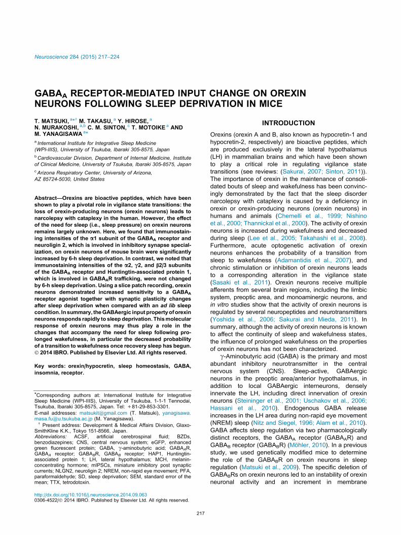
Changes in the expression of GABAARs on orexinneurons after sleep deprivation
To study the alteration in GABAergic input via GABAARs
on orexin neurons following SD, we sleep deprived adult
orexin-eGFP transgenic mice for 6 h. Control group
mice were allowed Ad lib sleep during this period.
Immunohistochemically, we studied the expression of
several GABAAR subunits on orexin neurons, as well as
molecules involved in GABAAR trafficking and inhibitory
synapse specialization. Based on previous reports that
GABAAR a1, a2, b2, b3, and c2 subunits are highly
expressed in the LH (Fritschy and Mohler, 1995;
Winsky-Sommerer, 2009), we examined the expression
of these subunits on orexin neurons from brains of mice
in the SD group in comparison with those from mice in
the Ad lib sleep group. The immunoreactivity of these
subunits was detected on the cell bodies and proximal
dendrites in GFP-positive orexin neurons as well as
GFP-negative non-orexin neurons. Although we found
no differences in the expression of the a2, c2, and b2/3subunits on orexin neurons of both groups (Fig. 1), the
intensity of GABAAR a1 subunit immunoreactivity was
visibly enhanced in orexin neurons from mice of the SD
group in comparison with those from the Ad lib sleep
group (Fig. 1A). Quantitative analyses of the luminance
intensities of subunit immunoreactivity showed that the
intensity of GABAAR a1 immunoreactivity was in fact sig-
nificantly higher on orexin neuron cell bodies from mice of
the SD group (Fig. 1B). Furthermore, after values were
normalized by the intensity of the same target proteins
on GFP negative neurons in the same photo view, values
remained significantly higher on orexin neurons from the
SD group (Fig. 1C). We also examined the expression
of two molecules in orexin neurons, HAP1 and NLGN2,
which are involved in GABAAR trafficking and inhibitory
synapse specialization, respectively. HAP1 is one of the
GABAAR trafficking molecules and is abundantly
expressed in orexin neurons in mouse brain (Lin et al.,
2010). The deletion of this gene in orexin neurons leads
to reductions in food intake, body weight and locomotor
activity (Lin et al., 2010). However, we did not observe
any changes in the expression of HAP1 in orexin neurons
from the SD group in comparison with those from the Ad
lib sleep group (Fig. 1B, C). In contrast, NLGN2
expression was significantly increased in orexin neurons
after SD (Fig. 1B, C). Neuroligins are postsynaptic cell
adhesion proteins known to interact with presynaptic
neurexins and postsynaptic PDZ domain-containing
scaffolding proteins (Craig and Kang, 2007). Among five
neuroligins, NLGN2 is exclusively localized to inhibitory
synapses and is believed to specify inhibitory synapses
(Varoqueaux et al., 2004; Chubykin et al., 2007).
Functional changes in orexin neurons followingsleep deprivation
We examined alterations in the electrophysiological
characteristics of orexin neurons from mice of the SD
group in comparison with those from the Ad lib sleep
group. We did not observe any significant difference
between the basal activities of orexin neurons from mice
of the two groups (Ad lib sleep: n= 6, 17 neurons; SD:
n= 6, 18 neurons). Resting membrane potentials were
�56.6 ± 1.2 mV versus �56.6 ± 0.4 mV, resting
discharge frequencies were 3.3 ± 0.5 Hz versus
3.2 ± 0.1 Hz, and membrane capacitances were 14.8 ±
1.8 pF versus 14.6 ± 1.2 pF in orexin neurons of the Ad
lib sleep versus SD groups, respectively.
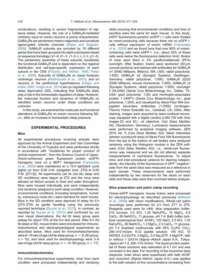
We next examined the response of orexin neurons to
the bath-applied GABAAR agonist, muscimol, under
whole-cell voltage clamping conditions (Vm= �50 mV).
Although we observed changes in current in neurons
from both groups following muscimol application at
concentrations higher than 0.4 lM, the current
amplitude following the same dose of muscimol (10 lM)
was significantly higher in the SD group than in the Ad
lib sleep group (Fig. 2A). The EC50 values from the
dose–response curve were 10.1 ± 3.3 lM in the Ad lib
sleep group and 5.7 ± 0.9 lM in the SD group
(Fig. 2B). These results suggested that the sensitivity of
orexin neurons to a GABAAR agonist was enhanced by
SD.
Alteration of mIPSCs on orexin neurons followingsleep deprivation
In order to examine the synaptic plasticity of orexin
neurons, we recorded mIPSCs on orexin neurons under
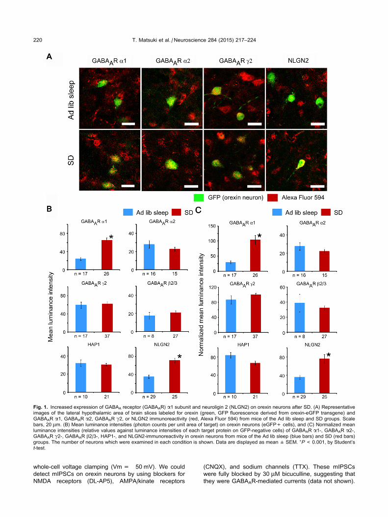
Fig. 1. Increased expression of GABAA receptor (GABAAR) a1 subunit and neuroligin 2 (NLGN2) on orexin neurons after SD. (A) Representative
images of the lateral hypothalamic area of brain slices labeled for orexin (green, GFP fluorescence derived from orexin-eGFP transgene) and
GABAAR a1, GABAAR a2, GABAAR c2, or NLGN2 immunoreactivity (red, Alexa Fluor 594) from mice of the Ad lib sleep and SD groups. Scale
bars, 20 lm. (B) Mean luminance intensities (photon counts per unit area of target) on orexin neurons (eGFP+ cells), and (C) Normalized mean
luminance intensities (relative values against luminance intensities of each target protein on GFP-negative cells) of GABAAR a1-, GABAAR a2-,GABAAR c2-, GABAAR b2/3-, HAP1-, and NLGN2-immunoreactivity in orexin neurons from mice of the Ad lib sleep (blue bars) and SD (red bars)
groups. The number of neurons which were examined in each condition is shown. Data are displayed as mean ± SEM. ⁄P< 0.001, by Student’s
t-test.
220 T. Matsuki et al. / Neuroscience 284 (2015) 217–224
whole-cell voltage clamping (Vm= �50 mV). We could
detect mIPSCs on orexin neurons by using blockers for
NMDA receptors (DL-AP5), AMPA/kinate receptors
(CNQX), and sodium channels (TTX). These mIPSCs
were fully blocked by 30 lM bicuculline, suggesting that
they were GABAAR-mediated currents (data not shown).
Fig. 2. Increased sensitivity of orexin neurons to a GABAAR agonist
after SD. (A) Representative traces from whole-cell patch recording
showing the effects of muscimol (10 lM, bath application) during
voltage clamp (Vm= �50 mV) on the current change of an orexin
neuron from mice of the Ad lib sleep and SD groups. Under these
conditions (i.e., ACSF-bath and K-Cl pipette solutions), current
changes were inward, as predicted. (B) Dose response curves of
muscimol on whole-cell currents during voltage clamp
(Vm= �50 mV) in orexin neurons from mice of the Ad lib sleep
(open circles, n= 8, each dose) and SD (closed circles, n= 8, each
dose) groups. Data are displayed as mean ± SEM. ⁄⁄P< 0.01,⁄P< 0.05 by a two-way ANOVA and Bonferroni post hoc test.
Fig. 3. Alteration of miniature inhibitory post synaptic currents
(mIPSCs) in orexin neurons after SD. (A) Representative data for
mIPSC recordings from whole-cell patch recording of orexin neurons
from mice of the Ad lib sleep and SD groups during voltage clamp
(Vm= �50 mV). Under these conditions (i.e., ACSF-bath and K-Cl
pipette solutions), currents were inward, as predicted. mIPSCs were
recorded in the presence of AP-5 (50 lM), CNQX (20 lM) and TTX
(1 lM). Summary data for mean mIPSC (B) frequency and (C)
amplitude from mice of the Ad lib sleep (n= 7) and SD (n= 6)
groups. Data are displayed as mean ± SEM. ⁄P< 0.05 by Student’s
t-test.
T. Matsuki et al. / Neuroscience 284 (2015) 217–224 221
We observed notable differences in frequency and
amplitude of the mIPSCs on orexin neurons between
the two groups (Fig. 3A). Both the frequency and
amplitude of mIPSCs were significantly enhanced in
orexin neurons from mice subjected to SD when
compared to orexin neurons from Ad lib sleeping
animals (Fig. 3B, C).
DISCUSSION
The orexin system is crucial for transitions between
vigilance states and thus for maintaining uninterrupted
periods of wakefulness and sleep (Sakurai, 2007). In this
study, we have demonstrated, both immunohistochemically
and functionally, that prolonged wakefulness affects the
GABAergic modulation of orexin neurons through
changes in GABAARs.
Expression of the a1 subunit of GABAARs in orexinneurons after sleep deprivation
Many hypnotics, including benzodiazepines (BZDs),
zopiclone and zolpidem, are known to target the asubunit of the GABAAR. Endogenous agonist binding
sites of GABAARs are directly modulated by these drugs
through allosteric mechanisms (Winsky-Sommerer,
2009; Rudolph and Knoflach, 2011). In this study, we
observed the up-regulation of GABAAR a1 subunit
expression in a specific functional nucleus following a
period of extended wakefulness. GABAARs containing
the a1 subunit show higher affinities and efficacies for
BZDs compared to other subunit-containing GABAARs
(Sieghart, 1995; Olsen and Sieghart, 2008). Thus the
up-regulation that we have demonstrated here will posi-
tively modulate endogenous agonist effects and enhance
GABAergic inhibitory currents in orexin neurons. Indeed,
our electrophysiological data showed enhancement of
the sensitivity for a GABAAR agonist in orexin neurons
after SD. Immunohistochemical data did not distinguish
those receptors located on the cell membrane or in the
cytoplasm. However, it is likely that the enhancement of
the sensitivity for a GABAAR agonist reflected an increase
in function of these receptors on the cell membrane.
These receptor alterations further suggest that orexin
neurons express the plasticity necessary to adapt rapidly
to ongoing changes in the need for sleep.
Our findings raise the possibility that SD might have
increased the total number of functional GABAARs on
orexin neurons. However, we did not observe any
differences between the Ad lib sleep and SD groups in
the expression of the other four subunits of GABAARs
including the c2 subunit. The c2 subunit usually occurs
in a one-to-one relationship with the a1 subunit and it is
therefore surprising that we did not observe an increase
in the c2 subunit. However, another subunit in a non-acontaining GABAAR could be replaced by the a1subunit following SD. Furthermore, it is known that the
majority (almost 90%) of GABAARs contain the c2subunit (Rudolph and Knoflach, 2011), and hence any
c2 subunit changes could be relatively insignificant. Over-
all therefore, these results suggest that the total amount
of GABAAR protein in each orexin neuron is unlikely to
222 T. Matsuki et al. / Neuroscience 284 (2015) 217–224
be much affected by SD. Instead, it is reasonable to
assume that subunit components of GABAARs were
altered and thus the proportion of a1 subunit-containing
GABAARs was increased following SD. One potential lim-
itation of this conclusion is that our antibody for the b2subunit did not differentiate the b3 subunit. This may be
of particular importance because a previous report
(Volgin et al., 2014), which found changes in GABAAR
mRNA expression in the perifornical hypothalamus follow-
ing SD in rats, reported that SD affected GABAAR b3expression but not b2 expression in this region. This
leaves open the possibility that we might have found
GABAAR b3 subunit up-regulation after SD in the present
study if our antibody had been sensitive to this differenti-
ation. However, apart from possible species and regional
differences, Volgin and colleagues did not examine
GABAAR mRNA expression at the level of specific cell
types (Volgin et al., 2014). Any potential changes to orex-
in neuron-specific a1 subunit expression in their study
might therefore have been negated by potentially larger
changes on a more numerous cell type in the region of
study. And, for the same reason, we found no difference
in GABAAR b2/b3 subunits because this change does
not occur on orexin neurons following SD.
Modulation of synaptic plasticity by sleep deprivation
Postsynaptic current changes are known to reflect
alterations in synapse number, presynaptic vesicle
formation and release, or post-synaptic receptor
modulation. While the change in the amplitude of
mIPSCs indicates alterations in the postsynaptic
component, including synaptic receptor sensitivity, the
change in the frequency of mIPSCs is thought to result
from modification to the presynaptic component of
synaptic transmission or formation of inhibitory
synapses (Turrigiano, 2012). Consequently, the change
in the amplitude of the mIPSCs that we found here would
be the basis for the enhancement of GABAAR agonist
sensitivity in orexin neurons from the SD group. Recently,
Ibata et al. reported that rapid changes in synaptic plastic-
ity were induced by changes in postsynaptic firing in cor-
tical neurons (Ibata et al., 2008). By extension, this
suggests that the change of orexin neuronal activity dur-
ing extended wakefulness might be sufficient to induce
the postsynaptic changes we found here and therefore
that the effect is not due to SD per se. In contrast, the
change in the frequency of mIPSCs would be the conse-
quence of increasing either the probability of presynaptic
vesicle release or synaptic formation. Since GABA
release is decreased in the LH during extended wakeful-
ness (Nitz and Siegel, 1996; Alam et al., 2010), the
enhanced frequency of mIPSCs found here indicates
therefore that GABAergic synapse formation increased
during SD.
Several studies have reported alteration in synaptic
plasticity with changes in vigilance state (McDermott
et al., 2003; Wang et al., 2011; Yan et al., 2011).
However, most studies have focused on hippocampal
and prefrontal cortical neurons and relatively little work
has addressed changes in hypothalamic neurons, includ-
ing orexin neurons. Orexin neurons remain active during
SD (Modirrousta et al., 2005) and it is likely therefore that
these neurons play a role, through innervation of all com-
ponents of the arousal system, in maintaining sufficient
alertness during long-term sleep loss. Rao et al. reported
that excitatory postsynaptic currents were enhanced in
mouse orexin neurons either by modafinil-induced pro-
longed wakefulness or by SD (Rao et al., 2007). These
authors hypothesized that orexin neurons maintain their
activity by means of excitatory synaptic plasticity changes
to adapt to homeostatic sleep pressure (Rao et al., 2007).
Similarly, the number of presynaptic output boutons of
orexin neurons was increased during prolonged wakeful-
ness in zebrafish (Appelbaum et al., 2010). Thus,
enhanced presynaptic excitatory neurotransmitter release
combined with changes to input mechanisms, including
synapse scaling and receptor accumulation at postsynap-
tic sites, maintain orexin neuronal activity under these
circumstances. Conversely, our findings now show that
SD also concurrently alters the sensitivity of orexin
neurons to inhibitory input by modulating GABAARs while
GABA release from presynaptic sites is reduced during
SD. This plasticity occurs over a duration that
corresponds approximately to the time that sleepiness
starts to intrude during prolonged wakefulness. This
suggests that a postsynaptic inhibitory mechanism on
orexin neurons is augmented during extended wakeful-
ness, presumably in preparation for eventual sleep onset.
If the orexin system plays an important role during
extended wakefulness to maintain vigilance, we have
shown here that it thus may also play a role afterextended wakefulness. The latter mechanism reduces
the probability of a transition to wakefulness once sleep
onset has occurred and so favors the maintenance of
sleep. Indeed, the latter has been confirmed in an optoge-
netic study (Carter et al., 2009), using a technique that
manipulates the postsynaptic response. Interestingly,
taken as a whole, these data suggest that it might eventu-
ally be possible to generate modulators for the orexin/
GABA system at presynaptic or postsynaptic sites that
have opposite functional effects, either excitatory or
inhibitory, depending on the need for sleep.
The expression of NLGN2 was also markedly
increased in orexin neurons after SD. A recent report
demonstrated that NLGN2 interacts with gephyrin and
collybistin, both of which are required for the
differentiation of inhibitory synapses (Poulopoulos et al.,
2009), and our present findings thus indicate that NLGN2
may modulate the inhibitory synapse on orexin neurons in
response to SD.
Potential experimental issues
For immunohistochemical analyses, the absolute intensity
of each marker was referenced to non-orexin cells (i.e.,
eGFP-negative neurons) to correct for intra- and inter-
procedural variance. These referenced cells were
selected randomly in the same slice and close to the
orexin target cells. The mean intensity of each target
protein and size of the non-orexin cells were found to be
similar between Ad lib sleep and SD groups, with the
exception of HAP1, which was significantly increased in
the SD group (data not shown). However, the relative
T. Matsuki et al. / Neuroscience 284 (2015) 217–224 223
intensity difference of HAP1 did not reach statistical
significance (Fig. 1C). In the LH, there are multiple
heterogeneous neuronal groups, containing, inter alia,
GABA, glutamate, histamine, and melanin-concentrating
hormone (MCH). Future studies, outside the scope of
the present work, will characterize how synaptic
modulation affects input to each of these types of
neuron during SD.
Extended wakefulness is inevitably associated with
increased motor activity and stress. Even a modest
duration of SD results in a systemic stress response,
including increased release of glucocorticoids and
proinflammatory cytokines. We have previously reported
that phosphorylated forms of dynamin 1 and N-myc
downstream-regulated gene 2 were increased by 6 h of
SD achieved by gentle handling even though these
proteins were not influenced by a restraint stress
(Suzuki et al., 2013). In that study, mice were subjected
to the restraint stress for 30 min and glucocorticoids levels
in the restraint stress group were higher than those of
mice in the SD group. Here mice were exposed to an
identical SD procedure and we can assume therefore that
glucocorticoid levels were increased to a similar extent.
We do not know, however, whether this level of stress
may have directly affected GABAARs on orexin neurons.
It is also unknown whether an increase in motor activity
that is inevitably associated with SD could have altered
the inhibitory input properties of orexin neurons. This is
especially relevant for the orexin system since the dis-
charge frequency of orexin neurons is increased in pro-
portion to the intensity of active wakefulness (Lee et al.,
2005; Takahashi et al., 2008). Further studies will there-
fore be required to confirm that the GABAAR change that
we have identified results from SD and not a correlated
but uncontrolled variable.
CONCLUSION
In this study, we have described a significant change to the
input properties of orexin neurons during SD. This change
has functional effects and indicates that orexin neurons
respond to extended wakefulness with a relatively rapid
change to GABAAR subunits that will favor the
maintenance of more prolonged sleep bouts once sleep
onset occurs. GABAARs on orexin neurons will therefore
contribute to at least one of the mechanisms by which
orexin modulates vigilance state transitions.
CONFLICT OF INTEREST
The authors declare that no competing interests exist.
AUTHOR CONTRIBUTIONS
T.Ma., M.T., Y.H., T.Mo., and M.Y. designed the
research. T. Ma., M.T., and Y.H. performed the
experiments and analyzed data. T.Ma., N.M., and M.Y.
contributed to analytic tools. T.Ma., C.M.S., T.Mo., and
M.Y. analyzed data and wrote the paper.
Acknowledgements—This study is supported by the Cabinet
Office, Government of Japan through the Funding Program for
World-Leading Innovative R&D on Science and Technology
(FIRST Program) (M.Y.), the Ministry of Education, Culture,
Sports, Science and Technology, World Premier International
Research Center Initiative (M.Y.), and the Perot Family
Foundation (M.Y.). M.Y. is a former Investigator of the Howard
Hughes Medical Institute.
REFERENCES
Adamantidis AR, Zhang F, Aravanis AM, Deisseroth K, De Lecea L
(2007) Neural substrates of awakening probed with optogenetic
control of hypocretin neurons. Nature 450:420–424.
Alam MN, Kumar S, Suntsova N, Bashir T, Szymusiak R, McGinty D
(2010) GABAergic regulation of the perifornical/lateral
hypothalamic neurons during non-rapid eye movement sleep in
rats. Neuroscience 167:920–928.
Appelbaum L, Wang G, Yokogawa T, Skariah GM, Smith SJ,
Mourrain P, Mignot E (2010) Circadian and homeostatic
regulation of structural synaptic plasticity in hypocretin neurons.
Neuron 68:87–98.
Carter ME, Adamantidis A, Ohtsu H, Deisseroth K, De Lecea L (2009)
Sleep homeostasis modulates hypocretin-mediated sleep-to-
wake transitions. J Neurosci 29:10939–10949.
Chemelli RM, Willie JT, Sinton CM, Elmquist JK, Scammell T, Lee C,
Richardson JA, Williams SC, Xiong Y, Kisanuki Y, Fitch TE,
Nakazato M, Hammer RE, Saper CB, Yanagisawa M (1999)
Narcolepsy in orexin knockout mice: molecular genetics of sleep
regulation. Cell 98:437–451.
Chubykin AA, Atasoy D, Etherton MR, Brose N, Kavalali ET, Gibson
JR, Sudhof TC (2007) Activity-dependent validation of excitatory
versus inhibitory synapses by neuroligin-1 versus neuroligin-2.
Neuron 54:919–931.
Craig AM, Kang Y (2007) Neurexin–neuroligin signaling in synapse
development. Curr Opin Neurobiol 17:43–52.
Fritschy J-M, Mohler H (1995) GABAA-receptor heterogeneity in the
adult rat brain: Differential regional and cellular distribution of
seven major subunits. J Comp Neurol 359:154–194.
Hassani OK, Henny P, Lee MG, Jones BE (2010) GABAergic
neurons intermingled with orexin and MCH neurons in the lateral
hypothalamus discharge maximally during sleep. Eur J Neurosci
32:448–457.
Ibata K, Sun Q, Turrigiano GG (2008) Rapid synaptic scaling induced
by changes in postsynaptic firing. Neuron 57:819–826.
Lee MG, Hassani OK, Jones BE (2005) Discharge of identified
orexin/hypocretin neurons across the sleep–waking cycle. J
Neurosci 25:6716–6720.
Lin Y-F, Xu X, Cape A, Li S, Li X-J (2010) Huntingtin-associated
protein-1 deficiency in orexin-producing neurons impairs neuronal
process extension and leads to abnormal behavior in mice. J Biol
Chem 285:15941–15949.
Mohler H (2010) Physiology and pharmacology of the GABA system:
focus on GABA receptors. In: Monti JM et al., editors. GABA and
sleep: molecular, functional and clinical aspects. Basel: Springer
Basel. p. 3–24.
Matsuki T, Nomiyama M, Takahira H, Hirashima N, Kunita S,
Takahashi S, Yagami K-i, Kilduff TS, Bettler B, Yanagisawa M,
Sakurai T (2009) Selective loss of GABAB receptors in orexin-
producing neurons results in disrupted sleep/wakefulness
architecture. Proc Natl Acad Sci 106:4459–4464.
McDermott CM, LaHoste GJ, Chen C, Musto A, Bazan NG, Magee
JC (2003) Sleep deprivation causes behavioral, synaptic, and
membrane excitability alterations in hippocampal neurons. J
Neurosci 23:9687–9695.
Modirrousta M, Mainville L, Jones B (2007) Dynamic changes in
GABAA receptors on basal forebrain cholinergic neurons following
sleep deprivation and recovery. BMC Neurosci 8:15.
Modirrousta M, Mainville L, Jones BE (2005) Orexin and MCH
neurons express c-fos differently after sleep deprivation vs.

224 T. Matsuki et al. / Neuroscience 284 (2015) 217–224
recovery and bear different adrenergic receptors. Eur J Neurosci
21:2807–2816.
Nishino S, Ripley B, Overeem S, Lammers GJ, Mignot E (2000)
Hypocretin (orexin) deficiency in human narcolepsy. Lancet
355:39–40.
Nitz D, Siegel JM (1996) GABA release in posterior hypothalamus
across sleep-wake cycle. Am J Physiol 271:R1707–R1712.
Olsen RW, Sieghart W (2008) International Union of Pharmacology.
LXX. Subtypes of c-aminobutyric acidA receptors: classification
on the basis of subunit composition, pharmacology, and function.
update. Pharmacol Rev 60:243–260.
Pirker S, Schwarzer C, Wieselthaler A, Sieghart W, Sperk G (2000)
GABAA receptors: immunocytochemical distribution of 13
subunits in the adult rat brain. Neuroscience 101:815–850.
Poulopoulos A, Aramuni G, Meyer G, Soykan T, Hoon M,
Papadopoulos T, Zhang M, Paarmann I, Fuchs C, Harvey K,
Jedlicka P, Schwarzacher SW, Betz H, Harvey RJ, Brose N,
Zhang W, Varoqueaux F (2009) Neuroligin 2 drives postsynaptic
assembly at perisomatic inhibitory synapses through gephyrin
and collybistin. Neuron 63:628–642.
Rao Y, Liu Z-W, Borok E, Rabenstein RL, Shanabrough M, Lu M,
Picciotto MR, Horvath TL, Gao X-B (2007) Prolonged
wakefulness induces experience-dependent synaptic plasticity in
mouse hypocretin/orexin neurons. J Clin Investig 117:4022–4033.
Rudolph U, Knoflach F (2011) Beyond classical benzodiazepines:
novel therapeutic potential of GABAA receptor subtypes. Nat Rev
Drug Discov 10:685–697.
Sakurai T (2007) The neural circuit of orexin (hypocretin): maintaining
sleep and wakefulness. Nat Rev Neurosci 8:171–181.
Sakurai T, Mieda M (2011) Connectomics of orexin-producing
neurons: interface of systems of emotion, energy homeostasis
and arousal. Trends Pharmacol Sci 32:451–462.
Sasaki K, Suzuki M, Mieda M, Tsujino N, Roth B, Sakurai T (2011)
Pharmacogenetic modulation of orexin neurons alters sleep/
wakefulness states in mice. PLoS One 6:e20360.
Sieghart W (1995) Structure and pharmacology of gamma-
aminobutyric acid A receptor subtypes. Pharmacol Rev
47:181–234.
Sinton CM (2011) Orexin/hypocretin plays a role in the response to
physiological disequilibrium. Sleep Med Rev 15:197–207.
Steininger TL, Gong H, McGinty D, Szymusiak R (2001) Subregional
organization of preoptic area/anterior hypothalamic projections to
arousal-related monoaminergic cell groups. J Comp Neurol
429:638–653.
Suzuki A, Sinton CM, Greene RW, Yanagisawa M (2013) Behavioral
and biochemical dissociation of arousal and homeostatic sleep
need influenced by prior wakeful experience in mice. Proc Natl
Acad Sci 110:10288–10293.
Takahashi K, Lin JS, Sakai K (2008) Neuronal activity of orexin and
non-orexin waking-active neurons during wake–sleep states in
the mouse. Neuroscience 153:860–870.
Thannickal TC, Moore RY, Nienhuis R, Ramanathan L, Gulyani S,
Aldrich M, Cornford M, Siegel JM (2000) Reduced number of
hypocretin neurons in human narcolepsy. Neuron 27:469–474.
Turrigiano G (2012) Homeostatic synaptic plasticity: local and global
mechanisms for stabilizing neuronal function. Cold Spring Harb
Perspect Biol 4:a005736.
Uschakov A, Gong H, McGinty D, Szymusiak R (2006) Sleep-active
neurons in the preoptic area project to the hypothalamic
paraventricular nucleus and perifornical lateral hypothalamus.
Eur J Neurosci 23:3284–3296.
Varoqueaux F, Jamain S, Brose N (2004) Neuroligin 2 is exclusively
localized to inhibitory synapses. Eur J Cell Biol 83:449–456.
Volgin DV, Kubin L (2007) Regionally selective effects of GABA on
hypothalamic GABAA receptor mRNA in vitro. Biochem Biophys
Res Commun 353:726–732.
Volgin DV, Lu JW, Stettner GM, Mann GL, Ross RJ, Morrison AR,
Kubin L (2014) Time- and behavioral state-dependent changes in
posterior hypothalamic GABAA receptors contribute to the
regulation of sleep. PLoS One 9:e86545.
Wang G, Grone B, Colas D, Appelbaum L, Mourrain P (2011)
Synaptic plasticity in sleep: learning, homeostasis and disease.
Trends Neurosci 34:452–463.
Winsky-Sommerer R (2009) Role of GABAA receptors in the
physiology and pharmacology of sleep. Eur J Neurosci
29:1779–1794.
Yamanaka A, Beuckmann CT, Willie JT, Hara J, Tsujino N, Mieda M,
Tominaga M, Yagami K-i, Sugiyama F, Goto K, Yanagisawa M,
Sakurai T (2003) Hypothalamic orexin neurons regulate arousal
according to energy balance in mice. Neuron 38:701–713.
Yan J, Li J-C, Xie M-L, Zhang D, Qi A-P, Hu B, Huang W, Xia J-X, Hu
Z-A (2011) Short-term sleep deprivation increases intrinsic
excitability of prefrontal cortical neurons. Brain Res 1401:52–58.
Yoshida K, McCormack S, Espana RA, Crocker A, Scammell TE
(2006) Afferents to the orexin neurons of the rat brain. J Comp
Neurol 494:845–861.
(Accepted 27 September 2014)(Available online 5 October 2014)