ox orexin/hypocretin receptor signaling through...
TRANSCRIPT
OX1 Orexin/Hypocretin Receptor Signaling through ArachidonicAcid and Endocannabinoid Release□S
Pauli M. Turunen, Maria H. Jantti, and Jyrki P. KukkonenBiochemistry and Cell Biology, Department of Veterinary Biosciences, University of Helsinki, Helsinki, Finland
Received February 1, 2012; accepted May 1, 2012
ABSTRACTWe showed previously that OX1 orexin receptor stimulationproduced a strong 3H overflow response from [3H]arachidonicacid (AA)-labeled cells. Here we addressed this issue with anovel set of tools and methods, to distinguish the enzymepathways responsible for this response. CHO-K1 cells heter-ologously expressing human OX1 receptors were used as amodel system. By using selective pharmacological inhibitors,we showed that, in orexin-A-stimulated cells, the AA-derivedradioactivity was released as two distinct components, i.e., freeAA and the endocannabinoid 2-arachidonoyl glycerol (2-AG).Two orexin-activated enzymatic cascades are responsible forthis response: cytosolic phospholipase A2 (cPLA2) and diacyl-glycerol lipase; the former cascade is responsible for part of theAA release, whereas the latter is responsible for all of the 2-AG
release and part of the AA release. Essentially only diacylglyc-erol released by phospholipase C but not by phospholipase Dwas implicated as a substrate for 2-AG production, althoughboth phospholipases were strongly activated. The 2-AG re-leased acted as a potent paracrine messenger through canna-binoid CB1 receptors in an artificial cell-cell communicationassay that was developed. The cPLA2 cascade, in contrast,was involved in the activation of orexin receptor-operated Ca2�
influx. 2-AG was also released upon OX1 receptor stimulation inrecombinant HEK-293 and neuro-2a cells. The results directlyshow, for the first time, that orexin receptors are able to gen-erate potent endocannabinoid signals in addition to arachi-donic acid signals, which may explain the proposed orexin-cannabinoid interactions (e.g., in neurons).
IntroductionThe peptide transmitters orexins/hypocretins, which act
through G-protein-coupled OX1 and OX2 receptors, are in-volved in the regulation of homeostatic brain functions, es-pecially wakefulness and sleep patterns and appetite (re-viewed in Kukkonen et al., 2002; Scammell and Winrow,
2011). Orexin receptor expression and signaling are alsofound in the periphery of the body, but the physiologicalsignificance of this is not known. The molecular mechanismsof orexin receptor signaling are very diverse (reviewed inKukkonen and Åkerman, 2005). Especially prominent seemsto be coupling to the production of messengers through phos-pholipase action (Johansson et al., 2008; Turunen et al.,2010a; Jantti et al., 2012).
Endocannabinoids are phospholipid-derived messengersthat contain arachidonic acid (AA). The most well establishedendocannabinoids are 2-arachidonoylglycerol (2-AG) andanandamide (N-arachidonoyl-ethanolamine) (reviewed inKano et al., 2009). Endocannabinoids act through G-protein-coupled CB1 and CB2 receptors; the CB1 receptor is ex-
This study was supported by the Academy of Finland, the Magnus Ehrn-rooth Foundation, the University of Helsinki Research Funds, the BiomedicumHelsinki Foundation, and the Research Foundation of the University of Hel-sinki.
Article, publication date, and citation information can be found athttp://molpharm.aspetjournals.org.
http://dx.doi.org/10.1124/mol.112.078063.□S The online version of this article (available at http://molpharm.
aspetjournals.org) contains supplemental material.
ABBREVIATIONS: AA, arachidonic acid; 2-AG, 2-arachidonoyl glycerol; AM-251, 1-(2,4-dichlorophenyl)-5-(4-iodophenyl)-4-methyl-N-1-piperidi-nyl-1H-pyrazole-3-carboxamide; CAY10499, [4-[5-methoxy-2-oxo-1,3,4-oxadiazol-3(2H)-yl]-2-methylphenyl]carbamic acid phenylmethyl ester;CAY10593, N-[2-[4-(5-chloro-2,3-dihydro-2-oxo-1H-benzimidazol-1-yl)-1-piperidinyl]-1-methylethyl]-2-naphthalenecarboxamide; CCPA, N-cyc-lohexanecarbonylpentadecylamine; CHO, Chinese hamster ovary; cPLA2, cytosolic (Ca2�-independent) phospholipase A2; DAG, diacylglycerol;DAGL, diacylglycerol lipase; FAAH, fatty acid amide hydrolase; FKGK11, 1,1,1,2,2-pentafluoro-7-phenyl-heptan-3-one; HBM, HEPES-bufferedmedium; HU-210, 3-(1,1�-dimethylheptyl)-6aR,7,10,10aR-tetrahydro-1-hydroxy-6,6-dimethyl-6H-dibenzo[b,d]pyran-9-methanol; IBMX, 3-isobu-tyl-1-methylxanthine; iPLA2, intracellular (Ca2�-independent) phospholipase A2; JZL184, 4-nitrophenyl-4-[dibenzo[d][1,3]dioxol-5-yl(hydroxy)m-ethyl]piperidine-1-carboxylate; MAFP, methyl arachidonyl fluorophosphonate; MAGL, monoacylglycerol lipase; PA, phosphatidic acid; PLA2,phospholipase A2; PLC, phospholipase C; PLD, phospholipase D; RHC-80267, 1,6-bis(cyclohexyloximinocarbonylamino)hexane; SB-334867,1-[2-methylbenzoxazol-6-yl]-3-[1,5]naphthyridin-4-yl-urea HCl; S-BSA, stripped bovine serum albumin; THL, tetrahydrolipstatin; TLC, thin-layerchromatography; U-73122, 1-[6-([(17b)-3-methoxyestra-1,3,5(10)-trien-17-yl]amino)hexyl]-1H-pyrrole-2,5-dione; URB597, [3-(3-carbamoylphe-nyl)phenyl] N-cyclohexylcarbamate.
1521-0111/12/8202-156–167$25.00MOLECULAR PHARMACOLOGY Vol. 82, No. 2Copyright © 2012 The American Society for Pharmacology and Experimental Therapeutics 78063/3781086Mol Pharmacol 82:156–167, 2012
156
http://molpharm.aspetjournals.org/content/suppl/2012/05/01/mol.112.078063.DC1Supplemental material to this article can be found at:
at ASPE
T Journals on July 31, 2018
molpharm
.aspetjournals.orgD
ownloaded from
pressed in central neurons. In addition to the receptors, theendocannabinoid system includes the enzymes that produceand metabolize endocannabinoids. Endocannabinoids aregenerally thought to be produced on demand and not storedin membrane vesicles like traditional neurotransmitters. En-docannabinoid signaling has been a subject of much interestfrom both the basic physiological perspective and the medicalone. In the central nervous system, endocannabinoids areinvolved in the regulation of, e.g., appetite, nociception, mem-ory, reward, and mood (reviewed in Kano et al., 2009). On thesynaptic level, endocannabinoids engage in retrograde trans-mission, in which postsynaptically produced endocannabi-noids act on the inhibitory, presynaptic CB1 receptors. Thisfunction may also act in homosynaptic feedback, but theassumed major function is heterosynaptic. Cannabinoid re-ceptors couple to G-proteins in the Gi family, and the presyn-aptic inhibition is likely to occur through G��-mediated in-hibition of voltage-gated Ca2� channels and inward rectifierK� channels (reviewed in Howlett, 2005; Kano et al., 2009).
There is much circumstantial evidence for interactions be-tween orexinergic and cannabinoidergic systems. These sys-tems show significant overlap on the gross neuroanatomicallevel, especially in particular nuclei of the hypothalamus.However, the possibility of interactions at the cellular levelhas not been verified for most nuclei. More-direct evidence isscarce. In the lateral hypothalamus, exogenous CB1 receptorstimulation inhibits orexinergic neurons by reducing the ex-citatory drive on them (Huang et al., 2007). In contrast, CB1
receptor block attenuates orexin-A-induced feeding (Crespoet al., 2008). It was proposed that CB1 and OX1 receptorsform heteromeric complexes, which may enhance orexin sig-naling (Hilairet et al., 2003; Ellis et al., 2006; Ward et al.,2011).
We showed previously that OX1 orexin receptor activationstrongly stimulates 3H overflow from [3H]AA-labeled cells(Turunen et al., 2010a). The full sensitivity of the response tothe reputed phospholipase A2 (PLA2) inhibitor methylarachidonyl fluorophosphonate (MAFP), in addition to par-tial sensitivity to inhibitors of other enzymes and intracellu-lar signal pathways (Turunen et al., 2010a), suggests thatthis effect is mediated by PLA2 enzymes. Although MAFP isvery commonly used as a PLA2 inhibitor, it is an analog of AAand is able to inhibit a number of serine hydrolases thathydrolyze ester- or amide-bound AA (De Petrocellis et al.,1997; Deutsch et al., 1997; Dinh et al., 2002; Savinainen etal., 2010). Therefore, does the 3H overflow observed takeplace solely through PLA2, and which isoform is involved?Another question left open is whether the 3H overflow iscomposed only of AA or whether other water-soluble andsecreted metabolites are involved. In the current study, weset out to resolve these questions. The results demonstratethat both free AA and AA incorporated into the endocannabi-noid 2-AG are released upon orexin receptor stimulation.Both AA and 2-AG are shown to have signaling roles of theirown.
Materials and MethodsDrugs. Human orexin-A and -B were obtained from NeoMPS (Stras-
bourg, France). 1-(2,4-Dichlorophenyl)-5-(4-iodophenyl)-4-methyl-N-1-piperidinyl-1H-pyrazole-3-carboxamide (AM-251), [4-[5-methoxy-2-oxo-1,3,4-oxadiazol-3(2H)-yl]-2-methylphenyl]carbamic acid phenylmethyl
ester (CAY10499), N-[2-[4-(5-chloro-2,3-dihydro-2-oxo-1H-benzimida-zol-1-yl)-1-piperidinyl]-1-methylethyl]-2-naphthalenecarboxamide(CAY10593) (compound 69 in Scott et al., 2009), N-cyclohexanecarbon-ylpentadecylamine (CCPA) (compound 17 in Tsuboi et al., 2004),1,1,1,2,2-pentafluoro-7-phenyl-heptan-3-one (FKGK11) (compound 10ain Baskakis et al., 2008), 3-(1,1�-dimethylheptyl)-6aR,7,10,10aR-tetrahydro-1-hydroxy-6,6-dimethyl-6H-dibenzo[b,d]pyran-9-methanol(HU-210), 4-nitrophenyl-4-[dibenzo[d][1,3]dioxol-5-yl(hydroxy)methyl-]piperidine-1-carboxylate (JZL184), lipid standards (2-AG and AA),MAFP, pyrrophenone [N-[[(2S,4R)-1-[2-(2,4-difluorobenzoyl)ben-zoyl]-4-[(triphenylmethyl)thio]-2-pyrrolidinyl]methyl]-4-[(Z)-(2,4-dioxo-5-thiazolidinylidene)methyl]benzamide] (compound 6 inSeno et al., 2001), and [3-(3-carbamoylphenyl)phenyl] N-cyclohexylcarbamate (URB597) were obtained from Cayman Eu-rope (Tallinn, Estonia). 1,6-Bis(cyclohexyloximinocarbonylamino)hexane (RHC-80267), 1-[2-methylbenzoxazol-6-yl]-3-[1,5]naphthyridin-4-yl-urea HCl (SB-334867), tetrahydrolipstatin(THL) (N-formyl-L-leucine-(1S)-1-[[(2S,3S)-3-hexyl-4-oxo-2-oxetanyl]methyl]dodecyl ester), and 1-[6-([(17b)-3-methoxyestra-1,3,5(10)-trien-17-yl]amino)hexyl]-1H-pyrrole-2,5-dione (U-73122) were obtained from Tocris Bioscience (Bristol, UK), fura-2acetoxymethyl ester from Invitrogen (Carlsbad, CA), and [5,6,8,9,11,12,14,15-3H]AA, [1-14C]AA, and [9,10-3H]oleic acid fromPerkinElmer Life and Analytical Sciences (Waltham, MA). Fors-kolin, probenecid [p-(dipropylsulfamoyl)benzoic acid], and3-isobutyl-1-methylxanthine (IBMX) were obtained from Sigma-Aldrich (St. Louis, MO).
Cell Culture. CHO-hOX1 cells expressing �500 fmol/mg ofprotein levels of high-affinity human OX1 receptors (as deter-mined from high-affinity 125I-orexin-A binding (P. M. Turunenand J. P. Kukkonen, unpublished data) were described previously,as were their culture conditions (Lund et al., 2000; Turunen et al.,2010a). CHO-hCB1 cells expressing human CB1a receptors (Grim-sey et al., 2010) were a kind gift from Dr. Michelle Glass (Univer-sity of Auckland, Auckland, New Zealand) via Drs. Jarmo Laiti-nen and Juha Savinainen (University of Eastern Finland, Kuopio,Finland); these cells were propagated under the same conditionsas CHO-hOX1 cells except that 0.25 mg/ml phleomycin (Zeocin;Invitrogen) was included. Neuro-2a-hOX1, PC12-hOX1, and wild-type HEK-293 cells were propagated as described previously (Hol-mqvist et al., 2002; Putula and Kukkonen, 2012) except that PC12cells received an additional supplement of 5% horse serum (Invit-rogen). For the 3H overflow experiments with [3H]AA- and [3H]o-leic acid-labeled cells and for the 2-AG reporter assays, the CHO-hOX1 cells were cultivated on 24-well plates (well bottom area,1.77 cm2; Greiner Bio-One GmbH, Frickenhausen, Germany)coated with polyethylenimine (25 �g/ml for 1 h at 37°C; Sigma-Aldrich); for Ca2� imaging, the cells were cultivated on polyeth-ylenimine-coated, circular, glass coverslips (diameter, 13 mm;Menzel-Glaser, Braunschweig, Germany). For the thin-layer chro-matography (TLC) assays, the cells were cultured on six-wellplates (bottom area, 9.6 cm2; Greiner Bio-One). For cAMP mea-surements (direct CB1 receptor assay or 2-AG reporter assay),CHO-hCB1 cells were cultured on plastic culture dishes (bottomarea, 56 cm2; Greiner Bio-One). HEK-293 cells were transientlytransfected with hOX1 receptor cDNA by using Fugene HD (RocheDiagnostics GmbH, Mannheim, Germany), as described previ-ously (Putula and Kukkonen, 2012).
3H Overflow from [3H]AA- or [3H]Oleic Acid-Labeled Cells.The experiments were largely performed as described previously(Turunen et al., 2010a). CHO-hOX1 cells were plated on 24-wellplates (20,000 cells per well) and left to grow for 24 h. The wells wereprecoated with polyethylenimine, which very much reduced cell de-tachment during the washes. Then, 0.1 �Ci of [3H]AA (or 0.2 �Ci of[3H]oleic acid, to compensate for the lower release levels and lower3H content) was added to each well, and the cells were cultured foranother 20 h. The incubation medium was removed, the cells werewashed twice with HEPES-buffered medium (HBM) (137 mM NaCl,
Orexin-Induced Arachidonic Acid and Endocannabinoid Release 157
at ASPE
T Journals on July 31, 2018
molpharm
.aspetjournals.orgD
ownloaded from
5 mM KCl, 1 mM CaCl2, 1.2 mM MgCl2, 0.44 mM KH2PO4, 4.2 mMNaHCO3, 10 mM glucose, and 20 mM HEPES, adjusted to pH 7.4with NaOH) supplemented with 2.4 mg/ml stripped bovine serumalbumin (S-BSA) (Turunen et al., 2010b), and the cells were left inHBM with S-BSA at 37°C. The cells were immediately stimulatedwith orexin-A or thapsigargin for 7 min, after which 200 �l of thetotal volume of 250 �l in each well was transferred to Eppendorftubes (VWR International, Radnor, PA) on ice. The samples werecentrifuged (16,000g for 1.5 min at 4°C), 100 �l of the medium wastransferred to a scintillation tube, scintillation cocktail (HiSafe 3;PerkinElmer Life and Analytical Sciences) was added, and the ra-dioactivity was measured with a Wallac 1414 liquid scintillationcounter (PerkinElmer Life and Analytical Sciences). It should benoted that total radioactivity released in the supernatant from the3H-preloaded cells was measured in this assay without any separa-tion with respect to molecular species.
In some cases, orexin stimulation was preceded by some inhib-itor preincubation. In such cases, the inhibitor used was added tothe cells after one wash with HBM plus S-BSA, and the cells werepreincubated for 30 min in HBM in the absence of S-BSA. HBMplus 10� S-BSA was added to a final concentration of S-BSA of 2.4mg/ml for the last 5 min, to chelate the radioactivity leaked duringthis period. The incubation solution was removed, fresh HBM plusS-BSA with the inhibitor was added, and the cells were stimulatedimmediately with orexin-A for 7 min. The control cells weretreated in the same way. The procedure effectively removed theradioactivity leaked during the preincubation period, and theS-BSA did not interfere with inhibitor entry into the cells.
Lipid Extraction and TLC. CHO-hOX1, neuro-2A-hOX1, PC12-hOX1, or transiently hOX1-transfected HEK-293 cells grown on six-well plates were labeled with [14C]AA (0.2 �Ci/ml) in cell culturemedium (Ham’s F12 medium) 16 h before the experiments. [14C]AAloading medium was removed, and the cells were washed twice withHBM plus S-BSA (2.4 mg/ml) and stimulated with orexin-A for 7min. Supernatants from the cells were rapidly removed and cen-trifuged (16,000g for 2 min at 4°C) to remove detached cells. Thelipids were extracted from the supernatant with a slight modifi-cation of the method described by Bligh and Dyer (1959). Briefly,800 �l of the total supernatant (1000 �l) was transferred to aKimax tube (Kimble Glass, Inc., Vineland, NJ), 2 ml of methanolwas added, followed by 1 ml of chloroform, and the tubes wereshaken thoroughly. After the addition of 1 ml of water and 1 ml ofchloroform, the tubes were shaken again and centrifuged (500g for5 min at room temperature). The lower phase was collected anddried under a stream of N2. The dried lipids were dissolved inchloroform, and the samples were applied to TLC plates (Silicagel60; Merck, Darmstadt, Germany), which were dried at 110°C for1 h. The plates underwent development with ethyl acetate/methanol (90:10) (Glass et al., 2005) in a chromatography tanklined with filter paper. Nonlabeled lipid standards were includedon each TLC plate, either in separate lanes or together with thesamples; the movement of the lipids of interest (2-AG and AA) inthe samples was not affected by the presence of the standards. Theaverage retardation factor values for AA and 2-AG were 0.58 �0.01 and 0.67 � 0.01, respectively.
TLC data were quantitated both through imaging plate analysisand through scraping and scintillation counting; the two methodsyielded comparable results (Jantti et al., 2012). For this reason,only the results from the imaging plate analysis are presented.After development, the plates were vacuum-dried and an imagingplate (BAS-MS; Fujifilm, Tokyo, Japan) was exposed overnight.The imaging plate was scanned with a Fujifilm FLA 5100 scanner,and the band areas and intensities were measured with ImageJ(http://rsbweb.nih.gov/ij). The plate background was subtractedfrom the band intensities.
2-AG Reporter Assay and cAMP Measurements. 2-AG re-lease from CHO-hOX1 cells upon orexin receptor stimulation wasdetected and quantitated by using CHO-hCB1 cells as detector
cells; the assay is conceptually reminiscent of, for instance, anitric oxide production assay (Hu and el-Fakahany, 1993). CHO-hCB1 cells on cell culture dishes were prelabeled with 5 �Ci/ml[3H]adenine in culture medium for 2 h, after which the cells werewashed with phosphate-buffered saline and detached with phos-phate-buffered saline plus 0.2% (w/v) EDTA. The cells were cen-trifuged, resuspended in HBM containing 500 �M IBMX (a cyclicnucleotide phosphodiesterase inhibitor), and dispensed on top ofCHO-hOX1 cells growing on 24-well plates that had been washedwith HBM. Different densities of CHO-hCB1 cells were tested, butwe finally chose a density of 150,000 cells per well. The plateswere gently centrifuged (100g for 3 min at room temperature) tosediment CHO-hCB1 cells on top of CHO-hOX1 cells in HBM. Thecells were allowed to rest at 37°C for 10 min, after which they werestimulated for 7 min with orexin-A or HU-210. The final volume ineach well was 250 �l. When inhibitors were included, these cells(and the control cells) were preincubated with the inhibitor orvehicle for 10 min (SB-334867 and AM-251) or 30 min (THL)before orexin stimulation. The reactions were interrupted throughrapid removal of the medium, addition of 300 �l of ice-cold 0.33 Mperchloric acid, and freezing. The insoluble fragments of thethawed samples were centrifuged (1100g for 10 min at roomtemperature), and the [3H]ATP/[3H]ADP and [3H]cAMP fractionsof the cell extracts were isolated through sequential Dowex/alu-mina chromatography (Holmqvist et al., 2005). Radioactivity wasdetermined by using scintillation counting; the conversion of[3H]ATP to [3H]cAMP was calculated as a percentage of the totaleluted [3H]ATP plus [3H]ADP. Because only CHO-hCB1 cells wereprelabeled with [3H]adenine, the radioactivity isolated was de-rived from those cells alone.
CHO-hCB1 cells were also subjected to cAMP measurements inthe absence of CHO-hOX1 cells. The assay was similar to thatdescribed above, except that the detached cells were preincubatedfor 10 min in HBM plus IBMX (with inhibitors, if they were used)and then dispensed on 96-well plates (105 cells per well) where thestimulants (orexin-A, HU-210, and 2-AG) were already present.The final volume was 150 �l per well. On the plates, 0.5 mg/mlS-BSA was included, because this helped to maintain 2-AG insolution (Savinainen et al., 2003). After 10 min of stimulation, thereactions were interrupted through rapid centrifugation (1100gfor 3 min at 4°C) of the cells, removal of the medium, addition of150 �l of ice-cold 0.33 M perchloric acid, and freezing. The sampleswere then treated as described above.
Ca2� Measurements. Cells plated on polyethylenimine-coatedglass coverslips were loaded for 20 min at 37°C with 4 �M fura-2
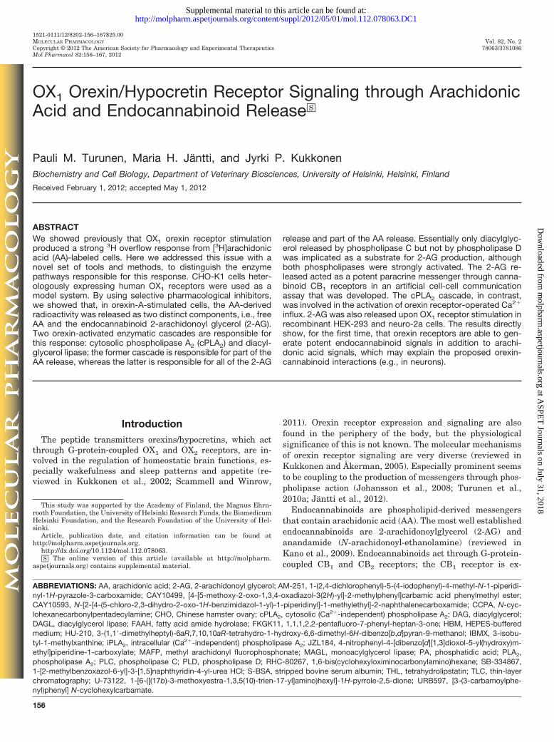
Fig. 1. Orexin-A-stimulated 3H overflow from [3H]AA-labeled CHO-hOX1cells.
158 Turunen et al.
at ASPE
T Journals on July 31, 2018
molpharm
.aspetjournals.orgD
ownloaded from
acetoxymethyl ester in HBM containing 1 mM probenecid, rinsedonce, and used immediately. Ca2� measurements were performedat 35°C with a Nikon TE2000 fluorescence microscope (20�/0.75air objective) and an Andor iXon 885 electron-multiplying charge-coupled device camera under the control of Nikon NIS ElementsAR software with 6D extension (Nikon, Tokyo, Japan). For Ca2�
imaging, the cells were excited with alternating 340- and 380-nmlight (Sutter DG4 Plus wavelength switcher; Sutter InstrumentCompany, Novato, CA), and the emitted light was collectedthrough a 400-nm dichroic mirror and a 450-nm long-pass filter.Additions were made with constant perfusion (HBM with probe-necid). When the inhibitors THL and pyrrophenone were tested,the cells were pretreated with the compounds for 20 min and thecompounds were included in the perfusion medium throughout theexperiment. Regions of interest were defined with NIS software,and the data were extracted to Microsoft Excel (Microsoft, Red-
mond, WA) for observation and quantitation. More than 30 cellswere measured in each experiment, and each experiment wasrepeated four times or more.
Data Analysis. All data are presented as mean � S.E.M.; Nrefers to the number of batches of cells. Each experiment wasperformed at least three times. The AA-release experiments wereperformed with six data points in parallel, imaging with 30 ormore, cAMP measurements with three or four, and TLC with two.Student’s two-tailed t test with Bonferroni correction was used forall pairwise comparisons except in Fig. 8, where, because of thenonparametric nature of cell counting, the �2 test also was used(Ekholm et al., 2007). Microsoft Excel was used for nonlinearcurve-fitting for the determination of EC50 values. The effects ofinhibitors on orexin-stimulated AA and 2-AG release were calculatedfrom the values by using a formula that compensated for the possibleeffects of the inhibitors on basal release, i.e., release [percentage of
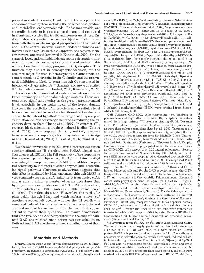
Fig. 2. Molecular mechanism of orexin-Aand thapsigargin-stimulated 3H overflowfrom [3H]AA-labeled CHO-hOX1 cells. A,the orexin response is inhibited by thePLC inhibitor U-73122 but not by thePLD1 inhibitor CAY10593. B and C, theorexin response is partially inhibited bythe DAGL inhibitors THL and RHC-80267 (B) and the cPLA2�/� inhibitor pyr-rophenone (C). D, the combination of THLand pyrrophenone produces full inhibi-tion of the orexin response. E, the re-sponse to 1 �M thapsigargin is nearlyfully blocked by pyrrophenone alone. Thedata were normalized so that each controlresponse (basal, 3 nM orexin-A, and 100nM orexin-A) amounts to 100% (see Ma-terials and Methods). Some differences inthe inhibitory efficacy can be seen in B, C,and D, because only experiments in whichall of the inhibitors were tested were in-cluded in the analysis in D, to allow directcomparison of the efficacy of THL andpyrrophenone. Comparisons are to thecorresponding control (ctrl) values (N �3–7); ns, not significant (p � 0.05); �, p �0.05; ��, p � 0.01; ���, p � 0.001.
Orexin-Induced Arachidonic Acid and Endocannabinoid Release 159
at ASPE
T Journals on July 31, 2018
molpharm
.aspetjournals.orgD
ownloaded from
control (noninhibited)] � (orexininhibitor basalinhibitor)/(orexincontrol basalcontrol) � 100% (see Figs. 2 and 4). In this manner, the nontreatedcontrol values (basal, 1 nM orexin-A, and 100 nM orexin-A) are set to100% and full inhibition to 0%.
ResultsOrexin Receptor Stimulation Induces [3H]AA-De-
rived 3H Overflow through Distinct Cytosolic PLA2
and Diacylglycerol Lipase Cascades. Our previous stud-ies showed strong release of radioactivity from [3H]AA-la-beled CHO-hOX1 cells upon OX1 receptor stimulation(Fig. 1), but the enzymes responsible for this could not beresolved because of the low selectivity of the inhibitors avail-able (Turunen et al., 2010a). We showed that OX1 receptorsalso potently activate both phospholipase C (PLC) and phos-pholipase D (PLD) cascades (Johansson et al., 2008; Jantti etal., 2012), producing diacylglycerol (DAG) and phosphatidicacid (PA), respectively. Both of these compounds could act assubstrates for AA release, through the DAG lipase (DAGL)-monoacylglycerol lipase (MAGL) and PA-PLA2 cascades, re-spectively, and PA can also be hydrolyzed to DAG by PAphosphohydrolase (reviewed in Kukkonen, 2011). Only PLD1and not PLD2 is activated by orexin receptor stimulation inCHO cells, and this isoform can be fully inhibited byCAY10593 (Jantti et al., 2012). CAY10593 produced veryweak (10–20%) inhibition of 3H overflow (Fig. 2A). In con-trast, the PLC inhibitor U-73122 produced full inhibition(Fig. 2A).
THL is used as a pancreatic triglyceride lipase inhibitor,but it is an even more-potent inhibitor of some related en-zymes, including DAGL (Lee et al., 1995; Bisogno et al.,2003), and it can be used at 1 �M as a relatively selectiveDAGL inhibitor (Bisogno et al., 2006) (V. Di Marzo, personalcommunication). THL produced significant inhibition of theorexin response even at 1 �M (Fig. 2B). RHC-80267, a less-potent DAGL inhibitor, showed a similar trend of inhibition(Fig. 2B).
Selective inhibitors for particular PLA2 isoforms have beenproduced during the past 10 years. The most well character-ized (e.g., for selectivity) of these are different pyrrolidineinhibitors for cytosolic PLA2 (cPLA2) � (Seno et al., 2000,2001). A commercially available inhibitor of this type, pyrro-phenone (Seno et al., 2001), at 1 �M produced significant
inhibition of the orexin response (Fig. 2C). Interestingly, theinhibition profile was the opposite of that for THL; whereasTHL apparently produced weaker inhibition with 3 nMorexin-A and stronger inhibition with 100 nM, pyrrophenonewas stronger with 3 nM orexin-A. We hypothesized thatDAGL and cPLA2 might represent two components of theorexin-stimulated 3H overflow from [3H]AA-labeled cells.The combination of THL and pyrrophenone produced fullinhibition of the orexin-A response (Fig. 2D). In contrast, theresponse to thapsigargin showed no sensitivity to THL butwas almost fully inhibited by pyrrophenone alone (Fig. 2E),in agreement with Ca2� activation of cPLA2 (reviewed inGhosh et al., 2006). Although Ca2� level elevation shouldalso stimulate DAGL (Bisogno et al., 2003), Ca2� level ele-vation (at least when triggered by thapsigargin or ionomycin)is a poor stimulant of PLD and an even poorer stimulant ofPLC in CHO cells (Lund et al., 2000; Johansson et al., 2007;Jantti et al., 2012); therefore, in the absence of DAG produc-tion, there was no DAGL activity.
The orexin response appears to rely on two different com-ponents. The relative contributions of the components mightbecome clearer with normalized raw data instead of inhibi-
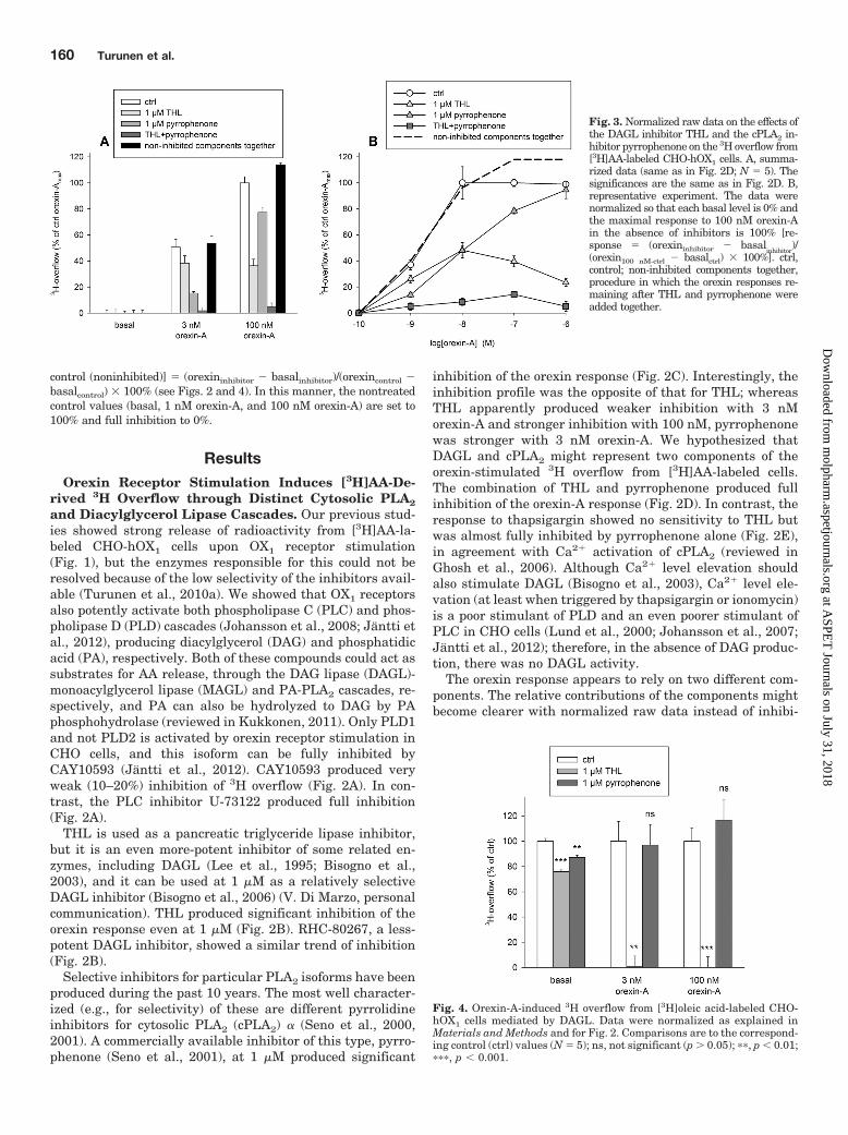
Fig. 3. Normalized raw data on the effects ofthe DAGL inhibitor THL and the cPLA2 in-hibitor pyrrophenone on the 3H overflow from[3H]AA-labeled CHO-hOX1 cells. A, summa-rized data (same as in Fig. 2D; N � 5). Thesignificances are the same as in Fig. 2D. B,representative experiment. The data werenormalized so that each basal level is 0% andthe maximal response to 100 nM orexin-Ain the absence of inhibitors is 100% [re-sponse � (orexininhibitor basal
inhibitor)/
(orexin100 nM-ctrl basalctrl) � 100%]. ctrl,control; non-inhibited components together,procedure in which the orexin responses re-maining after THL and pyrrophenone wereadded together.
Fig. 4. Orexin-A-induced 3H overflow from [3H]oleic acid-labeled CHO-hOX1 cells mediated by DAGL. Data were normalized as explained inMaterials and Methods and for Fig. 2. Comparisons are to the correspond-ing control (ctrl) values (N � 5); ns, not significant (p � 0.05); ��, p � 0.01;���, p � 0.001.
160 Turunen et al.
at ASPE
T Journals on July 31, 2018
molpharm
.aspetjournals.orgD
ownloaded from

tion data (Fig. 3A). The same finding could also be seen withmore complete concentration-response curves (Fig. 3B). Thepatterns of inhibition with THL and pyrrophenone were es-sentially not overlapping (Fig. 3). In the analyses of theconcentration-response data, the control pEC50 value was8.7 � 0.1 and the pEC50 values after pyrrophenone (putativeDAGL component) and THL (putative cPLA2 component)treatment were 8.4 � 0.1 and 9.3 � 0.1, respectively (N �3–5). Therefore, the putative cPLA2 component would beactivated with significantly higher potency (see Discussion).
We showed previously that 3H overflow from [3H]oleic acid-labeled cells was stimulated by OX1 receptor activation inCHO-hOX1 cells, although with significantly lower potencyand efficacy than in [3H]AA-labeled cells (Turunen et al.,2010a). The potency of this 3H overflow seemed to overlapbetter with the DAGL component of AA release than with AArelease in its entirety. Orexin-induced 3H overflow from[3H]oleic acid-labeled cells was inhibited fully by THL but notat all by pyrrophenone (Fig. 4). [3H]Oleic acid is likely to end
up in the sn1 position, which is not hydrolyzed by cPLA2.Even if some oleic acid would be found in the sn2 position,cPLA2� (in contrast to other PLA2 isoforms, includingcPLA2�) is rather specific for AA in the sn2 position (reviewedin Ghosh et al., 2006, 2007).
Endocannabinoid-Hydrolyzing Enzymes, Hormone-Sensitive Lipase, and Intracellular PLA2 in Orexin-Induced, AA-Derived 3H Overflow. DAG lipases arethought to show sn1 selectivity (3–8-fold in vitro) (Bisogno etal., 2003); therefore, DAGL might be thought to be unlikely torelease AA from its usual sn2 position. DAGL, however,works as a “gatekeeper” for MAGL, because no monoacylg-lycerol should be produced in the absence of DAGL activity.We tested a selective MAGL inhibitor, JZL184 (Long et al.,2009). JZL184 (10 �M) produced weak but significant inhi-bition (20 � 12% with 3 nM orexin-A, p � 0.01; 11 � 10%with 100 nM orexin-A, p � 0.05) (Supplemental Fig. 1). Wesimilarly tested inhibitors of fatty acid amide-type endocan-nabinoid-hydrolyzing enzymes, URB597 (100 nM; fatty acid
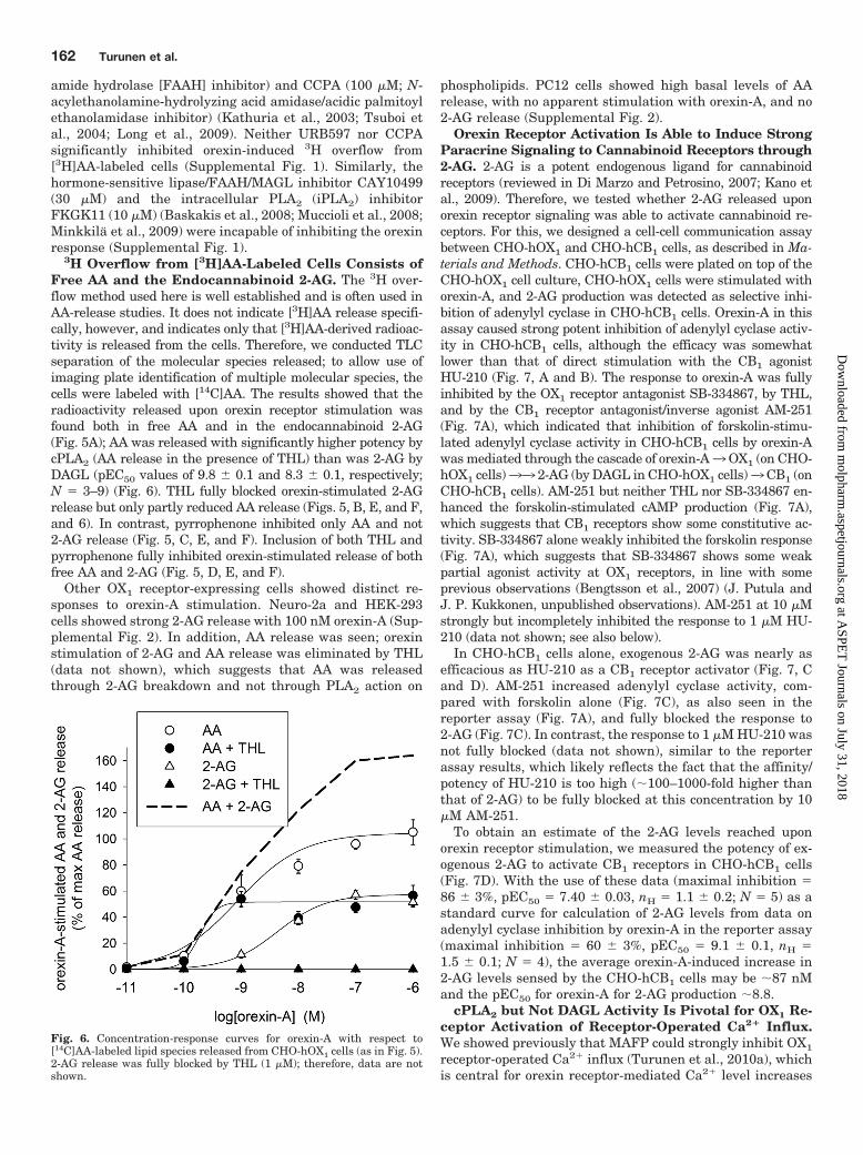
Fig. 5. TLC separation of the [14C]AA-labeled lipid species released from CHO-hOX1 cells. A, release of both free AA and2-AG upon orexin stimulation (N � 8). B,inhibitory actions of THL. C, inhibitoryactions of pyrrophenone. D, combined ef-fects of THL and pyrrophenone on AA and2-AG release upon OX1 receptor stimula-tion. E, summarized data for inhibition ofAA release (N � 3–5). F, summarizeddata for inhibition of 2-AG release (N �3–5). The data in the bar graphs werenormalized to the AA-release response to100 nM orexin-A (100%). Comparisonsare to the corresponding control (ctrl) val-ues; ns, not significant (p � 0.05); �, p �0.05; ��, p � 0.01; ���, p � 0.001.
Orexin-Induced Arachidonic Acid and Endocannabinoid Release 161
at ASPE
T Journals on July 31, 2018
molpharm
.aspetjournals.orgD
ownloaded from
amide hydrolase [FAAH] inhibitor) and CCPA (100 �M; N-acylethanolamine-hydrolyzing acid amidase/acidic palmitoylethanolamidase inhibitor) (Kathuria et al., 2003; Tsuboi etal., 2004; Long et al., 2009). Neither URB597 nor CCPAsignificantly inhibited orexin-induced 3H overflow from[3H]AA-labeled cells (Supplemental Fig. 1). Similarly, thehormone-sensitive lipase/FAAH/MAGL inhibitor CAY10499(30 �M) and the intracellular PLA2 (iPLA2) inhibitorFKGK11 (10 �M) (Baskakis et al., 2008; Muccioli et al., 2008;Minkkila et al., 2009) were incapable of inhibiting the orexinresponse (Supplemental Fig. 1).
3H Overflow from [3H]AA-Labeled Cells Consists ofFree AA and the Endocannabinoid 2-AG. The 3H over-flow method used here is well established and is often used inAA-release studies. It does not indicate [3H]AA release specifi-cally, however, and indicates only that [3H]AA-derived radioac-tivity is released from the cells. Therefore, we conducted TLCseparation of the molecular species released; to allow use ofimaging plate identification of multiple molecular species, thecells were labeled with [14C]AA. The results showed that theradioactivity released upon orexin receptor stimulation wasfound both in free AA and in the endocannabinoid 2-AG(Fig. 5A); AA was released with significantly higher potency bycPLA2 (AA release in the presence of THL) than was 2-AG byDAGL (pEC50 values of 9.8 � 0.1 and 8.3 � 0.1, respectively;N � 3–9) (Fig. 6). THL fully blocked orexin-stimulated 2-AGrelease but only partly reduced AA release (Figs. 5, B, E, and F,and 6). In contrast, pyrrophenone inhibited only AA and not2-AG release (Fig. 5, C, E, and F). Inclusion of both THL andpyrrophenone fully inhibited orexin-stimulated release of bothfree AA and 2-AG (Fig. 5, D, E, and F).
Other OX1 receptor-expressing cells showed distinct re-sponses to orexin-A stimulation. Neuro-2a and HEK-293cells showed strong 2-AG release with 100 nM orexin-A (Sup-plemental Fig. 2). In addition, AA release was seen; orexinstimulation of 2-AG and AA release was eliminated by THL(data not shown), which suggests that AA was releasedthrough 2-AG breakdown and not through PLA2 action on
phospholipids. PC12 cells showed high basal levels of AArelease, with no apparent stimulation with orexin-A, and no2-AG release (Supplemental Fig. 2).
Orexin Receptor Activation Is Able to Induce StrongParacrine Signaling to Cannabinoid Receptors through2-AG. 2-AG is a potent endogenous ligand for cannabinoidreceptors (reviewed in Di Marzo and Petrosino, 2007; Kano etal., 2009). Therefore, we tested whether 2-AG released uponorexin receptor signaling was able to activate cannabinoid re-ceptors. For this, we designed a cell-cell communication assaybetween CHO-hOX1 and CHO-hCB1 cells, as described in Ma-terials and Methods. CHO-hCB1 cells were plated on top of theCHO-hOX1 cell culture, CHO-hOX1 cells were stimulated withorexin-A, and 2-AG production was detected as selective inhi-bition of adenylyl cyclase in CHO-hCB1 cells. Orexin-A in thisassay caused strong potent inhibition of adenylyl cyclase activ-ity in CHO-hCB1 cells, although the efficacy was somewhatlower than that of direct stimulation with the CB1 agonistHU-210 (Fig. 7, A and B). The response to orexin-A was fullyinhibited by the OX1 receptor antagonist SB-334867, by THL,and by the CB1 receptor antagonist/inverse agonist AM-251(Fig. 7A), which indicated that inhibition of forskolin-stimu-lated adenylyl cyclase activity in CHO-hCB1 cells by orexin-Awas mediated through the cascade of orexin-A3OX1 (on CHO-hOX1 cells)33 2-AG (by DAGL in CHO-hOX1 cells)3CB1 (onCHO-hCB1 cells). AM-251 but neither THL nor SB-334867 en-hanced the forskolin-stimulated cAMP production (Fig. 7A),which suggests that CB1 receptors show some constitutive ac-tivity. SB-334867 alone weakly inhibited the forskolin response(Fig. 7A), which suggests that SB-334867 shows some weakpartial agonist activity at OX1 receptors, in line with someprevious observations (Bengtsson et al., 2007) (J. Putula andJ. P. Kukkonen, unpublished observations). AM-251 at 10 �Mstrongly but incompletely inhibited the response to 1 �M HU-210 (data not shown; see also below).
In CHO-hCB1 cells alone, exogenous 2-AG was nearly asefficacious as HU-210 as a CB1 receptor activator (Fig. 7, Cand D). AM-251 increased adenylyl cyclase activity, com-pared with forskolin alone (Fig. 7C), as also seen in thereporter assay (Fig. 7A), and fully blocked the response to2-AG (Fig. 7C). In contrast, the response to 1 �M HU-210 wasnot fully blocked (data not shown), similar to the reporterassay results, which likely reflects the fact that the affinity/potency of HU-210 is too high (�100–1000-fold higher thanthat of 2-AG) to be fully blocked at this concentration by 10�M AM-251.
To obtain an estimate of the 2-AG levels reached uponorexin receptor stimulation, we measured the potency of ex-ogenous 2-AG to activate CB1 receptors in CHO-hCB1 cells(Fig. 7D). With the use of these data (maximal inhibition �86 � 3%, pEC50 � 7.40 � 0.03, nH � 1.1 � 0.2; N � 5) as astandard curve for calculation of 2-AG levels from data onadenylyl cyclase inhibition by orexin-A in the reporter assay(maximal inhibition � 60 � 3%, pEC50 � 9.1 � 0.1, nH �1.5 � 0.1; N � 4), the average orexin-A-induced increase in2-AG levels sensed by the CHO-hCB1 cells may be �87 nMand the pEC50 for orexin-A for 2-AG production �8.8.
cPLA2 but Not DAGL Activity Is Pivotal for OX1 Re-ceptor Activation of Receptor-Operated Ca2� Influx.We showed previously that MAFP could strongly inhibit OX1
receptor-operated Ca2� influx (Turunen et al., 2010a), whichis central for orexin receptor-mediated Ca2� level increases
Fig. 6. Concentration-response curves for orexin-A with respect to[14C]AA-labeled lipid species released from CHO-hOX1 cells (as in Fig. 5).2-AG release was fully blocked by THL (1 �M); therefore, data are notshown.
162 Turunen et al.
at ASPE
T Journals on July 31, 2018
molpharm
.aspetjournals.orgD
ownloaded from
because it also drives PLC activity at low orexin concentra-tions (Lund et al., 2000; Johansson et al., 2007). BecauseMAFP is a potent inhibitor of both the cPLA2 and DAGLpathways, we tested selective inhibitors of these pathways,i.e., pyrrophenone and THL, in Ca2� imaging assays. Pyrro-phenone produced equally strong inhibition (Fig. 8, A and B),compared with MAFP (data not shown) (Turunen et al.,2010a), whereas THL was incapable of inhibiting the Ca2�
influx response (Fig. 8, A and C), which clearly indicates thatthe cPLA2 pathway is involved in orexin receptor-operatedCa2� influx. Inclusion of both pyrrophenone and THL did notproduce stronger inhibition than that observed with pyrro-phenone alone (Fig. 8, A, B, and D).
DiscussionWe targeted the [3H]AA-derived 3H overflow by using a
number of established or novel inhibitors of lipases and ami-dases that are capable of releasing AA. Most importantly,THL, an inhibitor of DAGL, and pyrrophenone, an inhibitorof cPLA2� and -� (Seno et al., 2001; Ghomashchi et al., 2010)produced complementary inhibition, together reaching full
inhibition. THL was selected as the DAGL inhibitor for de-tailed investigations because of its greater potency, efficacy,and stability and its likely greater selectivity, compared withRHC-80267. The pharmacological selectivity of the inhibitorsin CHO cells was confirmed with respect to thapsigargin-induced 3H overflow from [3H]AA-labeled cells (fully inhib-ited by pyrrophenone) and orexin-A-induced 3H overflowfrom [3H]oleic acid-labeled cells (fully inhibited by THL). Theconclusive evidence was obtained through TLC separation of[14C]AA-labeled lipid species, which showed that the appar-ent orexin-A-induced AA overflow was composed of free AAas well as 2-AG. 2-AG release was fully dependent on DAGL(inhibited by THL), whereas the free AA component wasdependent on both the cPLA2 and DAGL pathways (addi-tively inhibited by pyrrophenone and THL, respectively). Theresults are summarized in Fig. 9. The DAGL pathway islikely to follow from PLC activity, as suggested by bothinhibition by the PLC inhibitor U-73122 and only minorinhibition by the PLD inhibitor CAY10593. 2-AG productionclosely mirrors PLC activity, as judged from total inositolphosphate release and DAG generation in the presence of
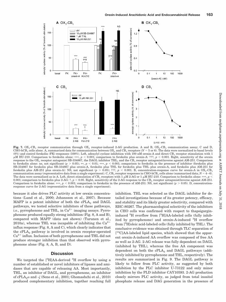
Fig. 7. OX1-CB1 receptor communication through OX1 receptor-induced 2-AG production. A and B, OX1-CB1 communication assay; C and D,CHO-hCB1 cells alone. A, summarized data for communication between OX1 and CB1 receptors (N � 5 or 6). The data were normalized to basal levels(0%) and control forskolin (FK) responses (100%). Left, adenylyl cyclase inhibition with 100 nM orexin-A and direct CB1 receptor stimulation with 1�M HU-210. Comparison to forskolin alone: ���, p � 0.001; comparison to forskolin plus orexin-A: †††, p � 0.001. Right, sensitivity of the orexinresponse to the OX1 receptor antagonist SB-334867, the DAGL inhibitor THL, and the CB1 receptor antagonist/inverse agonist AM-251. Comparisonto forskolin alone: ns, not significant (p � 0.05); ��, p � 0.01; ���, p � 0.001; comparison to forskolin in the presence of inhibitor (forskolin plusSB-334867 for forskolin plus SB-334867 plus orexin-A, forskolin plus THL for forskolin plus THL plus orexin-A, and forskolin plus AM-251 forforskolin plus AM-251 plus orexin-A): NS, not significant (p � 0.05); †††, p � 0.001. B, concentration-response curve for orexin-A in OX1-CB1communication assay (representative data from a single experiment). C, CB1 receptor responses in CHO-hCB1 cells alone (summarized data; N � 4–6).The data were normalized as in A. Left, direct stimulation of CB1 receptors with 1 �M 2-AG or 1 �M HU-210. Comparison to forskolin alone: ���, p �0.001; comparison to forskolin plus 2-AG: †, p � 0.05. Right, sensitivity of the 2-AG response to the CB1 receptor antagonist/inverse agonist AM-251.Comparison to forskolin alone: ���, p � 0.001; comparison to forskolin in the presence of AM-251: NS, not significant (p � 0.05). D, concentration-response curve for 2-AG (representative data from a single experiment).
Orexin-Induced Arachidonic Acid and Endocannabinoid Release 163
at ASPE
T Journals on July 31, 2018
molpharm
.aspetjournals.orgD
ownloaded from
PLD inhibition (Johansson et al., 2008). It is unclear, how-ever, whether DAGL in general passively follows PLC activ-ity or is actively regulated by, e.g., Ca2� level elevation(Bisogno et al., 2003) or phosphorylation. MAGL or anothercomponent with similar activity (Long et al., 2009), degrad-ing 2-AG to glycerol and AA, is likely to be responsible for theinhibition of free AA release by THL. We also used a numberof other inhibitors of enzymes with possible AA-releasingcapacity, including the MAGL inhibitor JZL184, the FAAHinhibitor URB597, the MAGL/FAAH/hormone-sensitive lipaseinhibitor CAY10499, the iPLA2 inhibitor FKGK11, and theN-acylethanolamine-hydrolyzing acid amidase/acidic palmitoylethanolamidase inhibitor CCPA. Whereas JZL184, URB597,and CAY10499 are rather well characterized, FKGK11 andCCPA are not (they have been used in only a few studies), and
it was difficult to estimate their effective concentrations. How-ever, none of the inhibitors except JZL184 produced any inhi-bition, which is logical in light of the studies clearly indicatingthe involvement of cPLA2 and DAGL only. It is unclear whyJZL184 produced a small but significant amount of inhibition of3H overflow. At the concentration used, CAY10499 also is aneffective inhibitor of MAGL (Muccioli et al., 2008; Minkkila etal., 2009), but it did not show any inhibition here; therefore, it ispossible that the inhibition seen with JZL184 is attributable toan effect on some target other than MAGL.
cPLA2 is activated with high potency by orexin receptorsand may be a potent physiological signal. In the currentstudy, we could verify that it was cPLA2 and not DAGLsignaling that was required for the activity of the receptor-operated Ca2� influx pathway. Our results do not reveal,
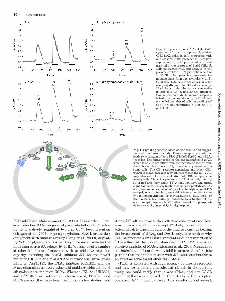
Fig. 8. Dependence on cPLA2 of the Ca2�
signaling of orexin receptors. A, controlCHO-hOX1 cells. B, cells pretreated withand assayed in the presence of 1 �M pyr-rophenone. C, cells pretreated with andassayed in the presence of 1 �M THL. D,cells pretreated with and assayed in thepresence of both 1 �M pyrrophenone and1 �M THL. Each panel is a representativeaverage trace from one coverslip with 31to 53 cells. S.E. values are shown only forevery eighth point, for the sake of clarity.Black bars under the traces, successiveadditions of 0.3, 3, and 30 nM orexin-A.Comparison to control, maximal response(t test): ns, not significant (p � 0.05); ���,p � 0.001; number of cells responding (�2
test): NS, not significant (p � 0.05); †††,p � 0.001.
Fig. 9. Signaling scheme based on the results and sugges-tions of the present study. Orexin receptor stimulationleads to activation of both PLC (PLC�?)-DAGL and cPLA2cascades. The former produces the endocannabinoid 2-AG,which is able to act either from the membrane face or fromthe extracellular side on CB1 receptors expressed in thesame cells. The CB1 receptor-dependent and other OX1-triggered signal cascades may interact within the cell. 2-AGmay also exit the cells and stimulate CB1 receptors onnearby cells. The other products of DAGL activity, mainlysaturated free fatty acids (FFA), may not have importantsignaling roles. cPLA2 likely acts on phosphatidylcholine(PC), leading to production of lysophosphatidylcholine (LPC)and polyunsaturated fatty acids (PUFAs) such as AA. Eitherlysophosphatidylcholine or polyunsaturated fatty acids ortheir metabolites centrally contribute to activation of theorexin receptor-operated Ca2� influx channel. PIs, phosphati-dylinositols; IP3, inositol-1,4,5-trisphosphate.
164 Turunen et al.
at ASPE
T Journals on July 31, 2018
molpharm
.aspetjournals.orgD
ownloaded from
however, whether it is AA or some other polyunsaturatedfatty acid, the other product of cPLA2 activity, lysophosphati-dylcholine, or some metabolite of those compounds that me-diates the response. All of those metabolites could be activeat transient receptor potential or ARC channels (reviewed inKukkonen, 2011).
cPLA2 enzymes are known to be sensitive to Ca2� levelincreases. The PLC inhibitor U-73122 fully inhibited 3Hoverflow from [3H]AA-labeled cells, which suggested thatPLC activity was required for both cPLA2 and DAGL path-ways. U-73122 is known to be toxic to CHO cells (Taylor andBroad, 1998; Lund et al., 2000) and it can fully block orexinreceptor-mediated Ca2� influx (Smart et al., 1999), althoughinositol-1,4,5-trisphosphate-dependent Ca2� influx is not in-volved in this response (Ekholm et al., 2007). In addition,cPLA2 activity occurs with a higher potency than PLC orDAGL activity (see below). Therefore, we doubt the conclusionregarding involvement of PLC in cPLA2 activation. Unfortu-nately, there are no other effective (and nontoxic) PLC inhibi-tors available (at least for CHO cells), and the activation mech-anism for cPLA2 thus remains unclear. However, we know thatthe increase in Ca2� levels (likely through influx) induced bythapsigargin is a potent stimulator of cPLA2 in these cells(present study) (Turunen et al., 2010a), and Ca2� influx isrequired for effective stimulation of 3H overflow from [3H]AA-labeled cells by orexin-A (Turunen et al., 2010a). It is possiblethat there is a feed-forward cycle in orexin receptor signalinginvolving cPLA2 and receptor-operated Ca2� influx. However,additional experiments are required to resolve this and alter-native options.
The potency of orexin-A for cPLA2 and DAGL activationwas determined in several ways. 2-AG would be released byDAGL with pEC50 values of 8.4 (3H overflow in the presenceof pyrrophenone), 8.3 (TLC assay), and 8.8 (2-AG releasereporter assay). AA would be released by cPLA2 with pEC50
values of 9.3 (3H overflow in the presence of THL) and 9.8(TLC assay in the presence of THL). Some of the variationmay depend on the resolution of the assays; for example, TLCseparation showed much less variation and thus greater sen-sitivity than the 3H overflow assay. cPLA2 was clearly acti-vated with significantly greater potency than DAGL in CHOcells. In the other cell types examined, the situation wasdifferent. It is interesting that, in all of these cell types,orexin receptors couple to PLC (Holmqvist et al., 2002;Putula and Kukkonen, 2012); therefore, the differences in2-AG release might be related to another component, such aslack of DAGL or the putative 2-AG transporter or effectivealternative metabolism of DAG or degradation of 2-AG.
Our previous results obtained with the reputed iPLA2 in-hibitor bromoenol lactone suggested partial involvement ofiPLA2 in the orexin-stimulated 3H overflow (Turunen et al.,2010a). Bromoenol lactone is known to act on different serinehydrolases (reviewed in Balsinde and Balboa, 2005), forminga soluble, reactive, molecular species that can form cysteineadducts with bystander proteins (Song et al., 2006). We areconvinced that the previous results obtained with bromoenollactone do not indicate the involvement of iPLA2 but repre-sent a result of nonspecific interactions of this inhibitor. Thisis supported by the fact that a novel iPLA2 inhibitor,FKGK11, did not inhibit 3H overflow.
Orexin-endocannabinoid interactions have been investi-gated in a few studies. CB1 receptor mRNA and preproorexin
mRNA are expressed in close apposition in the lateral hypo-thalamus, in part in the same cells (Cota et al., 2003). Thephysiological functions of orexins and endocannabinoids inappetite stimulation are similar, and leptin, a satiety mes-senger and negative regulator of orexinergic neurons (Hå-kansson et al., 1999; Lopez et al., 2000), is also a negativeregulator of endocannabinoid levels (Di Marzo et al., 2001).These systems may be involved in the same pathways. Astudy indicated endocannabinoid involvement in orexin-me-diated stimulation of appetite (Crespo et al., 2008), and sim-ilar findings are indicated for the analgesic action of orexin-A(Ho et al., 2011). In contrast, endocannabinoids inhibit orexinsignaling in the lateral hypothalamus by reducing the excit-atory glutamatergic drive to orexinergic neurons (Huang etal., 2007). Endocannabinoids may have different functions inthe lateral hypothalamus and in upstream regulatory centersof orexinergic neurons; whereas endocannabinoids in the lat-eral hypothalamus may suppress orexinergic neurons(Huang et al., 2007), endocannabinoids in the nucleus accum-bens shell may stimulate activation of orexinergic neurons(Kirkham et al., 2002; Zheng et al., 2003; Soria-Gomez et al.,2007), and orexinergic projections to the nucleus accumbensshell (Mori et al., 2011) may cause a positive feedback cycle.The few studies performed suggested interactions betweenthese systems, but the conclusions are not straightforward.Some of the problems arise from methodological obstacles,such as unreliable anti-orexin receptor antibodies (J. P. Kuk-konen, unpublished data). In addition, the studies conductedwere performed either with intact animals or with ex vivoslice preparations, where the neuronal circuitry involvedmay cause problems in identifying the actual sites of action oforexins and endocannabinoids.
In conclusion, the present study identified two enzymecascades, cPLA2 and DAGL, as potent orexin receptor targetsand as the enzymes entirely responsible for the previouslyreported orexin-induced 3H overflow from [3H]AA-labeledcells (Turunen et al., 2010a). The activation mechanisms foreither enzyme species remain unclear. Each enzyme activityproduces a signal of its own. Although cPLA2 appears essen-tial for the receptor-operated Ca2� influx, DAGL produces2-AG, which is able to activate CB1 cannabinoid receptors ina paracrine manner. This indicates that the suggested inter-actions between orexinergic and cannabinoidergic systems inthe brain may take place through paracrine endocannabinoidsignaling and that regulation of the some of the same phys-iological responses in appetite, for example, may take placethrough an arrangement such as this. Orexin receptors weresuggested previously to heterodimerize with CB1 receptors inheterologous coexpression systems (Hilairet et al., 2003; Elliset al., 2006; Ward et al., 2011). Our view is that interactions(alternatively or additionally) take place through 2-AG pro-duction, which represents a much more flexible signal systemthat does not require any molecular interactions betweenorexin and endocannabinoid receptors.
Acknowledgments
We gratefully acknowledge Drs. Michelle Glass (University ofAuckland, Auckland, New Zealand) and Jarmo Laitinen and JuhaSavinainen (University of Eastern Finland, Kuopio, Finland) for theCHO-hCB1 cells, Drs. Vincenzo Di Marzo (Institute of BiomolecularChemistry, Consiglio Nazionale delle Ricerche, Pozzuoli, Italy) and
Orexin-Induced Arachidonic Acid and Endocannabinoid Release 165
at ASPE
T Journals on July 31, 2018
molpharm
.aspetjournals.orgD
ownloaded from
Jarmo Laitinen for advice on the experiments, and Pirjo Purorantafor technical assistance.
Authorship Contributions
Participated in research design: Turunen, Jantti, and Kukkonen.Conducted experiments: Turunen and Jantti.Contributed new reagents or analytic tools: Kukkonen.Performed data analysis: Turunen, Jantti, and Kukkonen.Wrote or contributed to the writing of the manuscript: Turunen,
Jantti, and Kukkonen.
ReferencesBalsinde J and Balboa MA (2005) Cellular regulation and proposed biological func-
tions of group VIA calcium-independent phospholipase A2 in activated cells. CellSignal 17:1052–1062.
Baskakis C, Magrioti V, Cotton N, Stephens D, Constantinou-Kokotou V, Dennis EA,and Kokotos G (2008) Synthesis of polyfluoro ketones for selective inhibition ofhuman phospholipase A2 enzymes. J Med Chem 51:8027–8037.
Bengtsson MW, Makela K, Sjoblom M, Uotila S, Akerman KE, Herzig KH, andFlemstrom G (2007) Food-induced expression of orexin receptors in rat duodenalmucosa regulates the bicarbonate secretory response to orexin-A. Am J PhysiolGastrointest Liver Physiol 293:G501–G509.
Bisogno T, Cascio MG, Saha B, Mahadevan A, Urbani P, Minassi A, Appendino G,Saturnino C, Martin B, Razdan R, et al. (2006) Development of the first potent andspecific inhibitors of endocannabinoid biosynthesis. Biochim Biophys Acta 1761:205–212.
Bisogno T, Howell F, Williams G, Minassi A, Cascio MG, Ligresti A, Matias I,Schiano-Moriello A, Paul P, Williams EJ, et al. (2003) Cloning of the first sn1-DAGlipases points to the spatial and temporal regulation of endocannabinoid signalingin the brain. J Cell Biol 163:463–468.
Bligh EG and Dyer WJ (1959) A rapid method of total lipid extraction and purifica-tion. Can J Biochem Physiol 37:911–917.
Cota D, Marsicano G, Tschop M, Grubler Y, Flachskamm C, Schubert M, Auer D,Yassouridis A, Thone-Reineke C, Ortmann S, et al. (2003) The endogenous can-nabinoid system affects energy balance via central orexigenic drive and peripherallipogenesis. J Clin Invest 112:423–431.
Crespo I, Gomez de Heras R, Rodríguez de Fonseca F, and Navarro M (2008)Pretreatment with subeffective doses of Rimonabant attenuates orexigenic actionsof orexin A-hypocretin 1. Neuropharmacology 54:219–225.
De Petrocellis L, Melck D, Ueda N, Maurelli S, Kurahashi Y, Yamamoto S, MarinoG, and Di Marzo V (1997) Novel inhibitors of brain, neuronal, and basophilicanandamide amidohydrolase. Biochem Biophys Res Commun 231:82–88.
Deutsch DG, Omeir R, Arreaza G, Salehani D, Prestwich GD, Huang Z, and HowlettA (1997) Methyl arachidonyl fluorophosphonate: a potent irreversible inhibitor ofanandamide amidase. Biochem Pharmacol 53:255–260.
Di Marzo V, Goparaju SK, Wang L, Liu J, Batkai S, Jarai Z, Fezza F, Miura GI,Palmiter RD, Sugiura T, et al. (2001) Leptin-regulated endocannabinoids areinvolved in maintaining food intake. Nature 410:822–825.
Di Marzo V and Petrosino S (2007) Endocannabinoids and the regulation of theirlevels in health and disease. Curr Opin Lipidol 18:129–140.
Dinh TP, Carpenter D, Leslie FM, Freund TF, Katona I, Sensi SL, Kathuria S, andPiomelli D (2002) Brain monoglyceride lipase participating in endocannabinoidinactivation. Proc Natl Acad Sci USA 99:10819–10824.
Ekholm ME, Johansson L, and Kukkonen JP (2007) IP3-independent signalling ofOX1 orexin/hypocretin receptors to Ca2� influx and ERK. Biochem Biophys ResCommun 353:475–480.
Ellis J, Pediani JD, Canals M, Milasta S, and Milligan G (2006) Orexin-1 receptor-cannabinoid CB1 receptor heterodimerization results in both ligand-dependentand -independent coordinated alterations of receptor localization and function.J Biol Chem 281:38812–38824.
Ghomashchi F, Naika GS, Bollinger JG, Aloulou A, Lehr M, Leslie CC, and Gelb MH(2010) Interfacial kinetic and binding properties of mammalian group IVB phos-pholipase A2 (cPLA2�) and comparison with the other cPLA2 isoforms. J BiolChem 285:36100–36111.
Ghosh M, Loper R, Ghomashchi F, Tucker DE, Bonventre JV, Gelb MH, and LeslieCC (2007) Function, activity, and membrane targeting of cytosolic phospholipaseA2� in mouse lung fibroblasts. J Biol Chem 282:11676–11686.
Ghosh M, Tucker DE, Burchett SA, and Leslie CC (2006) Properties of the Group IVphospholipase A2 family. Prog Lipid Res 45:487–510.
Glass M, Hong J, Sato TA, and Mitchell MD (2005) Misidentification of prostamidesas prostaglandins. J Lipid Res 46:1364–1368.
Grimsey NL, Graham ES, Dragunow M, and Glass M (2010) Cannabinoid receptor 1trafficking and the role of the intracellular pool: implications for therapeutics.Biochem Pharmacol 80:1050–1062.
Håkansson M, de Lecea L, Sutcliffe JG, Yanagisawa M, and Meister B (1999) Leptinreceptor- and STAT3-immunoreactivities in hypocretin/orexin neurones of thelateral hypothalamus. J Neuroendocrinol 11:653–663.
Hilairet S, Bouaboula M, Carriere D, Le Fur G, and Casellas P (2003) Hypersensi-tization of the orexin 1 receptor by the CB1 receptor: evidence for cross-talkblocked by the specific CB1 antagonist, SR141716. J Biol Chem 278:23731–23737.
Ho YC, Lee HJ, Tung LW, Liao YY, Fu SY, Teng SF, Liao HT, Mackie K, and ChiouLC (2011) Activation of orexin 1 receptors in the periaqueductal gray of male ratsleads to antinociception via retrograde endocannabinoid (2-arachidonoylglycerol)-induced disinhibition. J Neurosci 31:14600–14610.
Holmqvist T, Akerman KE, and Kukkonen JP (2002) Orexin signaling in recombi-nant neuron-like cells. FEBS Lett 526:11–14.
Holmqvist T, Johansson L, Ostman M, Ammoun S, Akerman KE, and Kukkonen JP(2005) OX1 orexin receptors couple to adenylyl cyclase regulation via multiplemechanisms. J Biol Chem 280:6570–6579.
Howlett AC (2005) Cannabinoid receptor signaling. Handb Exp Pharmacol 168:53–79.
Hu J and el-Fakahany EE (1993) Role of intercellular and intracellular communi-cation by nitric oxide in coupling of muscarinic receptors to activation of guanylatecyclase in neuronal cells. J Neurochem 61:578–585.
Huang H, Acuna-Goycolea C, Li Y, Cheng HM, Obrietan K, and van den Pol AN(2007) Cannabinoids excite hypothalamic melanin-concentrating hormone but in-hibit hypocretin/orexin neurons: implications for cannabinoid actions on food in-take and cognitive arousal. J Neurosci 27:4870–4881.
Jantti MH, Putula J, Somerharju P, Frohman MA, and Kukkonen JP (2012) OX1orexin/hypocretin receptor activation of phospholipase D. Br J Pharmacol 165:1109–1123.
Johansson L, Ekholm ME, and Kukkonen JP (2007) Regulation of OX1 orexin/hypocretin receptor-coupling to phospholipase C by Ca2� influx. Br J Pharmacol150:97–104.
Johansson L, Ekholm ME, and Kukkonen JP (2008) Multiple phospholipase activa-tion by OX1 orexin/hypocretin receptors. Cell Mol Life Sci 65:1948–1956.
Kano M, Ohno-Shosaku T, Hashimotodani Y, Uchigashima M, and Watanabe M(2009) Endocannabinoid-mediated control of synaptic transmission. Physiol Rev89:309–380.
Kathuria S, Gaetani S, Fegley D, Valino F, Duranti A, Tontini A, Mor M, Tarzia G,La Rana G, Calignano A, et al. (2003) Modulation of anxiety through blockade ofanandamide hydrolysis. Nat Med 9:76–81.
Kirkham TC, Williams CM, Fezza F, and Di Marzo V (2002) Endocannabinoid levelsin rat limbic forebrain and hypothalamus in relation to fasting, feeding andsatiation: stimulation of eating by 2-arachidonoyl glycerol. Br J Pharmacol 136:550–557.
Kukkonen JP (2011) A menage a trois made in heaven: G-protein-coupled receptors,lipids and TRP channels. Cell Calcium 50:9–26.
Kukkonen JP and Åkerman KEO (2005) Intracellular signal pathways utilized bythe hypocretin/orexin receptors, in Hypocretins as Integrators of PhysiologicalSignals (de Lecea L and Sutcliffe JG eds) pp 221–231, Springer Science�BusinessMedia, Berlin.
Kukkonen JP, Holmqvist T, Ammoun S, and Akerman KE (2002) Functions of theorexinergic/hypocretinergic system. Am J Physiol Cell Physiol 283:C1567–C1591.
Lee MW, Kraemer FB, and Severson DL (1995) Characterization of a partiallypurified diacylglycerol lipase from bovine aorta. Biochim Biophys Acta 1254:311–318.
Long JZ, Li W, Booker L, Burston JJ, Kinsey SG, Schlosburg JE, Pavon FJ,Serrano AM, Selley DE, Parsons LH, et al. (2009) Selective blockade of2-arachidonoylglycerol hydrolysis produces cannabinoid behavioral effects. NatChem Biol 5:37– 44.
Lopez M, Seoane L, García MC, Lago F, Casanueva FF, Senarís R, and Dieguez C(2000) Leptin regulation of prepro-orexin and orexin receptor mRNA levels in thehypothalamus. Biochem Biophys Res Commun 269:41–45.
Lund PE, Shariatmadari R, Uustare A, Detheux M, Parmentier M, Kukkonen JP,and Akerman KE (2000) The orexin OX1 receptor activates a novel Ca2� influxpathway necessary for coupling to phospholipase C. J Biol Chem 275:30806–30812.
Minkkila A, Savinainen JR, Kasnanen H, Xhaard H, Nevalainen T, Laitinen JT,Poso A, Leppanen J, and Saario SM (2009) Screening of various hormone-sensitivelipase inhibitors as endocannabinoid-hydrolyzing enzyme inhibitors. ChemMed-Chem 4:1253–1259.
Mori K, Kim J, and Sasaki K (2011) Electrophysiological effects of orexin-B anddopamine on rat nucleus accumbens shell neurons in vitro. Peptides 32:246–252.
Muccioli GG, Labar G, and Lambert DM (2008) CAY10499, a novel monoglyceridelipase inhibitor evidenced by an expeditious MGL assay. Chembiochem 9:2704–2710.
Putula J and Kukkonen JP (2012) Mapping of the binding sites for the OX1 orexinreceptor antagonist, SB-334867, using orexin/hypocretin receptor chimaeras. Neu-rosci Lett 506:111–115.
Savinainen JR, Saario SM, Niemi R, Jarvinen T, and Laitinen JT (2003) An opti-mized approach to study endocannabinoid signaling: evidence against constitutiveactivity of rat brain adenosine A1 and cannabinoid CB1 receptors. Br J Pharmacol140:1451–1459.
Savinainen JR, Yoshino M, Minkkila A, Nevalainen T, and Laitinen JT (2010)Characterization of binding properties of monoglyceride lipase inhibitors by aversatile fluorescence-based technique. Anal Biochem 399:132–134.
Scammell TE and Winrow CJ (2011) Orexin receptors: pharmacology and therapeu-tic opportunities. Annu Rev Pharmacol Toxicol 51:243–266.
Scott SA, Selvy PE, Buck JR, Cho HP, Criswell TL, Thomas AL, Armstrong MD,Arteaga CL, Lindsley CW, and Brown HA (2009) Design of isoform-selectivephospholipase D inhibitors that modulate cancer cell invasiveness. Nat Chem Biol5:108–117.
Seno K, Okuno T, Nishi K, Murakami Y, Watanabe F, Matsuura T, Wada M, Fujii Y,Yamada M, Ogawa T, et al. (2000) Pyrrolidine inhibitors of human cytosolicphospholipase A2. J Med Chem 43:1041–1044.
Seno K, Okuno T, Nishi K, Murakami Y, Yamada K, Nakamoto S, and Ono T (2001)Pyrrolidine inhibitors of human cytosolic phospholipase A2. 2. Synthesis of potentand crystallized 4-triphenylmethylthio derivative ‘pyrrophenone.’ Bioorg MedChem Lett 11:587–590.Smart D, Jerman JC, Brough SJ, Rushton SL, Murdock PR, Jewitt F, ElshourbagyNA, Ellis CE, Middlemiss DN, and Brown F (1999) Characterization of recombi-nant human orexin receptor pharmacology in a Chinese hamster ovary cell-lineusing FLIPR. Br J Pharmacol 128:1–3.
Song H, Ramanadham S, Bao S, Hsu FF, and Turk J (2006) A bromoenol lactonesuicide substrate inactivates group VIA phospholipase A2 by generating a diffus-
166 Turunen et al.
at ASPE
T Journals on July 31, 2018
molpharm
.aspetjournals.orgD
ownloaded from
ible bromomethyl keto acid that alkylates cysteine thiols. Biochemistry 45:1061–1073.
Soria-Gomez E, Matias I, Rueda-Orozco PE, Cisneros M, Petrosino S, Navarro L, DiMarzo V, and Prospero-García O (2007) Pharmacological enhancement of theendocannabinoid system in the nucleus accumbens shell stimulates food intakeand increases c-Fos expression in the hypothalamus. Br J Pharmacol 151:1109–1116.
Taylor CW and Broad LM (1998) Pharmacological analysis of intracellular Ca2�
signalling: problems and pitfalls. Trends Pharmacol Sci 19:370–375.Tsuboi K, Hilligsmann C, Vandevoorde S, Lambert DM, and Ueda N (2004) N-
Cyclohexanecarbonylpentadecylamine: a selective inhibitor of the acid amidasehydrolysing N-acylethanolamines, as a tool to distinguish acid amidase from fattyacid amide hydrolase. Biochem J 379:99–106.
Turunen PM, Ekholm ME, Somerharju P, and Kukkonen JP (2010a) Arachidonicacid release mediated by OX1 orexin receptors. Br J Pharmacol 159:212–221.
Turunen PM, Putula J, and Kukkonen JP (2010b) Filtration assay for arachidonicacid release. Anal Biochem 407:233–236.
Ward RJ, Pediani JD, and Milligan G (2011) Hetero-multimerization of the canna-binoid CB1 receptor and the orexin OX1 receptor generates a unique complex inwhich both protomers are regulated by orexin A. J Biol Chem 286:37414–37428.
Zheng H, Corkern M, Stoyanova I, Patterson LM, Tian R, and Berthoud HR (2003)Peptides that regulate food intake: appetite-inducing accumbens manipulationactivates hypothalamic orexin neurons and inhibits POMC neurons. Am J PhysiolRegul Integr Comp Physiol 284:R1436–R1444.
Address correspondence to: Dr. Jyrki P. Kukkonen, Biochemistry and CellBiology, Department of Veterinary Biosciences, University of Helsinki, P.O.Box 66, FIN-00014, University of Helsinki, Helsinki, Finland. E-mail:[email protected]
Orexin-Induced Arachidonic Acid and Endocannabinoid Release 167
at ASPE
T Journals on July 31, 2018
molpharm
.aspetjournals.orgD
ownloaded from