organic and/or inorganic nanostructured biomaterial thin layers … · 2012-07-20 · organic...
TRANSCRIPT

Organic and/or inorganic
nanostructured biomaterial thin
layers for new applications in
drug delivery, biosensing or
biomimetic coatings
National Institute for Lasers, Plasma and Radiations Physics, Lasers Department, Magurele, Ilfov,
Romania
[email protected], http://lspi.inflpr.ro
Ion N. MIHAILESCU, Carmen RISTOSCU


Biomaterials
Key asset:
Meet minimal biological requirements: biocompatibility combined with the
absence of any adverse effect (non-toxic and non-allergic)
Other requests:
resistance to physiological fluids;
non-interference with the body’s natural immune system;
lifelong resistance to mechanical stress;
easy manufacturability in any desired shape.

Classification
1. Biologically inactive, nearly inert
2. Porous – They facilitate tissue in-growth into the pores
3. Bioactive – They firmly bind to tissues
4. Resorbable – They are gradually replaced by tissue over time
A. Inorganic
Metals, metals alloys, aluminates, silicates, phosphates, carbonates, …
B. Organic
Biopolymers, proteins, enzymes, …
C. Hybrid inorganic - organic

Why organic biomaterial thin films?
Organic biomaterials are generally: - rather expensive
- have modest/poor mechanical
and wear properties
Important note: only the outer layer of biomaterials enter in
contact with the biological/vivid (tissue) → possible solution:
application as thin films
Potential utilization: - coating of active substances for drug
delivery
- biosensors
- coating of biomimetic, generally metallic
implants

Thin films in drug delivery
Schematic representation of
reservoir diffusion controlled
drug delivery device
Thin films may be applied in order to:
• slow the rate of release of an active component;
• improve the dispersion / flow properties; or
• increase the absorption into the systemic circulation.

Biosensors
Biosensor:
1. biological receptor (the
recognition site for interaction with
the analyte to be detected)
2. transduction system, and
3. signal output
The biosensing mechanism is based upon the interaction between the
biologically active recognition element and the analytes.
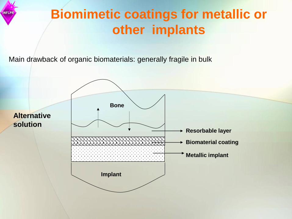
Bone
Resorbable layer
Biomaterial coating
Metallic implant
Implant
Main drawback of organic biomaterials: generally fragile in bulk
Alternative
solution
Biomimetic coatings for metallic or
other implants

How to deposit organic biomaterial thin films?
Plasma methods (plasma spraying, MS, ion and/or electron
bombardment) are inappropriate for deposition of organic biomaterial thin
films. The organic biomaterials suffer of irreversibly damage during
evaporation and transfer.
The same generally applies to chemical (sol-gel) or laser “classical”
methods: laser cladding, PLD.
New laser methods were developed and extended for the transfer of
organic biomaterials in form of thin films on different substrates: MAPLE
(after 1999), laser direct write methods (since 1969).
Organic biomaterial thin films must resemble the biological
ones in composition, structure, morphology, and functionality!
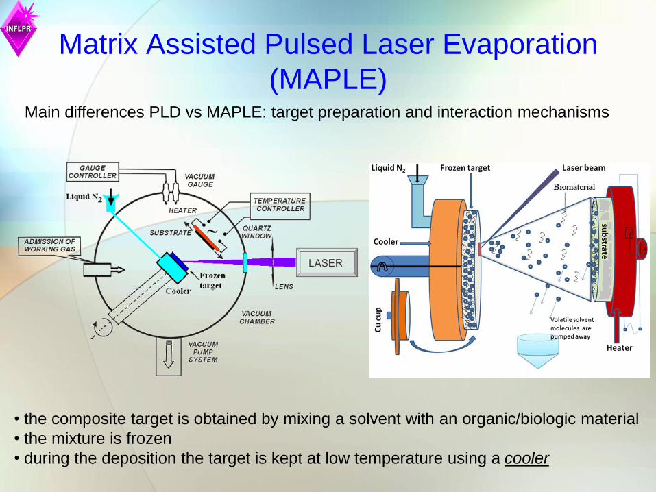
Matrix Assisted Pulsed Laser Evaporation
(MAPLE) Main differences PLD vs MAPLE: target preparation and interaction mechanisms
• the composite target is obtained by mixing a solvent with an organic/biologic material
• the mixture is frozen
• during the deposition the target is kept at low temperature using a cooler
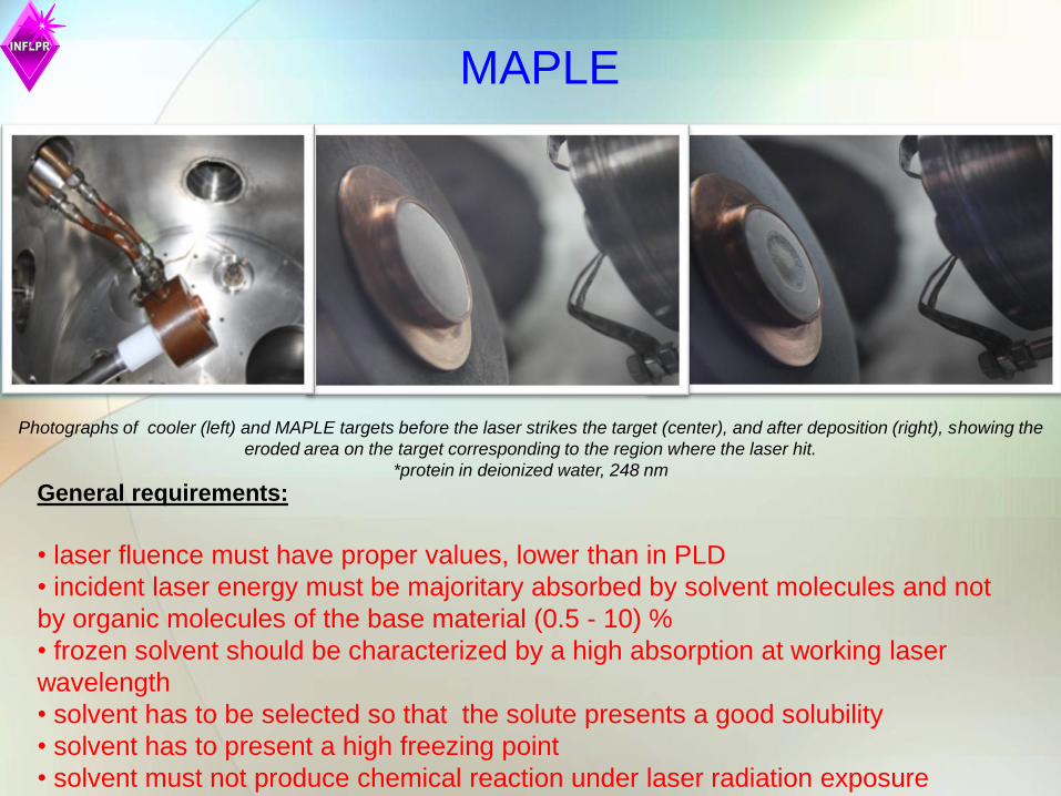
MAPLE
Photographs of cooler (left) and MAPLE targets before the laser strikes the target (center), and after deposition (right), showing the
eroded area on the target corresponding to the region where the laser hit.
*protein in deionized water, 248 nm
General requirements:
• laser fluence must have proper values, lower than in PLD
• incident laser energy must be majoritary absorbed by solvent molecules and not
by organic molecules of the base material (0.5 - 10) %
• frozen solvent should be characterized by a high absorption at working laser
wavelength
• solvent has to be selected so that the solute presents a good solubility
• solvent has to present a high freezing point
• solvent must not produce chemical reaction under laser radiation exposure

Biomaterials in this lecture
Urease
an enzyme that catalyzes the hydrolysis of urea into carbon dioxide and ammonia;
nitrogen concentration of the human serum in urea is a measure of kidney function
Triacetate-pullulan polysaccharide
a linear homopolysaccharide of glucose, often described as a linked polymer of maltotriose
subunits (1, 6)
has many potential food, pharmaceutical, and industrial applications
Levan
homopolysaccharide of d-fructofuranosyl residues joined by β-2,6 with multiple branches by
β-2,1 linkages;
great potential as a functional biopolymer in foods, feeds, cosmetics, pharmaceutical and
chemical industries
Alendronate-hydroxyapatite (HA)
Bisphosphonates (BPs) are widely used for the management of specific disorders of bone
metabolism, such as Paget bone disease, osteoporosis, fibrous dysplasia, myeloma and
bone metastases Dextran - γ-Fe2O3 NPs
Superparamagnetic γ-Fe2O3 NPs: biomedical applications as contrast agent in magnetic
resonance imaging, targeted destruction of tumor tissues through hyperthermia, and drug delivery;
cause inflammations, formation of apoptotic bodies, generation of reactive oxygen species,
chromosomal damage.
dextran prevents agglomeration and improves biocompatibility of NPs
IgG
antibodies, selective and chemical affinity molecules produced in response to the
introduction of a foreign molecule (antigen) in the body.

Extracellular matrix (ECM) proteins-HA
HA is capable to induce mineralization, while ECM proteins (as e.g. fibronectin or vitronectin)
are used for material bioactivation by adsorption and cell adhesion at the interface
Fibronectin (FN) is an ECM glycoprotein capable of binding to integrin receptors, which
mediate the attachment of cells to surrounding tissues
Vitronectin (VN) is a glycoprotein; about one-third of the protein's molecular mass is composed
of carbohydrates
Poly(methyl methacrylate) (PMMA) + Bioactive glasses (BG)
Biocompatible, bioinert polymer, often used as a light or shatter-resistant alternative to glass
increase the functionality and improve wear properties of the metallic implants
OCP; Sr:OCP; Mg:OCP
OCP: the most likely precursor of biological apatites due to its structural resemblance to HA;
- prospective alternative to HA coatings for metallic implants.
- greater solubility and easy hydrolysis into nanocrystalline apatite in physiological solution
Sr is present in mineral phase of bone with beneficial effect in the treatment of osteoporosis:
increase the number of hOB, decrease the number and activity of hOC
- increase in bone mass and strength by inhibition of bone resorption and augmentation of
bone formation
Mg occurs typically as the Mg2+ ion; it is an essential mineral nutrient for life and provide
strength to the bone structure
-it is present in every cell type of each organism and is indispensable for a good health
Biomaterials in this lecture

Triacetate-pullulan polysaccharide
Major differences between starting
material and the PLD (248 nm, 390
mJ/cm2 ) film which demonstrate the
degradation of the structure during
PLD.
a. Pullulan dropcast
b. 2% pullulan in deionized water
c. 2% pullulan in 3-butanol
d. filtered 2% pullulan in 3-butanol
e. filtered 2% pullulan in DMSO
Laser fluence 240 mJ/cm2
No decomposition by MAPLE of 2%
solutions in deionized water and 3-butanol!

Powder X-ray diffraction patterns of the thin films deposited from:
(a) HA, (b) HA-AL7(4%), (c) HA-AL28(7%)
The slight increase of the broadening of the diffraction peaks when
increasing alendronate concentration is indicative for a modest decrease
of the length of the crystalline domains as the alendronate content in the
apatite nanocrystals increases up to 7.1%.
HA –AL– XRD investigations
* 3.9 or 7% AL with HA; 0.25 g of HA-AL powder suspended in 5 ml deionized water, 248 nm, 750 mJ/cm2
Alendronate
HA: Ca10(PO4)6(OH)2
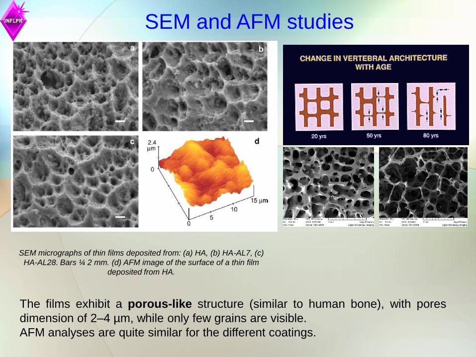
SEM micrographs of thin films deposited from: (a) HA, (b) HA-AL7, (c)
HA-AL28. Bars ¼ 2 mm. (d) AFM image of the surface of a thin film
deposited from HA.
The films exhibit a porous-like structure (similar to human bone), with pores
dimension of 2–4 µm, while only few grains are visible.
AFM analyses are quite similar for the different coatings.
SEM and AFM studies

Florescence microscopy images of hOB on
alendronate-HA coatings
Phallodin staining of culture after 24 hour from seeding: (a) Ti, (b) HA, (c) HA-AL7. Bars = 20 µm.
c b a
Presence of alendronate in HA thin films enhances osteointegration and
bone regeneration!

Proliferation of osteoclast (hOC) culture on
alendronate-HA coatings: 21 days
Presence of alendronate prevents the undesirable bone resorption!
Phallodin staining of culture after 21 days from seeding: (a) Ti, (b) HA, (c) HA-AL7.
Bars = 20 µm.
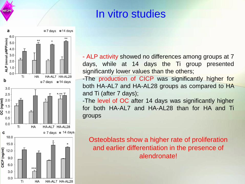
- ALP activity showed no differences among groups at 7
days, while at 14 days the Ti group presented
significantly lower values than the others;
-The production of CICP was significantly higher for
both HA-AL7 and HA-AL28 groups as compared to HA
and Ti (after 7 days);
-The level of OC after 14 days was significantly higher
for both HA-AL7 and HA-AL28 than for HA and Ti
groups
Osteoblasts show a higher rate of proliferation
and earlier differentiation in the presence of
alendronate!
In vitro studies

Hybrid maghemite Fe2O3 NPs - dextran
thin films
Maghemite nanoparticles
(mean size 8.3±0.3 nm)
Iron oxide NPs prepared by co-
precipitation of: FeCl2·4H2O,
FeCl3·6H2O, NaOH, Fe(NO3)3, HNO3
MAPLE target: iron oxide NP (0-5%),
dextran (10%), deionized water
F = 0.5 J/cm2
25000 subsequent laser pulses
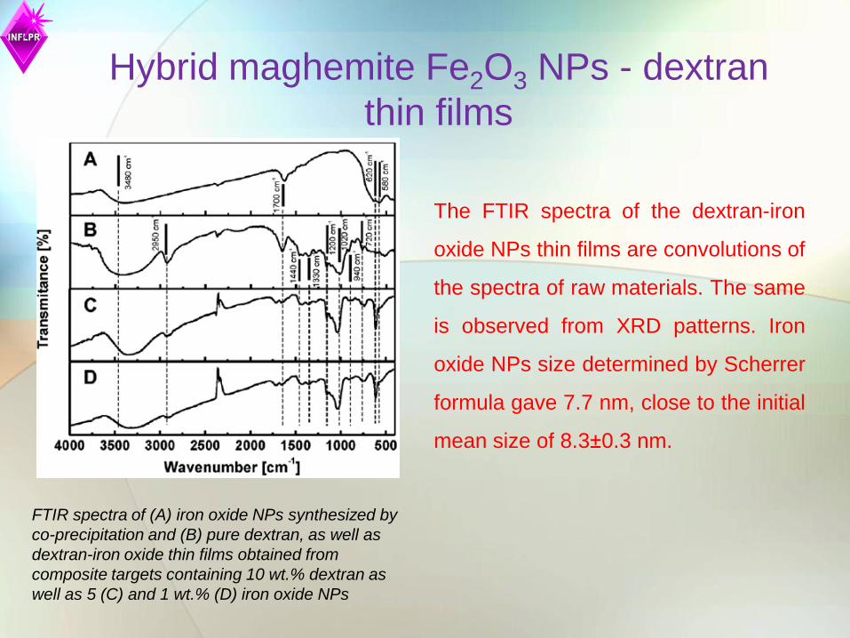
The FTIR spectra of the dextran-iron
oxide NPs thin films are convolutions of
the spectra of raw materials. The same
is observed from XRD patterns. Iron
oxide NPs size determined by Scherrer
formula gave 7.7 nm, close to the initial
mean size of 8.3±0.3 nm.
FTIR spectra of (A) iron oxide NPs synthesized by
co-precipitation and (B) pure dextran, as well as
dextran-iron oxide thin films obtained from
composite targets containing 10 wt.% dextran as
well as 5 (C) and 1 wt.% (D) iron oxide NPs
Hybrid maghemite Fe2O3 NPs - dextran
thin films

In-vitro tests
- only 8% drop of viability in case of dextran-iron oxide thin films after 24 h incubation
time
- drop increased slightly below 12% for the dextran-iron oxide thin films with 5% NPs
after 48 h
- HepG2 cells
- MTS colorimetric (viability) assay for fast counting of living cells
This evidence supports the utilization of composite
iron oxide – dextran thin films for applications in drug
delivery and as contrast agent in magnetic
resonance imaging!

LEVAN Sample Fluence
(J/cm2)
Solvent Concentration
(g/l)
Optimal parameters
(substrate temperature, pressure,
target-substrate separation distance)
Levan (L) 0.28 DMSO 5 100 oC, 5 Pa, 3.5 cm, 20000
Oxidized Levan (OL)* 0.35 DMSO 5 100 oC, 5 Pa, 3.5 cm, 20000
*Pure levan samples subjected to periodate oxidation by prolonged magnetic stirring in a beaker
500 1000 1500 2000 2500 3000 3500 4000
0.2
0.3
0.4
0.5
93
01
04
01
11
01
16
0
14
50
28
90
29
40
33
50
Ab
so
rba
nce
(a
.u.)
Wavenumber (cm-1)
levan by MAPLE
dropcast
500 1000 1500 2000 2500 3000 3500 4000
0.4
0.5
0.6
0.7
Wavenumber (cm-1)
Ab
so
rba
nce
(a
.u.)
dropcast
oxidized levan by MAPLE
FTIR absorption spectra of L and OL dropcast and L and OL thin films, respectively, deposited by MAPLE

LEVAN
Typical AFM images of sample surfaces for a) L and b) OL
coatings on Si
a) b)
XSEM of L thin films on glass by MAPLE
Sample Rms (nm) Ra (nm) Rz (nm)
L coating on Si 0.972 0.762 4.158
OL coating on
Si 1.203 0.962 4.818
Possible formation mechanims:
nanostructured assemebling!

LEVAN
SaOs2 cells adhesion on control, L and OL coatings on glass by
immunofluorescence microscopy
- similar coverage of the
control and tested surfaces
- no change in the filament
organization pattern. Actin is
uniformly spread throughout
cell cytosol in parallel
filaments sustaining cell
shape and motility.
- L and OL coatings have no detrimental
function over cells. Cells proliferation was
similar on L and control samples.
- OL induces an increase in the ability of cells
to divide and give rise to daughter cells. This
result is supported by CA measurements
where hydrophilic surfaces were revealed in
the case of OL.
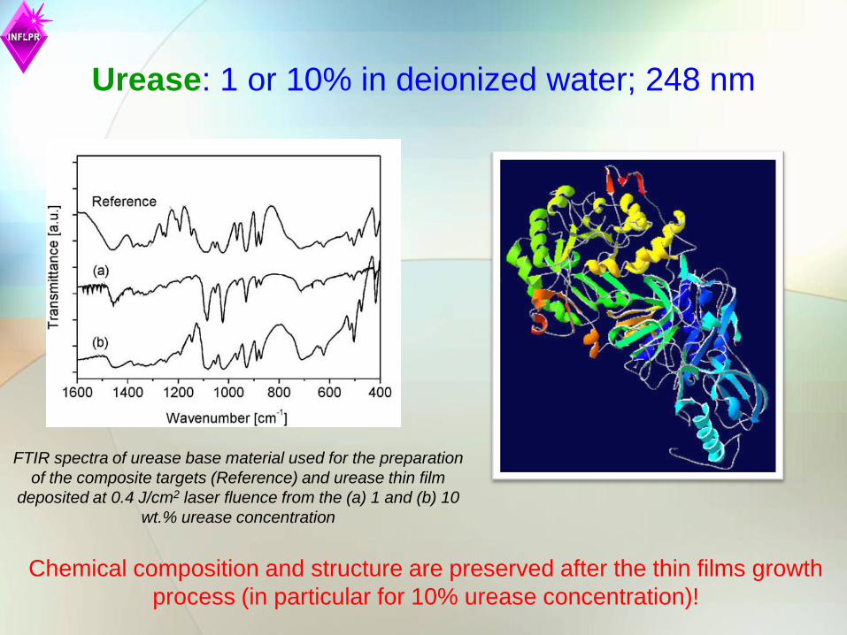
Urease: 1 or 10% in deionized water; 248 nm
FTIR spectra of urease base material used for the preparation
of the composite targets (Reference) and urease thin film
deposited at 0.4 J/cm2 laser fluence from the (a) 1 and (b) 10
wt.% urease concentration
Chemical composition and structure are preserved after the thin films growth
process (in particular for 10% urease concentration)!
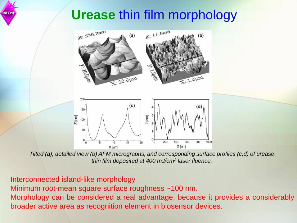
Urease thin film morphology
Tilted (a), detailed view (b) AFM micrographs, and corresponding surface profiles (c,d) of urease
thin film deposited at 400 mJ/cm2 laser fluence.
Interconnected island-like morphology
Minimum root-mean square surface roughness ~100 nm.
Morphology can be considered a real advantage, because it provides a considerably
broader active area as recognition element in biosensor devices.

Urease enzymatic activity and kinetics => determined by Worthington assay
method
urea [H2N-CO-NH2] + H2O 2NH4 + + CO2 (1)
2NH4
+ + 2α-Ketoglutarate + 2NADH 2Glutamate (C5H9NO4) +
2NAD+ + 2H2O (2)
The NADH molecule (reduced NAD) is oxidized to NAD+.
NADH absorbs UV light at 340 nm but NAD+ does not.
Urease enzymatic activity evaluation
If the number of NADH molecules drops, the absorbance at 340 nm
decreases – possible application to urea monitoring
The hydrolysis of urea was measured by coupling ammonia to NADH
(nicotinamide adenine dinucleotide) oxidation reaction
urease
glutamate dehydrogenase
Urease enzymatic activity evaluation
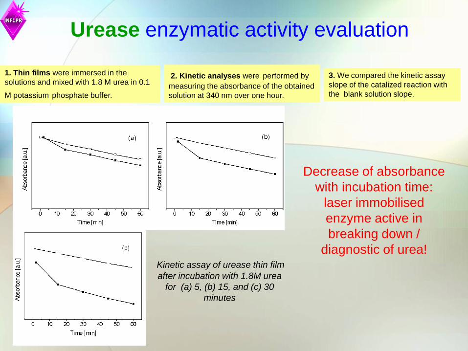
Kinetic assay of urease thin film
after incubation with 1.8M urea
for (a) 5, (b) 15, and (c) 30
minutes
1. Thin films were immersed in the
solutions and mixed with 1.8 M urea in 0.1
M potassium phosphate buffer.
3. We compared the kinetic assay
slope of the catalized reaction with
the blank solution slope.
Decrease of absorbance
with incubation time:
laser immobilised
enzyme active in
breaking down /
diagnostic of urea!
2. Kinetic analyses were performed by
measuring the absorbance of the obtained
solution at 340 nm over one hour.

IgG
Optical images of IgG structures
without lipid at fluences of (A) 0.33
J/cm2 , (B) 0.5 J/cm2, and (C) 0.67
J/cm2, respectively.
Optical images of IgG structures with
lipid at fluences of (A) 0.33 J/cm2 ,
(B) 0.5 J/cm2, and (C) 0.67 J/cm2,
respectively.
IL50 exhibited a well
protected,
encapsulate-like
material (Fig. B down),
as compared to I50
(Fig. B top) where
splashed and broken
particulates could be
seen besides intact
ones!

IgG
AFM images of IgG structures obtained by MAPLE deposition on fused silica substrates
from IgG dissolved in water and saline buffer without (I) and with (II) lipid at fluences of
0.33 J/cm2 (I33), 0.5 J/cm2 (I50) and 0.67 J/cm2 (I67), respectively.
I
II
Thickness values of 200, 350 and 500 nm for lipid free samples, and 180, 450 and 700
nm in the case of lipid containing samples. This corresponds to a deposition rate of
(0.02-0.05) nm/pulse in the first case and (0.018-0.07) nm/pulse in the second one,
respectively!

IgG
Fluorescence microscopy images of rabbit IgG samples after
incubation with 1:50 dilution of Alexa Fluor-conjugated secondary
antibody
Fluorescence microscopy images of rabbit IL50 versus I50 samples
after incubation with 1:50 dilution of Alexa Fluor-conjugated
secondary antibody
Quantification of IgG Protein Transferred by
MAPLE Using Lipid-Added Formulations
There is a ‘‘window’’ in laser
fluence (0.5 J/cm2) for which the
integer structure of the protein was
preserved and morphology can be
well controlled.

BG-PMMA Metals and alloys used in implants are susceptible to corrosion in body fluids
release of ions that accumulate in vital organs
Most common corrosion types of metallic medical devices:
- pitting
- galvanic
- crevice
Proposed solution: apply protective BG-PMMA coating
PMMA insulator and barrier against ions
BG+PMMA composite
Metallic substrate Metallic substrate Metallic substrate
Immersion in body fluids Continuous , compact layer of PMMA
Newly formed bioapatite
(C5O2H8)n

Experimental details
• Target preparation: 0.6g PMMA +0.08g BG in 19.3 ml chloroform
• Two types of BG :
6P61: SiO2 61,1%, Na2O 10.3%, K2O 2.8%, CaO 12.6%, MgO, 7.2%, P2O5 6%
6P57: SiO2 57%, Na2O 11%, K2O 3%, CaO 15%, MgO 8%, P2O5 6% Deposition substrates: Ti gr.4 disks
Laser parameters: λ=248 nm, τ= 25 ns, F=0.55J/cm2
Immersion in SBF: 28 days (for 6P57) or 42 days (for 6P61)

Results
SEM micrograph showing a typical surface
morphology of a 6P57-PMMA coating
1000 1500 2000 2500
0
10
20
30
40
50
60
70
PMMA powder
6P57-PMMA thin film
Ab
so
rba
nce
[a
.u.]
Wawenumber (cm-1)
FT-IR spectra of PMMA powders and of a
6P57-PMMA coating obtained by MAPLE
Fluorescence microscopy of cells cultured on
MAPLE deposited structures 6P57 – PMMA
Higher proliferation of cells on
6P57+PMMA!
1200 1000 800 600 400 200 0
Ti 3p
Na
2s
Si 2p
Si 2s
P 2
s
Ca
2s
N 1
s
C 1
s
Ca
2pT
i 2
p
O 1
s
O K
LL
Na
1s
C K
LL
6P57_PMMA sample
Inte
nsity (
a.u
.)
Binding energy (eV)
XPS survey spectrum of the 6P57-
PMMA coating
The film contains PMMA and bioglass.
Biological properties similar to simple 6P57
and 6P61 bioactive glass coatings
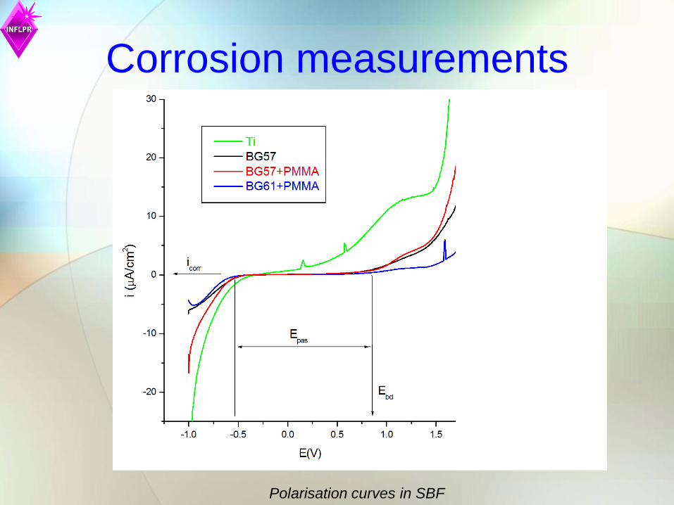
Corrosion measurements
Polarisation curves in SBF
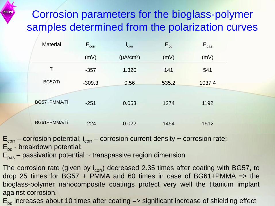
Material Ecorr
(mV)
icorr
(µA/cm2)
Ebd
(mV)
Epas
(mV)
Ti -357 1.320 141 541
BG57/Ti -309.3 0.56 535.2 1037.4
BG57+PMMA/Ti -251 0.053 1274 1192
BG61+PMMA/Ti -224 0.022 1454 1512
Corrosion parameters for the bioglass-polymer
samples determined from the polarization curves
The corrosion rate (given by icorr) decreased 2.35 times after coating with BG57, to
drop 25 times for BG57 + PMMA and 60 times in case of BG61+PMMA => the
bioglass-polymer nanocomposite coatings protect very well the titanium implant
against corrosion.
Ebd increases about 10 times after coating => significant increase of shielding effect
Ecorr – corrosion potential; icorr – corrosion current density ~ corrosion rate;
Ebd - breakdown potential;
Epas – passivation potential ~ transpassive region dimension
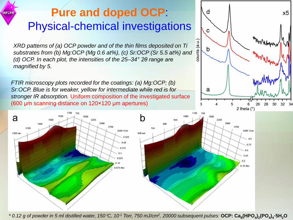
XRD patterns of (a) OCP powder and of the thin films deposited on Ti
substrates from (b) Mg:OCP (Mg 0.6 at%), (c) Sr:OCP (Sr 5.5 at%) and
(d) OCP. In each plot, the intensities of the 25–34° 2θ range are
magnified by 5.
Pure and doped OCP:
Physical-chemical investigations
FTIR microscopy plots recorded for the coatings: (a) Mg:OCP; (b)
Sr:OCP. Blue is for weaker, yellow for intermediate while red is for
stronger IR absorption. Uniform composition of the investigated surface
(600 μm scanning distance on 120×120 μm apertures)
* 0.12 g of powder in 5 ml distilled water, 150C, 10-1 Torr, 750 mJ/cm2, 20000 subsequent pulses: OCP: Ca8(HPO4)2(PO4)4·5H2O
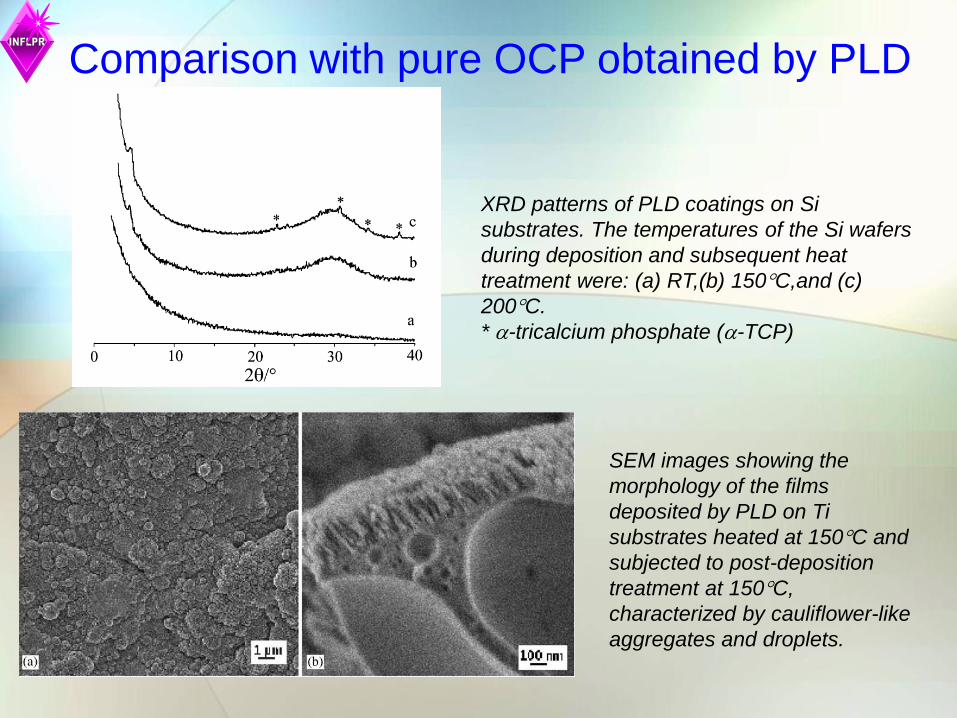
Comparison with pure OCP obtained by PLD
XRD patterns of PLD coatings on Si
substrates. The temperatures of the Si wafers
during deposition and subsequent heat
treatment were: (a) RT,(b) 150C,and (c)
200C.
* -tricalcium phosphate (-TCP)
SEM images showing the
morphology of the films
deposited by PLD on Ti
substrates heated at 150C and
subjected to post-deposition
treatment at 150C,
characterized by cauliflower-like
aggregates and droplets.

Morpho-chemical studies
SEM micrographs of thin
films deposited from OCP
sample. In the image at
higher magnification (b) the
presence of crystal
fragments, together with
cauliflower-like (c)
aggregates and droplets
(D) is clearly visible.
Energy dispersive X-ray
spectrometry (EDS)
maps recorded for the
coatings: (a) Mg:OCP;
(b) Sr:OCP
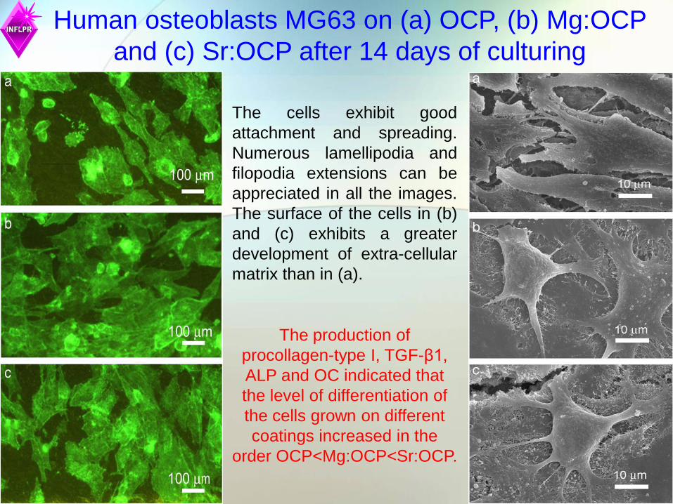
Human osteoblasts MG63 on (a) OCP, (b) Mg:OCP
and (c) Sr:OCP after 14 days of culturing
The cells exhibit good
attachment and spreading.
Numerous lamellipodia and
filopodia extensions can be
appreciated in all the images.
The surface of the cells in (b)
and (c) exhibits a greater
development of extra-cellular
matrix than in (a).
The production of
procollagen-type I, TGF-β1,
ALP and OC indicated that
the level of differentiation of
the cells grown on different
coatings increased in the
order OCP<Mg:OCP<Sr:OCP.

Fibronectin structure
HA-ECM proteins
ECM proteins: Fibronectin, vitronectin

Protein investigations: FT-IR studies
IR absorption bands of VN structures obtained on silicon substrates
1500 2000 2500 3000 3500 4000
0,0
0,1
0,2
0,3
0,4
0,5
0,6
saline buffer
saline buffer
saline buffer
Vitronectin
(1631 cm-1)
Vitronectin
2990 cm-1
Vitronectin
3186 cm-1
Ab
so
rba
nce
Wavenumber (cm-1)
VN MAPLE
VN dropcast
Si
C=O, CN stretch, NH bending
ν(CH3), ν(CH2)
(NH stretch)
The peaks of proteins deposited by MAPLE are matching very well the
ones from dropcast!
* 1.8 mg/ml in deionized water based saline buffer, 700 mJ/cm2, 15000 subsequent laser pulses
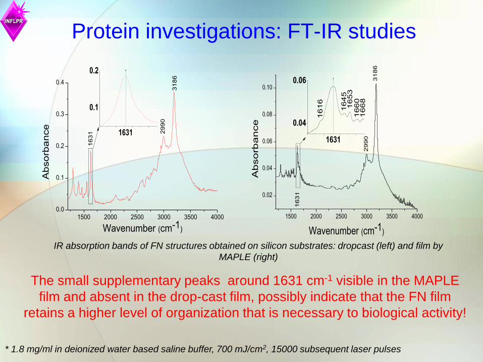
Protein investigations: FT-IR studies
IR absorption bands of FN structures obtained on silicon substrates: dropcast (left) and film by
MAPLE (right)
* 1.8 mg/ml in deionized water based saline buffer, 700 mJ/cm2, 15000 subsequent laser pulses
1500 2000 2500 3000 3500 40000.0
0.1
0.2
0.3
0.4
2990
1631
3186
Ab
so
rba
nce
Wavenumber (cm-1)
0.1
0.2
1631
1500 2000 2500 3000 3500 4000
0.02
0.04
0.06
0.08
0.10
Ab
so
rba
nce
Wavenumber (cm-1)
3186
2990
1631
0.04
0.06
1616
1668
1660
1653
1631
1645
The small supplementary peaks around 1631 cm-1 visible in the MAPLE
film and absent in the drop-cast film, possibly indicate that the FN film
retains a higher level of organization that is necessary to biological activity!
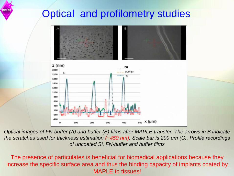
Optical images of FN-buffer (A) and buffer (B) films after MAPLE transfer. The arrows in B indicate
the scratches used for thickness estimation (~450 nm). Scale bar is 200 μm (C). Profile recordings
of uncoated Si, FN-buffer and buffer films
Optical and profilometry studies
The presence of particulates is beneficial for biomedical applications because they
increase the specific surface area and thus the binding capacity of implants coated by
MAPLE to tissues!
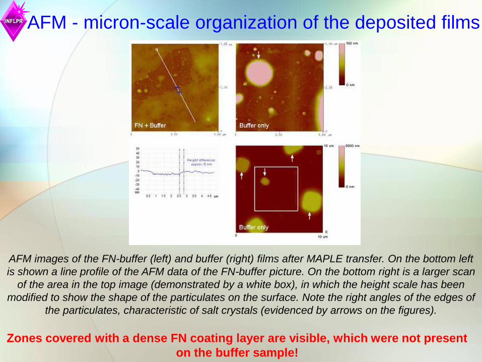
AFM - micron-scale organization of the deposited films
AFM images of the FN-buffer (left) and buffer (right) films after MAPLE transfer. On the bottom left
is shown a line profile of the AFM data of the FN-buffer picture. On the bottom right is a larger scan
of the area in the top image (demonstrated by a white box), in which the height scale has been
modified to show the shape of the particulates on the surface. Note the right angles of the edges of
the particulates, characteristic of salt crystals (evidenced by arrows on the figures).
Zones covered with a dense FN coating layer are visible, which were not present
on the buffer sample!
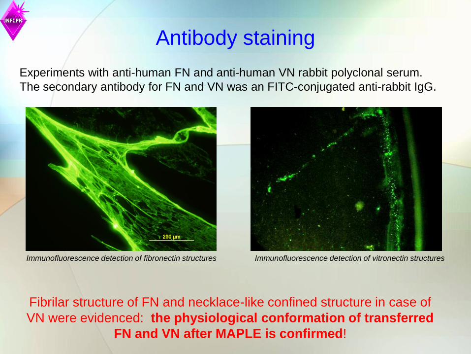
Antibody staining
Immunofluorescence detection of fibronectin structures
Experiments with anti-human FN and anti-human VN rabbit polyclonal serum.
The secondary antibody for FN and VN was an FITC-conjugated anti-rabbit IgG.
Fibrilar structure of FN and necklace-like confined structure in case of
VN were evidenced: the physiological conformation of transferred
FN and VN after MAPLE is confirmed!
Immunofluorescence detection of vitronectin structures

CTR Si Si/FN
I
II
Fig. 7
A B C
HOB precursor cells: Actin and nuclei staining 3 h
Characteristic cell round shape and cortical actin staining on glass control (A).
More evident actin fibers of cells grown on silicon (B). Patterns of parallel stress
fibers extending all throughout the cytoplasm from nucleus membrane to
plasma membrane are visible on FN films which confirm the structure and
functionality of deposited FN molecules (C).
HOB cell actin filament staining on (A) standard cover slips, (B) silicon and (C) FN
covered silicon by MAPLE after 3 hours in cell culture. Histograms of HOB cells perimeter and area
3 hours after seeding. Scale bar is 100 μm (I) and 50 μm (II).
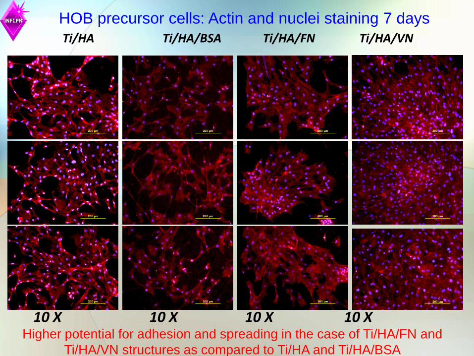
HOB precursor cells: Actin and nuclei staining 7 days
Ti/HA Ti/HA/BSA Ti/HA/FN Ti/HA/VN
10 X 10 X 10 X 10 X Higher potential for adhesion and spreading in the case of Ti/HA/FN and
Ti/HA/VN structures as compared to Ti/HA and Ti/HA/BSA
Ti/HA Ti/HA/BSA
Ti/HA/VN Ti/HA/FN
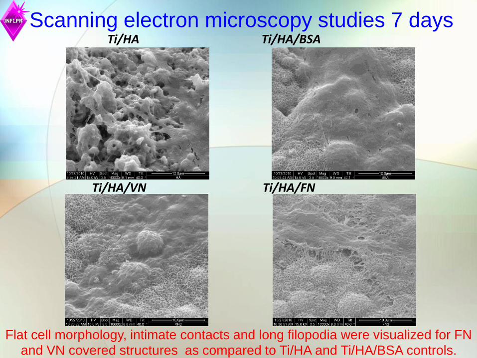
Scanning electron microscopy studies 7 days
Flat cell morphology, intimate contacts and long filopodia were visualized for FN and VN covered structures as compared to Ti/HA and Ti/HA/BSA controls.

HOB precursor cells: Actin (red), vinculin (green) and
nuclei (blue) staining 7 days: Ti/HA vs Ti/HA/VN
The HOB cells grown on Ti/HA/VN samples (D, E, F) exhibited improved
adherence, spreading and growth compared to cells grown on Ti/HA samples
(A, B, C), supporting a faster cell colonization and the physiological VN
functionality after MAPLE transfer.

Cell culture – human osteoprogenitor cells:
MTS assay
0.000
0.020
0.040
0.060
0.080
0.100
0.120
Day 1
Day 3
Day 7
Day 14
HA BSA FN VN
In vitro tests demonstrate a much larger bioactivity of Ti/HA/FN and Ti/HA/VN
as compared toTi/HA or Ti/HA/BSA

FN transfer by Laser Direct Write (LDW)
= 248 nm
= 25 ns
metallic mask with a circular pinhole of 200 µm diameter interposed in the central region of the focused laser beam
F = 400 mJ/cm2
Separation distance ribbon – acceptor: 20 µm
Ribbon: quartz lamellae covered with 25 nm Ti buffer layer obtained by PLD, FN
(~ 10 µm) by drop-cast
Acceptor: glass slide covered by PLD with 25 nm Ti layer

Fluorescence micrographs of
different patterns obtained by
excimer Laser Direct Write: (a)
a checkerboard-like pattern
and (b) a micrometric FN
features-based writing test.
Typical fluorescence
micrographs of LDW ribbon
and the corresponding
laser-transferred protein
spot stained by FITC-
Concanavalin A
FN transfer by LDW
Other results are in
publication.
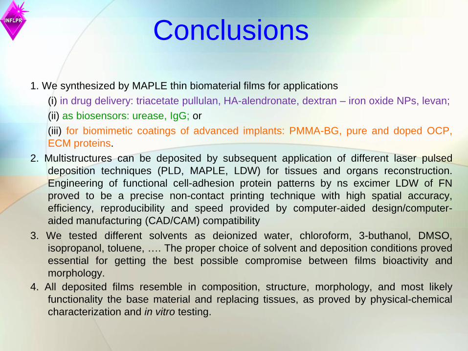
Conclusions
1. We synthesized by MAPLE thin biomaterial films for applications
(i) in drug delivery: triacetate pullulan, HA-alendronate, dextran – iron oxide NPs, levan;
(ii) as biosensors: urease, IgG; or
(iii) for biomimetic coatings of advanced implants: PMMA-BG, pure and doped OCP,
ECM proteins.
2. Multistructures can be deposited by subsequent application of different laser pulsed
deposition techniques (PLD, MAPLE, LDW) for tissues and organs reconstruction.
Engineering of functional cell-adhesion protein patterns by ns excimer LDW of FN
proved to be a precise non-contact printing technique with high spatial accuracy,
efficiency, reproducibility and speed provided by computer-aided design/computer-
aided manufacturing (CAD/CAM) compatibility
3. We tested different solvents as deionized water, chloroform, 3-buthanol, DMSO,
isopropanol, toluene, …. The proper choice of solvent and deposition conditions proved
essential for getting the best possible compromise between films bioactivity and
morphology.
4. All deposited films resemble in composition, structure, morphology, and most likely
functionality the base material and replacing tissues, as proved by physical-chemical
characterization and in vitro testing.

List of relevant publications 1. Magnesium and strontium doped octacalcium phosphate thin films by matrix assisted pulsed laser evaporation, E. Boanini, P. Torricelli, M.
Fini, F. Sima, N. Serban, I. N. Mihailescu, A. Bigi, Journal of Inorganic Biochemistry 107:65–72, 2012
2. Hybrid dextran-iron oxide thin films deposited by laser techniques for biomedical applications, D. Predoi, C.S. Ciobanu, M. Radu, M.
Costache, A. Dinischiotu, C. Popescu, E. Axente, I.N. Mihailescu, E. Gyorgy, Materials Science and Engineering: C, 32 (2), 296–302, 2012
3. Fibronectin layers by matrix assisted pulsed laser evaporation from saline buffer based cryogenic targets, Felix Sima; Patricia Davidson;
Emmanuel Pauthe; Livia E Sima; Olivier Gallet; Ion Mihailescu; Karine Anselme, Acta Biomaterialia, 7 (10) 3780-3788 (2011)
4. Thin films of vitronectin transferred by MAPLE, F. Sima, P. Davidson, E. Pauthe, O. Gallet, K. Anselme, I.N. Mihailescu, Applied Physics
A 105, 611–617 (2011)
5. Tailoring immobilization of immunoglobulin by excimer laser for biosensor applications, F. Sima, E. Axente, C. Ristoscu, I. N. Mihailescu, T.
V. Kononenko, I. A. Nagovitsin, G. Chudinova, V. I. Konov, M. Socol, I. Enculescu, L. E. Sima, St. M. Petrescu, Journal of
Biomaterials Research-A, 96A (2), 384 - 394, 2011
6. Levan thin films by MAPLE nanostructured assembling, Felix Sima, Esra Cansever Mutlu, Mehmet S. Eroglu , Livia E. Sima, Natalia
Serban, Carmen Ristoscu, Stefana M. Petrescu, Ebru Toksoy Oner, Ion N. Mihailescu, Biomacromolecules, 12(6) (2011) 2251–2256
7. Composite biocompatible hydroxyapatite–silk fibroin coatings for medical implants obtained by Matrix Assisted Pulsed Laser Evaporation,
F.M. Miroiu , G. Socol, A. Visan, N. Stefan, D. Craciun, V. Craciun, G. Dorcioman, I.N. Mihailescu, L.E. Sima, S.M. Petrescu, A. Andronie, I.
Stamatin, S. Moga and C. Ducu, Materials Science and Engineering B 169, 151–158, 2010
8. Biomolecular urease thin films grown by laser techniques for blood diagnostic applications, E. Gyorgy, F. Sima, I. N. Mihailescu, T.
Smauz, B. Hopp, D. Predoi, S. Ciuca, L. E. Sima, S. M. Petrescu, Materials Science and Engineering C, 30 (2010) 537–541
9. Functional porphyrin thin films deposited by matrix assisted pulsed laser evaporation, R. Cristescu, C. Popescu, A.C. Popescu, I.N.
Mihailescu, A.A. Ciucu, A. Andronie, S. Iordache, I. Stamatin, E. Fagadar-Cosma, D.B. Chrisey, Mat. Sci. Eng. B 169, 106–110 (2010)
10. Application of clean laser transfer for porphyrin micropatterning, T.V. Kononenko, I.A. Nagovitsyn, G.K. Chudinova, I.N. Mihailescu, Applied
Surface Science 256 (2010) 2803–2808
11. Comparative study on Pulsed Laser Deposition and Matrix Assisted Pulsed Laser Evaporation of urease thin films, T. Smausz, G. Megyeri,
R. Kékesi, C. Vass, E. György, F. Sima, I. N. Mihailescu, B. Hopp, Thin Solid Films, 517 (15), 4299-4302 , 2009
12. Specific biofunctional performances of the hydroxyapatite-sodium maleate copolymer hybrid coating nanostructures evaluated by in vitro
studies, L. E. Sima, A. Filimon, R.M. Piticescu, G.C. Chitanu, D.M. Suflet, M. Miroiu, G. Socol, I. N. Mihailescu, J. Neamtu, G.
Negroiu, Journal of Materials Science: Materials in Medicine, 20, 2305-2316, 2009
13. Bioglass –polymer thin coatings obtained by MAPLE for a new generation of implants, F. Sima, C. Ristoscu, A. Popescu, I. N. Mihailescu,
T. Kononenko, S. Simon, T. Radu, O. Ponta, R. Mustata, L. E. Sima, S. M. Petrescu, Journal of Optoelectronics and Advanced Materials,
11(9), 1170 – 1174 (2009)
14. Immobilization of urease by laser techniques: synthesis and application to urea biosensors, György E, Sima F, Mihailescu IN, Smausz T,
Megyeri G, Kékesi R, Hopp B, Zdrentu L, Petrescu SM., Journal of Biomedical Materials Research: 89A: 186–191, 2009
15. Biofunctional alendronate–Hydroxyapatite thin films deposited by Matrix Assisted Pulsed Laser Evaporation, A. Bigi, E. Boanini, C.
Capuccini, M. Fini, I. N. Mihailescu, C. Ristoscu, F. Sima, P. Torricelli, Biomaterials, Volume 30, Issue 31, October 2009, Pages 6168-6177
16. Thin Films of Polymer Mimics of Cross-Linking Mussel Adhesive Proteins Deposited by Matrix Assisted Pulsed Laser Evaporation, R
Cristescu, I.N. Mihailescu, I. Stamatin, A. Doraiswamy, R.J. Narayan, G. Westwood, J.J. Wilker, S. Stafslien, B. Chisholm, D.B.
Chrisey, Applied Surface Science, 255 (2009) 5496–5498

17. Functional polyethylene glycol derivatives nanostructured thin films synthesized by matrix-assisted pulsed laser evaporation, R. Cristescu, C.
Popescu, A. Popescu, S. Grigorescu, I.N. Mihailescu, D. Mihaiescu, S.D. Gittard, R.J. Narayan, T. Buruiana, I. Stamatin, D.B.
Chrisey, Applied Surface Science 255 (2009) 9873–9876
18. Functionalized Polyvinyl Alcohol Derivatives Thin Films for Controlled Drug Release and Targeting Systems: MAPLE Deposition and
Morphological, Chemical and In Vitro Characterization, R. Cristescu, C. Cojanu, A. Popescu, S. Grigorescu, L. Duta, G. Caraene, A.
Ionescu, D. Mihaiescu, R. Albulescu, T. Buruiana, A. Andronie, I. Stamatin, I. N. Mihailescu, D. B. Chrisey, Appl. Surf. Sci. 255 (2009)
5600–5604
19. Laser Processing of Polyethylene Glycol Derivative and Block Copolymer Thin Films, R. Cristescu, C. Cojanu, A. Popescu, S. Grigorescu, L.
Duta, O. Ionescu, D. Mihaiescu, T. Buruiana, A. Andronie, I. Stamatin, I, N. Mihailescu, D. B. Chrisey, Applied Surface Science, 255 (2009)
5605–5610
20. Creatinine biomaterial thin films grown by laser techniques, E.Gyorgy, E. Axente, I. N. Mihailescu, D. Predoi, S. Ciuca, J. Neamtu, Journal
of Material Science: Materials in Medicine,19(3), 1335-1339, 2008
21. Biocompatibility evaluation of a novel hydroxyapatite-polymer coating for medical implants (in vitro tests), G.Negroiu, R.M. Piticescu, G.C.
Chitanu, I.N. Mihailescu, L. Zdrentu, M. Miroiu, Journal of Materials Science: Materials in Medicine19 (4) 2008 1537-1544
22. Laser Processing of Natural Mussel Adhesive Protein Thin Films, A. Doraiswamy, R.J. Narayan, R. Cristescu, I.N. Mihailescu, D.B. Chrisey,
Materials Science and Engineering: C 27(3), (2007) 409-413
23. MAPLE Applications in Studying Organic Thin Films, M. Jelinek, T. Kocourek, J. Remsa, R. Cristescu, I.N. Mihailescu, D.B. Chrisey, Laser
Physics 17(2), (2007) 66-70(5)
24. Thin Films Growth Parameters in MAPLE; Application to Fibrinogen, M. Jelinek, R. Cristescu, T. Kocourek, V. Vorliček, J Remsa, L.
Stamatin, D. Mihaiescu, I. Stamatin, I.N. Mihailescu, D.B. Chrisey, Journal of Physics: Conference Series 59, (2007) 22-27
25. Matrix Assisted Pulsed Laser Evaporation of Pullulan Tailor-Made Biomaterials Thin Films for Controlled Drug Delivery Systems, R.
Cristescu, M. Jelinek, T. Kocourek, E. Axente, S. Grigorescu, A. Moldovan, D.E. Mihaiescu, M. Albulescu, T. Buruiana, J. Dybal, I.
Stamatin, I.N. Mihailescu, D.B. Chrisey, Journal of Physics: Conference Series 59, (2007) 144-149
26. Biomolecular papain thin films growth by laser techniques, E. Gyorgy, J. Santiso, A. Figueras, G. Socol, I. N. Mihailescu, Journal of Materials
Science: Materials in Medicine 18, 8, 1471 - 1663, 2007
27. Laser Processing of DOPA modified PEG Mussel Adhesive Protein Analog Thin Films, A. Doraiswamy, R.J. Narayan, C. Dinu, R. Cristescu,
P.B. Messersmith, S. Stafslien, D.B. Chrisey, Journal of Adhesion Science & Technology, 21(3-4), (2007) 287-299(13)
28. Processing of poly(1,3-bis-(p-carboxyphenoxy propane)-co-(sebacic anhydride)) 20:80 (P(CPP:SA)20:80) by matrix-assisted pulsed laser
evaporation for drug delivery systems, R. Cristescu, C. Cojanu, A. Popescu, S. Grigorescu, C. Nastase, F. Nastase, A. Doraiswamy, R.J.
Narayan, I. Stamatin, I.N. Mihailescu and D.B. Chrisey Applied Surface Science 254(4) 1169 - 1173 (2007)
29. Matrix Assisted Pulsed Laser Evaporation of Cinnamate- and Tosylate-Pullulan Polysaccharide Derivative Thin Films for Pharmaceutical
Applications M Jelinek, R Cristescu, E. Axente, T Kocourek, J Dybal, J Remsa, J Plestil, D. Mihaiescu, M. Albulescu, T. Buruiana, I.
Stamatin, I N Mihailescu, D B Chrisey, Applied Surface Science, 253(19), (2007) 7755-7760
30. Matrix Assisted Pulsed Laser Evaporation of Poly(D,L-Lactide) Thin Films for Controlled-Release Drug Systems, R. Cristescu, A.
Doraiswamy, T. Patz, G. Socol, S. Grigorescu, E. Axente, F. Sima, R.J. Narayan, D. Mihaiescu, A. Moldovan, I. Stamatin, I.N. Mihailescu,
B. J. Chislom, D.B. Chrisey, Applied Surface Science, 253(19), (2007) 7702–7706
31. Polycaprolactone Biopolymer Thin Films Obtained by Matrix Assisted Pulsed Laser Evaporation, R. Cristescu, A. Doraiswamy, G. Socol, S.
Grigorescu, E. Axente, F. Sima, R. J. Narayan, D. Mihaiescu, A. Moldovan, I. Stamatin, I. N. Mihailescu, B. J. Chisholm, D. B. Chrisey,
Applied Surface Science, 253 (2007), 6476–6479
30. …


Thank you for your attention!