original article uncoupling endothelial nitric...
TRANSCRIPT
Uncoupling Endothelial Nitric Oxide Synthase IsAmeliorated by Green Tea in Experimental Diabetes byRe-establishing Tetrahydrobiopterin LevelsAline M. Faria, Alexandros Papadimitriou, Kamila C. Silva, Jacqueline M. Lopes de Faria,
and José B. Lopes de Faria
The current study investigated the potential of green tea (GT) toimprove uncoupling of endothelial nitric oxide synthase (eNOS)in diabetic conditions. In rats with streptozotocin-induced dia-betes, nitric oxide (NO) bioavailability was reduced by uncou-pling eNOS, characterized by a reduction in tetrahydrobiopterin(BH4) levels and a decrease in the eNOS dimer-to-monomer ratio.GT treatment ameliorated these abnormalities. Moreover, immor-talized human mesangial cells (ihMCs) exposed to high glucose(HG) levels exhibited a rise in reactive oxygen species (ROS) anda decline in NO levels, which were reversed with GT. BH4 and theactivity of guanosine triphosphate cyclohydrolase I decreased inihMCs exposed to HG and was normalized by GT. Exogenousadministration of BH4 in ihMCs reversed the HG-induced rise inROS and the decline in NO production. However, coadministra-tion of GT with BH4 did not result in a further reduction in ROSproduction, suggesting that reduced ROS with GT was indeedsecondary to uncoupled eNOS. In summary, GT reversed the di-abetes-induced reduction of BH4 levels, ameliorating uncouplingeNOS, and thus increasing NO bioavailability and reducing oxi-dative stress, two abnormalities that are involved in the path-ogenesis of diabetic nephropathy. Diabetes 61:1838–1847, 2012
Oxidative stress has been seen as a critical un-derlying mechanism causing the microvascularcomplications of diabetes, including diabeticnephropathy (DN) (1–3). Hyperglycemia is known
to increase oxidative stress via the activation of multiplepathways, leading to the generation of superoxide anionsand other reactive oxygen species (ROS) in different renalcell types, which thus contributes to renal damage (1,2).Some of these pathways include enhanced activity of themitochondrial electron transport chain (3), activation ofNADPH-oxidase enzyme-induced superoxide formation(2–6), and uncoupling of endothelial nitric oxidase synthase(eNOS) (6). Uncoupled eNOS is a phenomenon character-ized by the diversion of electron transfer within the eNOSmolecule from L-arginine oxidation, resulting in a reductionof molecular oxygen to form superoxide instead of NO (7).Therefore, uncoupled eNOS contributes not only to increasesin ROS formation but also to decreases in NO bioavailability,two conditions involved in the pathogenesis of DN (8).
Indeed, eNOS uncoupling has been seen as a major sourceof local superoxide production in diabetic kidneys (6).
Three main pathways have been identified as the mecha-nism for uncoupling eNOS: oxidation of tetrahydrobiopterin(BH4), depletion of L-arginine, and accumulation of methyl-arginines (9). A recent study has suggested that the balancebetween NO and superoxide production by eNOS is de-termined by the levels of BH4 at its production and stabilitylevel (10). BH4 is synthesized via two main pathways—thede novo synthesis and salvage pathways. The first step in-volved in the de novo synthesis of BH4 formation includesa rate-limiting enzyme, such as guanosine triphosphate(GTP) cyclohydrolase I (GTPCH I), which catalyzes theformation of BH4 from GTP via a series of enzymaticreactions (11). An alternative pathway for BH4 synthesishas been documented, whereby 7,8-dihydrobiopterin (BH2)is reduced to BH4 via dihydrofolate reductase (DHFR), theso-called salvage pathway (12).
A recent study indicated that increased BH4 oxidation,rather than BH4 depletion, is the molecular trigger forNO insufficiency in high glucose (HG) conditions (13).Researchers have proposed that the mechanism of de-creased BH4 in diabetes is proteasome-dependent degra-dation of GTPCH I in BH4 synthesis (14). To this end, thereis evidence that the administration of BH4 may preventendothelial dysfunction (15). Therefore, maneuvers thatre-establish BH4 bioavailability with consequent eNOS cou-pling may be useful in treating DN, a disease characterizedby endothelial dysfunction (16).
Tea is considered the second most frequently consumedbeverage worldwide, after water (17). Green tea (GT;Camellia sinensis) is a rich source of polyphenols, par-ticularly flavonoids, which have been shown to positivelyaffect the modulation of endothelial NO (17). In a double-blind, placebo-controlled study, one of the main compo-nents of GT, (-)-epigallo-catechin gallate (EGCG), acutelyimproved flow-mediated dilation, an estimation of endo-thelial function in humans (18). In a recent study, GTameliorated oxidative stress in diabetic rat kidneys via re-duced expression of NADPH oxidase 4 (NOX4), and hencesuperoxide formation (5). The reduction in oxidative stressas a result of GT also contributed to the amelioration ofindices of renal injury, such as albuminuria and renal ac-cumulation of collagen IV (5). However, the role of GT inBH4 synthesis, coupling eNOS, and hence, NO bioavail-ability under diabetic conditions, has not been evaluated.Therefore, the aim of the present work was to assess thepotential of GT to ameliorate BH4 levels, uncouple eNOS,and NO bioavailability in diabetic conditions using an invivo model of diabetic spontaneously hypertensive (SHR)rats and an in vitro system of human kidney mesangial cells.
From the Renal Pathophysiology Laboratory, Investigation on Diabetes Com-plications, Faculty of Medical Sciences, State University of Campinas (Uni-camp), Campinas, São Paulo, Brazil.
Corresponding author: José B. Lopes de Faria, [email protected] 22 September 2011 and accepted 29 February 2012.DOI: 10.2337/db11-1241This article contains Supplementary Data online at http://diabetes
.diabetesjournals.org/lookup/suppl/doi:10.2337/db11-1241/-/DC1.� 2012 by the American Diabetes Association. Readers may use this article as
long as the work is properly cited, the use is educational and not for profit,and the work is not altered. See http://creativecommons.org/licenses/by-nc-nd/3.0/ for details.
1838 DIABETES, VOL. 61, JULY 2012 diabetes.diabetesjournals.org
ORIGINAL ARTICLE

RESEARCH DESIGN AND METHODS
Reagents. All reagents were purchased from Sigma (St. Louis, MO), unlessotherwise stated.Animals and study design. This study protocol was approved by the localcommittee for ethics in animal research (CEEA/IB/Unicamp). The SHR ratsused in the study were provided by Taconic (Germantown, NY). Experimentaldiabetes was induced in 12-week-old SHR rats via an intravenous injection ofstreptozotocin (50 mg/kg in sodium citrate buffer, pH 4.5). The day after di-abetes induction, the diabetic rats were randomly assigned to receive or notreceive GT (;5 g/kg body weight/day) instead of drinking water. Japanese GT(Midori Industria de Chá) was prepared daily, as we have described before (5).During the study, the diabetic rats received 2 units of insulin (human insulinHI-0310; Lilly), three times per week, subcutaneously. The control rats onlyreceived the vehicle. We chose to induce diabetes in SHR rats because theypresent a more progressive form of renal disease (19), we have a vast expe-rience with this model (2,5,20), and also because of the frequent association ofdiabetes with hypertension in human diabetic renal disease. After 12 weeks ofdiabetes induction, the rats were killed, the kidneys were decapsulated andremoved, and a piece of the cortex was used for protein isolation. Theremaining kidney cortex was snap-frozen at 280°C for future assays.Renal histopathology. The kidney was embedded in paraffin, and 3-mmsections were cut and stained with periodic acid–Schiff. Matrix mesangialexpansion, quantified by Leica Application Suite (LAS Image Analysis), wasderived from assessment of 30 glomeruli from each rat.Human mesangial cell culture. Immortalized human mesangial cells (ihMCs;passage 10 to 20) from Dr. Bernhard Banas (Nephrology Center, MedicalPoliclinic, Ludwig-Maximilian University of Munich, Germany) were providedby Dr. Nestor Schor (Department of Medicine, Nephrology Division, FederalUniversity of São Paulo, Brazil). The ihMCs were cultured as described
previously (21). The cells were kept without serum in normal glucose (NG, 5.5mmol/L) and HG (30 mmol/L) mediums in the presence of various treatmentsfor an additional 24 h. The concentrations of treatments used in the HG me-dium in all experiments were chosen after completing a thiazolyl blue tetra-zolium bromide assay (data not shown).Western blotting analysis. The kidney cortex or ihMCs were lysed ina radioimmunoprecipitation assay buffer supplemented with a protease in-hibitor cocktail (Complete; Boehringer-Mannheim, Indianapolis, IN). Thesamples and Western blots were prepared as previously described (5). TheBradford method (22) was used for protein quantification. The following pri-mary antibodies were used: rabbit polyclonal anti-eNOS (Santa Cruz) rabbitpolyclonal p-eNOS Thr495, and rabbit polyclonal p-eNOS Ser1177 (Cell Sig-naling Technology). Equal loading and transfer was achieved by reprobing themembranes for b-actin. To determine eNOS dimer-to-monomer ratio, samplepreparation and Western blot was performed as previously described (23).
TABLE 1Physiologic characteristics of studied animals
SHR groups
Body weight (g) SBP GlycemiaInitial Final mmHg mmol/L
CT 277.2 6 17 345.5 6 13 204.3 6 10 8.4 6 1DM 277.6 6 7 192.2 6 38* 203.5 6 8 29.5 6 2*DM+GT 269.7 6 15 194.9 6 25* 198.9 6 10 30.5 6 5*
CT, control; DM, diabetic; DM+GT, diabetic treated with GT; SBP,systolic blood pressure. *P , 0.0001 vs. CT group.
FIG. 1. A: Nitrite (NO22) and nitrate (NO3
2), the stable NO end products, were quantified as a measurement of NO levels in renal cortical
homogenates by Griess reaction in SHR rats: control (CT), diabetic (DM), and DM treated with GT (DM GT). Results were corrected for theprotein concentration and are expressed as NOx
2(mmol/mg of protein). *P = 0.02 vs. SHR CT; †P = 0.05 vs. SHR DM. B: Representative Western
blots of the renal cortical eNOS and p-eNOS (Thr495) expression from SHR CT rats, SHR DM rats, and SHR DM GT. Densitometric analysis of theeNOS–to–b-actin ratio (C), phosphorylated (p)-eNOS (Thr495)-to-b-actin ratio (*P = 0.002 vs. SHR CT, †P = 0.005 vs. SHR DM) (D), and p-eNOS(Ser1177)–to–b-actin ratio (*P = 0.03 vs. SHR CT, †P = 0.05 vs. SHR DM) (E), in the three SHR rat groups. Bars represent means 6 SD.F: Representative Western blots of the renal cortical of eNOS dimer and monomer expression from SHR CT rats, SHR DM rats, and SHR DM GTrats. G: Densitometric analysis of the percentage of eNOS dimer-to-monomer ratio. *P = 0.02 vs. SHR CT. †P = 0.05 vs. SHR DM.
A.M. FARIA AND ASSOCIATES
diabetes.diabetesjournals.org DIABETES, VOL. 61, JULY 2012 1839

NOX2analysis by Griess reaction. The analysis of NO end products, such as
nitrate and nitrite (NOX2), was evaluated by the Griess reaction, as previously
described (24). Briefly, the renal cortex was lysed in 300 mL extraction buffer(50 mmol/L Tris-HCl [pH 7.4], 1 mmol/L EDTA, 10 mmol/L dithiothreitol). Thekidney cortex lysate was then deproteinized by 0.6 mol/L trichloroacetic acidfor 1 h at 4°C, and the samples were incubated with chloride vanadium in 1:1proportion for 15 min. Then, 0.1% N-naftil-etilenodiamine and 2% sulfanilamidewere added to the samples for 30 min in the dark, at room temperature. Thesamples were read by a spectrophotometer (Powerwave XS2, Biotek) at 540-nmabsorbance.Measurement of intracellular levels of biopterins. Oxidized and reducedforms of biopterins were analyzed by the differential oxidation method andwere determined according to a previously described method (25), with somemodifications (26). The renal cortex and ihMCs were lysed in a 500 mL ex-traction buffer (50 mmol/L Tris-HCl [pH 7.4], 1 mmol/L EDTA, 10 mmol/Ldithiothreitol). The samples were injected into an ultraperformance liquidchromatography system (Waters). BH4 concentration, expressed as picomolesor nanomoles per milligram of protein, was calculated by subtracting BH2 plusbiopterin from total biopterins. The percentage of BH4 oxidation was calcu-lated using the following formula: Percentage of BH4 oxidation = 100 2 (BH4
levels 3 100/biopterin levels)Measurement of GTPCH activity. GTPCH activity was measured using thehigh performance liquid chromatography (HPLC) method, as described pre-viously (27).29,79-Dichlorodihydrofluorescein diacetate (H2DCF-DA) measurement
of ROS production. Intracellular ROS levels were measured by H2DCF-DA.Qualitative assessment of ROS was carried out in the ihMCs after they were keptfor 24 h with the NG and HGmediums alone or in the presence of GT (100 mg/mL)and different treatments. To quantify the ROS levels, the same procedureused for the qualitative analysis of ROS was applied. Relative fluorescencewas measured using a fluorescence plate reader (SynergyMx; Biotek) atexcitation and emission wavelengths of 485 and 528 nm, respectively. Therelative fluorescence values were corrected by the number of cells in eachtreatment.Diaminorhodamine-4M AM and 4,5 diamino-fluorescein diacetate (DAF)-
2DA measurement of NO. Intracellular NO was measured in ihMCs viadiaminorhodamine-4M AM (Alexis Biochemicals, Switzerland). To quantifyNO levels, the same procedure used for the qualitative analysis of NO was
applied, but using the probe DAF-2DA. Relative fluorescence was mea-sured using a fluorescence plate reader (SynergyMx; Biotek) at excitationand emission wavelengths of 495 and 515 nm, respectively. The relativefluorescence values were corrected by the number of cells in eachtreatment.Estimation of EGCG in GT. The estimation of EGCG in GT was assessed byHPLC (Waters) using an EGCG standard, as previously reported (28).Statistical analysis. All experiments were independently performed threetimes and the results are expressed as means6 SD. One-way ANOVA, followedby the Bonferroni test, were used to compare the groups. A value of P , 0.05was considered significant. All analyses were performed using StatView soft-ware (SAS Institute, Inc., Cary, NC).
RESULTS
Physiologic characteristics. Body weight gain was lowerin the diabetic rats than in the control rats. Systolic bloodpressure was similar in all groups. Blood glucose concen-tration was greater in the diabetic rats than in the controlrats but was not affected by GT (Table 1).Renal histopathology. Matrix mesangial expansion wasgreater in diabetic SHR rats than in control rats. Thisabnormality was reversed by GT treatment (P = 0.03;Supplementary Fig. 1A and B).NOX
2levels and eNOS expression in renal cortical
tissue. Kidney homogenates from the diabetic rats hadsignificantly lower levels of NOX
2 compared with thecontrol rats (P = 0.02), which was reversed by GT treat-ment (P = 0.05; Fig. 1A).
Modulation of NO bioavailability was also assessed viaanalysis of eNOS expression and its phosphorylation sta-tus. Renal cortical expression of eNOS did not differ be-tween the studied rats (Fig. 1B and C). Diabetic SHR ratsdemonstrated increased phospho-Thr495 eNOS expression(P = 0.002), which was reversed by GT treatment (P = 0.005;
FIG. 2. Ultraperformance liquid chromatography was used to analyze total biopterin and BH4 expression in SHR control (CT) rats, diabetic (DM)rats, and DM rats fed GT (DM GT) in the renal cortex (A) and urine (B). Results in the renal cortex were corrected for the protein concentrationand are expressed as nmol/mg protein. *P = 0.002 vs. SHR CT. †P< 0.0001 vs. SHR DM. ‡P< 0.0001 vs. SHR CT. Results in the urine were correctedfor 24-h urine volume. *P< 0.0001 vs. SHR CT. Representative graphs show the of percentage of BH4 to BH2 oxidation in the renal cortex (C) (*P =0.01 vs. SHR CT, †P = 0.03 vs. SHR DM) and in the urine (D) (*P = 0.004 vs. SHR CT, †P = 0.05 vs. SHR DM).
GT PREVENTS UNCOUPLING eNOS IN DIABETES
1840 DIABETES, VOL. 61, JULY 2012 diabetes.diabetesjournals.org

Fig. 1B and D). Thr495 represents the major negative reg-ulatory site of eNOS and is constitutively phosphorylated incultures in many endothelial cell types (29). However, theexpression of phospho-Ser1177 eNOS in renal corticalhomogenates increased in the diabetic SHR rats (P = 0.03),and GT consumption reduced its expression (P = 0.05; Fig.1B and E). Ser1177 thus appears to be the most importantpositive regulatory domain in eNOS. Finally, the percentageof the dimer-to-monomer ratio, an indicator of eNOSuncoupling, showed a significant decrease in diabetic rats(P = 0.02), which was re-established by GT treatment (P =0.05; Fig. 1F and G).Urinary and renal cortical levels of total biopterinand BH4 and the percentage of BH4 oxidation in thecontrol and diabetic animals. BH4 and total biopterinlevels in renal cortical homogenates (P , 0.002) and urine(P , 0.0001) decreased in the diabetic SHR rats comparedwith the control SHR rats (Fig. 2A and B). GT significantlyabrogated the reduction of BH4 in the renal cortex (P =0.0008), and the urinary levels of BH4 with GT tended toincrease in the diabetic SHR rats (Fig. 2A and B). Similar re-sults were obtained for total biopterin levels (Fig. 2A and B).Moreover, the percentage of oxidation of BH4 to BH2 wasincreased in the diabetic rats compared with the control
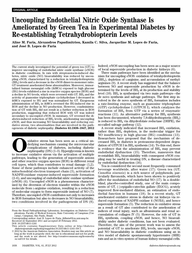
rats in the renal cortex (P = 0.03) and urine (P = 0.05),whereas GT treatment abolished the oxidation of BH4 (Fig.2C and D).Effects of HG and GT on NO production in ihMCs.Qualitative and quantitative analyses of NO productionshowed that the ihMCs kept in the HG medium for 24 h hada significant reduction (P = 0.03) in fluorescence intensitycompared with cells kept in the NG medium, whereas GT(100 mg/mL) reversed the HG-induced reduction in NO(P = 0.02; Fig. 3A and B). Western blot analysis revealedthat the eNOS expression was similar in ihMCs cultured inNG, HG, or HG with GT (Fig. 3C and D). However, theeNOS dimer-to-monomer ratio was decreased in ihMCscultured under HG (P = 0.04), and this alteration was re-versed by GT treatment (P = 0.05; Fig. 3E and F).Effects of HG in ROS production in ihMCs. Qualitativeand quantitative analyses (Fig. 4A and B) showed a signif-icant rise in ROS production after the ihMCs were exposedto HG levels for 24 h (P = 0.0001) compared with NGlevels. To evaluate the sources involved in the HG-inducedROS production in the ihMCs, we analyzed ROS pro-duction at HG in the presence of diphenyleneiodonium(DPI; 50 nmol/L, a blocker of NADPH-oxidase), L-NG-nitro-L-arginine methyl ester (L-NAME; 100 mmol/L, an inhibitor
FIG. 3. A: Representative photomicrographs of diaminorhodamine-4M AM indicating NO production. ihMCs were cultured for 24 h in NG (5.5mmol/L), HG (30 mmol/L), and HG with GT (100 mg/mL). B: Quantification of NO levels in ihMCs via DAF-2DA. Values are mean 6 SD andexpressed as the percentage of fluorescence. Values were corrected by the number of cells at the end of each treatment. *P = 0.03 vs. NG. †P = 0.02vs. HG. C: Representative Western blots of the ihMCs of eNOS expression from ihMCs cultured under NG, HG, and HG treated with GT for 24 h.D: Densitometric analysis of the eNOS–to–b-actin ratio. E: Representative Western blots of the ihMCs of eNOS dimer and monomer expressionfrom ihMCs cultured under NG, HG, and HG treated with GT for 24 h. F: Densitometric analysis of the percentage of eNOS dimer-to-monomer ratio.*P = 0.04 vs. NG. †P = 0.05 vs. HG. (A high-quality digital representation of this figure is available in the online issue.)
A.M. FARIA AND ASSOCIATES
diabetes.diabetesjournals.org DIABETES, VOL. 61, JULY 2012 1841
of NOS enzymes), and rotenone (10 mmol/L, an inhibitor ofmitochondria electron transport complex I). Fluorescentmicroscope (Fig. 4A) and fluorometer (Fig. 4B) datashowed that DPI reversed HG-induced ROS production tolevels lower than those of NG (P = 0.03), suggesting thatNADPH oxidase is one main source of superoxide inihMCs. DPI reduces ROS production even in cells culturedin NG. This observation may explain the reduction of ROSin cells under HG to below control levels (Fig. 4B). Fur-thermore, incubation of ihMC with L-NAME significantlyreduced HG-induced superoxide production (P = 0.04),suggesting that eNOS uncoupling is an important source ofROS production. Finally, incubation with rotenone re-duced HG-induced superoxide production, although itfailed to reach statistical significance (P = 0.07). Mannitol(30 mmol/L), which was used as an osmotic control, didnot alter ROS production. These results imply that NADPHoxidase and uncoupling eNOS are both important sourcesin the HG-induced ROS production in ihMCs.Effects of BH4 in ROS and NO production at HG inihMCs. We observed a significant reduction in totalbiopterin and BH4 levels in ihMCs kept in the HG medium(P = 0.05) compared with cells in the NG medium (Fig. 5A),which was reversed by GT (P = 0.04). Furthermore, therewas an increase in oxidation of BH4 to BH2 in cells kept inthe HG medium (P = 0.01), and this also was reversed afterGT treatment (P = 0.01; Fig. 5B). We next assessed the roleof BH4 in ROS and NO production in ihMCs. Exogenousadministration of BH4 in ihMCs kept in the HG medium(Fig. 5C) reduced the HG-induced rise in ROS productionin a concentration-dependent manner, although this onlyreached significance at 100 mmol/L (P = 0.004). Moreover,measurement of NO via DAF-2DA (Fig. 5D) showed thatBH4 at all concentrations used (P = 0.001) reversed theHG-induced decline in NO production (P = 0.007). These
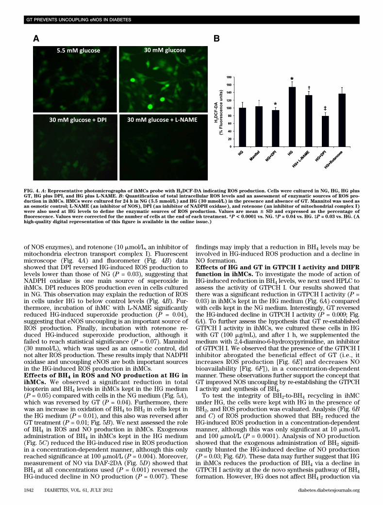
findings may imply that a reduction in BH4 levels may beinvolved in HG-induced ROS production and a decline inNO formation.Effects of HG and GT in GTPCH I activity and DHFRfunction in ihMCs. To investigate the mode of action ofHG-induced reduction in BH4 levels, we next used HPLC toassess the activity of GTPCH I. Our results showed thatthere was a significant reduction in GTPCH I activity (P =0.03) in ihMCs kept in the HG medium (Fig. 6A) comparedwith cells kept in the NG medium. Interestingly, GT reversedthe HG-induced decline in GTPCH I activity (P = 0.009; Fig.6A). To further assess the hypothesis that GT re-establishedGTPCH I activity in ihMCs, we cultured these cells in HGwith GT (100 mg/mL), and after 1 h, we supplemented themedium with 2,4-diamino-6-hydroxypyrimidine, an inhibitorof GTPCH I. We observed that the presence of the GTPCH Iinhibitor abrogated the beneficial effect of GT (i.e., itincreases ROS production [Fig. 6E] and decreases NObioavailability [Fig. 6F]), in a concentration-dependentmanner. These observations further support the concept thatGT improved NOS uncoupling by re-establishing the GTPCHI activity and synthesis of BH4.
To test the integrity of BH2-to-BH4 recycling in ihMCunder HG, the cells were kept with HG in the presence ofBH2, and ROS production was evaluated. Analysis (Fig. 6Band C) of ROS production showed that BH2 reduced theHG-induced ROS production in a concentration-dependentmanner, although this was only significant at 10 mmol/Land 100 mmol/L (P = 0.0001). Analysis of NO productionshowed that the exogenous administration of BH2 signifi-cantly blunted the HG-induced decline of NO production(P = 0.03; Fig. 6D). These data may further suggest that HGin ihMCs reduces the production of BH4 via a decline inGTPCH I activity at the de novo synthesis pathway of BH4formation. However, HG does not affect BH4 production via
FIG. 4. A: Representative photomicrographs of ihMCs probe with H2DCF-DA indicating ROS production. Cells were cultured in NG, HG, HG plusGT, HG plus DPI, and HG plus L-NAME. B: Quantification of total intracellular ROS levels and an assessment of enzymatic sources of ROS pro-duction in ihMCs. HMCs were cultured for 24 h in NG (5.5 mmol/L) and HG (30 mmol/L) in the presence and absence of GT. Mannitol was used asan osmotic control; L-NAME (an inhibitor of NOS), DPI (an inhibitor of NADPH oxidase), and rotenone (an inhibitor of mitochondrial complex I)were also used at HG levels to define the enzymatic sources of ROS production. Values are mean 6 SD and expressed as the percentage offluorescence. Values were corrected for the number of cells at the end of each treatment. *P< 0.0001 vs. NG. †P = 0.04 vs. HG. ‡P = 0.03 vs. HG. (Ahigh-quality digital representation of this figure is available in the online issue.)
GT PREVENTS UNCOUPLING eNOS IN DIABETES
1842 DIABETES, VOL. 61, JULY 2012 diabetes.diabetesjournals.org

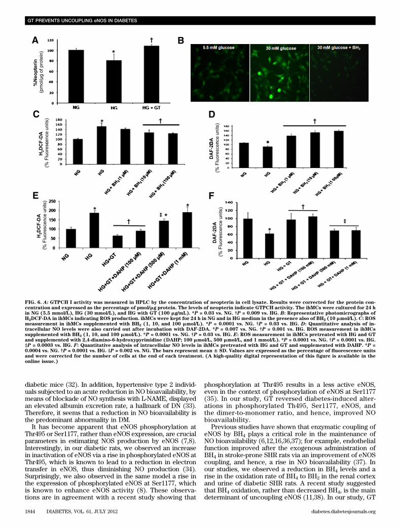
the recycling pathway because BH2 ameliorated both theHG-induced rise in ROS production and the decline in NOformation.Effects of GT in ROS production at HG in ihMCs. InihMCs, assessment of ROS production showed that GTreversed the HG-induced rise in ROS formation to NGlevels (Fig. 7A and B; P = 0.0001). Cotreatment of ihMCswith GT and BH4 (Fig. 7C) did not confer an additionalreduction in ROS production compared with GT treatmentalone. This suggests that GT may inhibit HG-induced ROSproduction through a rise in BH4 levels.Content of EGCG in GT and the EGCG effect inihMCs. When compared with the EGCG standard, weidentified this polyphenol as an important constituent of GT.The EGCG retention time was 8.891 min (SupplementaryFig. 2A), consistent with the main peak retention time of8.894 min observed in GT (Supplementary Fig. 2B). As-sessment of the effect of EGCG in ihMCs exposed to HGhas shown that this component of GT can reduce ROSproduction (Supplementary Fig. 2C) and increase NO bio-availability (Supplementary Fig. 2D). These observationssuggest that the main effect of GT may be attributed, at leastpartly, to EGCG.
DISCUSSION
The current study aimed to explore the potential of GT toameliorate kidney-uncoupling eNOS in diabetic conditions.We also investigated the mechanisms by which GT reverseduncoupling eNOS. We observed, both in vivo and in vitro, an
uncoupling of eNOS secondary to a reduction of BH4 andelevation of its oxidized form with a consequent decrease inNO and an increase in oxidative stress. GT re-establishedthe levels of BH4, reduced its oxidized form, coupled eNOS,and consequently, increased NO bioavailability and reducedoxidative stress. Our in vitro data also suggest that the re-establishment of GTPCH I activity is the main mechanismby which GT increased BH4 and coupled eNOS. Theseobservations are of great interest. First, they reinforce theimportance of uncoupling eNOS and BH4 synthesis/oxidationin reducing NO bioavailability, and increase oxidative stress,two conditions involved in the pathogenesis of DN (1–8). Inaddition, the mechanism by which GT can couple eNOS indiabetes is described for the first time. Together with pre-vious observations that GT can reduce oxidative stress andimprove indices of DN in rats (5), data from this translationalstudy demonstrate the possible beneficial use of GT or itsmain flavonol, EGCG, in patients with DN.
Bioavailability of NO in the diabetic kidney has beena subject of major controversy (7,8). Earlier studies havesuggested that NO production increases and contributes toglomerular hyperfiltration in short-term diabetes (30,31).More recently, it has been suggested that endothelial dys-function, which is often defined as a decrease in the bio-availability of endothelial-derived NO, is a preponderantfactor in diabetes and contributes to the pathogenesis of DN(6–8,17). To this end, researchers have demonstrated thatthe knockout of eNOS in diabetic mice leads to severe his-tologic lesions in the kidney that resemble the lesions seenin human DN but which are not seen in control wild-type
Pro
tein
(p
mo
l/m
g)
FIG. 5. A: Total biopterin and BH4 expression analysis by ultraperformance liquid chromatography in ihMC lysate. Results were corrected for theprotein concentration and expressed as nmol/mg protein. *P = 0.05 vs. NG. †P = 0.02 vs. HG. ‡P = 0.04 vs. HG. B: Representative graphs of per-centage of BH4 to BH2 oxidation in ihMC lysate. *P = 0.01 vs. NG. †P = 0.01 vs. HG. C: Quantification of total intracellular ROS levels by H2DCF-DAvia a fluorimeter after incubation of ihMCs for 24 h in NG and HG mediums in the presence also of BH4 (1, 10, and 100 mmol/L). *P = 0.0001 vs. NG.†P = 0.004 vs. HG. D: NO levels were also quantified after incubation with DAF-2DA via a fluorimeter. *P = 0.007 vs. NG. †P = 0.001 vs. HG. The barsrepresent mean 6 SD. Values are expressed as the percentage of fluorescence units and were corrected by the number of cells at the end of eachtreatment.
A.M. FARIA AND ASSOCIATES
diabetes.diabetesjournals.org DIABETES, VOL. 61, JULY 2012 1843
diabetic mice (32). In addition, hypertensive type 2 individ-uals subjected to an acute reduction in NO bioavailability, bymeans of blockade of NO synthesis with L-NAME, displayedan elevated albumin excretion rate, a hallmark of DN (33).Therefore, it seems that a reduction in NO bioavailability isthe predominant abnormality in DM.
It has become apparent that eNOS phosphorylation atThr495 or Ser1177, rather than eNOS expression, are crucialparameters in estimating NOS production by eNOS (7,8).Interestingly, in our diabetic rats, we observed an increasein inactivation of eNOS via a rise in phosphorylated eNOS atThr495, which is known to lead to a reduction in electrontransfer in eNOS, thus diminishing NO production (34).Surprisingly, we also observed in the same model a rise inthe expression of phosphorylated eNOS at Ser1177, whichis known to enhance eNOS activity (8). These observa-tions are in agreement with a recent study showing that
phosphorylation at Thr495 results in a less active eNOS,even in the context of phosphorylation of eNOS at Ser1177(35). In our study, GT reversed diabetes-induced alter-ations in phosphorylated Th495, Ser1177, eNOS, andthe dimer-to-monomer ratio, and hence, improved NObioavailability.
Previous studies have shown that enzymatic coupling ofeNOS by BH4 plays a critical role in the maintenance ofNO bioavailability (6,12,16,36,37); for example, endothelialfunction improved after the exogenous administration ofBH4 in stroke-prone SHR rats via an improvement of eNOScoupling, and hence, a rise in NO bioavailability (37). Inour studies, we observed a reduction in BH4 levels and arise in the oxidation rate of BH4 to BH2 in the renal cortexand urine of diabetic SHR rats. A recent study suggestedthat BH4 oxidation, rather than decreased BH4, is the maindeterminant of uncoupling eNOS (11,38). In our study, GT
FIG. 6. A: GTPCH I activity was measured in HPLC by the concentration of neopterin in cell lysate. Results were corrected for the protein con-centration and expressed as the percentage of pmol/mg protein. The levels of neopterin indicate GTPCH activity. The ihMCs were cultured for 24 hin NG (5.5 mmol/L), HG (30 mmol/L), and HG with GT (100 mg/mL). *P = 0.03 vs. NG. †P = 0.009 vs. HG. B: Representative photomicrographs ofH2DCF-DA in ihMCs indicating ROS production. ihMCs were kept for 24 h in NG and in HG medium in the presence also of BH2 (10 mmol/L). C: ROSmeasurement in ihMCs supplemented with BH2 (1, 10, and 100 mmol/L). *P = 0.0001 vs. NG. †P = 0.03 vs. HG. D: Quantitative analysis of in-tracellular NO levels were also carried out after incubation with DAF-2DA. *P = 0.007 vs. NG. †P = 0.001 vs. HG. ROS measurement in ihMCssupplemented with BH2 (1, 10, and 100 mmol/L). *P = 0.0001 vs. NG. †P = 0.03 vs. HG. E: ROS measurement in ihMCs pretreated with HG and GTand supplemented with 2,4-diamino-6-hydroxypyrimidine (DAHP; 100 mmol/L, 500 mmol/L, and 1 mmol/L). *P = 0.0001 vs. NG. †P = 0.0001 vs. HG.‡P = 0.0003 vs. HG. F: Quantitative analysis of intracellular NO levels in ihMCs pretreated with HG and GT and supplemented with DAHP. *P =0.0004 vs. NG. †P = 0.0001 vs. HG. ‡P = 0.002 vs. NG. The bars represent mean 6 SD. Values are expressed as the percentage of fluorescence unitsand were corrected for the number of cells at the end of each treatment. (A high-quality digital representation of this figure is available in theonline issue.)
GT PREVENTS UNCOUPLING eNOS IN DIABETES
1844 DIABETES, VOL. 61, JULY 2012 diabetes.diabetesjournals.org
treatment restored the levels of BH4 and the diabetes-induced oxidation of BH4 to BH2 in the renal cortex andurine of diabetic SHR rats. Therefore, GT seems to reversediabetes-induced eNOS uncoupling, thereby increasingNO bioavailability via a rise in BH4 availability. This isfurther supported by our observation that cotreatment ofGT and BH4 did not confer additional protection againstHG-induced ROS production compared with GT treat-ment alone. The ihMCs exposed to HG displayed a risein ROS production and a decline in intracellular NOproduction.
Two sources appeared to mediate HG-induced ROSproduction—NADPH oxidase and uncoupled eNOS—because blocking each one abolished the HG-induced risein ROS production. Our studies are in agreement with pre-vious work showing that uncoupling eNOS and NADPHoxidase in the glomeruli of rats with experimental DN arethe major sources of superoxide mediated by the lossof BH4 availability (6). In agreement with the importance ofreduction in BH4 availability, exogenous administration ofBH4 in ihMCs abolished ROS production and reversed thedecline of NO under HG levels. This finding suggests thatihMCs kept in HG mediums exhibit low levels of BH4,leading to uncoupling of eNOS, a subsequent rise in ROS,and a decline in NO levels. This is further supported by ourfinding that BH4 levels decreased and the oxidation of BH4rose to BH2 in ihMCs kept in the HG medium.
Our present work further showed that ihMCs kept in theHG medium exhibited reduced levels of BH4 comparedwith cells kept in the NG medium, due to the diminished denovo synthesis pathway of BH4 formation, because activityof GTPCH I was reduced in cells cultured in HG. Di-minished GTPCH I activity has also been reported in ratswith experimental DN, leading to reduced BH4 formationvia the de novo synthesis (39). In addition, the observationthat a GTCH I blocker abrogated the effect of GT in ROSand NO production in ihMCs exposed to HG and treatedwith GT further supports the concept that GT acts by im-proving GTPCH I activity. Our work also indicated that therecycling pathway of BH4 is probably preserved in ihMCsexposed to HG because BH2 supplementation decreasedROS and increased NO bioavailability by enhancing BH4levels via DHFR. These findings are in agreement withprevious in vivo (11) and in vitro studies (11,38) showingthat DHFR plays a key role in regulating the BH4-to-BH2ratio and eNOS coupling under conditions of low total BH4availability. For example, in endothelial cells (38,40)expressing eNOS with low BH4 levels, DHFR inhibition orknockdown further diminished the BH4-to-BH2 ratio andexacerbated eNOS uncoupling.
Reduced BH4 availability in ihMCs could also be attrib-uted to reduced BH4 stability. Diminished BH4 stability hasbeen reported in endothelial dysfunction (41) as well as inrats with DN (6). HG levels, as seen in diabetic conditions,
FIG. 7. A: Representative photomicrographs of H2DCF-DA in ihMCs indicating ROS production. ihMCs were kept for 24 h in NG and HG mediums inthe presence also of GT (100 mg/mL). B: Total intracellular ROS levels by H2DCF-DA was quantified via a fluorimeter. *P = 0.0001 vs. NG. †P =0.0001 vs. HG. C: ihMCs were cultured in HG and supplemented with GT (100 mg/mL) and BH4, followed by measurement of intracellular ROS byH2DCF-DA. *P < 0.0001 vs. NG. †P < 0.0001 vs. HG. The bars represent mean 6 SD. Values are expressed as the percentage of fluorescence units.(A high-quality digital representation of this figure is available in the online issue.)
A.M. FARIA AND ASSOCIATES
diabetes.diabetesjournals.org DIABETES, VOL. 61, JULY 2012 1845
increase the formation of superoxide through one mainsource in the kidney, NADPH oxidase activation. NO pro-duced by eNOS and superoxide combine to form peroxyni-trite anions. Oxidation of BH4 by ROS, such as peroxynitrite,results in BH2 formation, which inactivates the eNOS co-factor function, suggesting that reduced BH4 stabilityuncouples eNOS and leads to reduced NO bioavailabilityand a further rise in the formation of diabetic glomerulisuperoxides.
In conclusion, the current work indicates that GT reversesdiabetes-induced uncoupling eNOS as experienced in renalmesangial cells exposed to HG levels and SHR diabeticrats. GT seems to ameliorate uncoupling eNOS via a risein BH4 levels/reduction in BH4 oxidation, which occursas a result of the de novo synthesis of BH4. A rise in BH4levels would account, then, for reduced eNOS uncouplingleading to the amelioration of oxidative stress and enhancedNO availability.
ACKNOWLEDGMENTS
This work was supported by the Fundação de Amparo àPesquisa do Estado de São Paulo (Grant 2008/57560-0)and Conselho Nacional de Desenvolvimento Científicoe Tecnológico (CNPq). A.M.F. received a scholarship fromCNPq.
No potential conflicts of interest relevant to this articlewere reported.
A.M.F., A.P., and K.C.S. acquired the data and wrotethe manuscript. J.M.L.F. contributed to discussion andreviewed the manuscript. J.B.L.F. designed the study,reviewed the data, and wrote, reviewed, and edited themanuscript. J.B.L.F. is the guarantor of this work and,as such, had full access to all of the data in the studyand takes responsibility for the integrity of the data andthe accuracy of the data analysis.
The authors thank Luciana Cristina Teixeira (Depart-ment of Medicine, Nephrology Division, Federal Universityof São Paulo, Brazil) for helping with the culture of theihMC line and are grateful to Dr. Ricardo Pereira andDr. Marcelo Ganzarolli de Oliveira (Laboratory of LiquidChromatography, Institute of Chemistry, University ofCampinas [Unicamp], Campinas, SP, Brazil) for their tech-nical and scientific assistance. The authors are very grate-ful to the personnel from the Renal PathophysiologyLaboratory, Investigation on Diabetes Complications, Fac-ulty of Medical Sciences (FCM), Unicamp, for their invalu-able help with this work. In particular, the authors are verygrateful to Dr. Elisa E.M. Peixoto (Renal PathophysiologyLaboratory, Unicamp), for blindly estimating the extracel-lular matrix expansion in kidney slides.
REFERENCES
1. Brownlee M. The pathobiology of diabetic complications: a unifying mech-anism. Diabetes 2005;54:1615–1625
2. Lopes de Faria JB, Silva KC, Lopes de Faria JM. The contribution of hy-pertension to diabetic nephropathy and retinopathy: the role of inflammationand oxidative stress. Hypertens Res 2011;34:413–422
3. Nishikawa T, Edelstein D, Du XL, et al. Normalizing mitochondrial su-peroxide production blocks three pathways of hyperglycaemic damage.Nature 2000;404:787–790
4. Guzik TJ, Mussa S, Gastaldi D, et al. Mechanisms of increased vascularsuperoxide production in human diabetes mellitus: role of NAD(P)H oxi-dase and endothelial nitric oxide synthase. Circulation 2002;105:1656–1662
5. Ribaldo PDB, Souza DS, Biswas SK, Block K, Lopes de Faria JM, Lopes deFaria JB. Green tea (Camellia sinensis) attenuates nephropathy by down-regulating Nox4 NADPH oxidase in diabetic spontaneously hypertensiverats. J Nutr 2009;139:96–100
6. Satoh M, Fujimoto S, Haruna Y, et al. NAD(P)H oxidase and uncouplednitric oxide synthase are major sources of glomerular superoxide in ratswith experimental diabetic nephropathy. Am J Physiol Renal Physiol 2005;288:F1144–F1152
7. Komers R, Anderson S. Glomerular endothelial NOS (eNOS) expression intype 2 diabetic patients with nephropathy. Nephrol Dial Transplant 2008;23:3037
8. Komers R, Anderson S. Paradoxes of nitric oxide in the diabetic kidney.Am J Physiol Renal Physiol 2003;284:F1121–F1137
9. Zweier JL, Chen CA, Druhan LJ. S-glutathionylation reshapes our un-derstanding of endothelial nitric oxide synthase uncoupling and nitricoxide/reactive oxygen species-mediated signaling. Antioxid Redox Signal2011;14:1769–1775
10. Crabtree MJ, Hale AB, Channon KM. Dihydrofolate reductase protectsendothelial nitric oxide synthase from uncoupling in tetrahydrobiopterindeficiency. Free Radic Biol Med 2011;50:1639–1646
11. Thöny B, Auerbach G, Blau N. Tetrahydrobiopterin biosynthesis, regenerationand functions. Biochem J 2000;347:1–16
12. Schmidt TS, Alp NJ. Mechanisms for the role of tetrahydrobiopterin inendothelial function and vascular disease. Clin Sci (Lond) 2007;113:47–63
13. Crabtree MJ, Smith CL, Lam G, Goligorsky MS, Gross SS. Ratio of 5,6,7,8-tetrahydrobiopterin to 7,8-dihydrobiopterin in endothelial cells determinesglucose-elicited changes in NO vs. superoxide production by eNOS. AmJ Physiol Heart Circ Physiol 2008;294:H1530–H1540
14. Xu J, Wu Y, Song P, Zhang M, Wang S, Zou MH. Proteasome-dependentdegradation of guanosine 59-triphosphate cyclohydrolase I causes tetra-hydrobiopterin deficiency in diabetes mellitus. Circulation 2007;116:944–953
15. Werner ER, Blau N, Thöny B. Tetrahydrobiopterin: biochemistry andpathophysiology. Biochem J 2011;438:397–414
16. Nakagawa T, Tanabe K, Croker BP, et al. Endothelial dysfunction as a po-tential contributor in diabetic nephropathy. Nat Rev Nephrol 2011;7:36–44
17. Schmitt CA, Dirsch VM. Modulation of endothelial nitric oxide by plant-derived products. Nitric Oxide 2009;21:77–91
18. Widlansky ME, Hamburg NM, Anter E, et al. Acute EGCG supplementationreverses endothelial dysfunction in patients with coronary artery disease.J Am Coll Nutr 2007;26:95–102
19. Cooper ME, Allen TJ, Macmillan P, Bach L, Jerums G, Doyle AE. Genetichypertension accelerates nephropathy in the streptozotocin diabetic rat.Am J Hypertens 1988;1:5–10
20. Peixoto EB, Pessoa BS, Biswas SK, Lopes de Faria JB. Antioxidant SODmimetic prevents NADPH oxidase-induced oxidative stress and renaldamage in the early stage of experimental diabetes and hypertension. AmJ Nephrol 2009;29:309–318
21. Cristovam PC, Arnoni CP, de Andrade MC, et al. ACE-dependent andchymase-dependent angiotensin II generation in normal and glucose-stimulated human mesangial cells. Exp Biol Med (Maywood) 2008;233:1035–1043
22. Bradford MM. A rapid and sensitive method for the quantitation of mi-crogram quantities of protein utilizing the principle of protein-dye binding.Anal Biochem 1976;72:248–254
23. Komers R, Schutzer WE, Reed JF, et al. Altered endothelial nitric oxidesynthase targeting and conformation and caveolin-1 expression in the di-abetic kidney. Diabetes 2006;55:1651–1659
24. Miranda KM, Espey MG, Wink DA. A rapid, simple spectrophotometricmethod for simultaneous detection of nitrate and nitrite. Nitric Oxide 2001;5:62–71
25. Fukushima T, Nixon JC. Analysis of reduced forms of biopterin in biologicaltissues and fluids. Anal Biochem 1980;102:176–188
26. Fekkes D, Voskuilen-Kooijman A. Quantitation of total biopterin and tet-rahydrobiopterin in plasma. Clin Biochem 2007;40:411–413
27. Vann LR, Twitty S, Spiegel S, Milstien S. Divergence in regulation of nitric-oxide synthase and its cofactor tetrahydrobiopterin by tumor necrosisfactor-a. Ceramide potentiates nitric oxide synthesis without affectingGTP cyclohydrolase I activity. J Biol Chem 2000;275:13275–13281
28. Goto T, Yoshida Y, Kiso M, Nagashima H. Simultaneous analysis of in-dividual catechins and caffeine in green tea. J Chomatogr 1996;749:295–299
29. Förstermann U. Nitric oxide and oxidative stress in vascular disease.Pflugers Arch 2010;459:923–939
30. Mattar AL, Fujihara CK, Ribeiro MO, de Nucci G, Zatz R. Renal effects ofacute and chronic nitric oxide inhibition in experimental diabetes. Neph-ron 1996;74:136–143
31. Tolins JP, Shultz PJ, Raij L, Brown DM, Mauer SM. Abnormal renal he-modynamic response to reduced renal perfusion pressure in diabetic rats:role of NO. Am J Physiol 1993;265:F886–F895
32. Nakagawa T, Sato W, Glushakova O, et al. Diabetic endothelial nitric oxidesynthase knockout mice develop advanced diabetic nephropathy. J AmSoc Nephrol 2007;18:539–550
GT PREVENTS UNCOUPLING eNOS IN DIABETES
1846 DIABETES, VOL. 61, JULY 2012 diabetes.diabetesjournals.org

33. Ott C, Schneider MP, Delles C, Schlaich MP, Schmieder RE. Reduction inbasal nitric oxide activity causes albuminuria. Diabetes 2011;60:572–576
34. Thomas SR, Witting PK, Drummond GR. Redox control of endothelialfunction and dysfunction: molecular mechanisms and therapeutic oppor-tunities. Antioxid Redox Signal 2008;10:1713–1765
35. Oubaha M, Gratton JP. Phosphorylation of endothelial nitric oxide syn-thase by atypical PKC zeta contributes to angiopoietin-1-dependent in-hibition of VEGF-induced endothelial permeability in vitro. Blood 2009;114:3343–3351
36. Schulz E, Jansen T, Wenzel P, Daiber A, Münzel T. Nitric oxide, tetra-hydrobiopterin, oxidative stress, and endothelial dysfunction in hyper-tension. Antioxid Redox Signal 2008;10:1115–1126
37. Noguchi K, Hamadate N, Matsuzaki T, et al. Improvement of impairedendothelial function by tetrahydrobiopterin in stroke-prone spontaneouslyhypertensive rats. Eur J Pharmacol 2010;631:28–35
38. Crabtree MJ, Tatham AL, Hale AB, Alp NJ, Channon KM. Critical role fortetrahydrobiopterin recycling by dihydrofolate reductase in regulation ofendothelial nitric-oxide synthase coupling: relative importance of the denovo biopterin synthesis versus salvage pathways. J Biol Chem 2009;284:28128–28136
39. Okumura M, Masada M, Yoshida Y, et al. Decrease in tetrahydrobiopterinas a possible cause of nephropathy in type II diabetic rats. Kidney Int 2006;70:471–476
40. Sugiyama T, Levy BD, Michel T. Tetrahydrobiopterin recycling, a keydeterminant of endothelial nitric-oxide synthase-dependent signalingpathways in cultured vascular endothelial cells. J Biol Chem 2009;284:12691–12700
41. Landmesser U, Dikalov S, Price SR, et al. Oxidation of tetrahydrobiopterinleads to uncoupling of endothelial cell nitric oxide synthase in hyperten-sion. J Clin Invest 2003;111:1201–1209
A.M. FARIA AND ASSOCIATES
diabetes.diabetesjournals.org DIABETES, VOL. 61, JULY 2012 1847