orofacial granulomatosis (ofg) is a non-caseating ...discovery.ucl.ac.uk/1470143/3/ofg final draft...
TRANSCRIPT

1
Al-Hamad, A; Porter, S; Fedele, S; (2015) Orofacial Granulomatosis. Dermatol Clin , 33 (3) pp. 433-
446. 10.1016/j.det.2015.03.008. Downloaded from UCL Discovery: http://discovery.ucl.ac.uk/1470143
ARTICLE
Oro-facial Granulomatosis
Arwa Al-Hamad1, 2, Stephen Porter1, Stefano Fedele1, 3 1 University College London, UCL Eastman Dental Institute, Oral Medicine Unit, 256 Gray’s
Inn Road, WC1X 8LD, London UK. 2 Dental Services, King Abdulaziz Medical City-Riyadh, Ministry of National Guard, Riyadh,
Saudi Arabia. 3 NIHR University College London Hospitals Biomedical Research Centre, London, UK.
Acknowledgments: Part of this work was undertaken at University College
London/University College London Hospital, which received a proportion of funding from the
Department of Health’s National Institute for Health Research Biomedical Research Centre
funding scheme.
Conflicts of Interest: The authors declare that they have no affiliation with any organization
with a financial interest, direct or indirect, in the subject matter or materials discussed in the
manuscript that may affect the conduct or reporting of the work submitted.
Authorship: all authors named above meet the following criteria of the International
Committee of Medical Journal Editors:
1) Substantial contributions to conception and design, or acquisition of data, or analysis and
interpretation of data;
2) Drafting the article or revising it critically for important intellectual content;
3) Final approval of the version to be published.
Corresponding author: Dr. Stefano Fedele DDS, PhD
Clinical Senior Lecturer/Honorary Consultant in Oral Medicine
Programme Director - MSc in Oral Medicine
University College London and University College London Hospitals Trust
Eastman Dental Institute and Hospital
256 Gray's Inn Road,
London WC1X 8LD, UK
email: [email protected]
Tel:: +44 (0) 20 3456 1004
Fax: +44 (0) 20 3456 1105
Key Words: orofacial granulomatosis, Melkersson-Rosenthal syndrome, granulomatous
cheilitis, granulomas, Crohn’s disease,
2
Synopsis: Orofacial granulomatosis (OFG) is an uncommon chronic inflammatory disorder
of the orofacial region. It is characterised by sub-epithelial non-caseating granulomas and
has a spectrum of possible clinical manifestations ranging from subtle oral mucosal swelling
to permanent disfiguring fibrous swelling of the lips and face. Etiopathogenesis is unknown.
A range of systemic granulomatous disorders including Crohn’s disease and sarcoidosis may
cause orofacial manifestations that cannot be distinguished from those of OFG. Treatment of
OFG has proven difficult and unsatisfactory, with no single therapeutic model showing
consistent efficacy in reducing orofacial swelling and mucosal inflammation.
Key Points:
Orofacial granulomatosis (OFG) is an uncommon granulomatous disorder of the orofacial
tissues. Disease hallmarks include development of disfiguring labial and/or facial
enlargement and intra-oral mucosal swelling and ulceration. Etiopathogenesis remains
unknown.
Crohn’s disease, sarcoidosis and a range of other systemic disorders can present
orofacial features similar to those of OFG. However, a strict case definition of OFG
requires the exclusion of concomitant systemic granulomatous disease.
A small subgroup of OFG patients, especially those with disease onset during childhood,
will eventually develop intestinal Crohn’s disease or, more rarely, sarcoidosis.
Management of OFG is challenging and not evidence-based. Prolonged anti-inflammatory
and immune-modulatory systemic therapy is usually needed to obtain long-term control of
severe orofacial swelling and inflammation. However intralesional corticosteroid therapy
may provide notable long-term remission with no need of prolonged treatment.
The vast majority of OFG patients on therapy will eventually experience a variable degree
of reduction in clinical manifestations
Introduction
Orofacial granulomatosis (OFG) is an uncommon chronic inflammatory disorder that typically
affects the soft tissues of the orofacial region (1). It is histopathologically characterised by
sub-epithelial non-caseating granulomas and has a spectrum of possible clinical
manifestations ranging from subtle oral mucosal swelling to permanent disfiguring fibrous
swelling of the lips and face (2–6). Painful oral ulceration and neurological manifestations to
the head and neck region can also occur (3–5). The first cases of a disorder causing
recurrent/chronic orofacial swelling where initially reported in the nineteenth century
(Wiesenfeld et al. 1985; James et al. 1986); eventually Melkersson (9) and Rosenthal (10)
described the association between recurrent/chronic orofacial oedema, facial palsy and
fissured tongue (lingua plicata). The term Melkersson-Rosenthal syndrome (MRS) was
therefore introduced to describe individuals with the full triad of manifestations whereas those
with only labial swelling were referred as having cheilitis granulomatosa (Miescher’s cheilitis).
In 1985 Wiesenfeld et al introduced the term orofacial granulomatosis (OFG) to encompass
both MRS and Miescher’s chielits (Wiesenfeld et al. 1985). Over the years a number of
systemic granulomatous disorders (including Crohn’s disease, sarcoidosis, leprosy,
tuberculosis, chronic granulomatous disease and possibly deep fungal infections have been
reported to cause orofacial manifestations (12) similar to those of OFG. It remains

3
controversial whether it is appropriate to refer to them as OFG. The authors of the present
paper define OFG as an idiopathic granulomatous disease limited to the orofacial tissue,
namely affecting individuals who do not show any evidence of previous or concomitant
systemic granulomatous disease as per clinical, radiological, endoscopic, or serological
investigations; diagnosis of idiopathic OFG remains therefore one of exclusion (3,5,6,13). As
it is well established that some of these patients would eventually develop additional extra-
oral/facial manifestations of a systemic granulomatous disease (e.g. colonic Crohn’s
disease), we suggest that their diagnosis should be at that point revised and re-termed (e.g.
from OFG to oral and colonic Crohn’s disease). This classification has a pragmatic clinical
relevance, as individuals with OFG limited to orofacial tissues would benefit from therapeutic
interventions and monitoring that are significantly different from those with a systemic
granulomatous disease. Orofacial granulomatosis is an uncommon disease. No reliable
epidemiological data are available as most cases series report small single-centre groups of
patients (1). There remain few studies describing case series of 100 or more OFG patients,
which however represent retrospective analysis of cohorts that are often heterogeneous
(including patients with Crohn’s disease) and observed during two or more decades (1,14).
Orofacial granulomatosis seems to have no specific ethnic predilection, and most authors
report that both genders are equally affected (1,15). The disease occurs by the end of the
third decade of life in the vast majority of reported patients (1).
Orofacial granulomatosis can cause adverse effect upon the quality of life of patients due to
disfiguring chronic orofacial swelling, painful oral ulceration and occasional neurologic
involvement (16). Treatment of OFG has proven difficult and unsatisfactory, with no single
predictable therapeutic model showing consistent efficacy in reducing orofacial swelling and
mucosal inflammation (17).
The aim of the present paper is to present a comprehensive review of available literature
about etiopathogenesis, clinical manifestations, prognosis and management of OFG.
Etiopathogenesis
Although a number of possible causative factors have been associated to OFG (13), the
exact etiopathogenesis remains unknown. Existing literature has typically focussed upon the
role of (i) delayed hypersensitivity to food substances, food preservatives, or dental
materials, (ii) microbial infections and (iii) inflammatory/immunological factors (13). Recent
findings regarding the immunopathogenesis of granulomas in Crohn’s disease (18) and other
rare granulomatous disorders (19,20) could indicate that similar defects of the innate
immunity may also play part in the etiology of granulomas of OFG (21–23). Relevant studies
are ongoing (24).
Hereditary and genetic predisposition:
There is no adequate data in the literature that supports that OFG has a definite genetic
background (23,25). Reports of hereditary cases remain scarce and studies have not found
any convincing robust HLA association (26,27) in OFG patients versus population controls.
Inflammatory/immunological factors:
Characterisation of granulomatous inflammation of OFG has lead to conflicting and
inconsistent results. It remains unclear whether lesional T cells of OFG represent clonal
expansion as a result of chronic antigen stimulation (23,28,29). Studies on the expression of
4
cytokines and chemokines in OFG lesions have found a predominant Th1-mediated immune
response (23,30).
Hypersensitivity reactions:
A wide range of hypersensitivities have been reported in OFG patients including dental
restorative materials (31,32), toothpastes and other dental hygiene products (33,34), cocoa
and chocolate (33,34), cinnamon compounds, carvone, carbone piperitone, aspartate
(33,35–39), carmosine and sun yellow dye (35), monosodium glutamate (35,40), benzoates
(35,37–39,41,42) and tartrazine (41). Cinnamon and benzoate compounds have been
suggested to be most common triggers (33–35,38,41,42). A potential role of hypersensitivity
in OFG pathogenesis seems to be supported and confirmed by patient’s history of symptoms
aggravation associated with contact or ingestion of one or more of the above triggering
factors, positive response to elimination diet, and in some cases positive patch testing
(23,32). Further, a recent paper has reported OFG patients to have a higher prevalence of
allergy than the general population as demonstrated by their medical history, skin prick test,
and serum IgE (43). Nevertheless, other studies have failed to find convincing evidence of
sensitisation to foods, additives or contactants in OFG patients (44). Also outcomes of
avoidance diet remain controversial as they vary from 14% (14,45) to 70% (46) and seem not
to correlate with patch testing results (46). It is possible that delayed hypersensitivity
mechanisms may have a pathogenetic role in a small sub-group of OFG patients.
Microbial factors:
Several authors have investigated the potential role of microbial agents in triggering the
immune response of OFG, including M. tuberculosis, M. Paratuberculosis, Saccharomyces
Cerevisiae, Borrelia burgdorferi, Candida albicans, and Streptococcus mutans. Although
some studies have reported the presence of M. tuberculosis RNA in OFG samples (47) and
raised IgG antibody titers to the mycobacterial stress protein 65 (48) in OFG patients’ serum,
there remains little credible evidence to support a role of any of these agents in the
aetiopathogenesis of OFG (27,49–52).
Clinical presentation
Disfiguring lip swelling remains the clinical hallmark of OFG and the most common reason for
which OFG patients seek medical attention (53). Other possible clinical manifestations
include swelling and ulceration of the oral mucosa, swelling of facial (other than labial)
tissues, and neurological manifestations (3,54). It is probable that OFG represent a disease
with a spectrum of severity that ranges from localized granulomatous inflammation of the
lips, through orofacial swelling with mucosal ulceration to a disease with additional
neurological deficit (3,55–57). Available literature indicates that clinical manifestations at
disease onset can be highly variable and multiform, although permanent disfiguring labial
and/or facial swelling eventually develops in nearly all affected individuals.
Labial swelling
Available literature clearly shows that labial enlargement is the most common feature of
OFG, affecting more than 90% of patients (5). Lip enlargement is typically recurrent and
oedematous in the early stages of the disease, with each episode lasting few days or weeks.
(58–60). During the course of the disease and after a number of recurrent episodes the
swelling of the lips typically becomes persistent, firm and indurated, assuming the
characteristics of a granulomatous disorder (5). Lip fissuring, exfoliation and impetiginisation
can be associated, especially in severe cases, and intraoral labial mucosa may become

5
erythematous and granular (5,59). The peri-oral skin may become erythematous and
exfoliated and some patients may develop angular cheilitis (61). There is no site
predisposition for the labial swelling although it may be slightly more common on the lower lip
(3,5,7,62). The swelling rarely causes difficulties in speech or drooling (54).
Facial swelling:
Swelling of non-labial facial tissues has been described in OFG patients, sometimes in
absence of lip enlargement or other clinical manifestations, and can vary in severity. Patients
can develop recurrent or persistent enlargement of zygomatic, frontal, peri-orbital or
chin/submental region (3,63), as well as the cheek and eyelids, which represent a diagnostic
challenge as these are not favourable areas to obtain a biopsy specimen (3). Indeed
blepharitis granulomatosa (or granulomatous blepharitis) it is likely to represent OFG-like
disease (64,65). Swelling of submandibular and/or cervical areas due to persistent lymph
node enlargement can arise in about 25% of OFG individuals (5,54).
Intra-oral manifestations:
Generalised swelling of the buccal and or labial mucosa gives rise to a cobblestone-like
appearance (54), a common intra-oral features of OFG (66), particulary in the posterior
buccal mucosa (54,55). Localised mucosal swelling manifest as discreet painless tags
typically affect the vestibular buccal and labial mucosa or floor of the mouth (“stag-horning”)
(5). Deep chronic linear ulcers with raised margins can arise in the buccal and/or labial
sulcus and are often associated with significant pain (5). Less commonly flat and circular
aphthous-like ulcers can arise on any oral mucosal surface. Pyostomatitis vegetans, which
manifests with yellowish, linear pustules on the background of mucosal erythematous “snail
track ulcerations” has also been described, although the vast majority of reports refer to
individuals with evidence of inflammatory bowel disease (Crohn’s disease) (4,67–70).
Painless gingival swelling independent of plaque and calculus deposits may occur in up to
one third of patients with OFG (2,6,71,72). The swelling can affect the attached and/or free
gingivae, be localized or generalized, and is often associated with erythema and superficial
“granular” appearance (2,3). Generalised erythema/inflammation of the oral mucosa is
uncommonly described as a separate intraoral feature of OFG (73) possibly because it
usually develops in association with either manifestation including cobblestoning and
ulceration. The tongue may have superficial fissures that are most pronounced on the lateral
aspects of the dorsum (54). The fissuring may rarely cause food accumulation leading to
alteration in taste, oral malodour and a local burning sensation (71,74). Fissuring of the
tongue has been inconsistently associated with neurological manifestations (74).
Neurological manifestations:
A subgroup of patients with OFG may have lower motor nerve facial palsy at some point in
the disease course (71,75). Granuloma formation or inflammation within the course of the
main stem of the facial nerve is the most probable cause for the palsy (54). The exact
prevalence of facial palsy in OFG is unclear, but studies report a very wide range of 8-57%
(3,11,42,76). The palsy can be complete or partial but is typically unilateral. The palsy can
occur before, with or after (sometimes months to years) the facial swelling (77). It also may
be accompanied by otalgia and/or changes in hearing and taste (65). Complete recovery of
nerve function is usual but some residual weakness can occur (75,78,79). Facial palsy can
be considered as a feature of Melkersson-Rosenthan syndrome when associated with lip
swelling and fissured tongue, although most clinicians now categorize Melkersson-Rosenthal
6
syndrome as a sub- type of OFG. A number of less common neurological manifestations
have been reported to develop in up to 30% of OFG patients (2,3,7,76,79). These include
blepharospasm, migraine-like headache, hypogeusia, glossodynia, hyperacusia, lacrimation
and sweating (80,81). Relevant pathogenetic mechanisms remain unknown.
Clinical manifestations of early and advanced disease
Labial swelling is traditionally indicated as the most common clinical feature of OFG and was
previously reported as being the most frequent manifestation at disease presentation.
However, a number of authors have more recently suggested that OFG can in fact present
with multiple, temporary and variable clinical features affecting oral mucosa, gingivae, facial
tissues and the craniofacial nervous system (3), and that different clinical manifestations can
develop at different time points during the course of the disease (2,3). Zimmer et al (1992)
reported that labial swelling was the initial disease manifestation in only 43% of their 42
patients but this percentage increased to 74% during the course of the disease. Moreover,
the overall number of clinical manifestations increased during the years as the percentage of
patients with facial swelling increased from 26% to 50% and those with facial palsy from 19%
to 33% (2). Mignogna et al (2003) reported that about half of their 19 OFG patients (9/19)
had an “atypical” disease onset characterized by the absence of labial swelling, which
however developed in seven of these nine patients at later stage. Al Johani at el studied a
cohort of 49 OFG patients and confirmed that OFG present with lip swelling in only 50% of
cases, whereas the remaining individuals had intra-oral or neurological manifestations in
absence of labial or facial swelling (5). They also reported that the vast majority of patients
eventually developed a variety of additional features of OFG, with nearly all affected
individuals (>90%) ultimately developing lip/facial swelling (5).
Systemic association
Considering the strict definition and nomenclature that the authors of the present review have
adopted, the concomitant presence of orofacial and other systemic manifestations of a
specific and well-characterised generalised granulomatous disorder (e.g. Crohn’s disease or
sarcoidosis) should exclude the diagnosis of idiopathic OFG. Indeed, as mentioned above, a
diagnosis of “true” idiopathic OFG would require that detailed medical history, clinical
assessment and comprehensive investigations are performed so to rule out the presence of
these disorders and confirm that disease is limited to the orofacial tissues (23,25,60).
Nevertheless, potential associations with systemic disease can still exist in patients with
idiopathic OFG.
Subsequent development of systemic granulomatous disease
It is well described that a sub-group of OFG individuals would eventually develop
manifestations of systemic disease, typically intestinal Crohn’s disease or respiratory/multi-
organ sarcoidosis, even if at the moment of initial assessment there was no clinical,
serological or radiological evidence of any relevant extra-oral abnormality (59,73,82,83). As
discussed, it would be sensible at that stage to re-label these cases as, for example, having
oral and intestinal Crohn’s disease or oral and respiratory sarcoidosis rather than maintaining
the nomenclature of OFG in association with Crohn’s disease or sarcoidosis. It is difficult to
predict which OFG patients will eventually develop extra-oral manifestations of a
granulomatous systemic disease, although the vast majority of them are believed to have
disease that will remain limited to the orofacial tissues. Campbell et al reported that only 20%
of OFG patients followed-up in their cohort subsequently developed “true” symptomatic
7
Crohn’s disease (59). They also confirmed previous observations that childhood onset of
OFG carries at higher risk of subsequent Crohn’s disease development (61,84). There is no
convincing evidence that any particular clinical manifestation or haematological/histological
feature in OFG patients might be predictive of future Crohn’s disease development, including
early asymptomatic intestinal inflammation (see below).
Several cases of multi-systemic sarcoidosis developing in individuals with disease onset
limited to the orofacial region have been reported (85). Similarly to Crohn’s disease, there
remains no reliable clinical feature or test to predict development of systemic sarcoidosis in
individuals with OFG-like disease, with the possible exception of raised serum angiotensin
converting enzyme (sACE) levels (85).
Concomitant intestinal inflammation of unclear clinical significance
A variable portion of OFG individuals have been reported to show concomitant endoscopic
and histological features of intestinal inflammation in absence of specific gastrointestinal
symptoms and of unclear clinical significance. Both Scully et al in 1982 and Sanderson et al
in 2005 reported evidence of intestinal inflammation in subgroups of OFG patients with no
notable history of gastrointestinal symptoms. (34,45,73,86–89). The former study used rigid
sigmoidoscopy and barium radiology on 19 patients and reported evidence of likely intestinal
Crohn’s disease in 37% of cases, (82) whereas the latter used flexible endoscopy and
biopsies, and found discrete granulomatous intestinal inflammation (but no convincing
evidence of Crohn’s disease) in 54% of the 35 patients studied (73). Clinical significance of
asymptomatic gut inflammation in these sub-groups of OFG patients is unclear.
Unfortunately these studies were not followed by a long-term observation and it is unknown
whether the presence of discrete intestinal inflammation in OFG patients might be predictive
of subsequent development of symptomatic full-blown Crohn’s disease.
Allergy
It has been found that an history of IgE-mediated clinical allergy in the form of hay fever,
eczema, asthma, or oral allergy syndrome can be observed in up to 80% of OFG patients
compared with 15-20% of the general population (34,43,86–89). The most frequent skin prick
testing-confirmed sensitizations were to grass, silver birch, ragweed, mugwort, latex and
pollens (43,45). The clinical significance of IgE-mediated atopy in OFG patients is unclear as
dietary avoidance of cross-reactive foods failed to demonstrate significant improvements in
the vast majority of patients (45). Patients with OFG have also been described to have patch
test-confirmed delayed-type hypersensitivity to a number of food substances and additives
including wheat, dairy products, chocolates, eggs, peanuts, cinnamaldehyde, carbone
piperitone, cocoa, carvone, carmosine, sun yellow dye, monosodium glutamate, benzoate,
and cow’s milk (23). Delayed hypersensitivity to some dental materials including amalgam,
mercury, gold and cobalt has also been reported (23). Elimination diets and replacement of
the relevant dental material have been reported to improve clinical manifestations by some,
albeit not all, authors (23,44). Significant limitations of available studies about dietary
manipulation include concomitant use of immunosuppressive agents, open label design with
no controls, and surprising lack of correlation between patch testing result and dietary
outcome (59,88).
Evaluation and management
8
Diagnosis and assessment
Diagnosis of OFG requires (i) the presence of relevant oro-facial clinical features, and (ii) the
exclusion of systemic disorders causing similar manifestations through detailed medical
history and serological, radiological, and/or endoscopic investigations (where clinically
justified). Histopathological confirmation of non-caseating granulomas is not a required
criterion, although it may provide useful information contributing to exclude other causes of
granulomatous inflammmation (Table 1). There is no consensus with respect the most
appropriate measure or instrument to assess OFG disease severity/activity and monitor
response to treatment. Most authors have adopted a pragmatic, although highly subjective,
patient and/or clinician-centred assessment of swelling and inflammation, and some have
used standardised clinical photographs to support patients’ and clinicians’ judgement (6).
Disease severity/activity scores have been suggested by different groups (53,73) but are
limited by a lack of adequate validation. A newly developed quality of life questionnaire
known as COMDQ (Chronic Oral Mucosal Diseases Questionnaire) was demonstrated to be
a valid and reliable measure to assess quality of life in patients with chronic oral mucosal
diseases including OFG (90). However the number of OFG patients included in COMDQ
validation study was very small, and further confirmatory evidence is needed. Chiandussi et
al have proposed an objective method for assessing lip size and treatment-related
morphological changes based on lip impressions and measurement of related plaster models
(91).
Management
The principal goal of OFG therapy is to lessen cosmetically undesirable orofacial swelling
and control painful mucosal ulceration, however treatment may not be always needed if
symptoms and/or signs of OFG are mild. A number of treatment strategies have been
reported during the last 3 decades, but relevant outcomes remain variable and often
unpredictable. The overall evidence regarding the effectiveness of available therapeutic
options is not robust due to the lack of randomised control trials and use of inconsistent,
often subjective, outcome measures. Lack of multicentre collaborations recruiting large
groups of OFG patients adds further limitations to available data.
Available literature suggests that the treatment of the disfiguring orofacial swelling of OFG
has proven exceedingly difficult and remains unsatisfactory (17,53). Immunosuppressants,
tumour necrosis factor alpha inhibitors and other agents, as well as surgical cheiloplasty
have been used as single or combined therapy with some positive, although overall
inconsistent, results in a variety of cases reports and small case series (17). Similarly, the
encouraging results of a benzoate- and cinnamon-free diet reported by White et al. have
never been replicated by other groups and need further research. Recently a 3-week
regimen of intralesional triamcinolone acetonide was reported to provide long-term reduction
of disfiguring orofacial swelling of OFG.
Topical corticosteroid and immunosuppressants
Topical corticosteroids and tacrolimus applied directly onto the lips and oral mucosa have
been reported to induce reduction of the labial swelling and oral ulceration in small numbers
of patients (92,93), although benefits are often temporary and disease can quickly recur
(5,54). Topical application of corticosteroid and tacrolimus is reported as generally safe with
a low incidence of adverse side-effects including oral candidosis, mucosal burning sensation,
sore throat, transient taste disturbance, mucosal staining, and headache (94–99).

9
lntralesional corticosteroids
lntralesional injections of corticosteroids in the treatment of orofacial swelling of OFG were
originally introduced in 1971 (100). Initially low concentration triamcinolone acetonide
(10mg/mL) was used, requiring multiple sessions (12 to 20) of injections at approximately 2
week intervals in order to obtain a favourable, although transient, clinical response. Local
block anaesthesia at each session was required becasue of significant pain associated with
injecting 1-2mL of triamcinolone into affected tissues (101,102). In 1992, Sakuntabhai et al
suggested using a higher volume of triamcinolone in order to increase efficacy, reduce the
number of treatment sessions, and attempt long-term swelling remission (103,104). Under
mental and/or infraorbital nerve blocks with 2% lidocaine, they injected a high volume of
triamcinolone acetonide (mean 6mL) into the affected lip. Even if swelling increased
immediately after the injections, their regimen showed to be effective and led to nearly
complete clinical remission and a long-term swelling-free period (10-12 months). Intralesional
therapy was further modified in subsequent years with the introduction of highly concentrated
triamcinolone acetonide (40mg/mL) (105). This formulation allows injection of a high dose of
triamcinolone within a reduced drug volume, thereby increasing efficacy, reducing associated
pain and avoiding the need for anaesthetic block (105,106).
The largest cohort of OFG patients managed with triamcinolone injections was reported by
Fedele et al (107). They described the long-term outcomes of a homogeneous group of 22
OFG patients who had been managed with a standardised therapeutic regimen of
triamcinolone injections (107). The treatment led to a significant reduction in orofacial
swelling, with the majority of patients showing no disease recurrence after a single course of
therapy for up to 4 years. Those who experienced swelling recurrence responded well to a
second course of therapy. Of note, the vast majority of patients reached swelling-free status
at 2-week time point after the first course of therapy; all patients did so at 1-month time point.
Adverse side effects of intralesional therapy are uncommon and include local hematoma
(100,104), skin atrophy, mild transient swelling of the lip (103) hypo/hyper-pigmentation (105)
and rarely candidiasis (56).
Systemic corticosteroids and immunosuppressants
Short courses of moderate dosage prednisolone (25-50 mg/day; 0.3-0.7 mg/kg/day) or
deflazacort (108) (30-60 mg/day;1.2:1 therapeutic dosage ratio to prednisolone) can quickly
reduce orofacial swelling of OFG (55–57), but benefits are typically short-lived and followed
by disease recurrences. Long-term corticosteroid therapy is characterised by several
adverse side effects and therefore systemic immunosuppressants are likely to represent a
safer options in the long-term management of OFG. A recent report demonstrated a
significant improvement in lip swelling and erythema after one month of mycophenolate moftil
(MMF) 500mg twice daily, with sustained benefits and no notable toxicity after 1 year of
therapy (109). Although azathioprine is commonly used by many clinicians to achieve long-
term immunosuppression and swelling reduction in OFG patients, there remain no papers
reporting in detail the use of relevant therapeutic regimens and their outcomes (110,111).
Anti-tumour necrosis factor (TNF) agents
Thalidomide, infliximab and adalimumab have been occasionally used in the therapy of OFG
in small groups of patients with variable outcomes. Low-dose thalidomide (20-100 mg daily)
has been reported to induce notable reduction in OFG-related facial swelling (92,112–115)
11
even after previous failure of other topical and systemic immunosuppressant therapy (92).
The main limitating factor of thalidomide therapy is represented by its toxicity: in addition to
its teratogenicity, thalidomide can cause sensory and motor neuropathies, and skin rash
(92,113,116,117). Infliximab and adalimumab have been used in small groups of patient with
OFG and with oral manifestations of intestinal Crohn’s disease (118,119). Available evidence
suggests that infliximab can provide good-short term response in the majority of OFG
patients (up to 70%), however recurrences are common with only one third of patients being
still responsive after 2 years (118,119). It has been suggested that patients failing infliximab
therapy may benefit from adalimumab (118,119). Because of the association with potentially
serious adverse effects, which include infusion reactions, infection and increased risk of
malignancy (118) use of infliximab and adalimumab in OFG should mirror that for intestinal
CD, that is, severe disease and intolerant or resistant to standard systemic therapy.
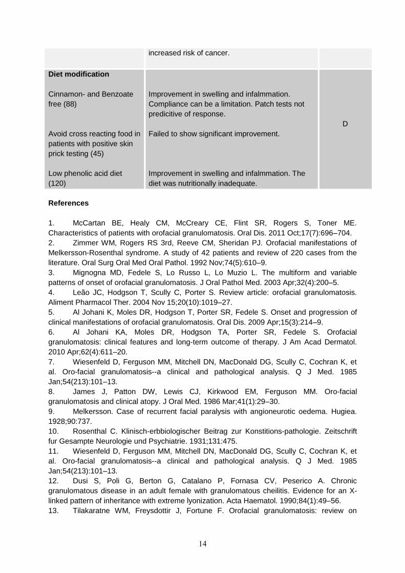
Diet modification
A number of studies have attempted treatment of OFG via elimination of potential allergens
from the diet of affected patients. In addition to multiple case reports of patch test-proven
hypersensitivity to a single antigen and relevant dietary avoidance (23), an UK group have
developed three separate dietary interventions aimed at reducing orofacial swelling and other
intra-oral manifestations of OFG: a cinnamon- and benzoate-free diet (88), a dietary
avoidance of cross reacting foods in OFG individuals with positive skin prick test to silver
birch, grass, mugwort, ragweed and latex (45), and a low phenolic acid diet (120). Although
the authors claim that some of these interventions can reduce orofacial inflammation of OFG
in up to 70% of cases, there remains little robust evidence regarding their actual
effectiveness due to methodological limitations of available studies.
Anti-leprotic agents
Dapsone and clofazimine that have been occasionally reported to reduce the orofacial
swelling of OFG (55,121–124). In one study, long-treatment with low-dosage clofazimine
(400-700mg weekly for 3–11 months) led to complete remission in 5 of 10 treated patients
and partial improvement in a further 3 patients (125). There are only few published cases of
dapsone therapy in OFG, with relevant results ranging from complete relief to ineffectiveness
(63). It seems that anti-leprotic agents may be more effective during the early stages of the
disease (121).
Miscellaneous drugs
There have been reports of small number of patients with OFG having clinical benefits with
methotrexate (126), sulphasalazine (127), lymecycline (128), minocycline (129), or 5-amino-
salicylic acid (130), metronidazole (115,131–133) hydroxychloroquine (122), and various
combinations of these agents with systemic, topical or intralesional corticosteroids.
Surgery and low-level laser therapy
OFG patients with long-standing and disfiguring fibrotic swelling that has proven to be
unresponsive to treatment may benefit from surgical correction. Relevant plastic surgery
procedures include cheiloplasty, commissuroplasty, facial liposuction and tangential
resection of labial mucosa, submucosa, and muscles (134). It has been recommended that
surgical correction of OFG should be undertaken during inactive phases of the disease
(25,122), and possibly in association with perioperative oral or intralesional corticosteroid
therapy (135–137). The long-term benefits of surgery are largely unknown although
11
recurrence of the labial swelling following surgery has been reported (138,139).
Merigo et al reported complete remission of labial swelling with the use of low-level laser
therapy (LLLT) in one OFG patient who had previusly failed to respond to topical and
systemic corticosteroid and immunosuppressive therapy (140). Low-level laser therapy is
believed to have anti-inflammatory and wound-healing properties (140). Of note, clinical
remission was maintained for two years after treatment (140).
Psychological support
When orofacial enlargement of OFG becomes persistent and aesthetically unacceptable,
typically in cases of absent or partial response to therapy, psychological support and
counselling may be beneficial in developing coping mechanisms and improving quality of life
(127). However there are no studies investigating potential benefits of psychological
interventions in OFG individuals.
Clinical outcomes
Although variable and multiform in its early stages, the natural history of OFG is ultimately
progressive, and permanent disfiguring labial and/or facial swelling eventually develops in
nearly all affected individuals (53). Spontaneous resolution is exceptionally rare and the vast
majority of patients would eventually require medical treatment. The long-term prognosis of
OFG individuals is largely unknown. Al-Jahani et al studied the long-term clinical outcomes of
49 patients with OFG who received a wide variety of treatments and reported that facial
swelling of OFG tends to improve over time in those who are on therapy, with approximately
78% of patients showing some reduction in their clinical manifestations. However, only
46.8% eventually experienced complete resolution of orofacial swelling (6). Historically a
variety of different topical and/or systemic agents have been used during the long-term
management of OFG patients with a variable incidence of development of new
manifestations, lack of response, and adverse side effects (6). However, more recent and
better designed studies seem to suggest that some single treatment modalities could
effectively provide long-term control of the disease, as in the case of intralesional
corticosteroid injections (53). A summary of clinical outcomes of available interventions is
provided in (Table 2).
Conclusion
Orofacial granulomatosis is an uncommon inflammatory disorder that typically affects
children and young adults and causes disfiguring facial swelling and painful oral ulceration.
Although a strict case definition of OFG requires the absence of any systemic granulomatous
disease, an unpredictable subgroup of individuals with OFG limited to the orofacial tissues
would eventually develop intestinal Crohn’s disease or, more rarely, sarcoidosis. A significant
number of OFG patients show evidence of concomitant IgE-mediated allergy and delayed-
type hypersensitivity to a number of food and other antigens, although relevant role in
etiopathogenesis and therapy remains unclear. Management of OFG is often difficult and not
evidence-based. Mild cases would benefit from topical immunosuppression whereas long-
term anti-inflammatory and immunosuppressive agents are needed to control more severe
facial swelling and painful oral ulceration. Intralesional corticosteroid therapy may provide
notable long-term remission with no need of prolonged treatment. It seems that the vast
majority of OFG patients on therapy would eventually experience a variable degree of
reduction in orofacial swelling.

12
Tables:
Table 1. Diagnostic investigations and criteria of orofacial granulomatosis
Investigations Results
Full blood cell count Should be normal
Hemoglobin Should be normal
Serum angiotensin converting enzyme levels* Should be normal
C-1 esterase inhibitor levelsy Should be normal
Serum iron and transferrin Should be normal
Tuberculin skin test (when clinically justified) Should be negative
Chest radiography (when clinically justified) Should be normal
GI endoscopy/histopathologyz Should be normal; if
inflammatory changes are
present, Crohn disease
should be excluded
Histopathology I: dilated lymphatics, edema of corium, slight
fibrosis, with/without multiple noncaseating granulomas with
Langerhans giant cell and lymphocytes
Should be present§
Histopathology II: PAS reaction and Ziehl-Neelsen stain (when
clinically justified)
Should be negative
Polarized light microscopy: identification of birefringent foreign-
body material (when clinically justified)
Should be negative
GI: Gastrointestinal; PAS: periodic acide-Schiff.
*To be performed when there are clinical features compatible with potential diagnosis of
sarcoidosis.
yTo be performed when orofacial swelling is recurrent and edematous without signs of
persistent tissue fibrosis.
zTo be performed when clinical or laboratory features increase suggestion of GI
inflammatory disease.
§Absence of histopathological features does not exclude orofacial granulomatosis diagnosis
if clinical features are compatible.

13
Table 2. Clinical outcomes of OFG therapy
yparehT Clinical outcome Level of
evidence
Topical agents:
Corticosteroids (3, 141, 142)
Tacrolimus (93)
Effective in managing moderate intra-oral lesions
and mild labial swelling.
Marked improvement in lip swelling. Benefits can
be transient.
-
D
lntralesional
corticosteroids
Low volume triamcinolone
acetonide [10 mg/mL] (103,
142, 143)
High volume triamcinolone
acetonide [40mg/ml] (53,
106)
Long term-effective in reducing lip swelling. It
requires multiple injection sessions and local
block aneasthesia. Adverse effects: acute pain
and transient worsening of swelling
Effective in long-term reduction of oro-facial
swelling. Requires one single 3-weekly course
and no block anaesthesia. Adverse effects: mild
discomfort, skin pigmentations.
D
Systemic corticosteroids
and
immunosuppressants
Prednisolone (57, 141)
Mycophenolate mofetil
(109)
Short-term courses effective in reducing facial
swelling and oral ulcers. Side effects from chronic
therapy: osteoporosis, diabetes, cataract,
Cushing’s syndrome.
Long-term effectiveness in reducing orofacial
swelling and ulceration. It requires monitoring of
toxicity.
D
Anti-TNF strategies
Thalidomide (92,117,144)
Infliximab (117,145–
148,119)
Adalimumab (149)
Effective in the short-term management of OFG.
Side effect: Neuropathy and teratogenesis.
Short-term reduction in orofacial swelling and oral
ulceration. It requires multiple infusions. Adverse
effects: possible infusion reaction, infection,
increased risk of cancer.
Satisfactory results for oral ulceration. Adverse
effects: possible infusion reaction, infection,
D
14
increased risk of cancer.
Diet modification
Cinnamon- and Benzoate
free (88)
Avoid cross reacting food in
patients with positive skin
prick testing (45)
Low phenolic acid diet
(120)
Improvement in swelling and infalmmation.
Compliance can be a limitation. Patch tests not
predicitive of response.
Failed to show significant improvement.
Improvement in swelling and infalmmation. The
diet was nutritionally inadequate.
D
References
1. McCartan BE, Healy CM, McCreary CE, Flint SR, Rogers S, Toner ME.
Characteristics of patients with orofacial granulomatosis. Oral Dis. 2011 Oct;17(7):696–704.
2. Zimmer WM, Rogers RS 3rd, Reeve CM, Sheridan PJ. Orofacial manifestations of
Melkersson-Rosenthal syndrome. A study of 42 patients and review of 220 cases from the
literature. Oral Surg Oral Med Oral Pathol. 1992 Nov;74(5):610–9.
3. Mignogna MD, Fedele S, Lo Russo L, Lo Muzio L. The multiform and variable
patterns of onset of orofacial granulomatosis. J Oral Pathol Med. 2003 Apr;32(4):200–5.
4. Leão JC, Hodgson T, Scully C, Porter S. Review article: orofacial granulomatosis.
Aliment Pharmacol Ther. 2004 Nov 15;20(10):1019–27.
5. Al Johani K, Moles DR, Hodgson T, Porter SR, Fedele S. Onset and progression of
clinical manifestations of orofacial granulomatosis. Oral Dis. 2009 Apr;15(3):214–9.
6. Al Johani KA, Moles DR, Hodgson TA, Porter SR, Fedele S. Orofacial
granulomatosis: clinical features and long-term outcome of therapy. J Am Acad Dermatol.
2010 Apr;62(4):611–20.
7. Wiesenfeld D, Ferguson MM, Mitchell DN, MacDonald DG, Scully C, Cochran K, et
al. Oro-facial granulomatosis--a clinical and pathological analysis. Q J Med. 1985
Jan;54(213):101–13.
8. James J, Patton DW, Lewis CJ, Kirkwood EM, Ferguson MM. Oro-facial
granulomatosis and clinical atopy. J Oral Med. 1986 Mar;41(1):29–30.
9. Melkersson. Case of recurrent facial paralysis with angioneurotic oedema. Hugiea.
1928;90:737.
10. Rosenthal C. Klinisch-erbbiologischer Beitrag zur Konstitions-pathologie. Zeitschrift
fur Gesampte Neurologie und Psychiatrie. 1931;131:475.
11. Wiesenfeld D, Ferguson MM, Mitchell DN, MacDonald DG, Scully C, Cochran K, et
al. Oro-facial granulomatosis--a clinical and pathological analysis. Q J Med. 1985
Jan;54(213):101–13.
12. Dusi S, Poli G, Berton G, Catalano P, Fornasa CV, Peserico A. Chronic
granulomatous disease in an adult female with granulomatous cheilitis. Evidence for an X-
linked pattern of inheritance with extreme lyonization. Acta Haematol. 1990;84(1):49–56.
13. Tilakaratne WM, Freysdottir J, Fortune F. Orofacial granulomatosis: review on

15
aetiology and pathogenesis. J Oral Pathol Med. 2008 Apr;37(4):191–5.
14. Campbell H, Escudier M, Patel P, Nunes C, Elliott TR, Barnard K, et al. Distinguishing
orofacial granulomatosis from crohn’s disease: two separate disease entities? Inflamm Bowel
Dis. 2011 Oct;17(10):2109–15.
15. Alawi F. Granulomatous diseases of the oral tissues: differential diagnosis and
update. Dent Clin North Am. 2005 Jan;49(1):203–221, x.
16. Somech R, Harel A, Rotshtein MS, Brazowski E, Reif S. Granulomatosis Cheilitis and
Crohn Disease. Journal of Pediatric Gastroenterology and Nutrition [Internet]. 2001;32(3).
Available from:
http://journals.lww.com/jpgn/Fulltext/2001/03000/Granulomatosis_Cheilitis_and_Crohn_Dise
ase.24.aspx
17. Banks T, Gada S. A comprehensive review of current treatments for granulomatous
cheilitis. Br J Dermatol. 2012 May;166(5):934–7.
18. Somasundaram R, Nuij VJAA, van der Woude CJ, Kuipers EJ, Peppelenbosch MP,
Fuhler GM. Peripheral neutrophil functions and cell signalling in Crohn`s disease. PLoS
ONE. 2013;8(12):e84521.
19. Korzenik JR, Dieckgraefe BK. Is Crohn’s Disease an Immunodeficiency? Dig Dis Sci.
2000 Jun 1;45(6):1121–9.
20. Petersen HJ, Smith AM. The Role of the Innate Immune System in Granulomatous
Disorders. Front Immunol [Internet]. 2013 May 24 [cited 2014 Jul 14];4. Available from:
http://www.ncbi.nlm.nih.gov/pmc/articles/PMC3662972/
21. Levy FS, Bircher AJ, Büchner SA. Delayed-type hypersensitivity to cow’s milk protein
in Melkersson-Rosenthal syndrome: coincidence or pathogenetic role? Dermatology (Basel).
1996;192(2):99–102.
22. Taibjee SM, Prais L, Foulds IS. Orofacial granulomatosis worsened by chocolate:
results of patch testing to ingredients of Cadbury’s chocolate. Br J Dermatol. 2004
Mar;150(3):595.
23. Tilakaratne WM, Freysdottir J, Fortune F. Orofacial granulomatosis: review on
aetiology and pathogenesis. J Oral Pathol Med. 2008 Apr;37(4):191–5.
24. H Petersen, Hodgson T, Porter S, Smith A. Defects of the innate immune system in
patients with orofacial franulomatosis. Oral Surgery, Oral Medicine, Oral Pathology, Oral
Radiology. 2014 May;117(5):e353.
25. Grave B, McCullough M, Wiesenfeld D. Orofacial granulomatosis--a 20-year review.
Oral Dis. 2009 Jan;15(1):46–51.
26. Satsangi J, Jewell DP, Rosenberg WM, Bell JI. Genetics of inflammatory bowel
disease. Gut. 1994 May;35(5):696–700.
27. Gibson J, Wray D. Human leucocyte antigen typing in orofacial granulomatosis. Br J
Dermatol. 2000 Nov;143(5):1119–21.
28. Lim SH, Stephens P, Cao QX, Coleman S, Thomas DW. Molecular analysis of T cell
receptor beta variability in a patient with orofacial granulomatosis. Gut. 1997 May;40(5):683–
6.
29. De Quatrebarbes J, Cordel N, Bravard P, Lenormand B, Joly P. [Miescher’s cheilitis
and lymphocytic clonal expansion: 2 cases]. Ann Dermatol Venereol. 2004 Jan;131(1 Pt
1):55–7.
30. Freysdottir J, Zhang S, Tilakaratne WM, Fortune F. Oral biopsies from patients with
orofacial granulomatosis with histology resembling Crohn’s disease have a prominent Th1
environment. Inflamm Bowel Dis. 2007 Apr;13(4):439–45.
31. Pryce DW, King CM. Orofacial granulomatosis associated with delayed
16
hypersensitivity to cobalt. Clin Exp Dermatol. 1990 Sep;15(5):384–6.
32. Lazarov A, Kidron D, Tulchinsky Z, Minkow B. Contact orofacial granulomatosis
caused by delayed hypersensitivity to gold and mercury. J Am Acad Dermatol. 2003
Dec;49(6):1117–20.
33. Patton DW, Ferguson MM, Forsyth A, James J. Oro-facial granulomatosis: a possible
allergic basis. Br J Oral Maxillofac Surg. 1985 Aug;23(4):235–42.
34. Haworth RJ, MacFadyen EE, Ferguson MM. Food intolerance in patients with oro-
facial granulomatosis. Hum Nutr Appl Nutr. 1986 Dec;40(6):447–56.
35. Sweatman MC, Tasker R, Warner JO, Ferguson MM, Mitchell DN. Oro-facial
granulomatosis. Response to elemental diet and provocation by food additives. Clin Allergy.
1986 Jul;16(4):331–8.
36. Reed BE, Barrett AP, Katelaris C, Bilous M. Orofacial sensitivity reactions and the
role of dietary components. Case reports. Aust Dent J. 1993 Aug;38(4):287–91.
37. McKenna KE, Walsh MY, Burrows D. The Melkersson-Rosenthal syndrome and food
additive hypersensitivity. Br J Dermatol. 1994 Dec;131(6):921–2.
38. Wray D, Rees SR, Gibson J, Forsyth A. The role of allergy in oral mucosal diseases.
QJM. 2000 Aug;93(8):507–11.
39. White A, Nunes C, Escudier M, Lomer MCE, Barnard K, Shirlaw P, et al.
Improvement in orofacial granulomatosis on a cinnamon- and benzoate-free diet. Inflamm
Bowel Dis. 2006 Jun;12(6):508–14.
40. Oliver AJ, Rich AM, Reade PC, Varigos GA, Radden BG. Monosodium glutamate-
related orofacial granulomatosis. Review and case report. Oral Surg Oral Med Oral Pathol.
1991 May;71(5):560–4.
41. Pachor ML, Urbani G, Cortina P, Lunardi C, Nicolis F, Peroli P, et al. Is the
Melkersson-Rosenthal syndrome related to the exposure to food additives? A case report.
Oral Surg Oral Med Oral Pathol. 1989 Apr;67(4):393–5.
42. Armstrong DK, Biagioni P, Lamey PJ, Burrows D. Contact hypersensitivity in patients
with orofacial granulomatosis. Am J Contact Dermatitis. 1997 Mar;8(1):35–8.
43. Patel P, Brostoff J, Campbell H, Goel RM, Taylor K, Ray S, et al. Clinical evidence for
allergy in orofacial granulomatosis and inflammatory bowel disease. Clin Transl Allergy.
2013;3(1):26.
44. Morales C, Peñarrocha M, Bagán JV, Burchés E, Pelaez A. Immunological study of
Melkersson-Rosenthal syndrome. Lack of response to food additive challenge. Clin Exp
Allergy. 1995 Mar;25(3):260–4.
45. Campbell H, Escudier MP, Brostoff J, Patel P, Milligan P, Challacombe SJ, et al.
Dietary intervention for oral allergy syndrome as a treatment in orofacial granulomatosis: a
new approach? J Oral Pathol Med. 2013 Aug;42(7):517–22.
46. Campbell HE, Escudier MP, Patel P, Challacombe SJ, Sanderson JD, Lomer MCE.
Review article: cinnamon- and benzoate-free diet as a primary treatment for orofacial
granulomatosis. Aliment Pharmacol Ther. 2011 Oct;34(7):687–701.
47. Apaydin R, Bahadir S, Kaklikkaya N, Kakklikkaya N, Bilen N, Bayramgürler D.
Possible role of Mycobacterium tuberculosis complex in Melkersson-Rosenthal syndrome
demonstrated with Gen-Probe amplified Mycobacterium tuberculosis direct test. Australas J
Dermatol. 2004 May;45(2):94–9.
48. Ivanyi L, Kirby A, Zakrzewska JM. Antibodies to mycobacterial stress protein in
patients with orofacial granulomatosis. J Oral Pathol Med. 1993 Aug;22(7):320–2.
49. Riggio MP, Gibson J, Lennon A, Wray D, MacDonald DG. Search for Mycobacterium
paratuberculosis DNA in orofacial granulomatosis and oral Crohn’s disease tissue by
17
polymerase chain reaction. Gut. 1997 Nov;41(5):646–50.
50. Muellegger RR, Weger W, Zoechling N, Kaddu S, Soyer HP, El Shabrawi-Caelen L,
et al. Granulomatous cheilitis and Borrelia burgdorferi: polymerase chain reaction and
serologic studies in a retrospective case series of 12 patients. Arch Dermatol. 2000
Dec;136(12):1502–6.
51. Handa S, Saraswat A, Radotra BD, Kumar B. Chronic macrocheilia: a clinico-
pathological study of 28 patients. Clin Exp Dermatol. 2003 May;28(3):245–50.
52. Savage NW, Barnard K, Shirlaw PJ, Rahman D, Mistry M, Escudier MP, et al. Serum
and salivary IgA antibody responses to Saccharomyces cerevisiae, Candida albicans and
Streptococcus mutans in orofacial granulomatosis and Crohn’s disease. Clin Exp Immunol.
2004 Mar;135(3):483–9.
53. Fedele S, Fung PPL, Bamashmous N, Petrie A, Porter S. Long-term effectiveness of
intralesional triamcinolone acetonide therapy in orofacial granulomatosis: an observational
cohort study. Br J Dermatol. 2014 Apr;170(4):794–801.
54. Leão JC, Hodgson T, Scully C, Porter S. Review article: orofacial granulomatosis.
Aliment Pharmacol Ther. 2004 Nov 15;20(10):1019–27.
55. Sciubba JJ, Said-Al-Naief N. Orofacial granulomatosis: presentation, pathology and
management of 13 cases. Journal of Oral Pathology & Medicine. 2003;32(10):576–85.
56. El-Hakim M, Chauvin P. Orofacial granulomatosis presenting as persistent lip
swelling: Review of 6 new cases. Journal of Oral and Maxillofacial Surgery. 2004
Sep;62(9):1114–7.
57. Kauzman A, Quesnel-Mercier A, Lalonde B. Orofacial granulomatosis: 2 case reports
and litrature review. J Can Dent Assoc. 2006;72:325–9.
58. Rogers RS 3rd. Melkersson-Rosenthal syndrome and orofacial granulomatosis.
Dermatol Clin. 1996 Apr;14(2):371–9.
59. Campbell H, Escudier M, Patel P, Nunes C, Elliott TR, Barnard K, et al. Distinguishing
orofacial granulomatosis from crohn’s disease: two separate disease entities? Inflamm Bowel
Dis. 2011 Oct;17(10):2109–15.
60. McCartan BE, Healy CM, McCreary CE, Flint SR, Rogers S, Toner ME.
Characteristics of patients with orofacial granulomatosis. Oral Dis. 2011 Oct;17(7):696–704.
61. Sainsbury CP, Dodge JA, Walker DM, Aldred MJ. Orofacial granulomatosis in
childhood. Br Dent J. 1987 Sep 5;163(5):154–7.
62. Odukoya O. Orofacial granulomatosis: report of two Nigerian cases. J Trop Med Hyg.
1994 Dec;97(6):362–6.
63. Ratzinger G, Sepp N, Vogetseder W, Tilg H. Cheilitis granulomatosa and Melkersson-
Rosenthal syndrome: evaluation of gastrointestinal involvement and therapeutic regimens in
a series of 14 patients. J Eur Acad Dermatol Venereol. 2007 Sep;21(8):1065–70.
64. Yeatts RP, White WL. Granulomatous blepharitis as a sign of Melkersson-Rosenthal
syndrome. Ophthalmology. 1997 Jul;104(7):1185–1189; discussion 1189–1190.
65. Cocuroccia B, Gubinelli E, Annessi G, Zambruno G, Girolomoni G. Persistent
unilateral orbital and eyelid oedema as a manifestation of Melkersson-Rosenthal syndrome.
J Eur Acad Dermatol Venereol. 2005 Jan;19(1):107–11.
66. Dupuy A, Cosnes J, Revuz J, Delchier JC, Gendre JP, Cosnes A. Oral Crohn
disease: clinical characteristics and long-term follow-up of 9 cases. Arch Dermatol. 1999
Apr;135(4):439–42.
67. Croft CB, Wilkinson AR. Ulceration of the mouth, pharynx, and larynx in Crohn’s
disease of the intestine. Br J Surg. 1972 Apr;59(4):249–52.
68. Chan SW, Scully C, Prime SS, Eveson J. Pyostomatitis vegetans: oral manifestation
18
of ulcerative colitis. Oral Surg Oral Med Oral Pathol. 1991 Dec;72(6):689–92.
69. Ruiz-Roca JA, Berini-Aytés L, Gay-Escoda C. Pyostomatitis vegetans. Report of two
cases and review of the literature. Oral Surg Oral Med Oral Pathol Oral Radiol Endod. 2005
Apr;99(4):447–54.
70. Kalman RS, Gjede JM, Farraye FA. Pyostomatitis vegetans in a patient with fistulizing
Crohn’s disease. Clin Gastroenterol Hepatol. 2013 Dec;11(12):A24.
71. Worsaae N, Christensen KC, Schiødt M, Reibel J. Melkersson-Rosenthal syndrome
and cheilitis granulomatosa. A clinicopathological study of thirty-three patients with special
reference to their oral lesions. Oral Surg Oral Med Oral Pathol. 1982 Oct;54(4):404–13.
72. Levenson MJ, Ingerman M, Grimes C, Anand KV. Melkersson-Rosenthal syndrome.
Arch Otolaryngol. 1984 Aug;110(8):540–2.
73. Sanderson J, Nunes C, Escudier M, Barnard K, Shirlaw P, Odell E, et al. Oro-facial
granulomatosis: Crohn’s disease or a new inflammatory bowel disease? Inflamm Bowel Dis.
2005 Sep;11(9):840–6.
74. Greene RM, Rogers RS 3rd. Melkersson-Rosenthal syndrome: a review of 36
patients. J Am Acad Dermatol. 1989 Dec;21(6):1263–70.
75. Alexander RW, James RB. Melkersson-Rosenthal syndrome: review of literature and
report of case. J Oral Surg. 1972 Aug;30(8):599–604.
76. Kanerva M, Moilanen K, Virolainen S, Vaheri A, Pitkäranta A. Melkersson-Rosenthal
syndrome. Otolaryngol Head Neck Surg. 2008 Feb;138(2):246–51.
77. Vistnes LM, Kernahan DA. The Melkersson-Rosenthal syndrome. Plast Reconstr
Surg. 1971 Aug;48(2):126–32.
78. Pino Rivero P, González Palomino A, Pantoja Hernández CG, Trinidad Ruíz G,
Pardo Romero G, Alvarez Domínguez J, et al. [Melkersson-Rosenthal syndrome. Report of a
case with bilateral facial palsy]. An Otorrinolaringol Ibero Am. 2005;32(5):437–43.
79. Khandpur S, Malhotra AK, Khanna N. Melkersson-Rosenthal syndrome with diffuse
facial swelling and multiple cranial nerve palsies. J Dermatol. 2006 Jun;33(6):411–4.
80. Hornstein OP. Melkersson-Rosenthal syndrome. A neuro-muco-cutaneous disease of
complex origin. Curr Probl Dermatol. 1973;5:117–56.
81. Stosiek N, Birolleau S, Capesius C, Hornstein OP. [Chronicity and diagnostic doubts
of Melkersson-Rosenthal syndrome. Analysis of developing ways in 5 cases]. Ann Dermatol
Venereol. 1992;119(9):635–8.
82. Scully C, Cochran KM, Russell RI, Ferguson MM, Ghouri MA, Lee FD, et al. Crohn’s
disease of the mouth: an indicator of intestinal involvement. Gut. 1982 Mar 1;23(3):198–201.
83. Zbar AP, Ben-Horin S, Beer-Gabel M, Eliakim R. Oral Crohn’s disease: Is it a
separable disease from orofacial granulomatosis? A review. Journal of Crohn’s and Colitis.
2012 Mar;6(2):135–42.
84. Saalman R, Mattsson U, Jontell M. Orofacial granulomatosis in childhood-a clinical
entity that may indicate Crohn’s disease as well as food allergy. Acta Paediatr. 2009
Jul;98(7):1162–7.
85. Al-Azri ARS, Logan RM, Goss AN. Oral Lesion as the first Clinical Presentation in
Sarcoidosis: A Case Report. Oman Med J. 2012 May;27(3):243–5.
86. Armstrong DK, Burrows D. Orofacial granulomatosis. Int J Dermatol. 1995
Dec;34(12):830–3.
87. Armstrong DK, Biagioni P, Lamey PJ, Burrows D. Contact hypersensitivity in patients
with orofacial granulomatosis. Am J Contact Dermatitis. 1997 Mar;8(1):35–8.
88. White A, Nunes C, Escudier M, Lomer MCE, Barnard K, Shirlaw P, et al.
Improvement in orofacial granulomatosis on a cinnamon- and benzoate-free diet. Inflamm

19
Bowel Dis. 2006 Jun;12(6):508–14.
89. Patel P, Campbell H, Brostoff J, et al. Allergy in orofacial granulomatosis and
inflammatory bowel disease. Gut. 2010;59:A99.
90. Riordain RN, Meaney S, McCreary C. Impact of chronic oral mucosal disease on
daily life: preliminary observations from a qualitative study. Oral Dis. 2011 Apr;17(3):265–9.
91. Chiandussi S, Tappuni AR, Watson TF, White A, Escudier MP, Sanderson JD, et al.
Lip impressions: a new method for monitoring morphological changes in orofacial
granulomatosis. Oral Dis. 2007 Jan;13(1):93–8.
92. Hegarty A, Hodgson T, Porter S. Thalidomide for the treatment of recalcitrant oral
Crohn’s disease and orofacial granulomatosis. Oral Surgery, Oral Medicine, Oral Pathology,
Oral Radiology, and Endodontology. 2003 May;95(5):576–85.
93. Casson DH, Eltumi M, Tomlin S, Walker-Smith JA, Murch SH. Topical tacrolimus may
be effective in the treatment of oral and perineal Crohn’s disease. Gut. 2000 Sep;47(3):436–
40.
94. Olivier V, Lacour J-P, Mousnier A, Garraffo R, Monteil RA, Ortonne J-P. Treatment of
chronic erosive oral lichen planus with low concentrations of topical tacrolimus: an open
prospective study. Arch Dermatol. 2002 Oct;138(10):1335–8.
95. Rozycki TW, Rogers RS 3rd, Pittelkow MR, McEvoy MT, el-Azhary RA, Bruce AJ, et
al. Topical tacrolimus in the treatment of symptomatic oral lichen planus: a series of 13
patients. J Am Acad Dermatol. 2002 Jan;46(1):27–34.
96. Shen JT, Pedvis-Leftick A. Mucosal staining after using topical tacrolimus to treat
erosive oral lichen planus. J Am Acad Dermatol. 2004 Feb;50(2):326.
97. Lozada-Nur FI, Sroussi HY. Tacrolimus powder in Orabase 0.1% for the treatment of
oral lichen planus and oral lichenoid lesions: an open clinical trial. Oral Surg Oral Med Oral
Pathol Oral Radiol Endod. 2006 Dec;102(6):744–9.
98. Albert MH, Becker B, Schuster FR, Klein B, Binder V, Adam K, et al. Oral graft vs.
host disease in children--treatment with topical tacrolimus ointment. Pediatr Transplant. 2007
May;11(3):306–11.
99. Hodgson TA, Sahni N, Kaliakatsou F, Buchanan JAG, Porter SR. Long-term efficacy
and safety of topical tacrolimus in the management of ulcerative/erosive oral lichen planus.
Eur J Dermatol. 2003 Oct;13(5):466–70.
100. Eisenbud L, Hymowitz SS, Shapiro R. Cheilitis granulomatosa. Report of case treated
with injection of triamcinolone acetonide aqueous suspension. Oral Surg Oral Med Oral
Pathol. 1971 Sep;32(3):384–9.
101. Krutchkoff D, James R. Cheilitis granulomatosa. Successful treatment with combined
local triamcinolone injections and surgery. Arch Dermatol. 1978 Aug;114(8):1203–6.
102. Allen CM, Camisa C, Hamzeh S, Stephens L. Cheilitis granulomatosa: report of six
cases and review of the literature. J Am Acad Dermatol. 1990 Sep;23(3 Pt 1):444–50.
103. Sakuntabhai A, MacLeod RI, Lawrence CM. Intralesional steroid injection after nerve-
block in orofacial granulomatosis. Lancet. 1992 Oct 17;340(8825):969.
104. Sakuntabhai A, MacLeod RI, Lawrence CM. Intralesional steroid injection after nerve
block anesthesia in the treatment of orofacial granulomatosis. Arch Dermatol. 1993
Apr;129(4):477–80.
105. Mignogna MD, Fedele S, Lo Russo L, Adamo D, Satriano RA. Effectiveness of small-
volume, intralesional, delayed-release triamcinolone injections in orofacial granulomatosis: a
pilot study. J Am Acad Dermatol. 2004 Aug;51(2):265–8.
106. Mignogna MD, Pollio A, Leuci S, Ruoppo E, Fortuna G. Clinical behaviour and long-
term therapeutic response in orofacial granulomatosis patients treated with intralesional

21
triamcinolone acetonide injections alone or in combination with topical pimecrolimus 1%. J
Oral Pathol Med. 2013 Jan;42(1):73–81.
107. Fedele S, Fung PPL, Bamashmous N, Petrie A, Porter S. Long-term effectiveness of
intralesional triamcinolone acetonide therapy in orofacial granulomatosis: an observational
cohort study. Br J Dermatol. 2013 Oct 1;
108. Van der Waal RI, Schulten EA, van de Scheur MR, Wauters IM, Starink TM, van der
Waal I. Cheilitis granulomatosa. J Eur Acad Dermatol Venereol. 2001 Nov;15(6):519–23.
109. Antonyan AS, Pena-Robichaux V, McHargue CA. Orofacial granulomatosis
successfully treated with mycophenolate mofetil. Journal of the American Academy of
Dermatology. 2014 Jun;70(6):e137–e139.
110. Plauth M, Jenss H, Meyle J. Oral manifestations of Crohn’s disease. J Clin
Gastroenterol. 1991;13:9–37.
111. Mahadevan U, Sandborn WJ. Infliximab for the treatment of orofacial Crohn’s
disease. Inflamm Bowel Dis. 2001 Feb;7(1):38–42.
112. Safa G, Joly P, Boullie MC, Thomine E, Lauret P. [Melkersson-Rosenthal syndrome
treated by thalidomide. 2 cases]. Ann Dermatol Venereol. 1995;122(9):609–11.
113. Odeka EB, Miller V. Thalidomide in oral Crohn’s disease refractory to conventional
medical treatment. J Pediatr Gastroenterol Nutr. 1997 Aug;25(2):250–1.
114. Weinstein TA, Sciubba JJ, Levine J. Thalidomide for the treatment of oral aphthous
ulcers in Crohn’s disease. J Pediatr Gastroenterol Nutr. 1999 Feb;28(2):214–6.
115. Medeiros M Jr, Araujo MI, Guimarães NS, Freitas LAR, Silva TMC, Carvalho EM.
Therapeutic response to thalidomide in Melkersson-Rosenthal syndrome: a case report. Ann
Allergy Asthma Immunol. 2002 Apr;88(4):421–4.
116. Thomas P, Walchner M, Ghoreschi K, Röcken M. Successful treatment of
granulomatous cheilitis with thalidomide. Arch Dermatol. 2003 Feb;139(2):136–8.
117. Eustace K. Thalidomide in the treatment of refractory orofacial granulomatosis.
Clowry J, Kirby B, Lally A, editors. Br J Dermatol. 2014;
118. O’Neill ID, Scully C. Biologics in oral medicine: oral Crohn’s disease and orofacial
granulomatosis. Oral Dis. 2012 Oct;18(7):633–8.
119. Elliott T, Campbell H, Escudier M, Poate T, Nunes C, Lomer M, et al. Experience with
anti-TNF-α therapy for orofacial granulomatosis. J Oral Pathol Med. 2011 Jan;40(1):14–9.
120. Campbell HE, Escudier MP, Milligan P, Challacombe SJ, Sanderson JD, Lomer MCE.
Development of a low phenolic acid diet for the management of orofacial granulomatosis. J
Hum Nutr Diet. 2013 Apr 10;
121. Podmore P, Burrows D. Clofazimine--an effective treatment for Melkersson-
Rosenthal syndrome or Miescher’s cheilitis. Clin Exp Dermatol. 1986 Mar;11(2):173–8.
122. Van der Waal RIF, Schulten EAJM, van der Meij EH, van de Scheur MR, Starink TM,
van der Waal I. Cheilitis granulomatosa: overview of 13 patients with long-term follow-up--
results of management. Int J Dermatol. 2002 Apr;41(4):225–9.
123. Camacho-Alonso F, Bermejo-Fenoll A, López-Jornet P. Miescher’s cheilitis
granulomatosa. A presentation of five cases. Med Oral Patol Oral Cir Bucal. 2004
Dec;9(5):427–429; 425–427.
124. Fdez-Freire LR, Serrano Gotarredona A, Bernabeu Wittel J, Pulpillo Ruiz A, Cabrera
R, Navarrete Ortega M, et al. Clofazimine as elective treatment for granulomatous cheilitis. J
Drugs Dermatol. 2005 Jun;4(3):374–7.
125. Sussman GL, Yang WH, Steinberg S. Melkersson-Rosenthal syndrome: clinical,
pathologic, and therapeutic considerations. Ann Allergy. 1992 Sep;69(3):187–94.
126. Tonkovic-Capin V, Galbraith SS, Rogers RS 3rd, Binion DG, Yancey KB. Cutaneous
21
Crohn’s disease mimicking Melkersson-Rosenthal syndrome: treatment with methotrexate. J
Eur Acad Dermatol Venereol. 2006 Apr;20(4):449–52.
127. Clayden AM, Bleys CM, Jones SF, Savage NW, Aldred MJ. Orofacial granulomatosis:
a diagnostic problem for the unwary and a management dilemma. Case reports. Aust Dent J.
1997 Aug;42(4):228–32.
128. Pigozzi B, Fortina AB, Peserico A. Successful treatment of Melkersson-Rosenthal
Syndrome with lymecycline. Eur J Dermatol. 2004 Jun;14(3):166–7.
129. Stein SL, Mancini AJ. Melkersson-Rosenthal syndrome in childhood: successful
management with combination steroid and minocycline therapy. J Am Acad Dermatol. 1999
Nov;41(5 Pt 1):746–8.
130. Girlich C, Bogenrieder T, Palitzsch K-D, Schölmerich J, Lock G. Orofacial
granulomatosis as initial manifestation of Crohn’s disease: a report of two cases. Eur J
Gastroenterol Hepatol. 2002 Aug;14(8):873–6.
131. Miralles J, Barnadas MA, de Moragas JM. Cheilitis granulomatosa treated with
metronidazole. Dermatology (Basel). 1995;191(3):252–3.
132. Dummer W, Lurz C, Jeschke R, Meissner N, Rose C, Bröcker EB. Granulomatous
cheilitis and Crohn’s disease in a 3-year-old boy. Pediatr Dermatol. 1999 Feb;16(1):39–42.
133. Coskun B, Saral Y, Cicek D, Akpolat N. Treatment and follow-up of persistent
granulomatous cheilitis with intralesional steroid and metronidazole. J Dermatolog Treat.
2004 Sep;15(5):333–5.
134. Tan O, Atik B, Calka O. Plastic Surgical Solutions for Melkersson-Rosenthal
Syndrome: Facial Liposuction and Cheiloplasty Procedures. Annals of Plastic Surgery. 2006
Mar;56(3):268–73.
135. Glickman LT, Gruss JS, Birt BD, Kohli-Dang N. The surgical management of
Melkersson-Rosenthal syndrome. Plast Reconstr Surg. 1992 May;89(5):815–21.
136. Camacho F, García-Bravo B, Carrizosa A. Treatment of Miescher’s cheilitis
granulomatosa in Melkersson-Rosenthal syndrome. J Eur Acad Dermatol Venereol. 2001
Nov;15(6):546–9.
137. Kruse-Lösler B, Presser D, Metze D, Joos U. Surgical treatment of persistent
macrocheilia in patients with Melkersson-Rosenthal syndrome and cheilitis granulomatosa.
Arch Dermatol. 2005 Sep;141(9):1085–91.
138. Ellitsgaard N, Andersson AP, Worsaae N, Medgyesi S. Long-term results after
surgical reduction cheiloplasty in patients with Melkersson-Rosenthal syndrome and cheilitis
granulomatosa. Ann Plast Surg. 1993 Nov;31(5):413–20.
139. Oliver DW, Scott MJL. Lip reduction cheiloplasty for Miescher’s granulomatous
macrocheilitis (Cheilitis granulomatosa) in childhood. Clin Exp Dermatol. 2002
Mar;27(2):129–31.
140. Merigo E, Fornaini C, Manfredi M, Meleti M, Alberici F, Corcione L, et al. Orofacial
granulomatosis treated with low-level laser therapy: a case report. Oral Surg Oral Med Oral
Pathol Oral Radiol. 2012 Jun;113(6):e25–29.
141. Sciubba JJ, Said-Al-Naief N. Orofacial granulomatosis: presentation, pathology and
management of 13 cases. J Oral Pathol Med. 2003 Nov;32(10):576–85.
142. Mignogna MD, Fedele S, Lo Russo L, Lo Muzio L. Orofacial granulomatosis with
gingival onset. J Clin Periodontol. 2001 Jul;28(7):692–6.
143. Kolokotronis A, Antoniades D, Trigonidis G, Papanagiotou P. Granulomatous cheilitis:
a study of six cases. Oral Dis. 1997 Sep;3(3):188–92.
144. Odeka EB, Miller V. Thalidomide in oral Crohn’s disease refractory to conventional
medical treatment. J Pediatr Gastroenterol Nutr. 1997 Aug;25(2):250–1.

22
145. Kaufman I, Caspi D, Yeshurun D, Dotan I, Yaron M, Elkayam O. The effect of
infliximab on extraintestinal manifestations of Crohn’s disease. Rheumatol Int. 2005
Aug;25(6):406–10.
146. Ottaviani F, Schindler A, Capaccio P, Petrone M, Bianchi Porro G. New therapy for
orolaryngeal manifestations of Crohn’s disease. Ann Otol Rhinol Laryngol. 2003
Jan;112(1):37–9.
147. Brunner B, Hirschi C, Weimann R, Seibold F. Tratment-resistant lingual Crohn’s
disease disappears after infliximab. Scand J Gastrorntrol. 2005;40:5–1259.
148. Cardoso H, Nunes A, Carneiro F, Tavarela Veloso F. Successful inflixmab therapy for
oral Crohn’s disease. Inflamm Bowel Dis. 2006;12:337–8.
149. Sánchez AR, Rogers RS 3rd, Sheridan PJ. Oral ulcerations are associated with the
loss of response to infliximab in Crohn’s disease. J Oral Pathol Med. 2005 Jan;34(1):53–5.