osteology of the pelvic limb of the african elephant (loxodonta
TRANSCRIPT
Onderstepoort Journal of Veterinary Research, 61 :51-66 (1994)
Osteology of the pelvic limb of the African elephant (Loxodonta africana)
MALlE M.S. SMUTS1 and A.J. BEZUIDENHOUT2
ABSTRACT
SMUTS, MALlE M.S. & BEZUIDENHOUT, A.J. 1994. Osteology of the pelvic limb of the African elephant (Loxodonta africana) . Onderstepoort Journal of Veterinary Research, 61 :51-66
The pelvic girdle was characterized by large, transversely-placed ilial wings. The femur was the longest bone of the skeleton and its fovea capitis was situated caudomedially between the epiphyseal line and the articular surface of the femoral head. A wedge-shaped patella articulated with the femoral trochlea. The bones of the crus were approximately half as long as the femur and consisted of the sturdy tibia and slender fibula. The condyles of the tibia were concave and the femoro-tibial joint was congruent with rudimentary menisci. The tarsus consisted of seven bones which were arranged in three rows. There were five metatarsal bones. Only four digits were present, the third and fourth consisted of three phalanges each while the second and fourth digits were smaller and consisted of two phalanges each. The first digit was represented by one proximal sesamoid bone only. A large, cartilagenous rod or prehallux was attached to the first tarsal and metatarsal bones. Proximal sesamoid bones were present on the plantar aspect of the trochleae of metatarsal bones 1- V. The pes was found to be digitigrade and the digits rested on a thick pad of elastic connective tissue and fat.
INTRODUCTION
The earliest publication on the osteology of an adult elephant is a detailed study by Blair (1706) of the measurements of the bones obtained from the skeleton of a female elephant. Unfortunately the latter author did not state whether it was an African or Indian elephant. Flower and Lydekker (1 891, cited by Gussgen 1988) , described the bones of the hind limb and Eales (1928) carried out a comprehensive study
1 Department of Anatomy, Faculty of Veterinary Science, University of Pretoria, Private Bag X04, Onderstepoort, 0110 South Africa
on the skeleton of an African elephant fetus, while Mariappa (1986) described and illustrated the bones of an Indian elephant fetus. Sikes (1971 ) gave a brief description of the bones of the African elephant. Due to the scarcity of material, the majority of studies were carried out on single specimens, mostly from zoological gardens. Because of a lack of material for comparison, the illustrations are often inaccurate and inadequate. This study was therefore undertaken to accurately describe and illustrate the osteology of the hind limb of the African elephant by utilizing material from animals of both sexes at different ages.
2 Department of Anatomy, Faculty of Veterinary Science, University of Pretoria, Private Bag X04, Onderstepoort, 01 10 South Africa
Received 18 November1993- Editor
MATERIALS AND METHODS
The hind-limb bones of five adult African elephants were examined. In addition, two pelvic bones and four
51

Osteology of the pelvic limb of the African elephant
femurs of adult animals were examined and the foot of a juvenile and an adult were dissected in order to study the topography of the bones of the pes in situ. All the material was collected in the Kruger National Park. In addition, the skeleton of a mature bull in the collection of the Zoology Department of the University of Pretoria and two pelvic bones of mature animals from the Hunterian Museum in the Department of Anatomy of the University of the Witwatersrand were studied. Each bone was illustrated from at least two aspects to ensure easy identification. The nomen'clature used is based on the Nomina Anatomica Veterinaria (1983).
RESULTS
Pelvic girdle, Cingulum membri pelvini
The most striking feature of the Ossa coxarum was the size of the wing of the ilium which completely overshadowed the other components, namely the ischium, pubis and the shaft of the ilium.
Os ilium (Fig. 1--4)
The ilium was characterized by a very large and curved three-sided wing, Ala ossis ilii, and a short, compressed shaft, the Corpus ossis ilii, which contributed to the formation of the acetabulum. The wings were placed more or less in the vertical plane. The gluteal surface, Facies glutea, presented a small dorsa-medial part (the part closest to the sacral tuberosity) and a greater convex ventro-lateral part. A number of indistinct gluteal lines were present on the ventrolateral part of the gluteal surface. The iliac crest, Crista iliaca , was represented by the rough lateral border of the iliac wing and formed an S-shaped curve when viewed from the lateral aspect. The ventrally directed Tuber coxae formed the expanded ventro-lateral extremity of the iliac crest. The dorsomedial extremity of the iliac crest curved caudally and slightly laterally to form the sacral tuberosity, Tuber sacra/e. The ventral border of the wing, between the tuber coxae and the shaft, was concave. A shallow vascular groove was present along the ventromedial surface at the junction of wing and shaft. Where the ventral border joined the shaft, a rough triangular area, the Area musculi recti femoris, occurred, which represented the attachment area for the rectus femoris muscle. The concave· dorsal border of the wing was longer than the ventral border and formed the greater ischiadic notch, Incisura ischiadica major. The ischiadic spine was situated caudally to the major ischiadic notch.
The inner or pelvic surface of the wing, the Facies sacropelvina , presented the following features:
• A medially facing, relatively smooth area for articulation with the sacrum, the Tuberositas iliaca.
52
• A rough, ear-shaped area ventral to the former for articulation with the sacrum, the Facies auricularis.
• A relatively smooth area cranially and laterally to the auricular surface for the attachment of lumbar muscles, the Facies iliaca.
The craniomedial border of the shaft showed a relatively inconspicuous line, the Linea arcuata. An elongated rough area along approximately the middle of the line represented the Tuberculum m. psoas minoris.
Os pubis (Fig. 1-5)
The pubis consisted of a body or Corpus ossis pubis which participated in the formation of the acetabulum, a transverse cranial ramus, Ramus crania/is assis pubis and a longitudinal Ramus cauda/is ossis pubis in the paramedian plane. The body and the cranial ramus formed the cranial border of the large, oval Foramen obturatum and the caudal ramus formed the craniomedial margin. The caudal ramus united with the ramus of the opposite side at the Symphysis pubis and continued caudally into the ramus of the ischium. Together the left and right cranial rami formed the Pecten ossis pubis that was sharp and well defined in the female and round and thick in the male (Fig. 5). The Eminentia iliopubica lay laterally on the pecten and was prominent in all specimens. In the middle of the pecten a prominent Tuberculum pubicum dorsa/e and Tuberculum pubicum ventrale were present in male specimens while the dorsal pubic tubercle was absent in the female.
Os ischii (Fig. 1-5)
The ischium consisted of the body or Corpus ossis ischii which participated in the formation of the acetabulum, the caudally directed table or Tabula ossis ischii and the paramedian Ramus ossis ischii. Cranially the ramus united with the caudal ramus of the pubis and the symphyseal surfaces of the two sides united variably at the Symphysis ischiadica. Even in old specimens the latter was not fully fused. The ischium formed the caudal, cauda-medial and lateral boundaries of the foramen obturatum. The medial border of the foramen was sharp, while the lateral border was rounded. Dorsally the body of the ischium was expanded to form the ischiadic spine, Spina ischiadica. The spine was a low crest, with a rough, tuberous area on its lateral aspect. This tuberous area was situated dorsal to the acetabulum and was separated from a smaller caudally situated tuberosity by a smooth intervening area. The relatively shallow Incisura ischiadica minor was situated caudally to the ischiadic spine. The lateral surface of the ischial body directly below the minor incisure may be rough. The ischiadic table formed a 45 o
slope with the ischiadic symphysis. The thickened

caudolateral extremity, the Tuber ischiadicum, therefore was situated dorsally and laterally to the level of the pelvic floor. It also resulted in the oblique placing of the obturator foramen. The caudal borders of the two ischiadic tables formed the concave ischial arch, Arcus ischiadicus. In immature specimens the epiphyseal line was not fused and the ischiadic tuber with the adjacent arch formed a loosely attached, rough border.
Acetabulum (Fig. 4)
The acetabulum was formed by the fusion of all three pelvic bones. The articular surface, Facies lunata, surrounded the irregularly shaped, rough area for ligamentous attachment, the Fossa acetabu/i, which was placed in line with the cranial border of the obturator foramen at the Incisura acetabuli. A number of large foramina occurred in the depth of the fossa. In some specimens, a shallow synovial fossa passed from the acetabular fossa in a ventromedial direction, interrupting the lunar surface in this region. This fossa was not consistently present in all specimens.
The bony pelvis (Fig. 6)
The cranial aperture was oval in outline, with the vertical diameter greater than the transverse. The Diameter conjugata, the line from the sacral promontory to the cranial end of the pubic symphysis, was 470 mm in an adult male and 525 mm in an adult female. The Diameter transversa, the line between the psoas tubercles, was 415 mm in the same male specimen and 390 mm in the female.
The floor of the pelvis was flat in the female. In the male there was a dorsal inclination to the level of the obturator foramen. From here caudally it was gently concave.
Femur (Fig. 7)
The femur was the longest bone of the elephant skeleton. The femur of a 53-year-old female specimen measured 1 m from the femoral head to the distal extremity and the femur of a mature male specimen of unknown age, with epiphyseal lines still clearly visible, measured 1 ,27m in length. The femur was a relatively slender bone, with the shaft slightly flattened proximately in a craniocaudal plane. From the femoral head to the distal extremity, the medial border was gently concave, while the lateral border was more or less straight.
The prominent head, Caput ossis femoris, was an almost perfect hemisphere. It faced proximomedially. The Fovea capitis, situated caudomedially directly above the epiphyseal line, was a triangular depression with its base along the epiphyseal line and its apex forming a notch in the substance of the head. The neck, Collum ossis femoris, sloped towards the shaft, Corpus ossis femoris.
MALlE M.S. SMUTS & A.J. BEZUIDENHOUT
The Trochanter major formed the lateral part of the proximal extremity of the femur. Its proximal border lay at the level of the base of the femoral head. When viewed from above, it was obliquely placed, with its rough surface facing craniolaterally. The major trochanter was divided by a relatively smooth, shallow groove into a cranial and a caudal part. A sloping flat surface connected the base of the head with the greater trochanter.
The Trochanter tertius was represented by the pointed distal continuation of the greater trochanter along the craniolateral border of the femoral shaft, distally to the epiphyseal line. In specimens with epiphyseal lines, the position was approximately a hand's width below the proximal extremity of the trochanter. This measurement may be used to determine the transition of major trochanter to third trochanter in specimens where the growth lines had already closed.
On the caudal aspect of the major trochanter there was a deep, elongated depression which faced caudomedially, the Fossa trochanterica. In growing specimens the epiphyseal line divided the fossa into a proximal epiphyseal part and a distal metaphyseal part. The Trochanter minor was not prominent. It was represented by a rough, elongated area along the proximal third of the medial border of the femoral shaft. It arose at the level of the distal border of the trochanteric fossa. In female specimens the minor trochanter was an elevated ridge; in old males it was pitted. Distally to the lesser trochanter muscular ridges continued along the medial border of the femoral shaft. A small nutrient foramen occurred along the middle or upper third of the medial border.
Cranially to the lesser trochanter, a rough elevated area occurred on the shaft. In male specimens it was not prominent. The cranial surface of the shaft had no special features except for a longitudinal muscular line which arose near the greater trochanter and petered out in the distal quarter. It was not distinct in old male specimens.
The caudal surface of the shaft presented a rough surface, the Facies aspera, along the second quarter of its length. This area was less prominent in male specimens than in females. A prominent line which arose from the caudal part of the greater trochanter formed the lateral border of the shaft. Distally the line was continuous with the elongated Tuberositas supracondylaris latera/is. Along the distal surface of the shaft, muscular ridges represented the Facies poplitea. On caudal view the distal extremity bore the Candy/us latera/is and Candy/us medialis which articulated with the tibia. The medial condyle was about twice as large as the lateral condyle and faced caudolaterally while the lateral condyle was placed sagittally and faced caudally. The Fossa intercondylaris between the condyles was narrow. On the contralateral aspects of the condyles a depression occurred
53

Osteology of the pelvic limb of the African elephant
-:;/
/ II
a
54

MALlE M.S. SMUTS & A.J. BEZUIDENHOUT
X
X
FIG. 1 Female pelvis, cranial view
FIG. 2 Female pelvis, caudal view
FIG. 3 Female pelvis, lateral view
FIG. 4 Acetabulum
FIG. 5 A. Female pelvic symphysis. B. Male pelvic symphysis
a. A/a ossis ilii. b. Corpus ossis ilii. c. Crista iliaca . d. Tuber coxae. e. Tuber sacra/e. f. Area musculi recti femoris. g.lncisura ischiadica major. h. Tuberositas iliaca. h'. Facies auricularis. h" . Facies iliaca. i. Facies lunata. j. Fossa acetabuli. j ' . Incisura acetabuli k. Linea arcuata. k'. Tuberculum musculi psoas minoris. I. Corpus ossis pubis. m. Ramus crania/is ossis pubis. n. Ramus cauda/is ossis pubis. o. Foramen obturatum. p. Symphysis pubis. q. Pecten ossis pubis. r. Eminentia iliopubica. s. Tuberculum pubicum dorsa/e . t. Tuberculum pubicum ventra/e . u. Corpus ossis ischii. v. Tabula ossis ischii. w. Ramus ossis ischii. x. Symphysis ischiadica . y. Spina ischiadica . z. Tuber ischiadicum
55

Osteology of the pelvic limb of the African elephant
A 8
b
FIG. 6 Pelvic inlet. a. Diameter conjugata. b. Diameter transversa FIG. 8 Left patella. A. Cranial surface. B. Articular surface
FIG. 7 Left femur. A. Dorsal (proximal) view. B. Cranial view. C.
56
Caudal view
a. Caput ossis femoris . b. Favia capitis. c. Collum ossis femoris. d. Corpus ossis femoris e. Trochanter major. e'. Epicondylus medialis. f. Trochanter tertius. g. Fossa trochanterica. h. Trochanter minor. i. Tuberositas supracondylaris. j. Facies poplitea. k. Candy/us latera/is. k'. Epicondylus latera/is. I. Candy/us medialis. m. Fossa intercondylaris. n. Fossa musculi poplitei. o. Fossa extensoria. p. Trochlea ossis femoris
a. Basis patellae. b. Apex patellae. c. Facies articularis. d. Facies crania/is
for the attachment of collateral ligaments, and proximally to each depression a rough protuberance was present, the Epicondylus latera/is and Epicondylus medialis. Distally the lateral condyle had two depressions for the origin of the popliteal and long digital extensor muscles. Laterally and directly above the articular surface was the Fossa musculi poplitei for the popliteal muscle. The shallow Fossa extensoria was found at the junction between the lateral lip of the femoral trochlea and the lateral condyle, directly cranial to the articular surface of the lateral condyle. Cranially the Trochlea ossis femoris, for articulation with the patella, consisted of two approximately equal ridges or lips separated by a groove. The trochlea was not placed exactly in the sagittal plane, but faced slightly craniomedially.
Patella (Fig. 8)
The patella was a sesamoid bone which articulated with the femoral trochlea. It presented a proximal base, Basis patellae, a distal apex, Apex patellae, an articular surface, Facies articularis and a cranial surface, Facies crania/is. In adults it was wedgeshaped, while its outline was more elongated and slender in juvenile specimens. The distally directed apex was rounded, particularly in the adult. The cranial aspect was strongly convex and rough for muscular attachment. The base was flattened and rough laterally, while it sloped down on the medial side. The articular surface was divided by a smooth longi~udinal ridge into slightly concave medial and lateral areas. The proximal third of the articular surface was convex from side to side. The medial articular area had a rounded border while the lateral border was straight.
Skeleton cruris
The skeleton of the leg was formed by the Tibia on the medial side and the Fibula laterally. The tibia was

B e c I
A
D
FIG. 9 Left tibia. A. Condylar articular surface. B. Cranial view. C. Caudal view. D. Cochlear articular surface
a. Margo crania/is. b. Tuberositas tibiae. c. Candy/us medialis. d. Candy/us latera/is . e. Tuberculum intercondylare mediale.f. Tuberculum intercondylare latera/e. g. Area intercondylaris centra/is . g'. Area intercondylaris crania/is. g". Area intercondylaris cauda/is. h. Incisura poplitea. i. Facies articularis fibularis. j. Linea muscularis. k. Sulcus malleolaris.l. Cochlea tibiae. m./ncisura fibularis . n. Malleolus medialis
stout and approximately half as long as the femur, while the fibula was a slender bone with a small head proximally and a prominent distal extremity, the lateral malleolus. Only the tibia articulated with the femur. The fibula articulated proximally and distally with the tibia, while both bones articulated with the
MALlE M.S. SMUTS & A.J. BEZUIDENHOUT
tarsal bones. The space between the two bones, Spatium interosseum cruris, was a fairly wide continuous gap, narrowing towards the distal end.
Tibia (Fig. 9)
As the principal weight bearer of the crus, this bone was sturdy and relatively short. The cranial border, Margo crania/is, was thickened proximally to form the deeply indented Tuberositas tibiae, while the distal end of the bone was flattened craniocaudally.
The proximal extremity was formed by two tibial condyles. In contrast to the position in domestic mammals, the condyles were concave, allowing for a congruent articulation with the femur. As a result of this arrangement the menisci were rudimentary. The medial condyle, Candy/us medialis, was larger and more circular than the lateral condyle and was placed more cranially. It slanted upwards mediocranially to form a pointed medial tubercle, Tuberculum intercondylare media/e. The lateral condyle, Candy/us latera/is, slanted upwards medially to form a less pronounced Tuberculum intercondylare latera/e. The medial and lateral tubercles were placed along a saggittal plane, with the medial tubercle more cranial in position than the lateral tubercle. In this way the Eminentia intercondylaris was represented by a sagittal crest with a cranial and a caudal prominence. The Area iotercondylaris centra/is was represented by a depressed or a rough, elevated area directly behind the medial intercondylar tubercle. Along the cranial aspect of the latter a relatively smooth surface occurred, with a small round depression laterally to it. These two areas represented the Areae intercandy/ares craniales for ligamentous attachment. At the caudal aspect of the intercondylar eminence the Area intercondylaris cauda/is occurred with the shallow Incisura poplitea below it. There was no extensor groove in the lateral condylar border. A clearly defined, round, articular facet, Facies articularis fibularis, was present along the caudolateral aspect of the lateral condyle.
In cranial view the shaft was divided by the cranial margin and the prominent, deeply indented tibial tuberosity into a flattened, moderately convex medial surface and a concave lateral surface. A prominent rough area for muscular attachment occurred about halfway down the medial surface. In caudal view the proximal half of the shaft was concave, with a rough surface in the region below the condyles, representing the popliteal notch while one or two nutrient foramina occurred variably above the middle of the shaft. The medial border formed a rounded, prominent ridge. The less prominent lateral border was also rough for muscular attachment. A vertical Linea muscularis was present along the middle of the distal half of this surface. A wide vascular groove which passed obliquely
57
Osteology of the pelvic limb of the African elephant
downwards from lateral to medial, interrupted the muscular line above the distal extremity of the tibia.
The distal epiphyseal region formed a rough border around the articular surfaces. A groove, the Sulcus malleolaris, marked the caudal surface of the medial malleolus, Malleolus medialis. The Cochlea tibiae was a shallow, concave articular surface which consisted of a clearly concave medial area and a smaller, flattened, lateral area, demarcated by an indistinct sagittal ridge. Directly adjacent to the lateral area and placed at an oblique angle to it, lay the relatively small articular facet for the fibula, Incisura fibularis.
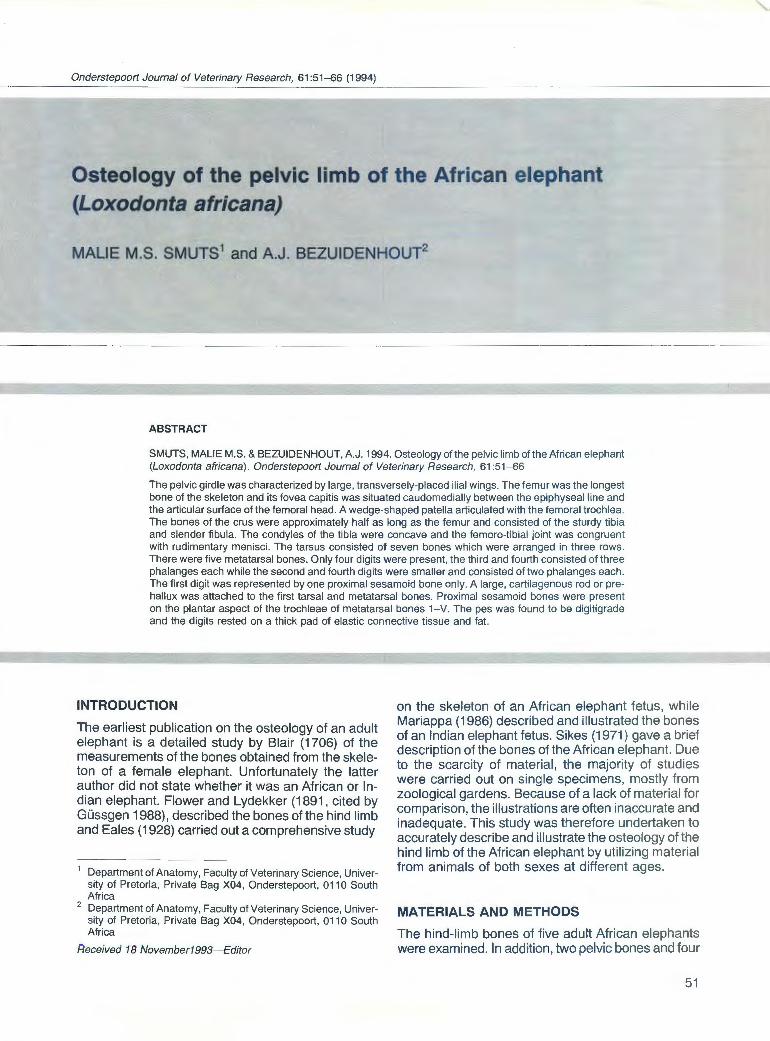
Fibula (Fig. 1 0)
The fibula was far smaller and more slender than the tibia and lay lateral to it. Proximally it was lower than the tibia, but distally it projected beyond the cochlea. The distal extremity, Malleolus latera/is, was expanded and relatively massive, while the proximally situated head, Caput fibulae, was small.
The head formed the rounded, proximal end of the bone. The small (approximately 15 mm), round, articular facet for the tibia, Facies articularis capitis fibulae, faced cranially. The caudal aspect was convex and rough. The neck, Collum fibulae, was flattened craniocaudally. Laterally it was rough and flattened, while the medial border was a sharp ridge. The shaft, Corpus fibulae, was a slightly twisted, slender region, compressed craniocaudally in the proximal third of its length while the distal part became progressively flattened from side to side. The medial border was sharp and rough in the proximal third and ended in an elongated tuberosity. A prominent muscular line ran longitudinally along the caudal border. On the cranial aspect a slightly curved muscular line was present from the neck to the distal extremity where it joined the lateral malleolus. A small nutrient foramen occurred in most specimens about halfway down the shaft, lateral to the cranial border. The lateral malleolus presented a medially placed articular surface, Facies articulares malleoli, and a rough lateral aspect without visible grooves. The articular facet for the tibia was a rounded, slightly convex area and was the most proximal of the articular facets on the medial aspect. It was placed almost horizontally, sloping downwards towards the medial side. Three other facets for articulation with the tarsal bones occurred distally to the facet for the tibia. A small, vertically placed area lay adjacent to the tibial facet, with a larger area obliquely below it. These two facets articulated with the talus. The largest facet was found distally and articulated with the calcaneus. It was gently concave and faced mediodistally.
Skeleton of the tarsus
There were seven tarsal bones, arranged in three rows. The proximal row consisted of the Talus (Os
58
A B
FIG. 10 Left fibula. A. Cranial view. B. Medial view
a. Malleolus latera/is. b. Caput fibulae. c. Facies articutaris capitis fibulae . d. Collum fibulae. e. Corpus fibulae. f. Facies articutaris malleoli. g. Articular facet for the talus and calcaneus
tarsi tibiale, Tt) and the large Calcaneus (Os tarsi fibulare , Tf). The middle row was represented by the Os tarsi centrale (Os naviculare, Tc). The distal row had a complement of four tarsal bones. From medial to lateral they were: Os tarsale I (Os cuneiforme mediate, T1 ), Os tarsale II (Os cuneiforme intermedium, T2), Os tarsale Ill (Os cuneiforme laterale, T3) and Os tarsale IV (Os cuboideum, T4).
A
8 c
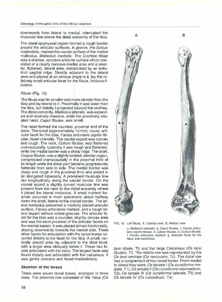
FIG. 11 Left talus. A. Proximal articular surface. B. Caudal articular surface C. Distal articular surface
a. Trochlea tali. b. Articular facet for the fibula. c. Articular facet for the calcaneus. d. Articular facet for Tc. bone
A
8
c
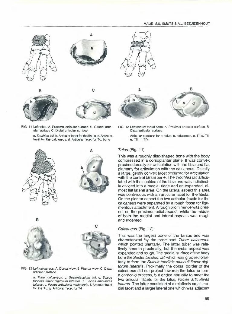
FIG. 12 Left calcaneus. A. Dorsal view. B. Plantar view. C. Distal articular surface
a. Tuber calcaneus. b. Sustentaculum tali. c. Sulcus tendinis flexor digitorum latera/is. d. Facies articulares talares. e. Facies articularis malleolaris. f. Articular facet for the Tc. g. Articular facet for T4
MALlE M.S. SMUTS & A.J. BEZUIDENHOUT
A 8
FIG. 13 Left central tarsal bone. A. Proximal articular surface. B. Distal articular surface
Articular surfaces for a. talus, b. calcaneus, c. Tl , d. Til , e. Till , f. TIV
Talus (Fig. 11)
This was a roughly disc-shaped bone with the body compressed in a dorsoplantar plane. It was convex proximodorsally for articulation with the tibia and flat plantarly for articulation with the calcaneus. Distally a large, gently convex facet occurred for articulation with the central tarsal bone. The Trochlea tali articulated with the cochlea of the tibia and was indistinctly divided into a medial ridge and an expanded, almost flat lateral area. On the lateral aspect this area was continuous with an articular facet for the fibula. On the plantar aspect the two articular facets for the calcaneus were separated by a rough fossa for ligamentous attachment. A rough prominence was present on the proximomedial aspect, while the middle of both the medial and lateral aspects was rough and indented.
Calcaneus (Fig. 12)
This was the largest bone of the tarsus and was characterized by the prominent Tuber calcaneus which pointed plantarly. The latter tuber was relatively smooth proximally, but the distal aspect was expanded and rough. The medial surface of the body bore the Sustentaculum tali which was grooved plantarly to form the Sulcus tendinis musculi flexor digitorum latera/is. Proximally the dorsal border of the calcaneus did not project towards the talus to form a coracoid process, but ended abruptly to meet the two articular facets for the talus, Facies articulares tala res. The latter consisted of a relatively small medial facet and a larger lateral one which was adjacent
59

Osteology of the pelvic limb of the African elephant
FIG. 14 Left distal row of tarsal bones (TI- IV) . A. Proximal articular surfaces. B. Distal articular surfaces
Articular surfaces for a. Tc. b. Calcaneus. c. Tl. d. Til. e. Till. f. TIV. g. Mtl. h. Mtll. i. Mtlll. j. MtiV. k. MtV
Tl
to a slightly convex lateral articular facet for the fibula, Facies articularis malleolaris. A deep, rough depression separated the two facets for the talus. Distally to the smaller medial facet and at an angle to it, a relatively small, flat facet occurred for articulation with the central tarsal bone. The slightly concave facet on the distal aspect was for articulation with T 4. The surface on its plantar side was tuberous and rough .
Os tarsi centrale (Fig. 13)
The central tarsal bone was a flat, slightly curved bone, with a concave, proximal, articular surface for the talus. In addition, there was a small plantar area
60
Til
adjoining it, for articulation with the calcaneus. The distal surface was gently convex, with four discernible articular facets from medial to lateral for the four tarsal bones. The facets for T3 and T 4 were separated by a ridge; the facets between T2 and T3 were separated by a shallow groove while the facets for T1 and T2 were not clearly separated. The articular area for T3 was the largest. The non-articular aspects were rough.
Os tarsale 1 (Fig. 14)
The first tarsal bone was the only bone of the distal row which was not wedge-shaped. It was flattened
and rough from side to side and presented two articular surfaces at each end. Proximally it articulated with the central tarsal bone and with T2. Distally, a large oval facet was present for Mt1, while a small facet occurred laterally for articulation with Mt2.
Os tarsale 2 (Fig. 14)
This was the smallest of the three wedge-shaped tarsal bones. Its medial border was irregularly convex, while the lateral border was straight. It bore a slightly concave facet proximally for articulation with Tc, while a facet for T1 was present medially. The distal articular surface was flat and occupied almost the entire distal surface of the bone. Along the lateral aspect two facets were present for articulation with T3. The plantar aspect of the bone was a rounded tuberosity.
Os tarsale 3 (Fig. 14)
T3 was about twice as large as T2. Proximally it articulated with Tc by means of a concave facet, and distally with T3 and part of T 4. The bone narrowed from dorsal to plantar, where a rounded tuberosity occurred. The medial and lateral borders each had two articular facets for T2 and T4, respectively.
Os tarsale 4 (Fig. 14)
This was the largest bone of the series. It was placed in such a way that the apex faced dorsally and the base plantarly. Its proximal surface presented two articular surfaces- a smaller concave facet for Tc and a large convex surface for the calcaneus. Distally there was a facet for part ofT 4 and a larger ovoid facet for Mt5. Along the medial border two facets were present for T3.
The skeleton of the metatarsus
There were five metatarsal bones. They showed typical characteristics of the bones of the metapodium, with a proximal base, a body which was roughly triangular to quadrilateral in cross section and a distal expanded extremity, the head, for articulation with the proximal phalanx. The largest bone in the series was Mt3, followed by Mt4 and Mt2. Metatarsal5 was a stout, almost quadrilateral bone, which was approximately half as long as Mt3. The smallest in the series was Mt1, which was approximately half the size of Mt2. Its base was more expanded than its head.
Os metatarsale I (Fig. 15)
This was the smallest of the metatarsal bones, and was characterized by a large, flat, oval articular surface on the base for articulation with T1 . The plantar aspect of the base formed an expanded, raised surface. The body was constricted and short, while the head formed a blunt point dorsally. A small, slightly
MALlE M.S. SMUTS & A.J. BEZUIDENHOUT
raised, round facet for articulation with the proximal sesamoid bone was present on the plantar side of the distal extremity.
Os metatarsale II (Fig. 15)
When viewed from above, the proximal extremity was convex medially and flat laterally. A spinous tuberosity divided the articular surface into a large, flat facet for T2 and a small, oblique facet for T3. On the flattened, lateral aspect of the base, two facets, or an elongated single facet, occurred for articulation with Mt3. Medially the base was slightly elongated and a small, oblique facet was present for articulation with T1 . The surface of the body was convex medially and flat laterally. On the plantar aspect the surface was rough proximally, while a deep fossa occurred distally above the head. The head was flattened on each side for ligamentous attachment. The distal articular surface could be divided into a dorsal convex area for the proximal phalanx, and a plantar articular area for the proximal sesamoid bones. An indistinct sagittal line divided the latter articular area into a large medial and a smaller lateral articular facet for the corresponding proximal sesamoid bones. The dorsal and plantar articular areas were separated by a transverse line.
Os metatarsale Ill (Fig. 15)
This was the largest bone of the metatarsal series. When viewed from above, the flattened proximal articular surface was triangular in shape, with the apex pointing plantarly. Along the medial border of the base of the bone two articular facets or a single elongated facet occurred for articulation with the corresponding facets on Mt2. Along the lateral border of the base there was a facet for Mt4. A prominent plantar process was present on the base. The body was flattened dorsally. The surface was rough proximally, with an oblique groove sloping from lateral to medial in the proximal half. The plantar surface of the body was flat and relatively smooth. Proximally the medial and lateral surfaces were rough; distally they were smoother and, in a dorsoplantar plane, narrower. The head or distal extremity was expanded and each side was flattened for ligamentous attachment. The articular surface consisted of an undivided dorsal area for the proximal phalanx and a plantar area which was divided into two approximately equal facets by a sagittal line for the proximal sesamoid bones. The dorsal and plantar articular areas were separated by a transverse line.
Os metatarsale IV (Fig. 15)
In adults this bone was approximately 200 mm shorter than Mt3 although its diameter was about the same. When viewed from above, the general outline of the base of the bone was triangular, with the apex
61

Osteology of the pelvic limb of the African elephant
FIG. 15 Left metatarsal bones. A. Dorsal view. B. Plantar view
Articular surfaces for a. Tl. b. Til. c. Till. d. TIV. e. Mtll. f . Mtlll. g. MtiV. h. MtV. i. proximal sesamoid bones. j. proximal phalanges
B
A
pointing plantarly. The articular surface, however, was not triangular, as a prominent notch was present along the medial border, towards the apex. The articular surface was confined chiefly to the dorsal side, with an elongation towards the plantar side along the lateral border. An indistinct line divided the surface into a small medial area for T3 and a large lateral area for T 4. Along the medial aspect of the base of the bone a facet occurred for T3 while on the lateral side there was a facet for T5. When viewed from the plantar side there was a prominent, rough tuberosity and, especially on the medial side, the surface was rough for ligamentous attachment. The diameter was more or less quadrilateral, with the dorsal width greater than the plantar. The distal extremity or head
62
f
was expanded, showing the same features as Mt3, with an articular surface for the proximal phalanx and two facets plantarly for the proximal sesamoid bones. The medial facet was smaller than the lateral. A curved transverse line separated the dorsal and plantar articular areas.
Os metatarsale V (Fig. 15}
This stout, stocky bone cannot be mistaken for any other. On proximal view it featured a large, ovoid, articular surface for T4 and a small, obliquely placed facet along the medial border for Mt4. The body was square to round in cross-section. The dorsal surface was concave, while the plantar surface was rounded

FIG. 16 Left pes, medial view
FIG. 17 Left manus, caudal view
a. Calcaneus. b. Talus. c. Tl. d. Mtl. e. Cartilagenous rod or prehallux. f . Cartilagenous rod or prepollux
for ligamentous attachment. The medial surface showed a transverse groove which was flanked by rough areas for attachment of ligaments proximally and distally to it. The lateral surface of the body was rounded and rough . The distal articular surface was extensive. The facet for the proximal phalanx was saddle-shaped. The two facets for the proximal sesamoids differed in size, the lateral facet was larger than the medial, and both facets were sepa-
. rated from the articular surface for P1 by a curved, transverse line.
The skeleton of the digits of the pes
Despite the fact that there were five metacarpal bones, only four digits consisting of phalanges were present in the hind foot. The first digit was represented by a single, proximal sesamoid bone. Attached to the plantar aspect of the first tarsal and metatarsal bones a firm, rib-shaped cartilage rod (prehallux) reached down to the sole, where it attached in the plantar quarter, just medial to the midline (Fig. 16). In the manus a similar cartilage rod (prepollux) was present along the mediopalmar aspect of Metacarpal I (Fig. 17). The chief digits belonged to the third and fourth radial units. They were complete in that they had three phalanges each. Digit 3 was the largest one and was clearly the chief weight bearer. Digits 2 and 4 consisted of two phalanges each. The digits rested on an extensive cushion of elastic connective tissue and fat and they were placed in a more slanting position than in the forelimb, where they were more upright. The cornified sole was narrower in outline
MALlE M.S. SMUTS & A.J. BEZUIDENHOUT
than that of the forelimb and was convex in a dorsaplantar plane, indicating a rolling movement. Three nails were present for digits 2-4 with the third the biggest, followed by the fourth and second.
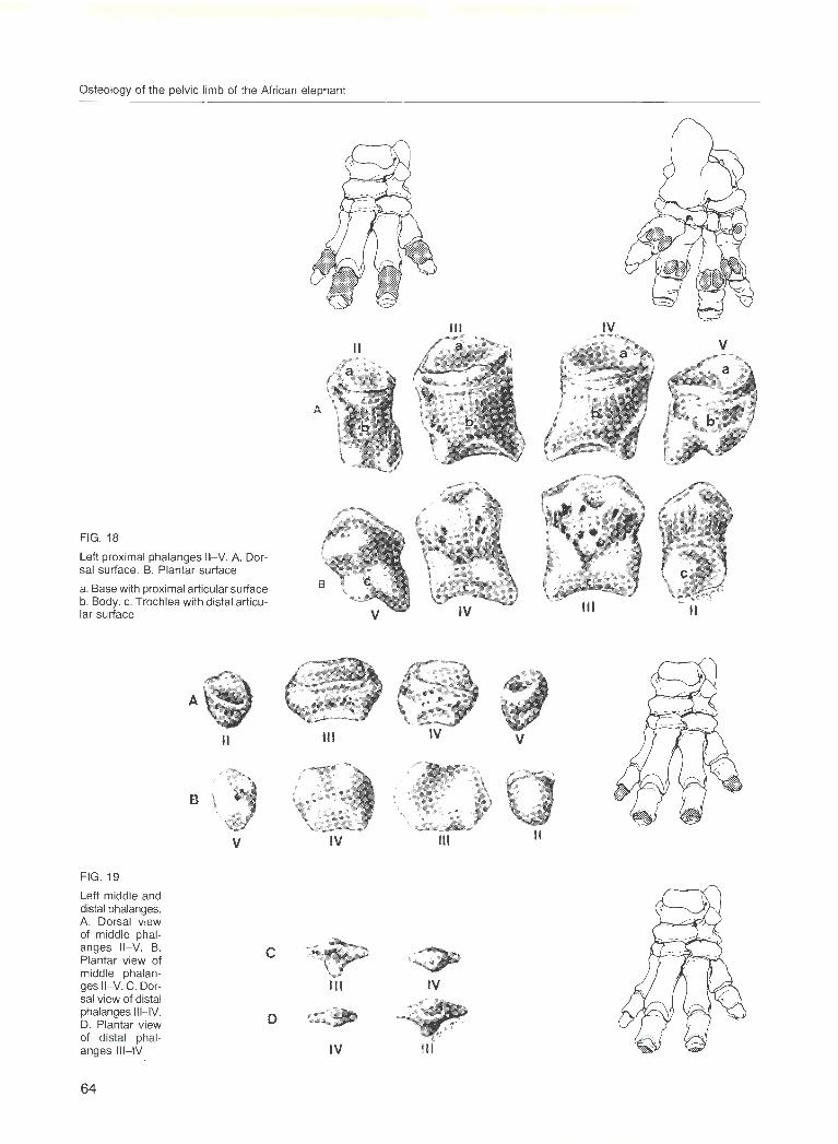
Phalanges proximales (Fig. 18)
The proximal phalanges of digits 3 and 4 were more or less similar in shape, with that of digit 3 the largest. The diameter of digit 2 was about half that of digit 4. Digit 5 was a short, stocky bone with a prominent distal process. Each bone consisted of a base with a single round to oval, slightly concave, proximal articular surface, a body which presented dorsal, medial, plantar and lateral surfaces and a distal trochlea. When viewed from the distal aspect, the trochlea of the proximal phalanx of digit 2 was roughly triangular in shape, with the apex pointing dorsally. The trochleae of digits 3 and 4 were concave from side to side and convex in the dorsoplantar plane. The distal extremity of the phalanx of digit 5 consisted of a rounded medial part and a pointed lateral part which projected distally. The plantar aspects of the bones were elevated proximally and the surfaces were relatively smooth and free from foramina except for the bones of digits 3 and 4, where large foramina occurred on the body. Numerous small foramina were present on the dorsal surfaces of all the bones.
Phalanges mediae (Fig. 19)
The middle phalanges were much smaller than the proximal ones and those of digits 2 and 5 differed markedly from those of digits 3 and 4.
63
Osteology of the pelvic limb of the African elephant
FIG. 18
Left proximal phalanges 11-V. A. Dorsal surface. B. Plantar surface
a. Base with proximal articular surface b. Body. c. Trochlea with distal articular surface
II
B ~~ ~
v
FIG. 19
Left middle and distal phalanges. A. Dorsal view of middle phalanges 11-V. B. Plantar view of middle phalanges 11- V. C. Dorsal view of distal phalanges III- IV. D. Plantar view of distal phalanges III- IV
c
D
64
A
Ill
IV
Ill
IV
Ill
II v
II
IV v
Ill II
~ IV

The middle phalanges of digits 2 and 5 were small, cone-shaped bones. Proximally a round to oval, articular surface occurred, while the distal extremity was pointed and lacked an articular surface.
The middle phalanges of digits 3 and 4 were short, relatively wide bones, which were roughly quadrilateral in shape and were flattened dorsoplantarly. The phalanx of digit 3 was bilaterally symmetrical whereas that of digit 4 was narrower and presented a distal projection medially. Proximally each bone had a large articular surface which was oval in outline and slightly saddle-shaped. The dorsal surface of each bone was rough and contained numerous foramina. The plantar surface was relatively smooth and devoid of foramina. The distal extremity was relatively smooth, but did not form a typical articular facet.
Phalanges distales (Fig . 19)
Only digits 3 and 4 possessed distal phalanges that articulated with the middle phalanges. In the available specimens the phalanx for digit 3 measured approximately 35 mm x 18 mm. This was about twice the size of the distal sesamoid bone for digit 4. The tiny bones were spindle-shaped, with a medial and
A
II II
B
v v
FIG. 20 Proximal sesamoid bones of Digits 1- V. A. Dorsal view with articular surface. B. Plantar view
Ill Ill
IV IV
MALlE M.S. SMUTS & A.J. BEZUIDENHOUT
a lateral horn that projected from a central body with its distally projecting tubercle. The proximal surface had a rounded , centrally positioned, articular facet, while the distal surface contained numerous foramina and was firmly attached to the corium of the nail.
Proximal sesamoid bones (Fig. 20)
The proximal sesamoid bones were found in pairs on the plantar facets of the trochlea of metatarsals 2- 5, while a single sesamoid bone was present on the plantar facet of metatarsal 1 . The relative size of each sesamoid corresponded to the articular facet on the trochlea of the corresponding metatarsal bone. Each sesamoid bone presented an articular surface for the metatarsal bone. In addition, there were also small facets along the contact area of each pair, with the exception of the sesamoids of digits 1 and 5. The plantar surfaces were convex and rough; the proximal aspects were larger than the distal and the shape varied from round (for digits 1 and 5) to elongated (for digits 2-4) . As can be expected, the sesamoids of digit 3 were the biggest, followed by digit 4.
DISCUSSION
It was noted that epiphyseal cartilages were present in many of the bones of all age groups of elephants studied. This implies that elephants have the potential to grow virtually througout life. The physical size of an elephant is largely dependent on the type of food available. Elephants from the northern part of the Kruger National Park, for instance, are known to be smaller than elephants from the southern part of the park. It is therefore difficult, if not impossible, to draw definite conclusions regarding the species (African or Indian), age or sex of an animal by measuring the size of various bones.
According to Eales (1928) the ilium of a foetal African elephant is large with a slender shaft and much
IV IV v v
Ill Ill II
65

Osteology of the pelvic limb of the African elephant
expanded crest, the greater sciatic notch is horseshoe-shaped, and it shows the vertical twist which brings the tuber sacrale and adjacent regions parallel with the vertebral column. Sikes (1971) noted the wide expansion of the ilia and the prominent iliac crest in the African elephant. The observations of both authors correspond to the findings of the present study, as does the description of the ilium of a foetal Asian elephant by Mariappa (1986} and the account of the bones of an adult elephant of unknown origin by Blair (1706).
Eales (1928) stated that the acetabulum was set far back and directed backwards, while Sikes (1971) stated that the acetabulum faced downwards, resting the full weight of the hindquarters directly on top of the femoral head. In the present study it was found that the acetabulum faced caudoventrally.
Blair (1706) stated that, in the mature female elephant he studied, the conjugata was 457 mm and the diameter transversa was 431 mm in the mature female elephant that he studied. In the present study the conjugata was 470 mm in mature females and 525 mm in an adult male, while the diameter transversa was 390 mm in the female and 415 mm in the male. From the measurements of Blair one is tempted to assume that he dissected an Indian elephant, but such an assumption might be totally erroneous.
Eales (1928) stated that there is no teres ligamentand therefore no fovea capitis-attaching the head of the femur to the acetubulum, and that the trochanter tertius was absent. In the present study the fovea capitis was found to lie directly above the epiphyseal line, forming a slight notch in the articular surface, and the trochanter tertius was represented by the distal continuation of the greater trochanter. It must be noted that there were considerable differences between the femurs of female and male specimens, e.g. in female animals the minor trochanter was an elevated ridge, in male animals a pitted area.
Blair (1706) identified six tarsal bones in the tarsus of a mature female animal, while all other investigations, including the present study, demonstrated seven tarsal bones in the tarsus of the African and Indian elephants.
The pes of the elephant seems to be the most controversial part of its anatomy. Mariappa (1986) claimed that elephants have six digits, while Eales (1928) and Mariappa (1986} described a cartilaginous prehallux. According to Sikes (1971) the hallux (first
66
digit) has a reduced metatarsal and vestigial phalanx, while Eales (1928) described a very small first digit. In the present study a large, rib-like cartilage was found to be attached to the first tarsal and metatarsal bones. A similar cartilage is also found in the manus of the front limb. The cartilage functions as an additional support for the manus and pes and represents the prehallux or sixth digit of the other authors. Contrary to the findings of Eales (1928) and Sikes (1971}, the first digit was found to consist of one proximal sesamoid bone only.
According to Sikes (1971) the pes of the elephant is semi-digitigrade and semi-plantigrade. Although the bones of the pes were in a more slanting position than those of the manus, dissections of the pes in situ showed that the pes of the African elephant is in a true digitigrade position.
ACKNOWLEDGEMENTS We gratefully acknowledge material provided by the Kruger National Park and the Hunterian Museum of the Department of Anatomy of the University of the Witwatersrand.
We are greatly indebted to Christine Seegers for the illustrations.
REFERENCES BLAIR, P. 1706. Osteographia Elephantina. A full and exact de
scription of all the bones of an elephant, which died near Dundee, April the 27th, 1706, with their several dimensions. Communicated in a letter to Dr. Hans Sloane, Royal Society's Secretary. Philosophical Transactions of the Royal Society of London, 27:51 - 168.
EALES, NELLIE B. 1928. The anatomy of a foetal African elephant. Ill. The contents of the thorax and abdomen and the skeleton. Transactions of the Royal Society of Edinburgh, 56: 203- 246.
GUSSGEN, BEATRIX 1988. Vergleichende Zusammenstellung der Literaturbefunde Ober die Anatomie des lndischen und Afrikanischen Elephanten als Grundlage tor Tierarzliches Handeln. D.Med.Vet. Dissertation, Tierarzliche Hochschule, Hannover.
MARIAPPA, D. 1986. Anatomy and histology of the Indian elephant, 1st ed. Michigan: Indira Publishing House.
HABEL, R.E., FREWEIN, J & SACK, W.O. (Eds.) 1983. Nomina Anatomica Veterinaria , 3rd ed. Ithaca, New York: International Anatomical Nomenclature Committee.
SIKES, SYLVIA K. 1971. The natural history of the African elephant. London: Weidenfeld & Nicolson.