otitis media in the tgif knockout mouse implicates tgfβ · otitis media in the tgif knockout mouse...
TRANSCRIPT

1
© The Author 2013. Published by Oxford University Press. This is an Open Access article distributed under the terms of the Creative Commons Attribution Non‐Commercial License (http://creativecommons.org/licenses/by‐nc/2.5), which permits unrestricted non‐commercial use, distribution, and reproduction in any medium, provided the original work is properly cited.
Otitis media in the Tgif knockout mouse implicates TGFβ
signalling in chronic middle ear inflammatory disease
Hilda Tateossian1, Susan Morse1, Andy Parker1, Philomena Mburu1, Nick
Warr1, Abraham Acevedo-Arozena1, Michael Cheeseman1, Sara Wells2 and
Steve DM Brown1,*
1MRC Mammalian Genetics Unit, Harwell, OX11 0RD, UK
2Mary Lyon Centre, MRC Harwell, Harwell, OX11 0RD, UK
Corresponding author: S.D.M Brown, MRC Mammalian Genetics Unit, Harwell, OX11 0RD, UK,
phone: +44 1235 841053, fax: +44 1235 841169, email: [email protected]
HMG Advance Access published March 3, 2013 by guest on January 14, 2014
http://hmg.oxfordjournals.org/
Dow
nloaded from
2
ABSTRACT
Otitis Media with effusion (OME) is the most common cause of hearing loss in children and
tympanostomy to alleviate the condition remains the commonest surgical intervention in children in
the developed world. Chronic and recurrent forms of OM are known to have a very significant genetic
component, however, until recently little was known of the underlying genes involved. The
identification of mouse models of chronic OM has indicated a role for TGFβ signalling and its impact
upon responses to hypoxia in the inflamed middle ear. We have therefore investigated the role of
TGFβ.signalling and identified and characterised a new model of chronic OM carrying a mutation in
the gene for Transforming Growth Inhibitory Factor 1 (Tgif1). Tgif1 homozygous mutant mice have
significantly raised auditory thresholds due to a conductive deafness arising from a chronic effusion
starting at around 3 weeks of age. The OM is accompanied by a significant thickening of the middle
ear mucosa lining, expansion of mucin secreting goblet cell populations and raised levels of VEGF,
TNF-α and IL-1β in ear fluids. We also identified downstream effects on TGFβ signalling in middle
ear epithelia at the time of development of chronic OM. Both phosphorylated SMAD2 and p21 levels
were lowered in the homozygous mutant, demonstrating a suppression of the TGFβ pathway. The
identification and characterisation of the Tgif mutant supports the role of TGFβ signalling in the
development of chronic otitis media and provides an important candidate gene for genetic studies in
the human population.
by guest on January 14, 2014http://hm
g.oxfordjournals.org/D
ownloaded from

3
INTRODUCTION
Otitis media with effusion (OME), inflammation of the middle ear, is the commonest cause of hearing
impairment in children and the commonest reason for surgery in children. Both chronic and recurrent
forms of OM are known to have a significant genetic component (1,2), but until recently little was
known about the genes or pathways involved (3,4). Several mouse models of OM have been reported
and some of them, including the mutants Jeff and Junbo, implicate the TGFβ signalling pathway in
susceptibility to OM (5-7). Both mutants were identified from a deafness screen as part of a large-
scale phenotype-driven mouse ENU mutagenesis program (8). Mice heterozygous for the Jeff
mutation develop chronic proliferative otitis media (9) and the gene mutated in Jeff was identified as
Fbxo11 (5). We found that Fbxo11 is involved in the regulation of the TGFβ signalling pathway by
regulating the levels of pSmad2 in the epithelial cells of different embryonic mouse tissues. Further,
we identified a genetic interaction between Fbxo11 and Smad2 (7). Junbo mice display chronic
suppurative otitis media and carry a mutation in the transcription factor Evi1 (6). Evi1 can repress the
TGFβ signalling pathway by interacting with Smad3 and can antagonize the growth-inhibitory effect
of the pathway (10). The identification of these mutants has focused studies on the role of
TGF�signalling in the development of chronic OM and the interaction of this pathway with other
molecular and cellular changes occurring in the inflamed middle ear (11).
The transforming growth factor beta (TGFβ) signalling pathway is involved in a variety of cellular
processes such as proliferation, differentiation and apoptosis (12,13). TGFβ pathway members act by
linking membrane receptors to specific target genes. TGFβ ligands initiate the signalling by binding to
a type II receptor on the cell surface, which in turn binds and phosphorylates a type I receptor. The
intracellular mediators R-Smads, Smad2 and Smad3, become phosphorylated by the TGFβ receptor I.
The activated Smads form a complex with the co-mediator Smad4. The Smad complex then
translocates into the nucleus and in conjunction with other nuclear cofactors regulates the
transcription of different target genes (12). The phosphorylation of the R-Smads is regulated by the
Smad anchor for receptor activation (SARA) protein, cytoplasmic promyelocytic leukaemia (cPML)
protein and transforming growth interacting factor (TGIF). SARA has a Smad binding domain and a
by guest on January 14, 2014http://hm
g.oxfordjournals.org/D
ownloaded from
4
domain interacting with the TGFβ receptor and recruits Smad2 and/or Smad3 to the receptor (14). In
the cytoplasm cPML physically interacts with Smad2/3 and SARA and is required for the association
of Smad2/3 with SARA (15).
TGIF1, also known as TGIF, belongs to a three amino acid loop extension (TALE) subgroup of
atypical homeodomain proteins (16,17). It has been found that TGIF functions through several routes
as a negative regulator of the TGFβ signalling pathway. TGIF was initially identified as a Smad2-
binding protein and a co-repressor of TGFβ-induced transcription. This repression is mediated by the
ability of TGIF to recruit to Smad2 a co-repressor containing histone deacetylases (HDACs) (18).
TGIF can also inhibit Smad2 phosphorylation by an alternative mechanism to its association with
Smad2, acting in partnership with c-Jun to sequester cPML in the nucleus. This prevents the
formation of the cPML-SARA complex that is required for the phosphorylation of Smad2 (19). In
addition, TGIF can restrict TGFβ signalling by recruiting TGIF-interacting ubiquitin ligase 1-WW-
containing protein (Tiul1-WWP1). Tiul1 is an E3 ubiquitin ligase that interacts with TGIF to induce
the degradation of Smad2 (20). Recently, PCTA (PML competitor for TGIF association) has been
found to compete with cPML for binding to TGIF, resulting in the accumulation of cPML in the
cytoplasm and inducing the phosphorylation of R-Smads by the TGFβ type I receptor. PCTA can
reverse the inhibitory activity of TGIF (21,22). TGIF2 is also a member of the TALE sub class of
homeobox proteins and shows distinct homology with TGIF1, especially in its DNA-binding domain
(23). It was reported that TGIF2 can perform many of the same functions as TGIF1 (24). Similarly to
TGIF1, TGIF2 binds to Smad and HDAC, acting as a repressor in the TGFβ signalling pathway (24).
Human TGIF1 maps to 18p11.3 (25). This chromosomal region contains a locus associated with
holoprosencephaly (HPE), a common developmental defect affecting the forebrain and the face (26).
Mutations in the human TGIF1 gene have been associated with HPE (27), but Tgif1 knockout mice on
a mixed background were reported to be indistinguishable from wild type mice possibly due to a
functional redundancy with Tgif2 (28,29). More recently it has been demonstrated that the Tgif1
mutation in mice can cause HPE when combined with a mutation in the closely related Tgif2 gene
(30). In humans, TGIF1 was found to be highly expressed in adult tissues like placenta, liver, kidney,
by guest on January 14, 2014http://hm
g.oxfordjournals.org/D
ownloaded from

5
prostate, ovary and testis (16). Mouse Tgif1 was found to be widely expressed in the E9.5 embryo,
with the highest expression in the forebrain, the branchial arches, the otic pit and the limb buds (28). It
was also detected at E10.5 and E12.5 in the ventricular neuroepithelium. The expression was found to
decline by E14.5 (28).
Given the role of TGIF as a regulator of the TGFβ signalling pathway, we decided to investigate if
Tgif knockout mice display a similar phenotype to Jeff and Junbo mice. We report here the Tgif
mutant mouse as a novel model of OM. Tgif mutants exhibit auditory deficits due to the development
of OM by weaning age. Our findings further elaborate the role of the TGFβ pathway in genetic
predisposition to OM.
RESULTS
A targeted knockout of Tgif1 was used to examine potential auditory phenotypes associated with the
gene, including OM. Tgif mutant mice had originally been generated and maintained on a mixed
background and were reported to be viable, fertile and with no obvious phenotype in the major organ
systems (28). We maintained the colony on a C57BL/6J background by recurrent backcrossing. To
generate mice homozygous for Tgif, heterozygous animals were intercrossed and the progeny were
genotyped by PCR assay as previously described (28). The surviving homozygotes at weaning age
comprised 10.84% (31/286) of the mice from this cross, which was less than the expected 25% (χ2 =
0.999857458, df1, p = 0.000000032).
In order to produce additional homozygous mice for this study we crossed heterozygote Tgif females
to Tgif/Tgif males. The surviving homozygotes at weaning age were 27.86% (39/140) which was less
than the expected 50% (χ2 = 0.999680234, df1, p = 0.00000016). About 50% of the homozygous mice
from both crosses were missing by weaning (post-natal day 21, P21), suggesting embryonic or
neonatal lethality (Table 1). Some of the mutant mice (4/199 heterozygotes and 11/67 homozygotes)
developed hydrocephalus at the age of one month, which may be one of the reasons why some of the
mutant mice do not survive (Fig. 1A). No wild type littermate mice (0/68) in this study developed
by guest on January 14, 2014http://hm
g.oxfordjournals.org/D
ownloaded from

6
hydrocephalus, in line with the low (0.029%) spontaneous rate of this condition in the background
C57BL/6J strain (http://jaxmice.jax.org/jaxnotes/archive/490f.html). We also set up a number of
homozygote intercrosses. Out of 5 plugged females only one (20%) gave birth to 7 pups, suggesting
that the null mothers may have a placental defect and lose their embryos before birth (Table 1).
Placental defects in Tgif null mothers
Because of the low number of pups born from homozygote intercrosses we searched for defects in the
placenta of the Tgif mutants. It has been described previously that Tgif null mice (29) on a relatively
pure C57BL/6J background have severe placental defects, primarily due to absence of TGIF function
in the mother (31). We studied the placental phenotype of the mice from homozygote intercrosses at
E9.5, 11.5, 13.5, 15.5 and 18.5. At E9.5 and 11.5 out of 8 embryos from each litter, 7 looked normal,
and only one was resorbed (Table 1). By E13.5 the number of the resorbed embryos had increased and
the number of embryos scored as normal decreased. Out of 16 embryos from two litters, only 9
(56.25%) appeared normal and we therefore proceeded to study the homozygote placental phenotype
at E13.5 and compare it with the phenotype of the placenta from heterozygote and wild type mothers.
93.33% and 81.48% of the embryos from wild type or heterozygous females respectively were scored
as normal, which was higher than that observed for homozygous females at this stage (73.68%),
however this did not achieve statistical significance (p = 0.3299 2x3 Fisher Exact test) (Table 1). We
examined placentas from heterozygous embryos for each maternal genotype at E13.5. We did not
detect a size difference in the placentas from mice with different genotypes (Fig. 1B), but after
sectioning and staining with hematoxylin and eosin (H&E) we observed a placental phenotype similar
to that previously described for Tgif null mice on a similar background to our mice (31). For both
heterozygous and homozygous mutants the central regions of the placental labyrinths were less well-
developed. There was a reduction in maternal blood spaces in mutant placentas and the maternal
blood spaces appeared larger compared to wild types with fewer surrounding fetal vessels (Fig. 1C).
by guest on January 14, 2014http://hm
g.oxfordjournals.org/D
ownloaded from

7
Craniofacial phenotype in Tgif mice
Tgif homozygous mice are smaller and appeared to have a shorter face when compared to their wild
type littermates (Fig. 2A). To investigate the craniofacial phenotype of the mice we took dorsoventral
X-ray images of the skulls and measured the length of the nasal bone. These measurements were
expressed relative to the full length of the skull in order to normalise for differences in body size (Fig.
2B,C and D). Although the results showed a significant difference between homozygous Tgif mice
and wild type mice (p = 0.000436), no significant difference between the heterozygous Tgif mice and
wild type mice was observed (p = 0.5617) and the magnitude of the difference between homozygotes
and wild types was small (<2%).
To investigate orientation and morphology of the Eustachian tube in mutant mice compared to wild
type the skulls were stained and the angle between the midline of the skull and the bony part of the
left and the right Eustachian tubes were measured (Fig. 2F and G). We did not detect statistically
significant differences in ET measurements between Tgif homozygotes (n = 3) and wild type mice (n
= 5). In addition, we measured the length and width of the bony part of the ET (Fig. 2E and G). The
mean length in wild type mice (n = 4) was 1.03mm, compared with 1.14mm in mutant homozygotes
(n = 4) and mean width for wild type mice was 1.43mm, compared with 1.46mm in the homozygous
mutant. These differences were not statistically significant.
Hearing deficiency in Tgif mutants
We proceeded to assess auditory function in Tgif mutant mice. A clickbox, that generates a brief 20
kHz soundburst at 90 dB sound pressure levels (SPL), was used initially to assess the hearing ability
of mice through the presence or absence of a Preyer reflex (32). At one month old, most of the Tgif
homozygote mice (86%, n = 14) showed a reduced Preyer reflex. At two months old, all the
homozygote mice demonstrated reduced hearing but to a variable degree. Five mice (36%, n = 14)
were scored deaf by clickbox as they did not display a Preyer reflex, seven mice (50%, n = 14) had
reduced hearing and two mice (14%, n = 14) had almost normal hearing, but still assessed as reduced
by guest on January 14, 2014http://hm
g.oxfordjournals.org/D
ownloaded from

8
compared to wild type mice. At the same age, heterozygous mice had normal or slightly reduced
hearing. Three of them (27%, n = 11) had reduced hearing and eight mice (73%, n = 11) had normal
hearing. By comparison 10 wild type mice all had normal hearing by clickbox.
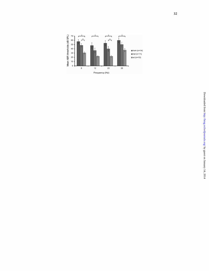
An auditory-evoked brainstem response (ABR) test was used to assess hearing thresholds in mutant
mice (32). We tested 14 homozygous, 11 heterozygous and 10 wild type mice at the age of two
months for ABR response at four frequencies 8, 12, 20 and 26 kHz. We showed that the average ABR
thresholds were elevated by about 30 dB SPL in homozygotes and by about 20 dB SPL in
heterozygotes, when compared with wild type mice, suggesting a conductive hearing loss (Fig. 3). At
all four frequencies the ABR thresholds of the homozygote mice were significantly different from the
wild types (at 8 kHz p = 0.000447; 12 kHz p = 0.003297; 20 kHz p = 0.000486; 26 kHz p =
0.004445). Significant differences were also seen between heterozygotes and wild types at two
frequencies (8 kHz p = 0.003229 and 20 kHz p = 0.023927).
We assessed if there was a sensorineural element to the hearing loss by dissecting inner ears from 2
one-month-old homozygous, heterozygous and wild type mice to examine the structure of the organ
of Corti and the sensory cells. We did not detect any abnormalities in the inner hair cell (IHC) or outer
hair cell (OHC) stereocilia bundles. Scanning electron micrographs showed normal cell and bundle
morphology in the mid (Fig. 4A), apical and basal (data not shown) turns of the cochlea in both
mutants and wild types. In all of the samples we could see well organised rows of OHCs in the organ
of Corti and normal sized IHCs. The histological examination of sagittal sections from the cochlea of
two month old mice also confirms that there were no obvious abnormalities in the cochlea or organ of
Corti of homozygote Tgif mice. We also observed no loss of spiral ganglion neurons (Fig. 4B).
Tgif mice display otitis media by 21 days after birth
Five-, thirteen- and twenty-one-day old as well as one-, two- and four-month-old adult heads were
sectioned to investigate the middle ear phenotype. Five- and thirteen-day-old Tgif/Tgif middle ears
were indistinguishable from wild types (Fig. 5A - D). However, we detected an OM phenotype in the
by guest on January 14, 2014http://hm
g.oxfordjournals.org/D
ownloaded from

9
middle ear of homozygote mice at three weeks. At that time point most of the homozygote mice
(67%, n = 6) had fluid in the middle ear cavity and a thickened epithelial lining in comparison with
wild type (n = 16) controls. There was only one wild type that demonstrated some fluid in the ear. The
ear effusions were cellular, with the presence of polymorphonuclear cells (PMNs) which are the first
responding inflammatory cells that migrate towards the site of inflammation (Fig. 5E - H). By
comparison three of the heterozygote mice at 21 days of age had fluid in one ear and thickened lining
(19%, n = 16) and some of them had a thickened epithelial lining only (37%, n = 16) (Supplementary
Fig. 1).
At one month, four homozygote mice had fluid in one ear (37%, n = 11) and only two had some fluid
in both ears (18%, n = 11). Five mice did not have any fluid, but some of these had a thickened
epithelial lining (27% of mice). Surprisingly, the effusions at one month did not appear cellular (Fig.
5I - L). Two of the heterozygote mice had ear fluid and a thickened lining (16%, n = 13) and five had
only a thickened lining (38%, n = 13). None of the wild type mice had OM at this stage (n = 7).
In two-month-old homozygote ears, the inflammation had progressed to a chronic inflammation with
effusion, as evidenced by fluid in the middle ear cavity along with a thickened epithelial lining. The
effusion content was variable. In some of the homozygote mice the middle ear fluid was more liquid
and in some thickened, more viscous (Fig. 5M,N,Q and R). At two months, five homozygote mice
(42%, n = 12) had unilateral OM, five (42%, n = 12) in both ears in each case associated with
thickening of the mucoperiosteum. Two homozygotes (17%, n = 12) did not have effusions, nor
thickened epithelial lining. Both of these mice showed relatively normal ABR thresholds. By
comparison two of the heterozygote mice had ear fluid (20%, n = 10) and three had their epithelial
lining thickened (30%, n = 10). Heterozygotes with ear fluid and heterozygotes with thickened lining
had elevated ABR thresholds. None of the wild type mice (n = 7) had OM. In homozygotes the
effusion was again cellular, and staining with F4/80 antibody confirmed the presence of macrophages
in the ear fluid at this stage (Fig. 5O). We also studied the OM phenotype of four-month-old
homozygote mice and observed that six of them (60%, n = 10) had unilateral OM, two (20%, n = 10)
had effusion in both ears and two (20%, n = 10) did not have any fluid in the ears. Three of the Tgif/+
by guest on January 14, 2014http://hm
g.oxfordjournals.org/D
ownloaded from

10
mice had fluid in the ears and thickened lining (25%, n = 12) and three had thickened lining only
(25%, n = 12). At both two and four months of age, we observed the appearance of cellular debris-like
aggregates in the ear effusions of the homozygote (Fig. 5P,W). In addition at four months we
observed polyps arising from the middle ear mucosa of the homozygote mice (Fig. 5V). The wild type
mice (n = 10) at this stage did not display OM.
To quantify the mucosal thickness of the middle ears at different stages we examined the histological
sections and took measurements at five equidistant points along a 1mm length of the mucosa. Four to
five homozygous and wild type mice were analysed at each stage. There were significant differences
between mutant and wild type in the thickness of the epithelial lining at each time point (21 days after
birth p = 0.004713728; 1 month p = 0.000598192; 2 months p = 0.0000443631; 4 months p =
0.004381456; see Fig. 6A).
In addition to histological analysis of the middle ear fluid, we analysed the cell types within the ear
fluids by Fluorescence activated cell sorting analysis (FACS) (Fig. 6B). Two antibodies were used for
the study. Gr-1 recognises granulocytes (PMNs) and monocytes; the other, F4/80, is a marker for
macrophages. The results from the FACS analysis confirmed the results obtained from the
histological study. At 21 days after birth 33.2% of cells were Gr-1 positive and only 1.71% were
F4/80 positive. Examination of ear fluids in histological sections did not reveal other populations of
cells such as lymphocytes so the >60% shortfall in stainable cells in the FACS analysis is probably
attributable to high proportion of apoptotic (Fig. 5F inset) and necrotic cells. At one month the ear
fluids contained few identifiable cells (Fig. 5J), and the percentage of cells positive for Gr-1 (4.4%)
and F4/80 (0.6%) were low (Fig. 6B). The proportion of F4/80 positive cells by FACS was
significantly lower at one month compared to 21 days after birth (p = 0.028). While there was a very
large reduction in Gr-1 cells at one month compared to 21 days after birth, this difference was not
significant. At 2 months, there were again more cells positive for Gr-1 (10.7%), but also more for
F4/80 (2.3%), confirming the result from the immunohistochemistry (IHC) (Fig 5O, Fig. 6B). The
proportion of F4/80 positive cells by FACS was significantly higher at two months compared to one
month (p = 0.026).
by guest on January 14, 2014http://hm
g.oxfordjournals.org/D
ownloaded from
11
Tgif homozygous mice have mucous effusions and elevated levels of cytokines in ear fluids
To study the type of the effusion in the middle ear of the homozygote mice we looked for the
expression of matrix metalloproteinase - 2 (MMP-2). The expression profile of MMPs has been found
to be specific to the type of middle ear effusion (33), with the active form of MMP-2 only found in
mucous effusions of human patients with OM (33). We looked for the expression of MMP-2 using
Western blot and gelatine zymography and detected both pro- and active MMP-2 in ear effusions of
two month old homozygote mice (Fig. 7A,B). The zymography demonstrated high levels of MMP-9
as well, which is specific to mucous effusions (33). The result suggested that the homozygote mice
have a mucous type ear effusion at two months.
This result was also confirmed by histological analysis of middle ears performed with a combined
Alcian blue/Periodic acid-schiff staining method (AB-PAS). Goblet cells are epithelial cells whose
function is to secrete mucin, which dissolves in water to form mucus. We detected a high density of
goblet cells, stained in purple by AB-PAS, amongst other cells in the epithelium of the middle ear
cavity of Tgif homozygote mice (Fig. 7C).
VEGF (or VEGF-A) is a member of the vascular permeability factor/vascular endothelial growth
factor (VPF/VEGF) family of cytokines that are critical for vasculogenesis and pathological and
physiologic angiogenesis. The low level of VEGF expression in normal tissues and the
overexpression in pathological angiogenesis is regulated by many factors, including hypoxia (34).
Occurrence of hypoxia and elevated levels of the VEGF protein has recently been reported for Jeff
and Junbo mice (11). For this reason, we assessed the level of VEGF protein in ear fluids, comparing
it to serum levels. VEGF protein was detected in all serum samples and there was no significant
difference between genotypes (Fig. 8A). Levels are very similar to the previously reported
concentration of VEGF in sera of Jeff heterozygote mice (78pg/ml). However, the level of the protein
in ear fluids of Tgif homozygote mice was significantly higher (between 356.7-25,923.1pg/ml) and
was detected in 8 samples out of 10 (two of the samples had levels below the detectable range). VEGF
by guest on January 14, 2014http://hm
g.oxfordjournals.org/D
ownloaded from

12
protein levels in Tgif/Tgif ear effusions were 66.6 fold elevated compared to Tgif/Tgif serum (Fig.
8A), similar to Jeff mice (74 fold) though less than Junbo mice (335 fold) (11).
TNF-α and Il-1β are cytokines involved in mediating inflammatory reactions. In wild type,
heterozygote and homozygote Tgif mice, TNF-α protein was detectable in all serum samples (except
one) in very low concentrations (n = 28, range 1.2-18.3pg/ml). The average concentration of TNF-α in
serum of homozygote mice was found to be 5.8pg/ml, heterozygote mice 5pg/ml and wild type
5.2pg/ml. Elevated concentrations of TNF-α was detected in ear fluids of homozygote mice (n = 11,
range between 480.9-3425.6pg/ml) which was 262 fold elevated compared to Tgif/Tgif serum
(Fig.8B). TNF-α levels are also highly raised in the ear fluids of Jeff (50 fold) and Junbo mice (78
fold).
Il-1β protein was detectable in all blood samples (n=28, range 1.8-24.6pg/ml) with no significant
difference between the genotypes and was also elevated (78 fold) in the ear effusions of Tgif/Tgif
homozygtoes (n = 10, range between 91.8-1748.7pg/ml) (Fig 8C). Il-1β levels are raised in Jeff (3
fold) and Junbo mice (26 fold).
The mutation in Tgif affects the TGFβ signalling pathway in the epithelial cells of the middle
ears
Finally, we proceeded to investigate the action of TGIF on TGFβ signalling in the epithelial cells of
the mouse middle ear at the time when Tgif mutant mice start to develop OM. We performed
immunohistochemical staining using TGIF, pSmad2, p21 and PML antibodies on ear sections from
three-week-old mice. TGIF protein was localised in the nucleus of middle ear cavity epithelial cells in
wild type ears, but as expected not in homozygous mutant ears. Phosphorylated Smad2 was also
localised in the epithelial cells. We detected a clear difference between the number of epithelial cells
positive for the pSmad2 antibody in wild type and homozygous mutant middle ears (Fig. 9). We
counted epithelial cells in eight mutant and four wild type ears and found a significant difference
between the percentage of cells positive for pSmad2 (p = 0.00029036). In wild type middle ear
by guest on January 14, 2014http://hm
g.oxfordjournals.org/D
ownloaded from

13
activated Smad2 was localised in 34.87% of the epithelial cells compared to 15.58% in the
homozygous middle ear. We also performed IHC with a p21 antibody to examine localisation of p21
in wild type and mutant middle ear epithelial cells and, consistent with raised levels of pSmad2,
detected more cells positive for p21 and much stronger staining in wild type cells (Fig. 9). Finally, we
examined labelling patterns of cPML, an activator of TGFβ signalling which is sequestered in the
nucleus by binding to TGIF. In wild type, the bulk of the labelling was observed in the cytoplasm, and
was unaffected in the homozygous mutant. We conclude from these observations that TGIF induces
TGFβ mediated transcriptional regulation in the epithelial cells of the mouse middle ear at time when
the mutant mice develop OM, and that this regulation does not involve significant changes in the
distribution of the cPML activator protein.
DISCUSSION
Our study has identified the Tgif knockout mouse as a novel model of OM, providing further insights
into the genetic basis for chronic OM. Tgif mutants have a craniofacial defect and reduced hearing by
weaning age. Mice homozygous for Tgif do not have any obvious inner ear abnormalities, but
demonstrate raised auditory thresholds detected by ABR. OM is the major cause of hearing
impairment in Tgif homozygotes. The histological analysis revealed that they develop OM by 21 days
after birth. At that stage, inflammatory cells are detectable in the middle ear cavity of the mutant
mice. By two months, the inflammation had progressed to a chronic state accompanied by a thickened
middle ear epithelial lining and the presence of inflammatory cells and macrophages within the
middle ear space. The inflammatory cell populations varied over time and this may reflect episodes of
bacterial infection and clearance. Eustachian tube angle and morphology in homozygote mutants was
normal ruling out Eustachian tube dysfunction as a contributory factor to the OM that develops. While
Tgif homozygotes demonstrate a highly penetrant OM, our analysis of heterozygotes reveals chronic
OM but at a markedly lower frequency. Thus, Tgif is a semi-dominant mutation of OM.
by guest on January 14, 2014http://hm
g.oxfordjournals.org/D
ownloaded from

14
Previously, we have identified Fbxo11 and Evi1 as the genes underlying two other mouse models of
OM: Jeff and Junbo (5,6). Both of these models are also associated with defects in the regulation of
TGFβ signalling. Jeff has a chronic proliferative otitis media (9) and it was shown that Fbxo11 affects
TGFβ signalling by regulating the levels of phosphoSmad2 in the epithelial cells of palatal shelves,
eyelids and airways of the lungs (7). Mice heterozygous for Evi1 have a hearing loss due to chronic
suppurative OM (6). Evi1 is a zinc-finger protein, which represses TGFβ signalling by interacting
with the MH2 domain of Smad3 (10). However, for the first time we have identified OM arising from
a lesion in a protein within the TGFβ signalling pathway and directly involved in its regulation and
control.
Recently, the role of hypoxia and HIF-mediated VEGF has been highlighted as a playing a key role in
the pathogenesis of OM in the Junbo and Jeff mutants (11). Protein levels of VEGF, as well as the
inflammatory cytokines, modulators of HIF-1α, are significantly elevated in middle ear fluids in the
Junbo and Jeff mutants. There is considerable cross-talk between TGFβ and HIF-1α pathways and, for
example, Smad3 and HIF-1α cooperate with TGFβ to induce VEGF expression (35,36). We might
therefore expect TGFβ pathway mutants to impact upon the middle ear’s response to the hypoxia that
develops in chronic OM. It is striking therefore that the elevation of protein titers of VEGF, IL-1β and
TNFα that were observed in the Junbo and Jeff mutants are also found in the Tgif mutant. Overall,
both the OM phenotype in Tgif, along with the molecular pathogenesis observed in the middle ear,
provides considerable support to the view that defects in TGFβ signalling may impact generally on
susceptibility to chronic OM.
TGIF is implicated in the regulation of TGFβ signalling through a number of routes. Initially it was
found that TGIF can inhibit the signalling pathway but more recently there is evidence that it can also
induce the pathway. TGIF can repress transcription by recruiting to SMAD2 a corepressor containing
HDACs (37), or alternatively CtBP (carboxyl terminus binding protein) (38) to an activated Smad
complex. TGIF may also inhibit TGF-β signalling by participating in the degradation of Smad2. TGIF
can bind to TI1UL and this interaction allows the ubiquitin ligase to target Smad2 for degradation
(20). Recently, it has been shown that TGIF may inhibit TGFβ signalling independent of its
by guest on January 14, 2014http://hm
g.oxfordjournals.org/D
ownloaded from

15
association with Smads. The inhibition involves interaction with cPML in the nucleus thereby
suppressing the phosphorylation of Smad2 by the TGFβ receptor (19). The interaction between TGIF
and cPML can be influenced by PCTA as PCTA competes with cPML for binding with TGIF and
reverses the inhibitory activity of TGIF (21,22). PCTA binding to TGIF causes the release of cPML
from the nucleus to the cytoplasm where it binds to the TGFβ receptors and promotes the
phosphorylation of Smad2. Induction of PCTA can enhance the ability of TGFβ to induce expression
of endogenous ADAM12 and p21, two TGFβ target genes. In addition, PCTA depletion suppresses
their expression and can also blunt the growth inhibitory action of TGFβ (21).
We have shown that TGIF is localised in the epithelial cells of the middle ear of wild type mice at the
time when the Tgif mutant begins to develop OM. Moreover, pSmad2 was also localised in the same
cells. In the Tgif homozygous mutant, as expected there are no epithelial cells positive for TGIF and
significantly fewer cells positive for pSmad2. This suggests that TGIF is regulating the TGFβ
signalling pathway by promoting the phosphorylation of Smad2 in middle ear epithelia. Indeed,
consistent with this, we have observed down regulation of the TGFβ target gene, p21, in the
homozygous mutant. It is not clear that these effects in the mutant are mediated by cPML as we have
not observed any changes to levels of cPML that would account for the observed down regulation of
p21 in the Tgif mutant. It is possible that changes to TGFβ pathway activity occur via another route
not involving PCTA and cPML.
In conclusion, the discovery of an OM mouse model with a mutation in a gene regulating the TGFβ
signalling pathway highlights the role of this pathway in the genetic predisposition to OM. Indeed,
two candidate gene studies (3,39) have demonstrated that FBXO11, a regulator of TGFβ signalling, is
significantly associated with chronic and recurrent OM in three independent cohorts. Recently, a
GWAS analysis of association with OM in the Western Australian Pregnancy Cohort (Raine) study
identified 32 regions with significant evidence of association, and pathway analysis demonstrated a
connection between top candidates and the TGFβ pathway (40). The identification of the Tgif mutant
provides support for the role of TGFβ signalling and its effects on responses to hypoxia in the
by guest on January 14, 2014http://hm
g.oxfordjournals.org/D
ownloaded from

16
chronically inflamed middle ear. Moreover, it provides a new candidate gene to explore the genetic
contributions to chronic and recurrent OM in the human population.
MATERIALS AND METHODS
Mice
Tgif mutant mice were imported from The Jackson Laboratory. They were backcrossed onto a
C57BL/6J background for at least eight generations. The colony was then maintained on a C57BL/6J
background and genotyped as previously described (28). Mice of all three possible genotypes were
produced by intercrossing heterozygous Tgif deficient mice.
Histology
Mouse adult 5, 13 and 21 day old; 1, 2 and 4 month old heads from wild type, heterozygote and
homozygote for Tgif mice were fixed in 10% buffered formaldehyde, decalcified in Kristenson fluid
for four days and embedded in paraffin following routine procedures. Three-micrometer-thick sections
were obtained, de-paraffinized in xylene substitute and rehydrated via a graded ethanol. For
morphological observations, sections were stained with haematoxylin and eosin stain. Goblet cells
were identified by a combined AB-PAS schiff staining method.
Clickbox
One-month and two-month old mice were tested for a hearing defect using a clickbox (Institute of
hearing Research, Nottingham, UK), that generates a brief 20 kHz soundburst at 90 dB SPL, for a
presence or a lack of Preyer reflex as previously described (32).
Auditory-evoked brainstem response (ABR) analysis
Two-month-old wild type, heterozygote and homozygote mice, at least ten from each group, were
used for this hearing test. The mice were anaesthetised and placed in an audiometric chamber. The
acoustic stimuli was delivered to the right ear and the test was analysed as previously described (32).
by guest on January 14, 2014http://hm
g.oxfordjournals.org/D
ownloaded from
17
After the test the mice were sacrificed by overdose of anaesthetic, the heads were skinned and used
for further analysis.
X-ray analysis
Radiography was performed using a Faxitron Mx-20 DC-4 specimen X-ray System. Image analysis
programme ImageJ was used to conduct measurements of the two-month-old skulls. To study the
craniofacial defect of the Tgif mutant mice we compared the ratio between the full dorsal length of the
skull and the dorsal length of the nasal bone of ten wild type, heterozygote and homozygote mice.
Scanning Electron Microscopy
Inner ears, dissected from two one-month-old wild type, heterozygote and homozygote mice were
fixed, washed and decalcified as previously described (41). After the decalcification, the organ of
Corti was exposed, the ears dehydrated in ethanol, dried, sputter coated with gold and then viewed on
a JEOL 6010 LV scanning electron microscope under high vacuum conditions.
Western blot
Total protein was extracted from the ear effusions of two-month-old Tgif homozygote mice. Briefly,
the effusions were collected in phosphate buffered saline, containing a cocktail of protease inhibitors
(cOmplate mini, Roche), centrifuged at 10 000 x g for 30min at 4oC. The albumin was removed from
the supernatants using Qproteome Murine Albumin depletion kit (QIAGEN). The samples were
subjected to 4-12% SDS NuPAGE (Invitrogen) and immunoblotted onto nitrocellulose membrane
(Invitrogen). The antibody for MMP2 (Abcam) was used in 1:500 dilution. ECL Plus (GE Healthcare)
was used as detection system. Have to repeat the western with a new antibody!
Zymography
The enzyme activity of MMP-2 was assessed by zymography using gelatine impregnated gels. Total
protein from ear effusion, prepared the same way as for the Western blots, was loaded onto 10% SDS-
PAGE gels containing gelatine (Invitrogen). After electrophoresis the gels were incubated in 1X
by guest on January 14, 2014http://hm
g.oxfordjournals.org/D
ownloaded from

18
Zymogram Renaturing Buffer (Invitrogen) for 30 min, followed by two incubations in 1X Zymogram
Developing Buffer (Invitrogen) – the first one for 30 min at room temperature and the second
overnight at 37oC. The gels were stained with SimplyBlue Safestain (Invitrogen) and destained in
water.
Blood and ear effusion collection for Vegf, TNF-α and Il-1β protein assays
Blood from 8-10 wild type, heterozygote and homozygote mice was collected into serum-gel clotting
activator tubes (Sarstedt) from the retro-orbital sinus of mice under terminal anaesthesia. The samples
were left to clot for 2 hours at room temperature before centrifuging for 20 min at 2000 x g. The
serum was removed, aliquoted and stored at -20oC until required.
Measured volumes of ear fluids from 10 homozygote mice were transferred in cold phosphate
buffered saline, vortexed and centrifuged for 10 min at 500 x g, 4oC. Supernatant ear fluids were
stored at -80oC until required.
Quantikine mouse VEGF, IL-1β and TNF-α ELISA kits (R&D System) were used to compare the
levels of these three cytokines in the blood and ear fluid of the Tgif mutant mice.
Ear effusion collection for FACS analysis
Ear effusions from homozygote mice age 21 days after birth, 1 and 2 months were collected in 100
microliters cold FACS buffer (1% BSA in PBS) and half of the amount was processed for the
analysis. The samples were washed, blocked and incubated with a mixture of the two antibodies Gr1
(PerCP-Cy 5.5 Rat anti-mouse Ly6G and Ly-6C, BD Pharminogen) and F4/80 (Rat anti mouse mAb
APC conjugated, Invitrogen) in 1:400 dilution for 20 min. After washing, the samples were processed
on BD FACS Cantoll and set to acquire 20000 events in FSC Vs SSC gate with a flow rate of 0.5-1
microliter/ second.
by guest on January 14, 2014http://hm
g.oxfordjournals.org/D
ownloaded from

19
Immunohistochemistry
For immunohistochemical analysis, the avidin–biotin complex (ABC) method was used to look for the
localization of Tgif, pSmad2 and p21 in wild type and mutant mouse middle ears. The sections
through the ears of the three-week-old mice were de-paraffinized and endogenous peroxidase activity
was quenched with 3% hydrogen peroxide in isopropanol for 20 minutes. Vectastain Elite ABC kit
(Vector Laboratories, PK 6101) was used to perform the IHC. The antibodies were as follows: rabbit
polyclonal TGIF (H-172; sc-9084 Santa Cruz Biotechnology), rabbit polyclonal anti-phospho-Smad2
(Ser465/467) (AB3849 Chemicon International) and rabbit polyclonal p21 (C-19; sc-397 Santa Cruz
Biotechnology). The sections were incubated with the antibodies in 1:200 dilutions over night at 4oC.
DAB+ chromogen system (DAKO K3468) was used to develop the specific signals. The slides were
counterstained with haematoxylin.
Data analysis
We used the chi squared test to compare the difference between the observed and the expected
number of the mutant mice from crosses and Fisher Exact tests to test proportions of genotypes. To
evaluate the probability of the calculated chi squared value the chidist function in Excel was used.
Two-tailed t test was used for comparing mean ABR and FACS thresholds. A value of p < 0.05 was
considered significant.
by guest on January 14, 2014http://hm
g.oxfordjournals.org/D
ownloaded from

20
ACKNOWLEDGEMENTS
The authors would like to thank Caroline Barker, Jennifer Corrigan, Adele Seymour and Elizabeth
Darley for histology services, David Shipston, Kate Vowell and Jim Humphreys for necropsy skills,
Paras Pathak, Helen Natukunda and Tertius Hough for the protein assays and the FACS analysis,
Sarah Carter, Andrew Hinton, Lisa Ireson and Lucie Vizor for the technical support and Steve
Thomas and Kevin Glover for preparing the figures. The authors are also grateful to Christopher
Walsh and Jun Shen for the Tgif knockout mice.
CONFLICT OF INTEREST STATEMENT
None declared.
FUNDING
This work was funded by the Medical Research Council, UK.
by guest on January 14, 2014http://hm
g.oxfordjournals.org/D
ownloaded from

21
REFERENCES
1. Casselbrant, M.L., Mandel, E.M., Rockette, H.E., Kurs-Lasky, M., Fall, P.A., Bluestone, C.D.
and Ferrell, R.E. (2004) The genetic component of middle ear disease in the first 5 years of
life. Arch Otolaryngol Head Neck Surg, 130, 273-278.
2. Daly, K.A., Brown, W.M., Segade, F., Bowden, D.W., Keats, B.J., Lindgren, B.R., Levine,
S.C. and Rich, S.S. (2004) Chronic and recurrent otitis media: a genome scan for
susceptibility loci. Am J Hum Genet, 75, 988-997.
3. Segade, F., Daly, K.A., Allred, D., Hicks, P.J., Cox, M., Brown, M., Hardisty-Hughes, R.E.,
Brown, S.D., Rich, S.S. and Bowden, D.W. (2006) Association of the FBXO11 gene with
chronic otitis media with effusion and recurrent otitis media: the Minnesota COME/ROM
Family Study. Arch Otolaryngol Head Neck Surg, 132, 729-733.
4. Rye, M.S., Bhutta, M.F., Cheeseman, M.T., Burgner, D., Blackwell, J.M., Brown, S.D. and
Jamieson, S.E. (2011) Unraveling the genetics of otitis media: from mouse to human and back
again. Mamm Genome, 22, 66-82.
5. Hardisty-Hughes, R.E., Tateossian, H., Morse, S.A., Romero, M.R., Middleton, A.,
Tymowska-Lalanne, Z., Hunter, A.J., Cheeseman, M. and Brown, S.D. (2006) A mutation in
the F-box gene, Fbxo11, causes otitis media in the Jeff mouse. Hum Mol Genet, 15, 3273-
3279.
6. Parkinson, N., Hardisty-Hughes, R.E., Tateossian, H., Tsai, H.T., Brooker, D., Morse, S.,
Lalane, Z., MacKenzie, F., Fray, M., Glenister, P. et al. (2006) Mutation at the Evi1 locus in
Junbo mice causes susceptibility to otitis media. PLoS Genet, 2, e149.
7. Tateossian, H., Hardisty-Hughes, R.E., Morse, S., Romero, M.R., Hilton, H., Dean, C. and
Brown, S.D. (2009) Regulation of TGF-beta signalling by Fbxo11, the gene mutated in the
Jeff otitis media mouse mutant. Pathogenetics, 2, 5.
8. Nolan, P.M., Peters, J., Strivens, M., Rogers, D., Hagan, J., Spurr, N., Gray, I.C., Vizor, L.,
Brooker, D., Whitehill, E. et al. (2000) A systematic, genome-wide, phenotype-driven
mutagenesis programme for gene function studies in the mouse. Nat Genet, 25, 440-443.
by guest on January 14, 2014http://hm
g.oxfordjournals.org/D
ownloaded from

22
9. Hardisty, R.E., Erven, A., Logan, K., Morse, S., Guionaud, S., Sancho-Oliver, S., Hunter,
A.J., Brown, S.D. and Steel, K.P. (2003) The deaf mouse mutant Jeff (Jf) is a single gene
model of otitis media. J Assoc Res Otolaryngol, 4, 130-138.
10. Kurokawa, M., Mitani, K., Irie, K., Matsuyama, T., Takahashi, T., Chiba, S., Yazaki, Y.,
Matsumoto, K. and Hirai, H. (1998) The oncoprotein Evi-1 represses TGF-beta signalling by
inhibiting Smad3. Nature, 394, 92-96.
11. Cheeseman, M.T., Tyrer, H.E., Williams, D., Hough, T.A., Pathak, P., Romero, M.R., Hilton,
H., Bali, S., Parker, A., Vizor, L. et al. (2011) HIF-VEGF pathways are critical for chronic
otitis media in Junbo and Jeff mouse mutants. PLoS Genet, 7, e1002336.
12. Shi, Y. and Massague, J. (2003) Mechanisms of TGF-beta signaling from cell membrane to
the nucleus. Cell, 113, 685-700.
13. Massague, J. and Chen, Y.G. (2000) Controlling TGF-beta signaling. Genes Dev, 14, 627-
644.
14. Tsukazaki, T., Chiang, T.A., Davison, A.F., Attisano, L. and Wrana, J.L. (1998) SARA, a
FYVE domain protein that recruits Smad2 to the TGFbeta receptor. Cell, 95, 779-791.
15. Lin, H.K., Bergmann, S. and Pandolfi, P.P. (2004) Cytoplasmic PML function in TGF-beta
signalling. Nature, 431, 205-211.
16. Bertolino, E., Reimund, B., Wildt-Perinic, D. and Clerc, R.G. (1995) A novel homeobox
protein which recognizes a TGT core and functionally interferes with a retinoid-responsive
motif. J Biol Chem, 270, 31178-31188.
17. Holland, P.W., Booth, H.A. and Bruford, E.A. (2007) Classification and nomenclature of all
human homeobox genes. BMC Biol, 5, 47.
18. Wotton, D., Lo, R.S., Lee, S. and Massague, J. (1999) A Smad transcriptional corepressor.
Cell, 97, 29-39.
19. Seo, S.R., Ferrand, N., Faresse, N., Prunier, C., Abecassis, L., Pessah, M., Bourgeade, M.F.
and Atfi, A. (2006) Nuclear retention of the tumor suppressor cPML by the homeodomain
protein TGIF restricts TGF-beta signaling. Mol Cell, 23, 547-559.
by guest on January 14, 2014http://hm
g.oxfordjournals.org/D
ownloaded from

23
20. Seo, S.R., Lallemand, F., Ferrand, N., Pessah, M., L'Hoste, S., Camonis, J. and Atfi, A.
(2004) The novel E3 ubiquitin ligase Tiul1 associates with TGIF to target Smad2 for
degradation. EMBO J, 23, 3780-3792.
21. Faresse, N., Colland, F., Ferrand, N., Prunier, C., Bourgeade, M.F. and Atfi, A. (2008)
Identification of PCTA, a TGIF antagonist that promotes PML function in TGF-beta
signalling. EMBO J, 27, 1804-1815.
22. Liu, F. (2008) PCTA: a new player in TGF-beta signaling. Sci Signal, 1, pe49.
23. Imoto, I., Pimkhaokham, A., Watanabe, T., Saito-Ohara, F., Soeda, E. and Inazawa, J. (2000)
Amplification and overexpression of TGIF2, a novel homeobox gene of the TALE superclass,
in ovarian cancer cell lines. Biochem Biophys Res Commun, 276, 264-270.
24. Melhuish, T.A., Gallo, C.M. and Wotton, D. (2001) TGIF2 interacts with histone deacetylase
1 and represses transcription. J Biol Chem, 276, 32109-32114.
25. Edwards, M.C., Liegeois, N., Horecka, J., DePinho, R.A., Sprague, G.F., Jr., Tyers, M. and
Elledge, S.J. (1997) Human CPR (cell cycle progression restoration) genes impart a Far-
phenotype on yeast cells. Genetics, 147, 1063-1076.
26. Overhauser, J., Mitchell, H.F., Zackai, E.H., Tick, D.B., Rojas, K. and Muenke, M. (1995)
Physical mapping of the holoprosencephaly critical region in 18p11.3. Am J Hum Genet, 57,
1080-1085.
27. Gripp, K.W., Wotton, D., Edwards, M.C., Roessler, E., Ades, L., Meinecke, P., Richieri-
Costa, A., Zackai, E.H., Massague, J., Muenke, M. et al. (2000) Mutations in TGIF cause
holoprosencephaly and link NODAL signalling to human neural axis determination. Nat
Genet, 25, 205-208.
28. Shen, J. and Walsh, C.A. (2005) Targeted disruption of Tgif, the mouse ortholog of a human
holoprosencephaly gene, does not result in holoprosencephaly in mice. Mol Cell Biol, 25,
3639-3647.
29. Bartholin, L., Powers, S.E., Melhuish, T.A., Lasse, S., Weinstein, M. and Wotton, D. (2006)
TGIF inhibits retinoid signaling. Mol Cell Biol, 26, 990-1001.
by guest on January 14, 2014http://hm
g.oxfordjournals.org/D
ownloaded from
24
30. Taniguchi, K., Anderson, A.E., Sutherland, A.E. and Wotton, D. (2012) Loss of Tgif function
causes holoprosencephaly by disrupting the SHH signaling pathway. PLoS Genet, 8,
e1002524.
31. Bartholin, L., Melhuish, T.A., Powers, S.E., Goddard-Leon, S., Treilleux, I., Sutherland, A.E.
and Wotton, D. (2008) Maternal Tgif is required for vascularization of the embryonic
placenta. Dev Biol, 319, 285-297.
32. Hardisty-Hughes, R.E., Parker, A. and Brown, S.D. (2010) A hearing and vestibular
phenotyping pipeline to identify mouse mutants with hearing impairment. Nature protocols,
5, 177-190.
33. Moon, S.K., Linthicum, F.H., Jr., Yang, H.D., Lee, S.J. and Park, K. (2008) Activities of
matrix metalloproteinases and tissue inhibitor of metalloproteinase-2 in idiopathic
hemotympanum and otitis media with effusion. Acta Otolaryngol, 128, 144-150.
34. Levy, A.P., Levy, N.S., Iliopoulos, O., Jiang, C., Kaplin, W.G., Jr. and Goldberg, M.A.
(1997) Regulation of vascular endothelial growth factor by hypoxia and its modulation by the
von Hippel-Lindau tumor suppressor gene. Kidney Int, 51, 575-578.
35. Sanchez-Elsner, T., Botella, L.M., Velasco, B., Corbi, A., Attisano, L. and Bernabeu, C.
(2001) Synergistic cooperation between hypoxia and transforming growth factor-beta
pathways on human vascular endothelial growth factor gene expression. J Biol Chem, 276,
38527-38535.
36. Jeon, S.H., Chae, B.C., Kim, H.A., Seo, G.Y., Seo, D.W., Chun, G.T., Kim, N.S., Yie, S.W.,
Byeon, W.H., Eom, S.H. et al. (2007) Mechanisms underlying TGF-beta1-induced expression
of VEGF and Flk-1 in mouse macrophages and their implications for angiogenesis. Journal of
leukocyte biology, 81, 557-566.
37. Wotton, D., Lo, R.S., Swaby, L.A. and Massague, J. (1999) Multiple modes of repression by
the Smad transcriptional corepressor TGIF. J Biol Chem, 274, 37105-37110.
38. Melhuish, T.A. and Wotton, D. (2000) The interaction of the carboxyl terminus-binding
protein with the Smad corepressor TGIF is disrupted by a holoprosencephaly mutation in
TGIF. J Biol Chem, 275, 39762-39766.
by guest on January 14, 2014http://hm
g.oxfordjournals.org/D
ownloaded from

25
39. Rye, M.S., Wiertsema, S.P., Scaman, E.S., Oommen, J., Sun, W., Francis, R.W., Ang, W.,
Pennell, C.E., Burgner, D., Richmond, P. et al. (2011) FBXO11, a regulator of the TGFbeta
pathway, is associated with severe otitis media in Western Australian children. Genes and
immunity, 12, 352-359.
40. Rye, M.S., Warrington, N.M., Scaman, E.S., Vijayasekaran, S., Coates, H.L., Anderson, D.,
Pennell, C.E., Blackwell, J.M. and Jamieson, S.E. (2012) Genome-wide association study to
identify the genetic determinants of otitis media susceptibility in childhood. PloS one, 7,
e48215.
41. Mburu, P., Romero, M.R., Hilton, H., Parker, A., Townsend, S., Kikkawa, Y. and Brown,
S.D. (2010) Gelsolin plays a role in the actin polymerization complex of hair cell stereocilia.
PloS one, 5, e11627.
by guest on January 14, 2014http://hm
g.oxfordjournals.org/D
ownloaded from

26
Figure 1. Hydrocephalus and placental defect in Tgif mutant mice.
A. One-month-old Tgif/Tgif homozygous mutant mouse with hydrocephalus (indicated with an
arrow).
B. Placentas from wild type and mutant E13.5 embryos. No differences in size or morphology were
observed.
C. Representative images of H&E stained sections through the centre of the placenta for each
genotype combination. Eight M+/+E+/-, ten M+/- E+/- and nine M-/- E+/- placentas were analysed.
Arrows and arrowheads indicate fetal and maternal blood spaces. The genotype of the mother (M) and
the embryo (E) are indicated in the pictures and the sections
Figure 2. Craniofacial defect of Tgif mutant mice.
A. Size comparison of a male Tgif/Tgif mutant (bottom) and a wild type mouse (top).
B. X-ray analysis of two-month-old skulls from homozygous (hom, Tgif/Tgif), heterozygous (het,
Tgif/+) and wild type (wt, +/+) mice. Lateral X-rays show the craniofacial defect of the mutant mice.
C. Dorsoventral view of a two-month-old wild-type mouse skull showing the measurements used in
this study. Scale bar = 2cm.
D. Graphic comparison of the ratio of the nasal bone length (NL) to the full dorsal length of the skull
(FL) for each Tgif genotype. n: number of mice used in the measurements from each genotype. Bars:
standard error of mean. *: p < 0.05.
E. Ventral view of the middle ear and dissected bulla showing the Eustachian tube of two-month-old
homozygous (hom) and wild type (wt) mice. ET: Eustachian tube; solid double-ended arrows: length
of the tube; open double-ended arrows: width of the tube.
F. Dissected and stained skulls of wild type and homozygous mutant two-month-old mice. Eustachian
tube angle measurements are indicated by white lines. R: right ear; L: left ear.
G. Mean length and width of wild type (wt) and homozygous mutant (hom) Eustachian tubes along
with mean angle between the midline of the skull and the bony part of the left and the right Eustachian
tubes in wild type and homozygous mutants. Bars: standard error of mean.
by guest on January 14, 2014http://hm
g.oxfordjournals.org/D
ownloaded from

27
Figure 3. Deafness phenotype by ABR.
ABR thresholds in the right ears of two-month-old Tgif/Tgif homozygote (hom), Tgif/+ heterozygote
(het) and wild type (wt) mice. n: number of mice in ABR testing for each genotype. Bars: standard
error of mean. *: p < 0.05.
Figure 4. Normal bundle morphology of the hair cells.
A. Scanning electron micrographs showing hair cell morphology in the mid turn of the cochlea of
wild type (on the left) and homozygous Tgiif/Tgif (on the right) mice at two magnifications, age one
month. Three rows of outer hair cells and a row of inner hair cells are observed along the length of the
organ of Corti in both wild type and mutant mice. Normal bundle morphology for both OHCs and
IHCs is observed in wild-type and mutants. Scale bars = 10μm for the top panel and 1μm for other
panels; OHC: outer hair cell; IHC: inner hair cell.
B. Histological analysis of the hair cells in wild type (left panels) and homozygous Tgif/Tgif (right
panels) mice, aged two months. Scale bars = 1mm for the top panel and 100μm for other panels;
OHC: outer hair cell; IHC: inner hair cell; OC: organ of Corti; SG: spiral ganglion, RM: Reissner’s
membrane.
Figure 5. Middle ear histology in Tgif homozygous mice.
Each group of panels consists of representative images (transverse sections) of the middle ears of
homozygous (Tgif/Tgif) and wild type (+/+) mice at different time points. A,B 5 days after birth
(DAB) (mutants analysed n = 8, controls analysed n = 5); C,D 14 DAB (mutants n = 8, controls n =
10); E-H 21 DAB (mutants n = 6, controls n = 16); I-L 1 month old (mutants n = 11, controls n = 7);
M-T 2 months old (mutants n = 12, controls n = 7); U-X 4 months old (mutants n = 10, controls n =
10). F and G enlarged views of E; J and K enlarged views of I; N and P enlarged views of M; R and
S enlarged views of Q; V and W enlarged views of U.
F inset viable and apoptotic PMNs.
by guest on January 14, 2014http://hm
g.oxfordjournals.org/D
ownloaded from

28
O Immunohistochemical staining of a two-month-old homozygous mouse ear section using an F4/80
antibody. Macrophages are indicated with an arrow.
Where indicated the thickness of the epithelial lining is shown with a double arrow. The presence of
cellular aggregates is indicated by an asterisk (P) and an inflamed polyp by an arrowhead (V). A-E, I,
M, Q, U scale bar = 4mm; F, J, N, O, R scale bar = 100μm; G, H, K, L, P, S, T, V, W, X scale bar =
200μm.
Pie charts represent percentage of homozygote mice from different time points with no effusion and
with effusion in one or in both ears.
Figure 6. Thickened epithelial lining and cytology of Tgif homozygous middle ears.
A. Measurements of mucosal thickness of the middle ears of homozygous (hom, Tgif/Tgif) and wild
type (wt, +/+) mice at different stages: 21 DAB (mutant ears measured n = 6, controls measured n =
7); 1 month old (mutants n = 6, controls n = 6); 2 months old (mutants n = 7, controls n = 7); 4
months old (mutants n = 7, controls n = 6). Five measurements of the epithelial lining of each ear
were taken at a spacing of 250μm.
B. FACS analysis of the ear effusions of 21 DAB, one- and two-month old mice. Two antibodies were
used: Gr1 which recognises granulocytes (neutrophils and eosinophils) and monocytes and F4/80
which recognises macrophages. n: number of mice used in the analysis for each time point.
Bars: standard error of mean. *: p < 0.05.
Figure 7. OM phenotype of the two–month-old Tgif mutants.
A. Western blot analysis of total protein from the ear fluid of a two-month-old homozygous
(Tgif/Tgif) mouse before (1) and after (2) removing the albumin from the sample. The blot was probed
with a MMP-2 antibody, which detected both pro- and active forms of MMP-2.
B. Gelatine zymography demonstrating enzyme activity of MMP-2 and MMP-9 in Tgif/Tgif ear
effusions.
by guest on January 14, 2014http://hm
g.oxfordjournals.org/D
ownloaded from

29
C. Histological images of middle ear sections stained with Alcian blue/Periodic acid-schiff, showing
mucus-producing goblet cells, stained in pink, in the epithelium of the middle ear cavity (arrow) of a
homozygous (right panel) and a wild-type (left panel) mouse. Scale bar = 20μm.
Figure 8. Protein composition of middle ear fluids in Tgif mutant mice.
Levels of mouse VEGF (A), TNF-α (B) and IL-1β (C) in serum samples and ear fluid from wild type
(wt, +/+), heterozygote (het, Tgif/+) and homozygote (hom, Tgif/Tgif) mice, determined by
Quantikine Immunoassays. n: number of mice used in the analysis from each genotype. 25% quartile,
median and 75% quartile are indicated on the box plots and the whiskers represent the data range.
Figure 9. Protein expression in middle ear epithelia in Tgif mutant mice.
A. Immunohistochemistry of middle ear sections of wild type (wt, +/+) and homozygous (hom,
Tgif/Tgif) mice, age 21 days after birth, stained with TGIF, phosphoSmad2, and p21 and cPML
antibodies. Arrows indicate nuclear localisation in the epithelial cells. cPML was localised in the
cytoplasm of the cells. No difference in the localisation between wild type and homozygous cells was
observed with the cPML antibody. MEC: middle ear cavity. Scale bars = 1mm and 20μm.
B. Graphic comparison of the percentage of epithelial cells in the middle ear positive for pSmad2.
Bars: standard error of mean. *: p < 0.05.
Table 1. Genotyping data from Tgif mutant crosses
The genotype of the mother (M) and the father (F) are indicated in the table.
The number of the embryos and mice were analysed at different embryonic days (E) and at weaning
age, post-natal day 21 (P21).
by guest on January 14, 2014http://hm
g.oxfordjournals.org/D
ownloaded from

30
by guest on January 14, 2014http://hm
g.oxfordjournals.org/D
ownloaded from

31
by guest on January 14, 2014http://hm
g.oxfordjournals.org/D
ownloaded from
32
by guest on January 14, 2014http://hm
g.oxfordjournals.org/D
ownloaded from

33
by guest on January 14, 2014http://hm
g.oxfordjournals.org/D
ownloaded from

34
by guest on January 14, 2014http://hm
g.oxfordjournals.org/D
ownloaded from

35
by guest on January 14, 2014http://hm
g.oxfordjournals.org/D
ownloaded from

36
by guest on January 14, 2014http://hm
g.oxfordjournals.org/D
ownloaded from

37
by guest on January 14, 2014http://hm
g.oxfordjournals.org/D
ownloaded from

38
by guest on January 14, 2014http://hm
g.oxfordjournals.org/D
ownloaded from

39
by guest on January 14, 2014http://hm
g.oxfordjournals.org/D
ownloaded from