tgfβ signaling enhances wound healing inflammation in post...
TRANSCRIPT
TGFβ signaling enhances wound healing inflammation in post-partum breast cancer
By
Andrew John Williams
Thesis
Submitted to the Faculty of the
Graduate School of Vanderbilt University
In partial fulfillment of the requirements
For the degree of
MASTERS OF SCIENCE
in
Cancer Biology
December, 2014
Nashville, Tennessee
Approved:
Rebecca S. Cook, Ph.D. Jin Chen, M.D., Ph.D.

ii
To my wife Lehanna, Mentor, Family, Friends
And,
As with all endings come new beginnings,
I dedicate this work to Bianca and Zuly Marrero
iii
LIST OF FIGURES
Figure/Table Page
1: Breast cancers of premenopausal women are affected by reproductive
events……………………………………………………………………………………………………………………………………………………….…………..………2
2: TGFβ signaling transiently regulates metastasis during
involution…………………………………………………………………………………………………………………………………………………………...……….4
3:TGFβ transiently enhances TH2 cytokine mRNA exp in
ppBC…………………………………………………………………………………………………………………………………………………….………………………..6
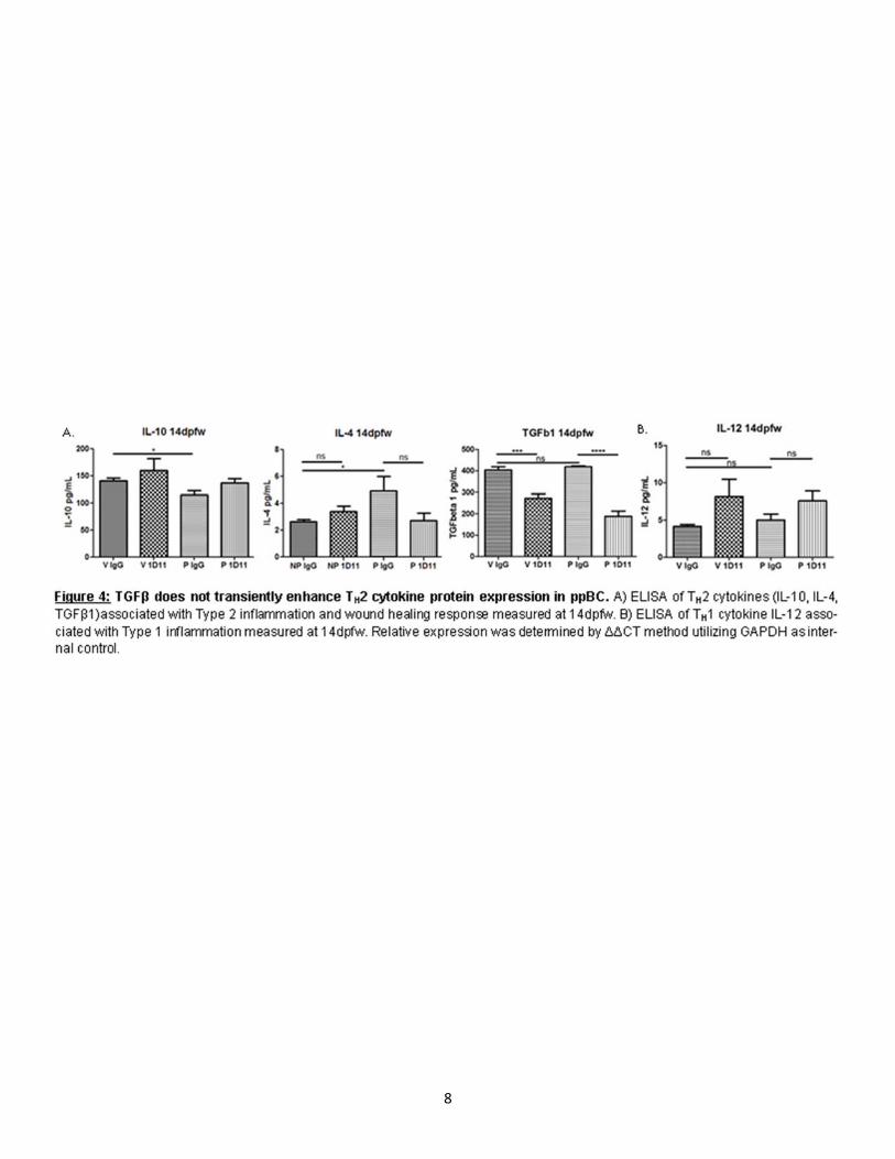
4: TGFβ does not transiently enhance TH2 cytokine expression in
ppBC……………………………………………………..………………………………………………………….…………………………………..……………….………8
5: TGFβ regulates immune cell infiltration and inhibition promotes
prolonged immune infiltration………………………………………………………………………………………………………………..…….……9
LIST OF TABLES
1: qPCR Primer sets…………………………………………………………………………………..……………………………………………………………11
iv
TABLE OF CONTENTS
DEDICATION......................................................................................................................................................................ii
LIST OF FIGURES & TABLES..................................................................................................................................iii
INTRODUCTION..............................................................................................................................................................1
RESULTS...........................................................................................................................................................................3
TGFβ regulates metastasis in ppBC transiently during involution................................................................................................3
TGFβ transiently enhances TH2 cytokine expression in ppBC........................................................................................................5
Transient TGFβ expression negatively regulates immune cell infiltration while inhibition promotes long term immune infiltration............................................................................................................................................................................................5
DISCUSSION....................................................................................................................................................................7
MATERIALS AND METHODS...................................................................................................................................14
Mice.......................................................................................................................................................................................................................14
Western analysis and ELISA.......................................................................................................................................................................14
Histological analysis and IHC.....................................................................................................................................................................14
qRT-PCR.............................................................................................................................................................................................................16
Statistics.............................................................................................................................................................................................................16
Study approval.................................................................................................................................................................................................16
REFERENCES................................................................................................................................................................17
1
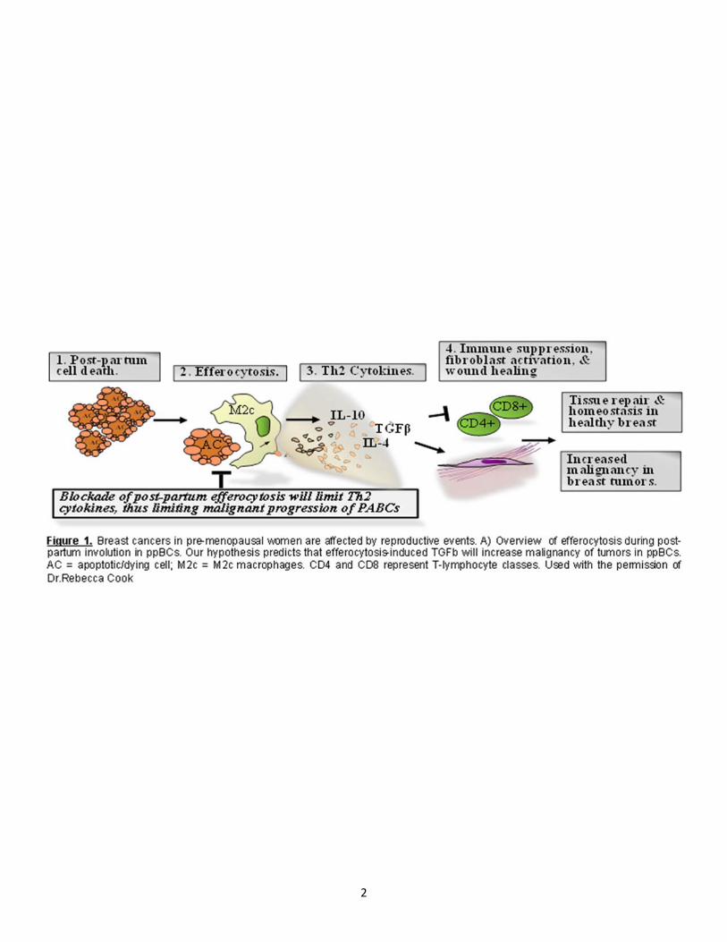
INTRODUCTION The breast is a dynamic tissue throughout the reproductive phases of a woman’s life (puberty, pregnancy, lactation, post-partum involution and post-menopausal involution). Each phase uniquely shapes cancer susceptibility, formation, and progression[1]. Although pregnancy at a young age decreases lifetime breast cancer risk[2, 3], the first five years following pregnancy are associated with increased breast cancer risk regardless of the woman’s age, and with even greater risk with increasing age at the woman’s first pregnancy [1, 4-7]. Increasingly, women are postponing child-birth, which may increase the incidence of post-partum breast cancer (ppBC), defined as those breast cancers diagnosed 2-5 years after pregnancy. These ppBCs are distinguishable from those breast cancers that are diagnosed and treated during pregnancy, and which are never exposed to post-partum/post-lactational involution. Currently, ppBC accounts for nearly 25% of all breast cancers in young (pre-menopausal) women [8]. In contrast to breast cancers diagnosed during pregnancy, which correlate with a favorable prognosis, ppBCs are highly aggressive and metastatic even when corrected for molecular breast cancer subtype and age of the woman at diagnosis [4, 8, 9]. Although the molecular mechanisms underlying the increased lethality of post-partum breast cancers are not fully understood, M2 macrophage activity during post-partum mammary involution actively promotes ppBC tumor malignancy [6, 10, 11]. The biological events triggering this shift in mammary macrophage behavior during post-partum involution are unclear. We recently developed two fully immune competent models of ppBC, one which is orthotopically transplantable and one which is spontaneous[12]. Both models recapitulate many aspects of clinical ppBCs, including profoundly increased metastasis, which was not due to alterations in transgenic oncogene expression, tumor latency, or tumor growth rate, but were specific to the mammary microenvironment of post-partum involution, when milk production ceases and widespread cell death eliminates the large population of milk-producing cells[12, 13]. We previously demonstrated that ppBCs exhibit widespread cell death during early post-partum involution, greater than what is seen in tumors harvested at other reproductive stages in parous or nulliparous mice[12]. These dying tumor cells are recognized for clearance by tumor associated macrophages, which engulf the dying tumor cells through a process termed efferocytosis[14]. MerTK, a receptor tyrosine kinase expressed on macrophages, is required for efferocytosis[15, 16]. We recently demonstrated that blockade of MerTK in ppBCs, using genetic MerTK ablation or pharmacologic MerTK kinase inhibition, decreased efferocytosis and decreased ppBCs metastasis to levels seen in nulliparous mice [12]. It will be important to determine the mechanism by which MerTK-mediated efferocytosis enhances tumor metastasis in ppBCs. We and others have shown efferocytosis robustly induces TH2 cytokines [12, 17, 18] including TGF1, a pleiotropic cytokine which suppresses innate and adaptive cytotoxic immunity[19, 20], promotes M2 macrophage polarization[21], and enhances mammary tumor cell motility. We have found that blockade of efferocytosis in ppBCs impairs TGF1 induction [12]. Results have shown cytotoxic activities of macrophages promote expression of TH2 and wound healing cytokines and that correlates to cell death within the post-partum/post-lactational mammary gland, demonstrating that efferocytosis promotes mammary repair and remodeling during post-partum involution[14]. We have applied this knowledge to examine how tumor cell death during post-partum involution affects TGF-mediated tumor progression and metastasis. TGFβ has a key role in recruiting and regulating leukocytes in the TME, including macrophages, neutrophils, natural killer (NK) cells and dendritic cells (DCs) [22]. TGFβ induces neutrophils to produce factors that enhance tumor metastasis, including MMP9 and CXCL1 [23]. NK cells, which harbor potent anti-tumor cytotoxic activity, are neutralized by TGFβ in the TME, preventing their maturation and their ability to recognize tumor cells [24]. TGFβ suppresses production of type I interferons (IFNs) from NK cells and DCs, and prevents IL-12 production from DCs [25]. Type I IFNs and IL-12 support antigen-mediated clonal expansion of CD8+ T-lymphocytes, thus their depletion from the TME decreases the presence of CD8+ T-cells in the TME. TGFβ enhances the presence of TH2-like CD4+ T-lymphocytes and TRegs in the TME. Notably, TGFβ is a critical driver of M2 macrophage polarization [26].
2
3
TH2 cytokine response is often seen in wound healing environments [27, 28] and associated with anti-inflammatory activity, inhibiting infiltration and maturation of cytotoxic T cells [25, 29]. In normal tissue these cytokines are required for the appropriate recruitment of macrophages, angiogenesis, extracellular matrix remodeling, and re-epithelialization of tissues [27, 28]. IL-4, IL-13, and TGFβ proteins have been shown to enhance infiltration of neutrophils, M2 macrophages, and basophils in order to remove debris [27, 28]. TGFβ proteins and IL-10 efficiently inhibit cytotoxic inflammatory responses [27, 28]. However, TH2 cytokine response is also associated with poor prognosis, immune cell polarization and maturation, immune infiltration and metastasis[5, 7, 12, 22, 25, 26, 30-37]. Many cell types have the ability to produce TH2 cytokines within the tumor TME. CD4+ TH2 cells are characterized by their ability to produce TH2 cytokines [38, 39]. Inversely, signaling induced by these cytokines activate signaling pathways, such as the STAT pathways, which regulate methylation and gene expression patterns of naïve T helper cells driving phenotype commitment [40]. Antigen presenting cells such as dendritic cells and macrophages also have the ability to secrete TH2 cytokines when appropriate to mount a specific immune response based on the initiating effector molecules which includes cellular debris [12, 26, 41, 42]. During involution over 80% of secretory mammary epithelium undergoes apoptosis [43] producing a wound healing immune environment which is regulated by efferocytosis by M2 macrophages [6, 12, 44]. Our studies investigate how efferocytosis-induced TGFβ in ppBCs affects tumor leukocyte populations, cytokine expression, and metastasis (Fig. 1). In ppBCs, efferocytosis induces transcription and secretion of TGFβ1[45, 46], a pleiotropic cytokine that suppresses cytotoxic immune responses, and which correlates with decreased survival in breast cancer patients[14, 46-49]. Like TGF1, several other TH2-like cytokines (IL-4, IL-10, IL-13, and others) are similarly up-regulated in ppBCs (Fig. 3,4), and are decreased upon inhibition of efferocytosis [12]. However, several of these cytokines are known to be influenced by TGF in the TME. Thus, it is unclear if these TH2-like cytokines are induced by efferocytosis, per se, or if they are induced by TGF signaling, secondary to efferocytosis. This information is critical for discerning the mechanism by which efferocytosis and/or TGF enhances ppBC metastasis, and will be an important for determining molecular targets for treating patients with ppBC. We have shown that blockade of TGF signaling in ppBCs decreases tumor metastasis (Fig. 2). In contrast, blockade of IL-4 had no impact on ppBC metastasis, underscoring the important role of TGF signaling in efferocytosis-enhanced metastasis [12]. We and others have demonstrated that increased expression of TGF1, or increased TGF receptor type I (TRI) activity, increases mammary tumor cell motility and enhances metastasis[25, 30, 32, 35, 36, 50]. Conversely, inhibition of TGF signaling using antibody inhibitors or kinase inhibitors of TRI or TRII decreases mammary tumor metastasis[25, 30, 35, 51]. This may be due to enhanced tumor cell motility and invasion. Further, TGF may enhance epithelial-mesenchymal transition (EMT) in a fraction of tumor epithelial cells, since several EMT associated genes are TGF-dependent [32, 34, 35, 51, 52]. EMT associated gene products often increase cancer stem cell-like activity, further contributing to the ability of TGF signaling to support tumor metastasis [53, 54].
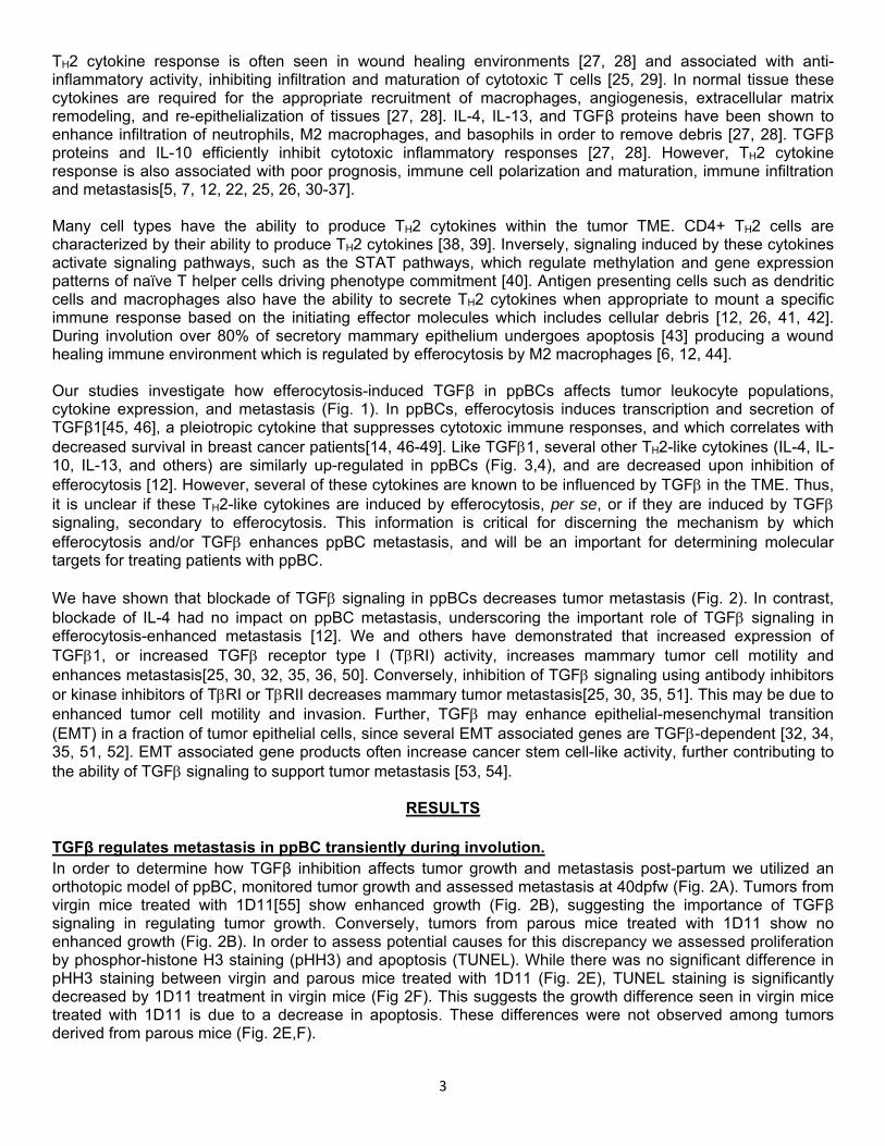
RESULTS TGFβ regulates metastasis in ppBC transiently during involution. In order to determine how TGFβ inhibition affects tumor growth and metastasis post-partum we utilized an orthotopic model of ppBC, monitored tumor growth and assessed metastasis at 40dpfw (Fig. 2A). Tumors from virgin mice treated with 1D11[55] show enhanced growth (Fig. 2B), suggesting the importance of TGFβ signaling in regulating tumor growth. Conversely, tumors from parous mice treated with 1D11 show no enhanced growth (Fig. 2B). In order to assess potential causes for this discrepancy we assessed proliferation by phosphor-histone H3 staining (pHH3) and apoptosis (TUNEL). While there was no significant difference in pHH3 staining between virgin and parous mice treated with 1D11 (Fig. 2E), TUNEL staining is significantly decreased by 1D11 treatment in virgin mice (Fig 2F). This suggests the growth difference seen in virgin mice treated with 1D11 is due to a decrease in apoptosis. These differences were not observed among tumors derived from parous mice (Fig. 2E,F).
4

6
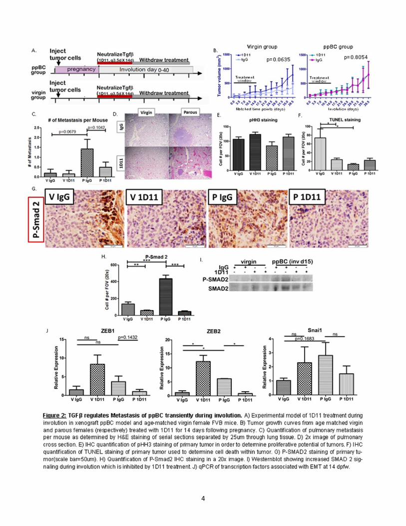
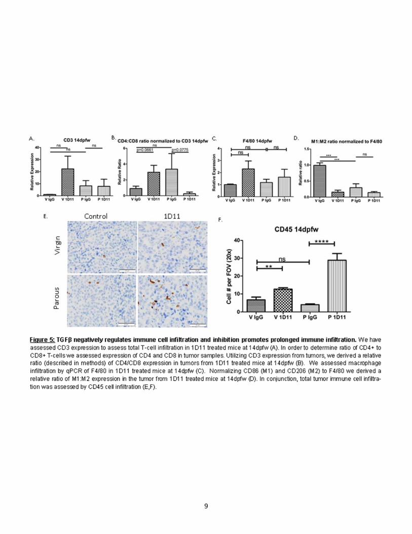
In order to determine how TGFβ regulates metastasis post-partum we assessed intralobular pulmonary metastasis in virgin and parous mice treated with IgG and 1D11. In our model of ppBC, we observed few metastases in age-matched virgin mice than those of parous mice at 40dpfw (Fig. 2C,D). However, we observed a trending increase in metastasis in parity, which is rescued upon 1D11 treatment (Fig. 2C, D). We confirmed the inhibition of canonical TGFβ signaling in 1D11 treated tumors by assessing down-stream Smad 2 signaling by IHC in the primary tumor (Fig. 2G,H) and western blot (Fig. 2I). We also confirmed an increase in Smad 2 in tumors from parous females compared to virgin. We next assessed the expression of genes commonly associated with EMT which has been associated with motility of transformed epithelial cells and metastasis. Interestingly, parous tumors showed a transient increase in EMT associated gene expression relative to tumors from virgin female mice (Fig. 2J,K). However, tumors from female mice treated with 1D11 showed reduced expression of these EMT associated transcription factors (Fig. 2J,K). These data suggest that TGFβ may produce an EMT-like phenotype transiently during post-partum involution in turn enhancing metastatic capacity. TGFβ transiently enhances TH2 cytokine expression in ppBC. It has previously been identified that during post-partum involution, the mammary gland microenvironment expresses gene signatures which are similar to wound healing [6, 33, 56]. Wound healing cyotokines and immune responses are associated with poor prognosis in many cancer types, including breast cancer [6, 8, 12, 31, 44]. Following weaning, TGFβ has been shown to be temporally regulated within 6 hrs in rodent models [57-59]. In order to determine how TGFβ proteins regulate cytokine expression within the TME we assessed TH2 and TH1 cytokine expression in primary tumors of our allograft model. Tumors harvested 14dpfw showed a significant increased TH2 cytokine expression by qPCR (Fig. 3A). 1D11 treatment reduced expression of most TH2 cytokines as compared to what was seen in IgG-treated post-partum tumors, with the exception or TGFβ1 (Fig. 3A). This result could be due to TGFβ1’s inability to regulate its own expression post-partum or TGFβ1 gene expression response to new stimuli, which arise upon 1D11 treatment, although this hypothesis has not yet been tested. While most TH1 cytokines remained relatively unchanged by parity, we observed decreased expression of IFNγ at the mRNA level (Fig. 3B). However, upon 1D11 treatment there is an increase in IFNγ (Fig. 3B). However, these observed changes in cytokine expression were not observed at the protein level as assessed by ELISA (Fig. 4A,B). Transient TGFβ expression negatively regulates immune cell infiltration while inhibition promotes long term immune infiltration. In order to address immune infiltration more directly we assessed total immune infiltration by CD45 Immunohistochemistry (IHC) and CD3 and F4/80 gene expression. We observed no significant difference in intratumoral infiltration of CD45+ immune cells between parous and virgin female mice. However, upon 1D11 treatment there was an increased number of intratumoral CD45 staining cells (Fig.5I,J). Tumors assessed for CD3 and F4/80 mRNA levels show only trending differences in expression between parous controls when compared to 1D11 treated tumors (Fig.5A,C). However, assessment of CD4:CD8 relative ratios normalized to CD3 show significant infiltration of CD4 expression relative to CD8 expression in parous mice (Fig.5B). In parous mice treated with 1D11, this ratio is rescued to a level similar to virgin tumors (Fig.5B). These data suggest balance between CD8+ T-cells and CD4+ T-helper cells are regulated by TGFβ signaling in post-partum tumors and could play a role in promoting metastasis in ppBCs.
Interestingly, comparison of M1 (CD86) and M2 (CD206) marker expression normalized to F4/80 show an increase in M2 ratio in parous mice relative to virgin controls (Fig.5D). Unexpectedly, parous mice were treated with 1D11 this ratio is further enhanced with a higher expression of CD206 in the tumor (Fig.5D).
6
7
DISCUSSION
Premenopausal breast cancers diagnosed during post-partum involution are more frequently diagnosed at metastatic stages as compared with premenopausal breast cancers diagnosed in nulliparous women, pregnant women, or women whose pregnancies occurred more than 10 years before diagnosis [7, 8, 60-62]. Using our allograft model of ppBC we assessed the role of TGFβ signaling during involution in regulating metastasis post-partum. Post-partum involution increased tumor metastasis in this tumor model. During involution approximately 80-90% of secretory mammary epithelium undergoes apoptosis[43]. A widespread burden of apoptotic cells in any healthy or injured tissue requires a mechanism to clear dying cells [12, 63, 64].
MerTK is required for efferocytosis and is known to produce a shift in macrophage phenotypes toward M2-like characteristics and further induce transcription of TH2-like cytokines, including Il10 and Tgfb1 [65, 66]. Other studies demonstrated that MerTK-mediated efferocytosis of dying neutrophils or injured cardiomyocytes, liver cells, or lung epithelial cells induces the expression of wound-healing cytokines that promote resolution of inflammation and tissue repair [63, 67-72].Genetically engineered mouse models demonstrated that IL-4 and TGF-β signaling also enhances M2 macrophage polarization [26, 37, 38], suggesting that MerTK may initiate M2 macrophage polarization through efferocytosis-mediated induction of IL-4 and TGFβ. We have shown that MerTK is necessary for the increased metastasis of ppBCs and for a robust M2-like macrophage presence in post-partum tumors, without affecting total tumor macrophage content [12]. While these results are promising, the mechanisms by which efferocytosis promotes malignant cancer progression remain to be elucidated. In ppBC’s, efferocytosis further produces TH2 cytokines[12] enhancing tumor promoting microenvironment. Similarly, M2-associated cytokines, increase during involution[73].
Results from the present study suggest a potential role of TGFβ signaling expressed in the post-partum tumor microenvironment, which may enhance metastasis of ppBC’s. This may occur through effects on transformed epithelial cells, the surrounding stromal microenvironment, or by regulating cytokine signals within the tumor environment, which correlate with advanced disease and reduced disease free survival in BC patients[6, 39, 74, 75]. Our results suggest TGFβ signaling post-partum may reduce TH2 cytokine and immune response. TH2 cytokine response is associated with wound-healing, poor prognosis, immune cell polarization and maturation, immune infiltration and metastasis[5, 7, 12, 22, 25, 26, 30-33]. While inhibition of other cytokines have shown promising data in regulating the wound healing response and metastasis [37, 76], our data suggests that TGFβ may be a primary driver of TH2 cytokine production and metastasis in ppBC[12].
Results from our study show significantly increased tumor growth in tumors from virgin mice treated with 1D11 which appears to be due to a decrease in apoptotic cells, which has been shown previously[77]. This contrasts to studies in normal mammary gland epithelial cells, which show that upon expression of TGFβ proteins normal mammary epithelium undergo enhanced apoptosis[58, 78]. However, tumors from parous females do not show this enhanced tumor growth upon 1D11 treatment, nor did they show a significant increase in apoptosis upon 1D11 treatment. This data contradicts our previous research showing that there is increased apoptosis in post-partum breast cancers[12]. This could suggest a difference between our spontaneous and allograph models of ppBC which requires elucidation.
In rodent models, M2 macrophages are recruited within 3 days of weaning to the post-partum mammary gland in order to clear apoptotic alveolar cell populations which are no longer required[6]. We previously identified that MerTK, a critical regulator of efferocytosis in physiological post-partum involution [14], required for efferocytosis in post-partum mammary tumors during involution, driving M2 macrophage polarization and wound-healing cytokine production. However, we unexpectedly found that upon neutralization of TGFβ ligands there was an increase in M2 macrophage relative ratio in post-partum tumors compared to M1. Conversely there appears to be an increase in CD4:CD8 relative ratio in tumors treated with 1D11 post-partum. While these results will be further validated by flow cytometry, this suggests that TGFβ signaling in the microenvironment of ppBC may regulate cytokine production and through altering CD4+ T-cell response. This is supported by data which suggests that high ratios of CD4/CD8 or TH2/TH1 T lymphocytes in primary tumors and draining lymph nodes correlate with tumor grade, stage, and patient survival [75, 79, 80]. While evidence suggests that macrophage presence and phenotype is heavily involved in ppBC[5, 31, 44, 73], TGFβ signaling may have a larger role regulating T helper cell infiltration and regulation specifically in ppBC. Similar results
8
9
10
have recently been shown with the inhibition of IL10 in a ppBC model[31]. These data suggest the role the adaptive immune system may play a significant role in controlling antitumor immunity and metastasis of ppBC’s. Further research must be done to define the specific action of TGFβ signaling and how it controls the immune microenvironment of ppBC.
Based on our findings herein, we believe that TGFβ signaling during post-partum involution enhances changes in tumor promoting TH2 cytokine levels (IL-4, IL-10, and IL-13) in post-partum mammary tumors. TGFβ inhibition could also alter immune cell populations within the tumor microenvironment which could play an important role in antitumor immunity by promoting TH2 cells expressing TH2 cytokines and inhibiting cytotoxic T-cells which produce IFNγ. Alternatively, TGFβ could enhance M2 macrophage polarization leading to the production of TH2 cytokines upon efferocytosis. Additional studies will be required to determine how specific populations of immune cells are regulated by TGFβ and if these cells are responsible for directly driving metastasis in ppBC, how these populations regulate cytokine production, or if TGFβ directly regulates transformed epithelial cells within ppBC’s. It is important to address that TGFβ neutralization did not change tumor growth in our model of ppBC but showed a statistical trend towards decreased metastasis when mice were treated for a 2-week period post-partum, providing a potential treatment window to limit metastatic potential of ppBC cases. Additional research performed herein suggests that a transient treatment utilizing a TGFβ inhibitor similar to GM1[81] or LY299[30] may be used to reduce metastatic potential of tumors which arise during involution.
MATERIALS AND METHODS
Mice WT FVB [82], were purchased from The Jackson Laboratory. Female virgin mice were randomized into 4 groups: (a) 1 group that remained virgin treated with IgG, (b) 1 group that remained virgin treated with 1D11, (c) 1 group that was bred from 42 to 44 days of age with WT male mice treated with IgG mAb, and (d) 1 group that was bred from 42 to 44 days of age with WT male mice treated with 1D11 mAb. Pregnancies were timed according to identification of a vaginal semen plug, indicating 0.5 dpc. PyVmT primary mammary tumor cells harvested from MMTV PyVmT polyclonal tumors (previously described in ref. [83]) were collected by trypsinization, suspended 1:1 in growth factor–reduced Matrigel, and 2 × 10⁶ cells were injected into the inguinal mammary fat pads of 45- to 48-day-old virgin or pregnant (3–6 dpc) WT female mice. Mice were monitored daily for tumor formation by manual palpation. Pups were withdrawn at parturition to initiate involution, such that parturition was deemed Inv d0. Mice were maintained until no longer than 104 days of age, corresponding to Inv d40. Where indicated in the figures, mice were treated twice weekly 1D11 or isotype-matched IgGs (10 mg/kg) for 2 weeks beginning at Inv d0. 1D11 (HB-9811) was purchased from ATCC. Hybridomas were cultured, and antibodies were harvested and purified by the Vanderbilt Antibody and Protein Shared Resource.
Western analysis and ELISA. Tissues were homogenized in ice-cold lysis buffer (50 mM Tris, pH 7.4, 100 mM NaF, 120 mM NaCl, 0.5% NP-40, 100 μM Na3VO4, 1X protease inhibitor cocktail [Roche]), sonicated for 10 seconds, and cleared by centrifugation at 4°C, 13,000 g for 5 minutes. Protein concentration was determined using the BCA Protein Assay (Pierce Biotechnology). Proteins were separated by SDS-PAGE, transferred to nitrocellulose membranes, blocked in 3% gelatin in TBS-T (Tris-buffered saline, 0.1% Tween 20), incubated in primary antibody overnight and in HRP-conjugated anti-rabbit or anti-mouse for 1 hour, and developed using ECL substrate (Pierce Biotechnology). The following primary antibodies were used: Smad2 and S465/467 P-Smad2(1:1000 and 1:500 respectively; Cell Signaling Technology). Protein lysates were generated from primary tumors from female mice as described previously. 40ug of protein quantified by BCA were used to quantify murine IL-12p70, TGFbeta1, IL-10 and IL-4 by ELISA (BioLegend) according to the manufacturer’s protocol.
Histological analysis and IHC. Tumors and mammary glands were fixed in 10% formalin (VWR Scientific), paraffin-embedded, sectioned (5 μm), and stained with H&E at the Vanderbilt University Medical Center Translational Pathology Shared Resource. IHC using rabbit antibodies against and developed using the Vectastain kit (Vector Laboratories). TUNEL analysis was performed with the TUNEL kit (Millipore). Phospho-Histone H3 (Santa Cruz Biotechnology Inc.) and S465/467 P-Smad2 (Cell Signaling Technology). Lungs were

11
12
perfused and fixed with 10% formalin (VWR Scientific), paraffin-embedded, and serial sectioned (5um) separated by 25um between sections, and stained with H&E at the Vanderbilt University Medical Center Translational Pathology Shared Resource. Metastasis per mouse were manually assessed in H&E stained serial sections.
qRT-PCR. Whole-tumor RNA was harvested with an RNeasy kit (QIAGEN), and cDNA was synthesized (High Capacity; Applied Biosystems) and amplified using an equal ratio of murine cDNA-specific Oligo-dT and Random Hexamer primers (Integrated DNA Technologies), along with SYBR Green Supermix (Bio-Rad). Primer sets are listed in Table 1. Target gene Ct values were normalized to GAPDH (housekeeping gene) Ct values according to the formula: 2^-[(Cttarget gene-CtGAPDH) Sample A – (Cttarget gene-CtGAPDH)Sample B]. Values were analyzed as the mean in fold differences (± SE, n = 4). Relative ratios were derived utilizing ∆CT values of CD206 (M2), CD86 (M1), CD4 (Thelper), and CD8 (CTL) and normalizing each sample to ∆CT values of F4/80(Macrophage) and CD3 (T-cell) expression respectively.
Statistics. All statistical analysis was carried out using GraphPad Prism software. Kaplan-Meier tumor-free survival analysis was used to assess tumor latency. One-way ANOVA or an unpaired 2-tailed Student’s t test, with a 95% confidence interval, was used to determine significance for all other data. A P value less than 0.05 was considered significant.
Study approval. Mice were maintained in AAALAC-approved animal facilities at Vanderbilt University. The protocols performed herein were reviewed and approved by the IACUC of Vanderbilt University.
16
REFERENCES
1. Martinson, H.A., et al., Developmental windows of breast cancer risk provide opportunities for targeted chemoprevention. Exp Cell Res, 2013.
2. Innes, K.E. and T.E. Byers, First pregnancy characteristics and subsequent breast cancer risk among young women. Int J Cancer, 2004. 112(2): p. 306‐11.
3. Pathak, D.R., Dual effect of first full term pregnancy on breast cancer risk: empirical evidence and postulated underlying biology. Cancer Causes Control, 2002. 13(4): p. 295‐8.
4. Lyons, T.R., P.J. Schedin, and V.F. Borges, Pregnancy and breast cancer: when they collide. J Mammary Gland Biol Neoplasia, 2009. 14(2): p. 87‐98.
5. Schedin, P., Pregnancy‐associated breast cancer and metastasis. Nat Rev Cancer, 2006. 6(4): p. 281‐91. 6. O'Brien, J., et al., Alternatively activated macrophages and collagen remodeling characterize the postpartum
involuting mammary gland across species. Am J Pathol, 2010. 176(3): p. 1241‐55. 7. Schedin, P.J. and C.J. Watson, The complexity of the relationships between age at first birth and breast cancer
incidence curves implicate pregnancy in cancer initiation as well as promotion of existing lesions. Preface. J Mammary Gland Biol Neoplasia, 2009. 14(2): p. 85‐6.
8. Callihan, E.B., et al., Postpartum diagnosis demonstrates a high risk for metastasis and merits an expanded definition of pregnancy‐associated breast cancer. Breast Cancer Res Treat, 2013. 138(2): p. 549‐59.
9. Faupel‐Badger, J.M., et al., Postpartum remodeling, lactation, and breast cancer risk: summary of a National Cancer Institute‐sponsored workshop. J Natl Cancer Inst, 2013. 105(3): p. 166‐74.
10. Lyons, T.R., et al., Postpartum mammary gland involution drives progression of ductal carcinoma in situ through collagen and COX‐2. Nat Med, 2011. 17(9): p. 1109‐15.
11. McDaniel, S.M., et al., Remodeling of the mammary microenvironment after lactation promotes breast tumor cell metastasis. Am J Pathol, 2006. 168(2): p. 608‐20.
12. Stanford, J.C., et al., Efferocytosis produces a prometastatic landscape during postpartum mammary gland involution. J Clin Invest, 2014. 124(11): p. 0‐0.
13. Kreuzaler, P.A., et al., Stat3 controls lysosomal‐mediated cell death in vivo. Nat Cell Biol, 2011. 13(3): p. 303‐9. 14. Sandahl, M., et al., Epithelial cell‐directed efferocytosis in the post‐partum mammary gland is necessary for
tissue homeostasis and future lactation. BMC Dev Biol, 2010. 10: p. 122. 15. Seitz, H.M., et al., Macrophages and dendritic cells use different Axl/Mertk/Tyro3 receptors in clearance of
apoptotic cells. J Immunol, 2007. 178(9): p. 5635‐42. 16. Zizzo, G., et al., Efficient clearance of early apoptotic cells by human macrophages requires M2c polarization and
MerTK induction. J Immunol, 2012. 189(7): p. 3508‐20. 17. Crane, M.J., et al., The monocyte to macrophage transition in the murine sterile wound. PLoS One, 2014. 9(1): p.
e86660. 18. Nishi, C., et al., Tim4‐ and MerTK‐ mediated engulfment of apoptotic cells by mouse resident peritoneal
macrophages. Mol Cell Biol, 2014. 19. Jinushi, M., et al., MFG‐E8‐mediated uptake of apoptotic cells by APCs links the pro‐ and antiinflammatory
activities of GM‐CSF. J Clin Invest, 2007. 117(7): p. 1902‐13. 20. Miyanishi, M., et al., Identification of Tim4 as a phosphatidylserine receptor. Nature, 2007. 450(7168): p. 435‐9. 21. Cook, R.S., et al., MerTK inhibition in tumor leukocytes decreases tumor growth and metastasis. J Clin Invest,
2013. 123(8): p. 3231‐42. 22. Flavell, R.A., et al., The polarization of immune cells in the tumour environment by TGFbeta. Nat Rev Immunol,
2010. 10(8): p. 554‐67. 23. Fridlender, Z.G., et al., Polarization of tumor‐associated neutrophil phenotype by TGF‐beta: "N1" versus "N2"
TAN. Cancer Cell, 2009. 16(3): p. 183‐94. 24. Marcoe, J.P., et al., TGF‐beta is responsible for NK cell immaturity during ontogeny and increased susceptibility to
infection during mouse infancy. Nat Immunol, 2012. 13(9): p. 843‐50. 25. Pickup, M., S. Novitskiy, and H.L. Moses, The roles of TGFbeta in the tumour microenvironment. Nat Rev Cancer,
2013. 13(11): p. 788‐99. 26. Gong, D., et al., TGFbeta signaling plays a critical role in promoting alternative macrophage activation. BMC
Immunol, 2012. 13: p. 31.
16
27. Gause, W.C., T.A. Wynn, and J.E. Allen, Type 2 immunity and wound healing: evolutionary refinement of adaptive immunity by helminths. Nat Rev Immunol, 2013. 13(8): p. 607‐614.
28. Braiman‐Wiksman, L., et al., Novel Insights into Wound Healing Sequence of Events. Toxicologic Pathology, 2007. 35(6): p. 767‐779.
29. de Oliveira, C.M.B., et al., Cytokines and Pain. Brazilian Journal of Anesthesiology, 2011. 61(2): p. 255‐265. 30. Bhola, N.E., et al., TGF‐β inhibition enhances chemotherapy action against triple‐negative breast cancer. J Clin
Invest, 2013. 123(3): p. 1348‐1358. 31. Martinson, H.A., et al., Wound healing‐like immune program facilitates postpartum mammary gland involution
and tumor progression. International Journal of Cancer, 2014: p. n/a‐n/a. 32. Oft, M., K.‐H. Heider, and H. Beug, TGFβ signaling is necessary for carcinoma cell invasiveness and metastasis.
Current Biology, 1998. 8(23): p. 1243‐1252. 33. Stein, T., et al., A Mouse Mammary Gland Involution mRNA Signature Identifies Biological Pathways Potentially
Associated with Breast Cancer Metastasis. Journal of Mammary Gland Biology and Neoplasia, 2009. 14(2): p. 99‐116.
34. Bhowmick, N.A., et al., Transforming Growth Factor‐β1 Mediates Epithelial to Mesenchymal Transdifferentiation through a RhoA‐dependent Mechanism. Molecular Biology of the Cell, 2001. 12(1): p. 27‐36.
35. Muraoka, R.S., et al., Blockade of TGF‐β inhibits mammary tumor cell viability, migration, and metastases. J Clin Invest, 2002. 109(12): p. 1551‐1559.
36. Muraoka‐Cook, R.S., et al., Conditional Overexpression of Active Transforming Growth Factor β1 In vivo Accelerates Metastases of Transgenic Mammary Tumors. Cancer Research, 2004. 64(24): p. 9002‐9011.
37. DeNardo, D.G., et al., CD4+ T Cells Regulate Pulmonary Metastasis of Mammary Carcinomas by Enhancing Protumor Properties of Macrophages. Cancer Cell, 2009. 16(2): p. 91‐102.
38. DeNardo, D.G., et al., Leukocyte complexity predicts breast cancer survival and functionally regulates response to chemotherapy. Cancer Discov, 2011. 1(1): p. 54‐67.
39. Teschendorff, A.E., et al., Improved prognostic classification of breast cancer defined by antagonistic activation patterns of immune response pathway modules. BMC Cancer, 2010. 10: p. 604.
40. Hirahara, K., et al., Mechanisms underlying helper T‐cell plasticity: Implications for immune‐mediated disease. Journal of Allergy and Clinical Immunology, 2013. 131(5): p. 1276‐1287.
41. León, B., A. Ballesteros‐Tato, and F.E. Lund, Dendritic Cells and B Cells: Unexpected Partners in Th2 Development. The Journal of Immunology, 2014. 193(4): p. 1531‐1537.
42. Galdiero, M.R., et al., Tumor associated macrophages and neutrophils in tumor progression. Journal of Cellular Physiology, 2013. 228(7): p. 1404‐1412.
43. Jindal, S., et al., Postpartum breast involution reveals regression of secretory lobules mediated by tissue‐remodeling. Breast Cancer Res, 2014. 16(2): p. R31.
44. O'Brien, J., et al., Macrophages are crucial for epithelial cell death and adipocyte repopulation during mammary gland involution. Development, 2012. 139(2): p. 269‐275.
45. Lemke, G. and C.V. Rothlin, Immunobiology of the TAM receptors. Nat Rev Immunol, 2008. 8(5): p. 327‐36. 46. Rothlin, C.V. and G. Lemke, TAM receptor signaling and autoimmune disease. Curr Opin Immunol, 2010. 22(6): p.
740‐6. 47. Clarkson, R.W., et al., Gene expression profiling of mammary gland development reveals putative roles for death
receptors and immune mediators in post‐lactational regression. Breast Cancer Res, 2004. 6(2): p. R92‐109. 48. Stein, T., et al., Involution of the mouse mammary gland is associated with an immune cascade and an acute‐
phase response, involving LBP, CD14 and STAT3. Breast Cancer Res, 2004. 6(2): p. R75‐91. 49. Fadok, V.A., et al., Macrophages that have ingested apoptotic cells in vitro inhibit proinflammatory cytokine
production through autocrine/paracrine mechanisms involving TGF‐beta, PGE2, and PAF. J Clin Invest, 1998. 101(4): p. 890‐8.
50. Muraoka‐Cook, R.S., et al., Activated type I TGF[beta] receptor kinase enhances the survival of mammary epithelial cells and accelerates tumor progression. Oncogene, 2005. 25(24): p. 3408‐3423.
51. Bakin, A.V., et al., Phosphatidylinositol 3‐Kinase Function Is Required for Transforming Growth Factor β‐mediated Epithelial to Mesenchymal Transition and Cell Migration. Journal of Biological Chemistry, 2000. 275(47): p. 36803‐36810.
16
52. Moustakas, A. and P. Heldin, TGFβ and matrix‐regulated epithelial to mesenchymal transition. Biochimica et Biophysica Acta (BBA) ‐ General Subjects, 2014. 1840(8): p. 2621‐2634.
53. Martin, T., et al., Expression of the Transcription Factors Snail, Slug, and Twist and Their Clinical Significance in Human Breast Cancer. Annals of Surgical Oncology, 2005. 12(6): p. 488‐496.
54. Elloul, S., et al., Snail, Slug, and Smad‐interacting protein 1 as novel parameters of disease aggressiveness in metastatic ovarian and breast carcinoma. Cancer, 2005. 103(8): p. 1631‐1643.
55. Edwards, J.R., et al., Inhibition of TGF‐β signaling by 1D11 antibody treatment increases bone mass and quality in vivo. Journal of Bone and Mineral Research, 2010. 25(11): p. 2419‐2426.
56. Hughes, K., et al., Conditional deletion of Stat3 in mammary epithelium impairs the acute phase response and modulates immune cell numbers during post‐lactational regression. The Journal of Pathology, 2012. 227(1): p. 106‐117.
57. Robinson, S.D., A.B. Roberts, and C.W. Daniel, TGF beta suppresses casein synthesis in mouse mammary explants and may play a role in controlling milk levels during pregnancy. The Journal of Cell Biology, 1993. 120(1): p. 245‐251.
58. Nguyen, A.V. and J.W. Pollard, Transforming growth factor beta3 induces cell death during the first stage of mammary gland involution. Development, 2000. 127(14): p. 3107‐3118.
59. Bierie, B., et al., TGF‐β promotes cell death and suppresses lactation during the second stage of mammary involution. Journal of Cellular Physiology, 2009. 219(1): p. 57‐68.
60. Schedin, P., Pregnancy‐associated breast cancer and metastasis. Nat Rev Cancer, 2006. 6(4): p. 281‐291. 61. Schedin, P. and C. Watson, Preface. Journal of Mammary Gland Biology and Neoplasia, 2009. 14(2): p. 85‐86. 62. Faupel‐Badger, J.M., et al., Postpartum Remodeling, Lactation, and Breast Cancer Risk: Summary of a National
Cancer Institute–Sponsored Workshop. Journal of the National Cancer Institute, 2013. 105(3): p. 166‐174. 63. Vandivier, R.W., P.M. Henson, and I.S. Douglas, Burying the dead*: The impact of failed apoptotic cell removal
(efferocytosis) on chronic inflammatory lung disease. Chest, 2006. 129(6): p. 1673‐1682. 64. Erwig, L.P. and P.M. Henson, Clearance of apoptotic cells by phagocytes. Cell Death Differ, 2007. 15(2): p. 243‐
250. 65. Fadok, V.A., et al., Macrophages that have ingested apoptotic cells in vitro inhibit proinflammatory cytokine
production through autocrine/paracrine mechanisms involving TGF‐beta, PGE2, and PAF. J Clin Invest, 1998. 101(4): p. 890‐898.
66. Huynh, M.‐L.N., V.A. Fadok, and P.M. Henson, Phosphatidylserine‐dependent ingestion of apoptotic cells promotes TGF‐β1 secretion and the resolution of inflammation. J Clin Invest, 2002. 109(1): p. 41‐50.
67. Thorp, E., M. Subramanian, and I. Tabas, The role of macrophages and dendritic cells in the clearance of apoptotic cells in advanced atherosclerosis. Eur J Immunol, 2011. 41(9): p. 2515‐8.
68. Filardy, A.A., et al., Proinflammatory clearance of apoptotic neutrophils induces an IL‐12(low)IL‐10(high) regulatory phenotype in macrophages. J Immunol, 2010. 185(4): p. 2044‐50.
69. Wan, E., et al., Enhanced efferocytosis of apoptotic cardiomyocytes through myeloid‐epithelial‐reproductive tyrosine kinase links acute inflammation resolution to cardiac repair after infarction. Circ Res, 2013. 113(8): p. 1004‐12.
70. Choi, J.Y., et al., Upregulation of Mer receptor tyrosine kinase signaling attenuated lipopolysaccharide‐induced lung inflammation. J Pharmacol Exp Ther, 2013. 344(2): p. 447‐58.
71. Gautier, E.L., et al., Gene‐expression profiles and transcriptional regulatory pathways that underlie the identity and diversity of mouse tissue macrophages. Nat Immunol, 2012. 13(11): p. 1118‐28.
72. Patin, E., et al., Genome‐wide association study identifies variants associated with progression of liver fibrosis from HCV infection. Gastroenterology, 2012. 143(5): p. 1244‐52 e1‐12.
73. O'Brien, J., et al., Alternatively Activated Macrophages and Collagen Remodeling Characterize the Postpartum Involuting Mammary Gland across Species. The American Journal of Pathology, 2010. 176(3): p. 1241‐1255.
74. Kristensen, V.N., et al., Integrated molecular profiles of invasive breast tumors and ductal carcinoma in situ (DCIS) reveal differential vascular and interleukin signaling. Proc Natl Acad Sci U S A, 2012. 109(8): p. 2802‐7.
75. Faghih, Z., et al., Immune profiles of CD4+ lymphocyte subsets in breast cancer tumor draining lymph nodes. Immunol Lett, 2014. 158(1‐2): p. 57‐65.
76. Martinson, H.A., et al., Wound healing‐like immune program facilitates postpartum mammary gland involution and tumor progression. Int J Cancer, 2014.
16
77. Ehata, S., et al., Transforming Growth Factor‐β Promotes Survival of Mammary Carcinoma Cells through Induction of Antiapoptotic Transcription Factor DEC1. Cancer Research, 2007. 67(20): p. 9694‐9703.
78. Kordon, E.C., et al., Ectopic TGFβ1 Expression in the Secretory Mammary Epithelium Induces Early Senescence of the Epithelial Stem Cell Population. Developmental Biology, 1995. 168(1): p. 47‐61.
79. Bates, G.J., et al., Quantification of regulatory T cells enables the identification of high‐risk breast cancer patients and those at risk of late relapse. J Clin Oncol, 2006. 24(34): p. 5373‐80.
80. Kohrt, H.E., et al., Profile of immune cells in axillary lymph nodes predicts disease‐free survival in breast cancer. PLoS Med, 2005. 2(9): p. e284.
81. Araujo‐Jorge, T.C., et al., The TGF‐[beta] Pathway as an Emerging Target for Chagas Disease Therapy. Clin Pharmacol Ther, 2012. 92(5): p. 613‐621.
82. Scott, R.S., et al., Phagocytosis and clearance of apoptotic cells is mediated by MER. Nature, 2001. 411(6834): p. 207‐211.
83. Cook, R.S., et al., ErbB3 ablation impairs PI3K/Akt‐dependent mammary tumorigenesis. Cancer Res, 2011. 71(11): p. 3941‐51.