our 5 major sensory systems vision - the detection of light olfaction- (sense of smell) the...
TRANSCRIPT
OUR 5 MAJOR SENSORY SYSTEMS
Vision - the detection of light
Olfaction- (sense of smell) the detection of small molecules in the air
Taste or Gustation- the detection of selected organic compounds and ions by the tongue
Hearing-The detection of sound (or pressure wave in the air)
Touch- the detection of changes in pressure, temp. and other factors by the skin
S E N S O R Y S Y S T E M S
When fully adapted to darkness our eyes allow us to sense very low levels of light, down to a limit of less than 10 photons.
With more light we are able to distinguish millions of colors.
Through our senses of smell and taste we are able to detect thousands of chemicals
and sort them into distinct categories

Each of these primary sensory systems contains specialized sensory neurons that transmit nerve
impulses to the CNS
In the CNS theses signals are processed and combined with other information to
yield a perception that may trigger a change in behavior.
By these means, our senses allow us to detect changes in our environments and
adjust our behavior appropriately
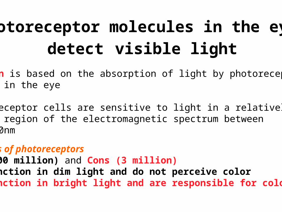
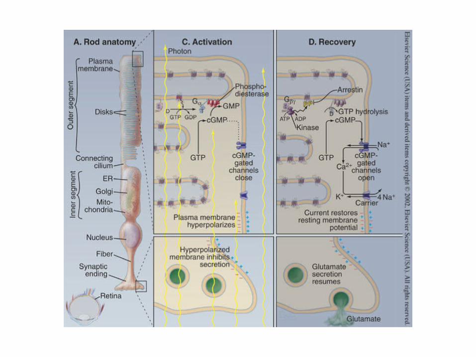
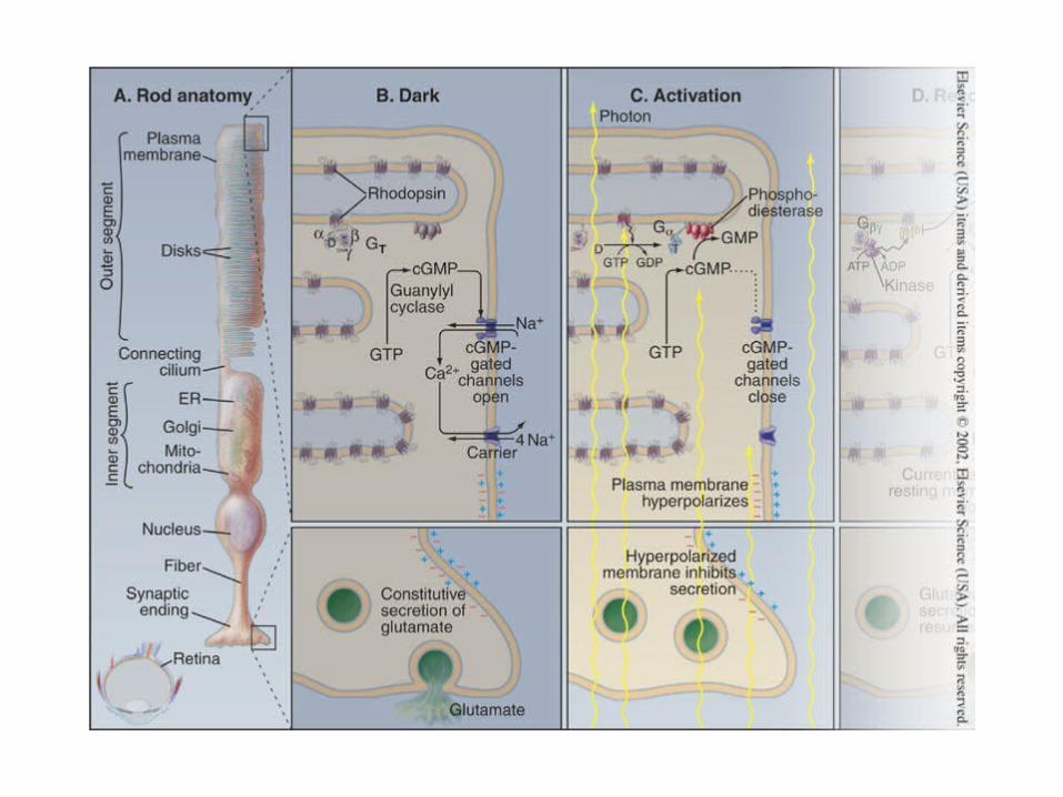
Vision is based on the absorption of light by photoreceptor cells in the eye
Photoreceptor cells are sensitive to light in a relativelynarrow region of the electromagnetic spectrum between 300-850nm
Photoreceptor molecules in the eye
detect visible light
Two kinds of photoreceptorsRods (100 million) and Cons (3 million)Rods function in dim light and do not perceive colorCons function in bright light and are responsible for color vision
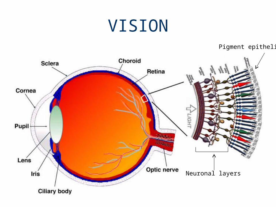
VISIONPigment epithelium
Neuronal layers
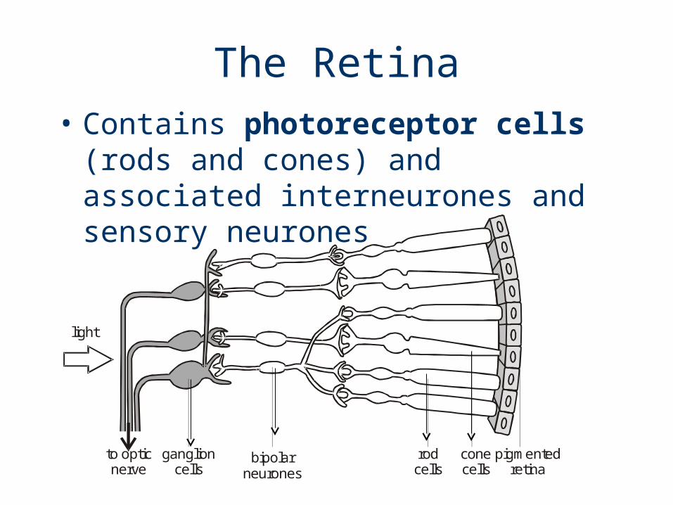
The Retina• Contains photoreceptor cells (rods
and cones) and associated interneurones and sensory neurones
light
to opticnerve
ganglioncells
bipolarneurones
rodcells
conecells
pigmentedretina
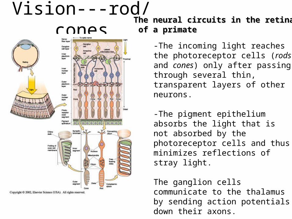
Vision---rod/cones-The incoming light reaches the photoreceptor cells (rods and cones) only after passing through several thin, transparent layers of other neurons. -The pigment epithelium absorbs the light that is not absorbed by the photoreceptor cells and thus minimizes reflections of stray light.
The ganglion cells communicate to the thalamus by sending action potentials down their axons.
However, the photoreceptor cells and other neurons communicate by graded synaptic potentials that are conducted electronically.
The neural circuits in the retinaThe neural circuits in the retina of a primateof a primate

The Rod Cell
Scanning electron micrographs of retinal rod cells
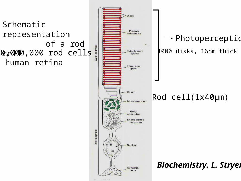
Schematic representation of a rod cell
Biochemistry. L. Stryer
(1x40µm)
100,000,000 rod cellsin human retina
Photoperception
1000 disks, 16nm thick
Rod cell
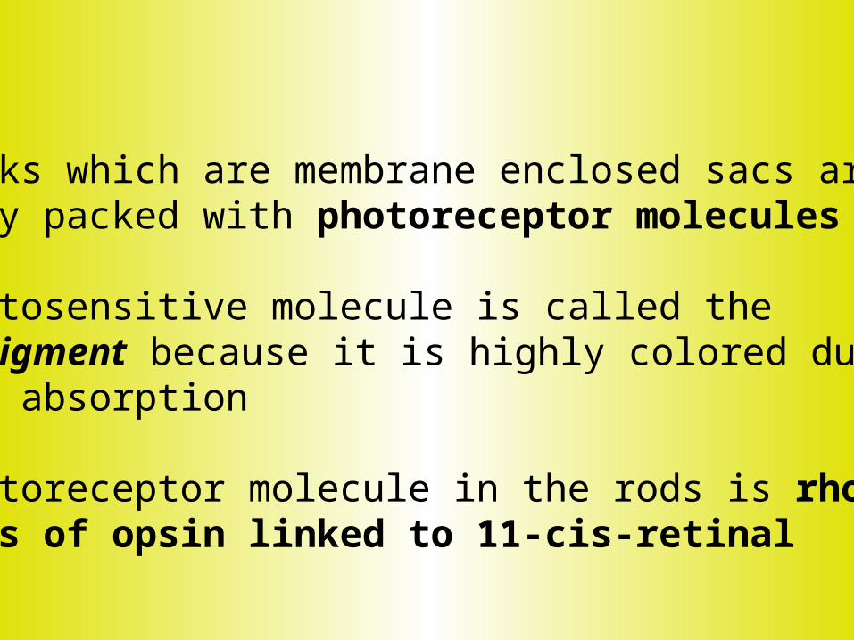
The disks which are membrane enclosed sacs are densely packed with photoreceptor molecules
The photosensitive molecule is called the visual pigment because it is highly colored due to light absorption
The photoreceptor molecule in the rods is rhodopsinconsists of opsin linked to 11-cis-retinal

The electromagnetic spectrum300-850nm
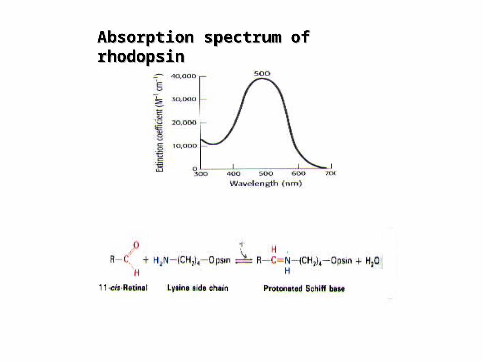
Absorption spectrum of rhodopsinAbsorption spectrum of rhodopsin

Questions
How does the cell respond to How does the cell respond to photons?photons?
What mechanism converts light What mechanism converts light into a cellular signal?into a cellular signal?
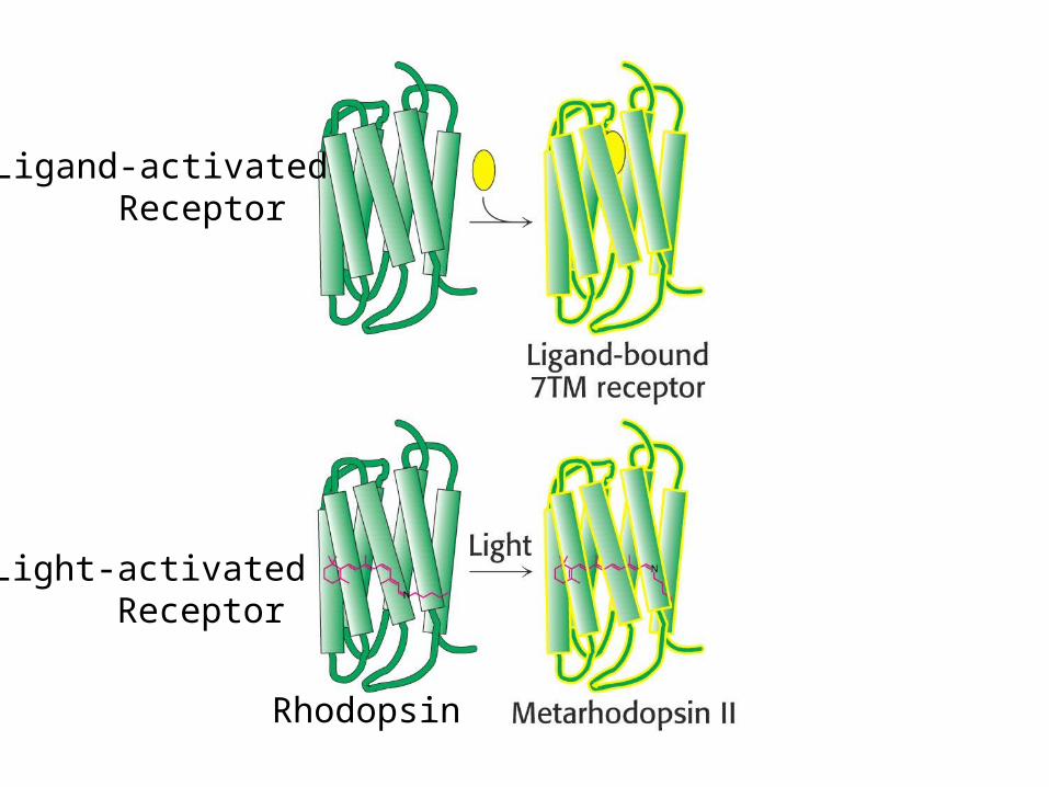
Rhodopsin
Ligand-activated Receptor
Light-activated Receptor

(polyene- with 6 alternating double and single bonds)

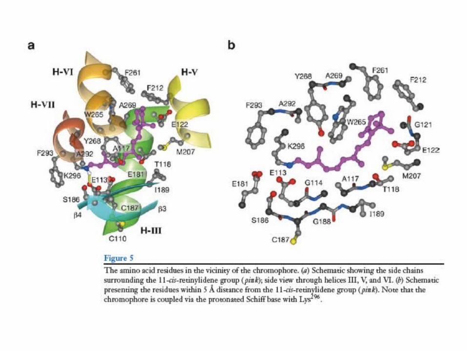
Illustration of Rhodopsin (blue)with11-cis retinal (red)


The protonated form of the 11-cis retinal absorbs at 440nmUnlike 380nm of the non-protonated.The positive charge of Lys296(VII) is compensated by Glu113(II)
(440nm absorption)
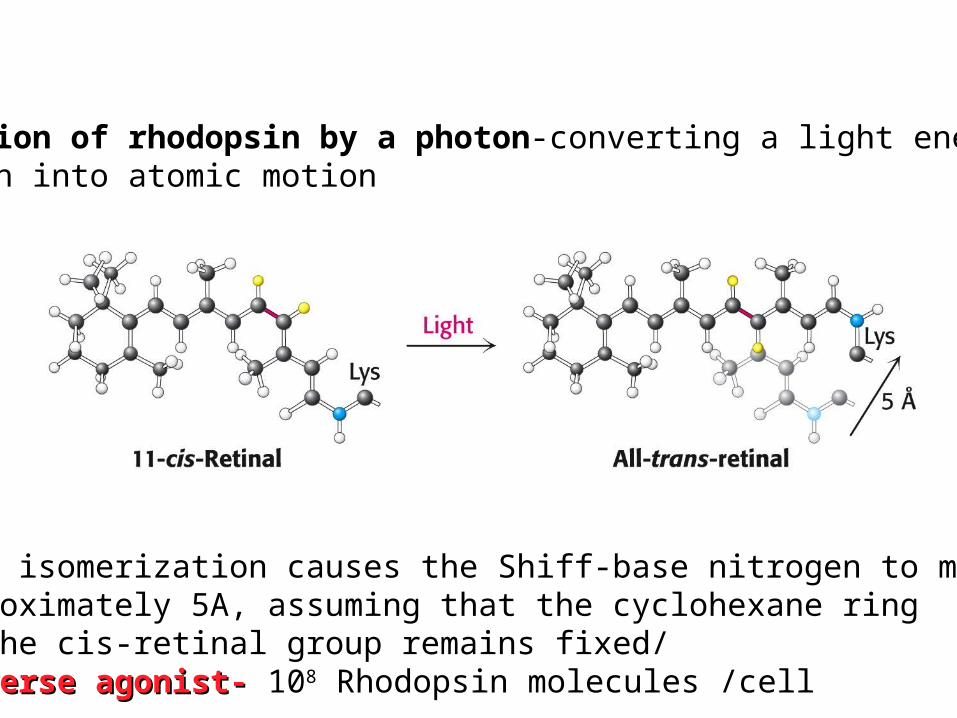
Activation of rhodopsin by a photon-converting a light energy of A photon into atomic motion
-The isomerization causes the Shiff-base nitrogen to moveapproximately 5A, assuming that the cyclohexane ring of the cis-retinal group remains fixed/-Inverse agonist--Inverse agonist- 108 Rhodopsin molecules /cell
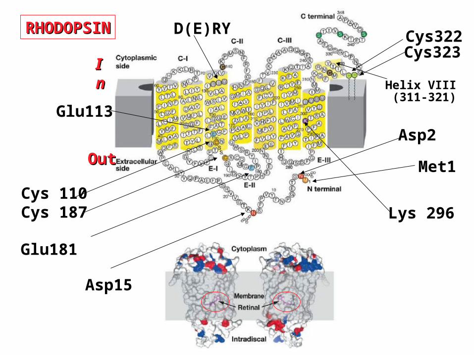
Cys 110Cys 187 Lys 296
Glu113
OutOut
InIn
Asp2
Asp15
Glu181
Met1
Cys322Cys323
D(E)RY
Helix VIII (311-321)
RHODOPSINRHODOPSIN

Rhodopsin 2.8A resolution; Science 389,739 (2000)
Science 289, 739-745 (2000)
The three dimensional structure of rhodopsin

PalmitoylPalmitoylat Helix 8at Helix 8
Retinal
Three dimensional Model of Rhodopsin
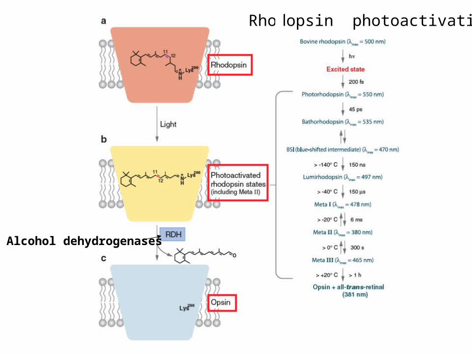
Alcohol dehydrogenases
Rhodopsin photoactivation
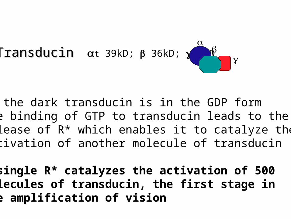
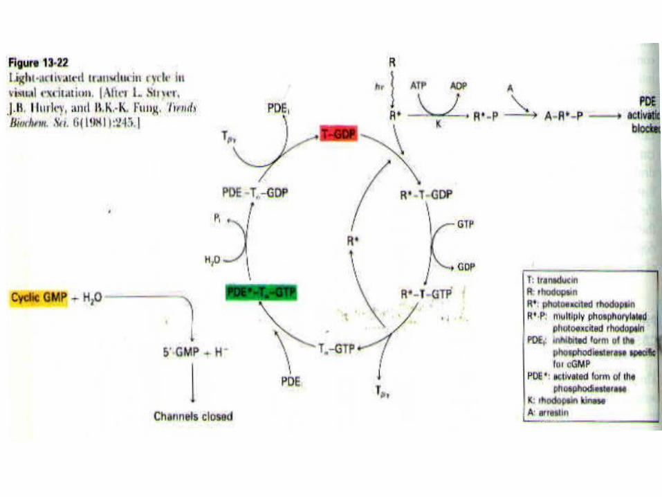
Transducin Transducin t 39kD; 36kD; 8kD
In the dark transducin is in the GDP formthe binding of GTP to transducin leads to therelease of R* which enables it to catalyze theActivation of another molecule of transducin
A single R* catalyzes the activation of 500molecules of transducin, the first stage in the amplification of vision
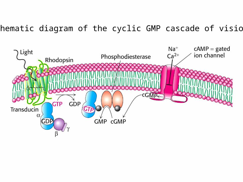
Schematic diagram of the cyclic GMP cascade of vision
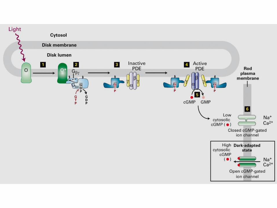


The binding of GTP switches on the phosphodiesterase (PDE) by relieving an inhibitory constraint. In the dark the two catalytic subunits and are held in check by a pair of
inhibitory subunits ().By binding of Gt to the enzyme it removes the inhibitory
subunits and the enzyme is activated
Activation of phosphodiesterase
by Gt
Gt
GtGt
Inactive Active
The hydrolysis of cGMP by phosphodiesterase is the second stage of of amplification
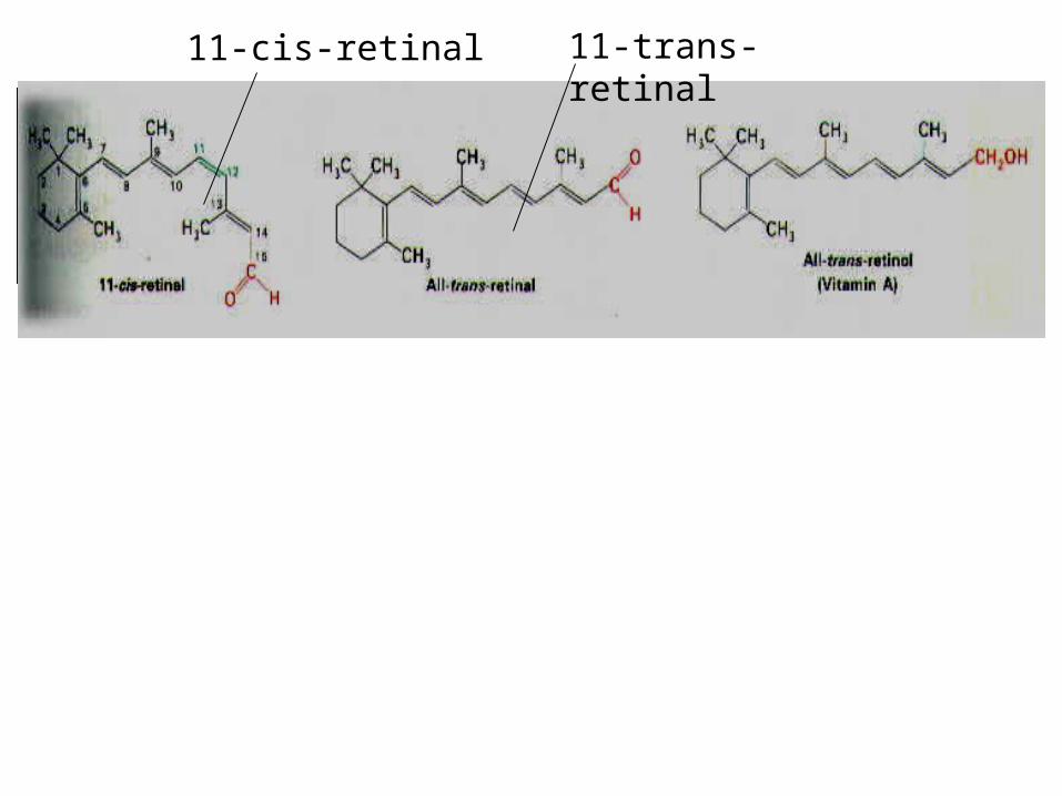
11-cis-retinal 11-trans-retinal

mV
Mem
bran
e po
tent
ial
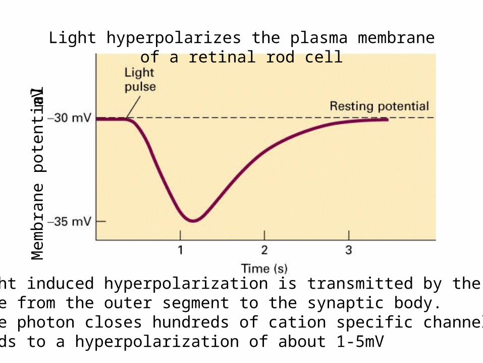
Light hyperpolarizes the plasma membrane of a retinal rod cell
The light induced hyperpolarization is transmitted by the plasmamembrane from the outer segment to the synaptic body.A single photon closes hundreds of cation specific channels (~500)and leads to a hyperpolarization of about 1-5mV
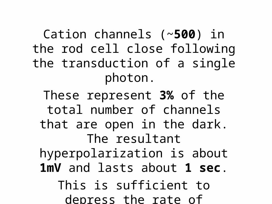
Cation channels (~500) in the rod cell close following the transduction of a
single photon.
These represent 3% of the total number of channels that are open in the dark. The resultant hyperpolarization is about 1mV
and lasts about 1 sec.
This is sufficient to depress the rate of neurotransmitter release that transmits
the onward signal

The high-degree of co-operativity (3 molecules of cGMP) to open the channel increases the sensitivity of the channel for small changes in cGMP which enable it to act as a switch.
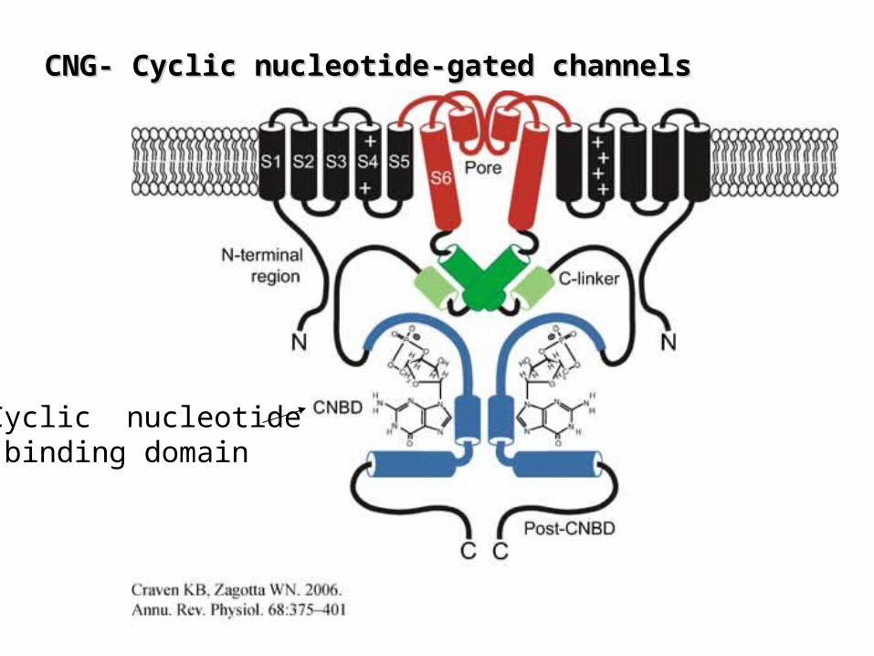
CNG- Cyclic nucleotide-gated channelsCNG- Cyclic nucleotide-gated channels
Cyclic nucleotide binding domain
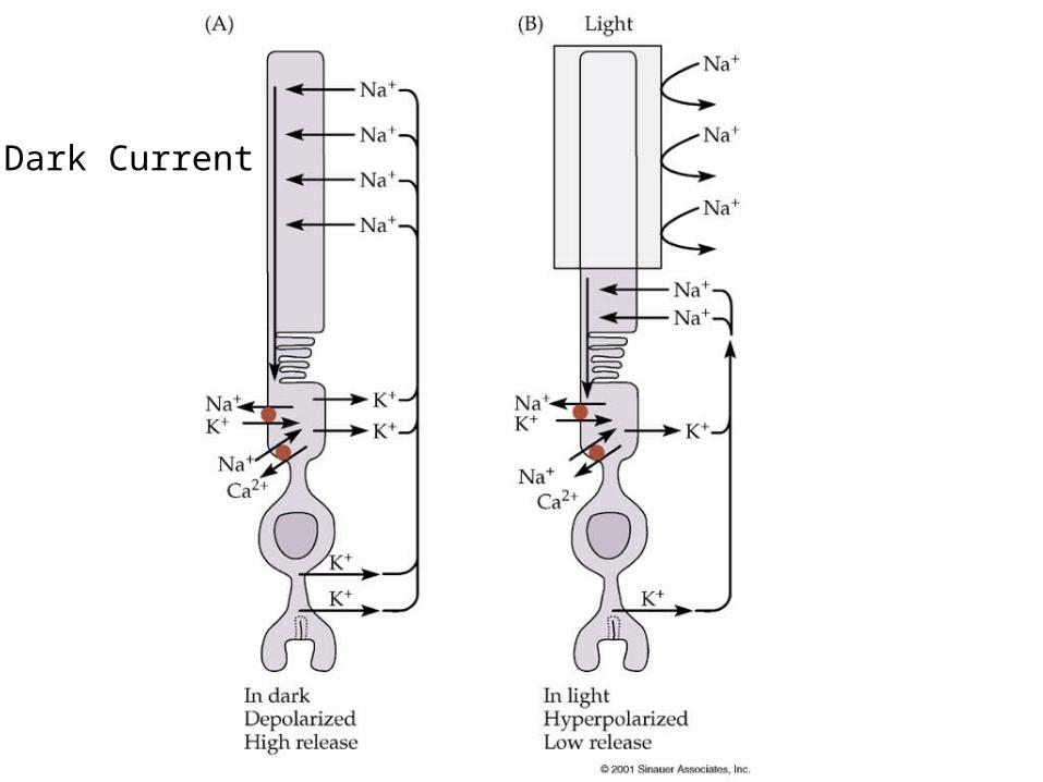
Dark Current

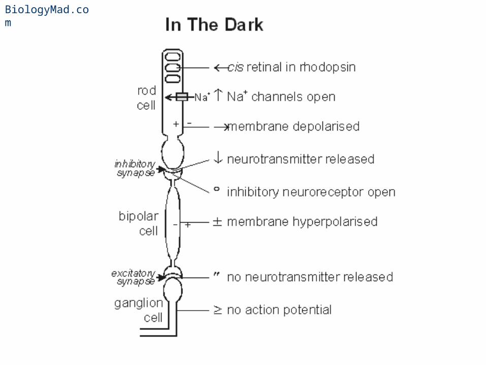
In the Dark…
• In the dark the channel is open Na+ flow in can cause rod cells to depolarise.– Therefore in total darkness, the membrane of a rod cell is
polarised
• Therefore rod cells release neurotransmitter in the dark
• However the synapse with bipolar cells is an inhibitory synapse i.e. the neurotransmitter stops impulse
BiologyMad.com
BiologyMad.com
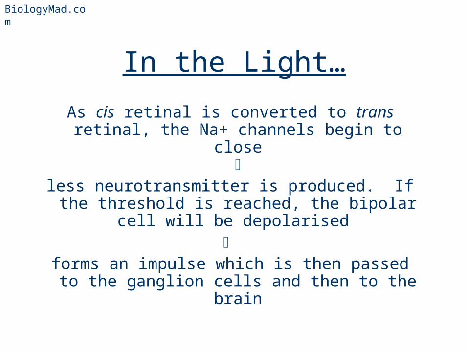
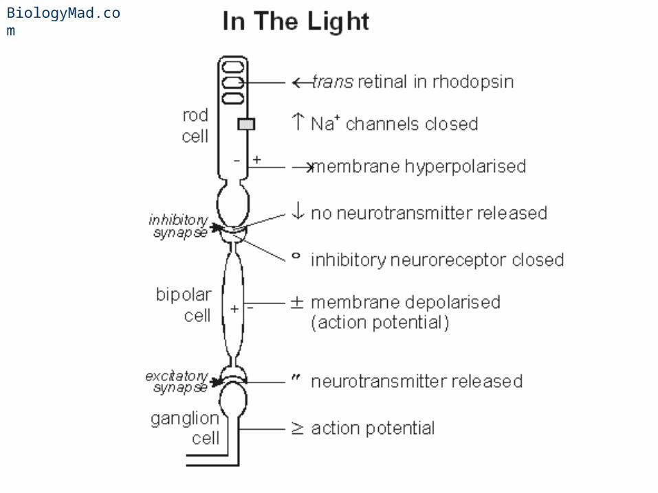
In the Light…
As cis retinal is converted to trans retinal, the Na+ channels begin to close
less neurotransmitter is produced. If the threshold is reached, the bipolar cell will be
depolarised
forms an impulse which is then passed to the ganglion cells and then to the brain
BiologyMad.com
BiologyMad.com
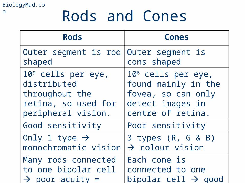
Rods and ConesRods Cones
Outer segment is rod shaped
Outer segment is cons shaped
109 cells per eye, distributed throughout the retina, so used for peripheral vision.
106 cells per eye, found mainly in the fovea, so can only detect images in centre of retina.
Good sensitivity Poor sensitivity
Only 1 type monochromatic vision
3 types (R, G & B) colour vision
Many rods connected to one bipolar cell poor acuity = poor resolution
Each cone is connected to one bipolar cell good acuity = good resolution
BiologyMad.com
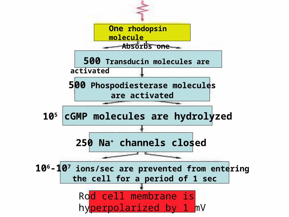
One rhodopsin molecule Absorbs one photon
500 Transducin molecules are activated
500 Phospodiesterase moleculesare activated
105 cGMP molecules are hydrolyzed
250 Na+ channels closed
106-107 ions/sec are prevented from entering the cell for a period of 1 sec
Rod cell membrane is hyperpolarized by 1 mV
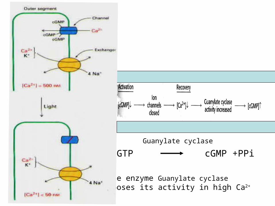
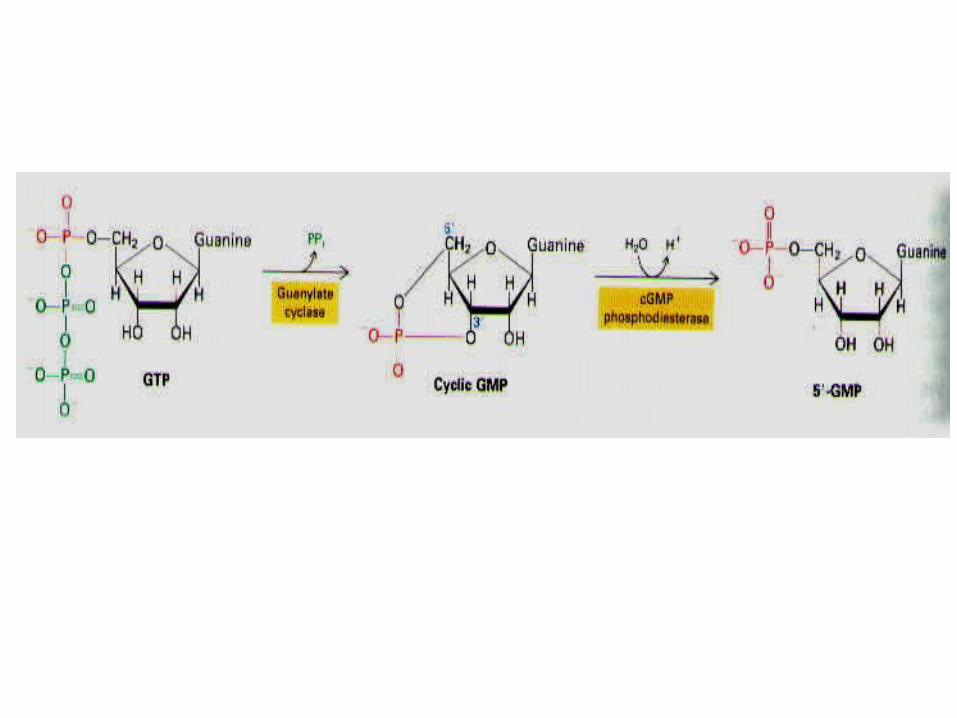
GTP cGMP +PPi
Guanylate cyclase
The enzyme Guanylate cyclase looses its activity in high Ca2+

Color Vision
• 3 different cone cells. Each have a different form of opsin (they have the same retinal)
• 3 forms of rhodopsin are sensitive to different parts of the spectrum– 10% red cones – 45% blue cones – 45% blue cones
BiologyMad.com
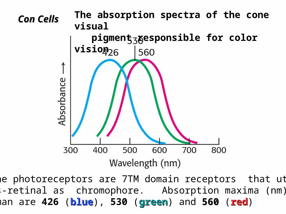
The absorption spectra of the cone visual pigment responsible for color vision
The cone photoreceptors are 7TM domain receptors that utilize 11-cis-retinal as chromophore. Absorption maxima (nm) in human are 426 (blueblue), 530 (greengreen) and 560560 (redred)
Con CellsCon Cells
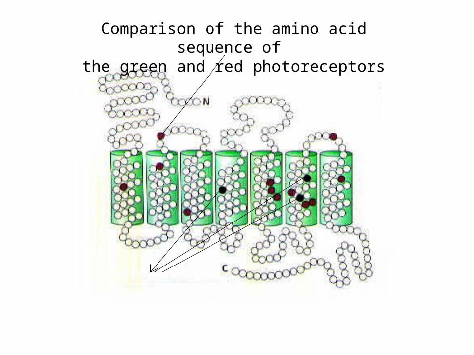
Comparison of the amino acid sequence of the green and red photoreceptors

• Colored light will stimulate these 3 cells differently - by comparing the nerve impulses from the 3 kinds of cones the brain can detect any colour– Red light stimulates R cones– Yellow light stimulates R and G cones equally– Cyan light stimulates B and G cones equally– White light stimulates all 3 cones equally
• Called the trichromatic theory of color vision
Color Vision

• When we look at something the image falls on the fovea and we see it in color and sharp detail.
• Objects in the periphery of our field of view are not seen in colour, or detail.
• The fovea has high density of cones.
• Each cone has a synapse with one bipolar cell and one ganglion each cone sends impulses to the brain about its own small area of the retina high visual acuity
Color Vision
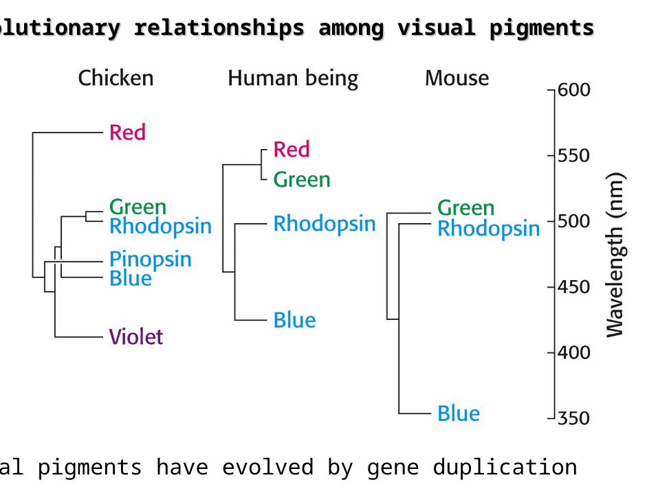
Evolutionary relationships among visual pigmentsEvolutionary relationships among visual pigments
Visual pigments have evolved by gene duplication
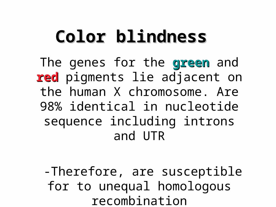
Color blindnessColor blindness
The genes for the greengreen and redred pigments lie adjacent on the human X chromosome. Are 98% identical in nucleotide sequence
including introns and UTR
-Therefore, are susceptible for to unequal homologous recombination
-5% of males have this form of blindness

Recombination pathways leading to color blindness
Rearrangements in the course of DNA replicationA) Loss of visual pigment B) The formation of hybrid pigemnt genes that encode photoreceptors with anomalous abs. spectra
A homologous recombination: the exchange of DNA segment at equivalent positions between chromosomes with substantial similarity
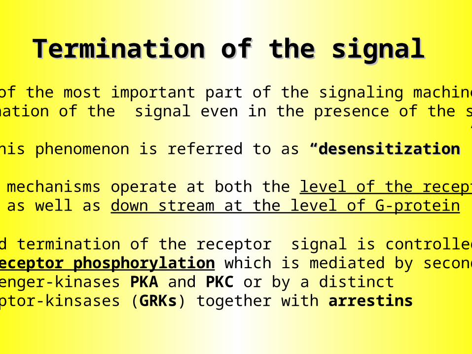
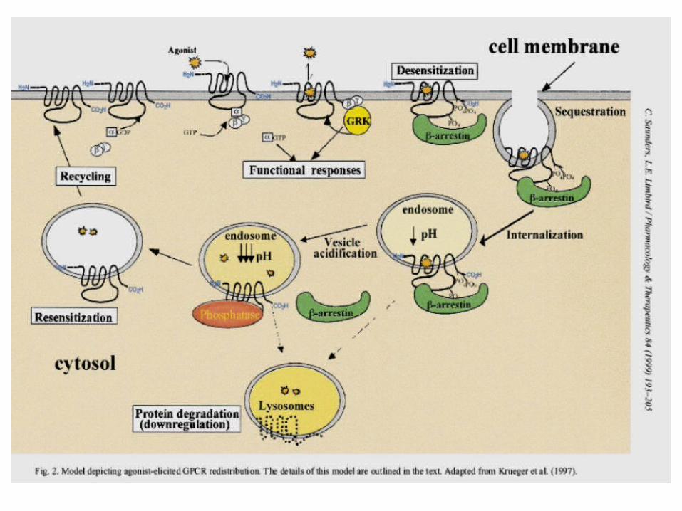
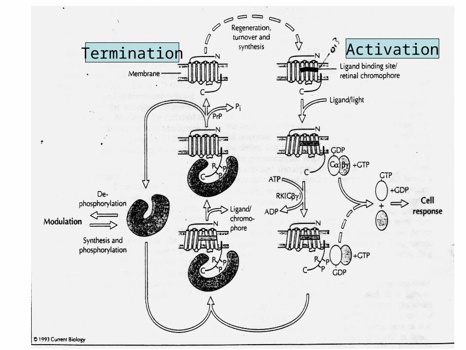
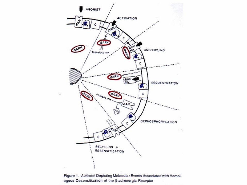
Termination of the signalTermination of the signal
One of the most important part of the signaling machineryis termination of the signal even in the presence of the stimulus
This phenomenon is referred to as “desensitization”“desensitization”
Such mechanisms operate at both the level of the receptor as well as down stream at the level of G-protein
Rapid termination of the receptor signal is controlledby receptor phosphorylation which is mediated by secondmessenger-kinases PKA and PKC or by a distinct Receptor-kinsases (GRKs) together with arrestins


Second-messenger kinase regulationSecond-messenger kinase regulation
PKA and PKC uncouple receptors from their respective G-proteins and serve as negative-feed-back regulatory loops.
Feed back regulation by the 2nd messenger-stimulated kinases PKA and PKC.
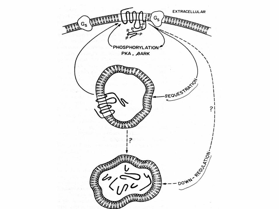
The phosphorylated receptor changes its conformation and no longer can activate the G-proteins. It is an agonist non-specific desensitization
Heterologous desensitizationHeterologous desensitization
Homologous desensitizationGRK(G-ptrotein-receptor kinase)-mediated desensitization(G-ptrotein-receptor kinase)-mediated desensitization
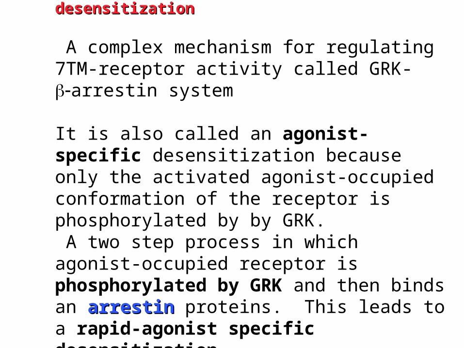
A complex mechanism for regulating 7TM-receptor activity called GRK-arrestin system
It is also called an agonist-specific desensitization because only the activated agonist-occupied conformation of the receptor is phosphorylated by by GRK. A two step process in which agonist-occupied receptor is phosphorylated by GRK and then binds an arrestinarrestin proteins. This leads to a rapid-agonist specific desensitization

Heterogous and homologous desensitization
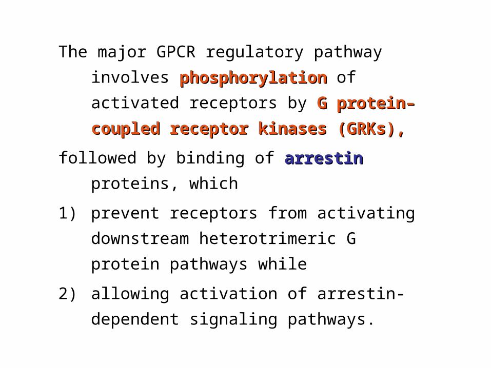
The major GPCR regulatory pathway
involves phosphorylationphosphorylation of activated
receptors by G protein–coupled G protein–coupled
receptor kinases (GRKs),receptor kinases (GRKs),
followed by binding of arrestinarrestin proteins,
which
1) prevent receptors from activating
downstream heterotrimeric G protein
pathways while
2) allowing activation of arrestin-
dependent signaling pathways.

GRK - GRK - G-protein–coupled receptor kinaseG-protein–coupled receptor kinase
As long as the agonist remains bound to the receptor, the activated receptor can continue to activate G proteins.
GRK which is catalytically activated by this interaction, also recognizes the activated conformation of the receptor.
Activated GRKs phosphorylate (P) intracellular domains of the receptor and are then released. The agonist-activated, GRK-phosphorylated receptor binds tightly to an arrestinarrestin protein, which desensitizesdesensitizes further G protein activation and couples the receptor to the clathrin-coated-pit internalization pathway and to arrestin-scaffolded (and G protein–independent) signaling pathways.

GRK-GPCR-kinaseGRK-GPCR-kinaseThe role of GRK-phosphorylation of the receptors in the sequestration process is to facilitate arrestin binding
Experiments to prove this idea
1)A mutated -adrenergic receptor Y326A is a poor
substrate for -Adrenergic receptor-kinase, and is not sequestered. Over-expression of -arrestin restores sequestration
2) Removal of C-terminal tail (sites for GRK sites) prevents sequestration
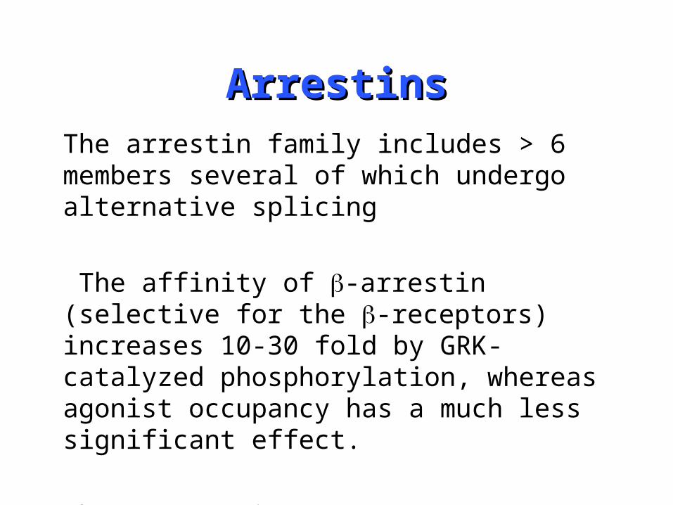
ArrestinsArrestinsThe arrestin family includes > 6 members several of which undergo alternative splicing
The affinity of -arrestin (selective for the -receptors) increases 10-30 fold by GRK-catalyzed phosphorylation, whereas agonist occupancy has a much less significant effect.
The -arrestins promote internalization by binding to clatherin
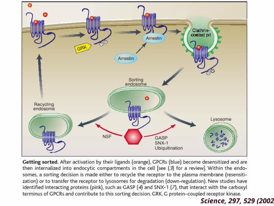
Science, 297, 529 (2002)

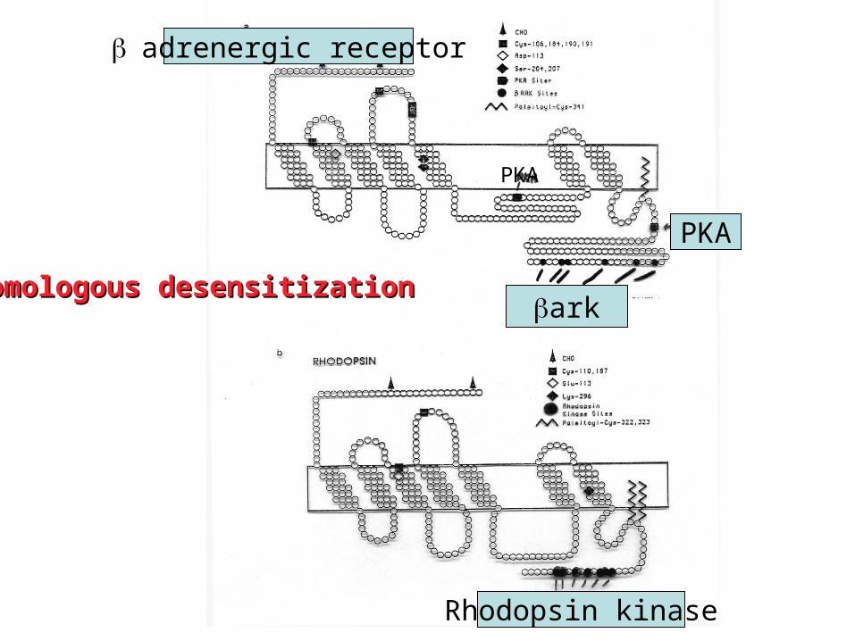
Rhodopsin
ark
Rhodopsin kinase
adrenergic receptor
PKA
PKA
Homologous desensitizationHomologous desensitization
ActivationTermination
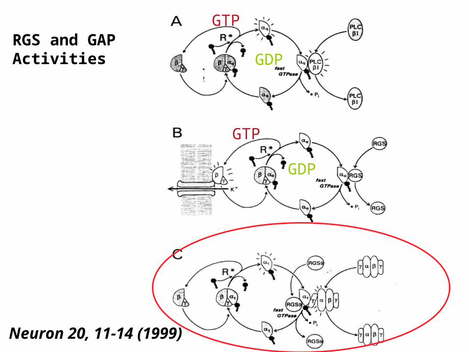
RGS and GAP Activities
Neuron 20, 11-14 (1999)
GTP
GDP
GTP
GDP

11-11-ciscis vs. all- vs. all-transtrans retinal retinal

