outcome of pediatric cataract surgeries in a tertiary...
TRANSCRIPT
Research ArticleOutcome of Pediatric Cataract Surgeries in a TertiaryCenter in Switzerland
Sarah Claudia Ambroz ,1 Marc Töteberg-Harms ,2 James V. M. Hanson ,2,3
Jens Funk ,2 Daniel Barthelmes ,2 and Christina Gerth-Kahlert 2
1University Children’s Hospital Zurich, Steinwiesstrasse 75, 8032 Zürich, Switzerland2Department of Ophthalmology, University Hospital Zurich, Frauenklinikstrasse 24, 8091 Zürich, Switzerland3Neuroimmunology and Multiple Sclerosis Research, Clinic for Neurology, University Hospital Zurich, Frauenklinikstrasse 26,8091 Zurich, Switzerland
Correspondence should be addressed to Christina Gerth-Kahlert; [email protected]
Received 23 August 2017; Revised 27 November 2017; Accepted 1 January 2018; Published 25 February 2018
Academic Editor: Tamer A. Macky
Copyright © 2018 Sarah Claudia Ambroz et al. This is an open access article distributed under the Creative Commons AttributionLicense, which permits unrestricted use, distribution, and reproduction in any medium, provided the original work isproperly cited.
Purpose. To determine and to analyze the outcome of pediatric cataract surgery. Methods. A retrospective chart review ofindividuals aged up to 10 years who underwent cataract surgery between January 1, 2004, and December 31, 2014, at theUniversityHospital Zurich, Switzerland. Results. 63 children (94 affected eyes) with bilateral (68/94) or unilateral (26/94) cataractwere identified. Surgery was performed at a median age of 1.5 months (IQR: 1.3–2.6 months) for the aphakic group (45/94)and of 50.7 months (IQR: 38.0–78.4 months) for the IOL group (49/94). At the last follow-up visit (median 31.1 months, IQR:18.4–50.2 months), visual acuity was better in bilateral than in unilateral cataract cases. Posterior capsular opacification (PCO)was diagnosed in 30.9% of eyes without a significant difference in the IOL and aphakic groups (p = 0 12). Aphakic glaucomawas diagnosed in 12/45 eyes at a median of 6.8 months (IQR 2.1–13.3 months) after surgery. Microcornea (5/12) and anteriorsegment anomalies (8/12) were associated with glaucoma development (p < 0 05). Conclusion. Laterality and timing of surgeryinfluence the outcome of pediatric cataract surgery. PCO was the most frequent postoperative complication. Aphakic glaucomais often associated with ocular developmental abnormalities and a poor visual outcome.
1. Introduction
Congenital cataract is the main reason for preventable blind-ness in children worldwide, with a prevalence of 3 to 4 per10,000 children in Europe [1, 2]. Although cataracts in chil-dren are mostly congenital, acquired cataracts (e.g., followingocular trauma) are also relatively common [3, 4]. Etiology ofcongenital or acquired childhood cataract includes ocularabnormalities, ocular trauma, intrauterine infections, associ-ated syndromes, or hereditary causes [5].
The long-term outcome of pediatric cataract surgerydepends on multiple factors, for example, age at first presen-tation of cataract and age at surgery, associated ocular anom-alies, and development of aphakic glaucoma [6, 7]. A varietyof factors determines the likelihood of a successful functionaland morphological outcome after pediatric cataract surgery.
According to Wu et al., the treatment of congenital cataractpatients is among the most difficult and cost-intensive inter-ventions in ophthalmology [1].
Early detection of cataract in children and rapid referralto a pediatric ophthalmology center in order to evaluate sur-gical indications are essential for successful management [6].The appropriate timing of surgery is crucial and should bebalanced between the severity of amblyopia and the risk ofglaucoma development after cataract surgery [7–9]. Theimplementation of new surgical techniques, such as 23-gaugevitrectomy, has led to reduced surgical trauma [10]. Pediatriccataract often leads to the development of nystagmus, strabis-mus, and amblyopia [5, 11]. Therefore, careful postoperativecare includes therapy for amblyopia and accurate opticalrehabilitation [6]. During the follow-up period, it is criticalto detect postoperative complications. These complications
HindawiJournal of OphthalmologyVolume 2018, Article ID 3230489, 10 pageshttps://doi.org/10.1155/2018/3230489

can manifest long after the procedure and can be severe andfrequent [9]. Development of glaucoma after pediatric cata-ract surgery is one of the most severe complications [12],which depends on such factors as age at surgery, presenceof associated ocular pathologies, and whether or not an intra-ocular lens is primarily implanted [8, 13].
The aim of this study is to gain a better understanding ofthe multiple factors that influence the outcome of pediatriccataract surgery. We analyzed the patients’ characteristicsas well as surgical techniques and their associations withpostoperative complications, such as posterior capsular opa-cification (PCO) and aphakic glaucoma.
2. Methods
A retrospective review of medical charts and surgical reportswas performed on all children diagnosed with cataract whounderwent cataract surgery within the first 10 years of life.Surgery was performed between January 1, 2004, andDecember 31, 2014, at the Department of Ophthalmologyat the UniversityHospital Zurich, Switzerland. The studywas approved by the Cantonal Ethics Committee of Zurich(protocol number 2015-0324). All surgical records of thedepartment were reviewed to identify cases. Search stringswere “cataract” as diagnosis and “age ≤10 years” at the timeof surgery. The results were filtered to exclude all cases thathad prior cataract surgery and therefore had an interven-tion other than cataract surgery during the time of thereview. Patients without any follow-up examination betweenJanuary 1, 2004, and December 31, 2015, were excluded fromthis analysis.
Data collection included demographic, morphological,and functional data at initial presentation and last follow-up (i.e., gender, age, personal and family history, type ofcataract, results of anterior and posterior segment examina-tion, presence of strabismus, nystagmus, best corrected visualacuity (BCVA), and intraocular pressure (IOP)), as well astype and techniques of surgery, postoperative management(i.e., amblyopia therapy), and complications. Exclusion cri-teria were diagnosis of glaucoma, traumatic globe perfora-tion, or retinal detachment prior to cataract surgery. Visualacuity was assessed according to the age and performanceof the patient (e.g., preferential-looking charts at ages up to6 months, Cardiff charts at ages up to 2 years, Patti Picscharts at ages up to 4 years, or Snellen acuity thereafter)and is presented in Snellen acuity notation. Intraocular pres-sure was measured by Goldmann applanation tonometry orusing the TONO-PEN XL (Haag-Streit, Koeniz, Switzerland)or Perkins tonometer (Haag-Streit, Essex, UK), dependingon the age and cooperation of patients during examina-tion. Digital palpation was employed in completely uncoop-erative children; however, no patient was diagnosed withglaucoma based on digital palpation-based measures of intra-ocular pressure.
Microcornea was defined as horizontal corneal diame-ter< 9mm for newborn children and <10mm for children >2years of age. Microphthalmia was defined as an eye with anaxial length of <19mm in a 1-year-old child or <21mm inan adult [14, 15].
Aphakic glaucoma refers to the glaucoma that occursafter congenital cataract surgery [8]. According to the9th consensus report of the World Glaucoma Association,a diagnosis of childhood glaucoma should be made if twoor more of the following characteristics are present: “(1)IOP >21mmHg, (2) optic disc cupping (a progressiveincrease in cup-disc ratio, cup-disc asymmetry of ≥0.2 whenthe optic discs are of similar size, or focal rim narrowing),(3) corneal findings (i.e. Haab’s striae, corneal edema, ordiameter ≥11mm in newborn, >12mm in child <1 yearof age, or >13 mm at any other age), (4) progressive myo-pia or myopic shift coupled with an increase in ocular dimen-sions out of keeping with normal growth, and/or (5) areproducible visual field defect that is consistent with glauco-matous optic neuropathy with no other observable reason forthe visual field defect” [16].
The technique used during surgery was adapted accord-ing to the age and properties of the lens material. In case ofsoft lens material, a simple aspiration of the lens materialwith a bimanual irrigation-aspiration system could be used.In case of harder lens material, either a 25 g vitrectomy probeor a phaco tip was used. In older children (usually of age 2years and older), an IOL was placed in the bag and a posteriorcapsulotomy was performed via a pars plana access using a25 g vitrectomy probe. In younger children, eyes were leftaphakic and a posterior capsulotomy was performed usingeither a bent needle or a 25 g vitrectomy probe using theestablished paracentesis. The use of nonabsorbable sutureswas replaced in 2013 by absorbable vicryl sutures, with theresult that suture removal was no longer required.
2.1. Statistical Analysis. Statistical analyses were performedusing SPSS statistical software versions 22 and 23 forWindows (IBM Corp., Armonk, NY, USA) and MicrosoftExcel version 14.0.0 for MAC/Windows (Microsoft Corp.,Redmond, WA, USA) and http://www.vassarstats.net/index.html (accessed between November 2, 2017, 8 p.m. andNovember 5, 2017, 8 p.m.). The Fisher exact test was usedfor categorical variables. Kaplan-Meier survival analysis wasused to calculate time to event with event being glaucoma.Results are expressed as p values or median and IQR (dueto nonnormal distribution of data to avoid skewing byextremely large or small values). p values< 0.05 were consid-ered statistically significant.
3. Results
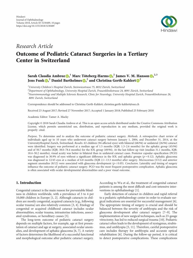
Medical charts of 69 patients were identified. The followingcharts were excluded from the study: one patient presentedwith retinal detachment, one patient with posterior perfo-ration of the globe before surgery, and three patients thatsuffered from combined congenital glaucoma and cataract.Furthermore, one patient had no documented follow-upvisits. Ultimately, the data of 63 children who had surgeryduring the first 10 years of life were included, whichencompasses 94 affected eyes: 35 boys (55.6%) and 28 girls(44.4%) with bilateral (68/94) or unilateral (26/94) cataracts(Figure 1). We included only one eye of six patients withbilateral cataracts due to the second eye undergoing
2 Journal of Ophthalmology
surgery during a time outside of the analyzed period or ata different institution.
3.1. Classification of Cataract. Diagnosis of cataract waseither congenital or juvenile, with congenital cataract beingdiagnosed in 82 out of 94 eyes. The diagnosis of juvenile cat-aract accounted for 12 out of 94 eyes, with 8 instances beingacquired cataracts. The details of associated ocular anomaliesor secondary causes are listed in Table 1.
3.2. First Presentation and Examination. Median age at firstpresentation of all patients was 15.0 months (IQR: 1.2–50months), with 18 patients (28.6%) presenting during the firstmonth of life. Ophthalmologists or primary eye centersreferred 28 patients (44.4%). The remaining patients werereferred by a pediatrician, family physician, a children’s hos-pital (22/63; 13.9%), the parents (3/63), the school physician(1/63), or by the maternity clinic (1/63). In 8/63 cases, nomedical chart information regarding the patient’s referralwas located.
Signs at first presentation were leucocoria (59/94), stra-bismus (21/94), or nystagmus (9/94). A positive familyhistory of early onset cataracts was documented in sevenpatients (11.1%). Table 2 provides an overview of the pre-senting ocular and systemic anomalies.
3.3. Surgery. Surgery was performed at a median age of 22.2months (IQR: 1.6–50.9 months). An intraocular lens (IOL)was implanted in 49 of 94 eyes (median age 50.7 months atsurgery, IQR: 38.0–78.4 months). There were no cases of sec-ondary IOL implantation.
No IOL was implanted in 45 of 94 eyes (median age 1.5months at surgery, IQR: 1.3–2.6 months). An anterior vitrec-tomy was performed in 63 eyes (67.0%), and the posteriorcapsular bag was opened in 81 eyes (86.2%). A microinci-sional cataract surgery technique (23-gauge) was used in 12eyes (12.8%) with an IOL implantation in 7/12 eyes. Para-centesis incisions were usually placed in 10 and 2 o’clockpositions. Primary posterior capsulotomy was performed inall cases as well as anterior vitrectomy.
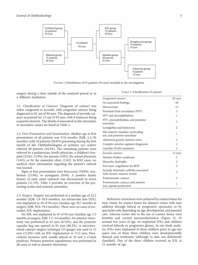
Refractive corrections were achieved by contact lenses fornear vision, by contact lenses for distance vision with nearaddition through bifocal or progressive spectacles, or byspectacles only depending on age, development, and parentalcare. Adverse events due to the use of contact lenses werekeratitis and corneal neovascularization (Figure 2). Ataround two years of age, we implanted IOLs and childrenreceived bifocals or progressive glasses. In our study analy-sis, IOLs were implanted in three children prior to age twoyears: two of these three children were developmentallydelayed and syndromic (although to date not definitivelyclassified). One of the three children received an IOL at21 months of age.
63 patients94 eyes
Unilateral group26 patients26 eyes
Bilateral group37 patients68 eyes
IOL group35 patients49 eyes
Aphakia group28 patients45 eyes
Glaucoma group9 patients12 eyes
Nonglaucoma group19 patients33 eyes
Figure 1: Distribution of 63 patients (94 eyes) included in the investigation.
Table 1: Classification of cataract.
Congenital cataract 82 eyes
No associated findings 56
Microcornea 12
Persistent fetal vasculature (PFV) 3
PFV and microphthalmia 1
PFV, microphthalmia, and posteriorsynechiae
1
Lentiglobus and lenticonus 2
Flat anterior chamber, protrudingiris, and posterior synechiae
1
Abnormal greyish anterior sclera 2
Complex anterior segment dysgenesis 2
Aniridia (PAX6 mutation) 2
Juvenile cataract 12 eyes
Marden-Walker syndrome 2
Muscular dystrophy 2
Post laser coagulation for ROP 3
Juvenile rheumatic arthritis associatedwith chronic anterior uveitis
2
Posttraumatic cataract 2
Posttraumatic cataract and anteriorlens capsule perforation
1
3Journal of Ophthalmology
Amblyopia therapy was recommended in all patientswith surgery for unilateral cataract. 37 patients (48 eyes)received therapy for amblyopia. 23/37 of these patients(62.2%) had undergone surgery for unilateral cataractand 14/37 (37.8%) for bilateral cataract. The success rateof amblyopia therapy is limited by compliance and mor-phological/anatomical factors. Patients who had bilateralcataract surgery and developed an interocular differencein visual acuity or patients who presented with manifestunilateral strabismus as a sign of amblyopia also receivedpatching therapy.
3.4. Postoperative Complications. During the follow-upperiod (median 31.1 months, IQR: 18.4–50.2 months), 52eyes (55.3%) developed complications, which are summa-rized in Figure 3. Complete retinal detachment and phthisisin one eye was described in a patient with bilateral cataractwho developed complicated aphakic glaucoma requiring sev-eral glaucoma surgeries in both eyes. Vitreous hemorrhage
and retinal detachment was documented in one patient withDown syndrome at 13.5 months post cataract surgery fortraumatic cataract.
PCO occurred in 10/45 eyes (22.2%) in the aphakic groupand 19/49 eyes (38.8%) in the IOL group. There was no sig-nificant difference in the incidence of PCO in the IOL andaphakic groups (p = 0 12). A posterior capsular bag openingand an anterior vitrectomy were performed in 82.8% (24/29eyes) and 65.5% (19/29 eyes), respectively, of these eyes. Incomparison, eyes without documented PCO had a posteriorcapsular bag opening and an anterior vitrectomy in 87.7%(57/65 eyes) and 67.7% (44/65 eyes), respectively. Therewas no statistically significant relationship between anteriorvitrectomy or opening of the posterior bag and the subse-quent development of PCO (p = 1 00 and 0.75, resp.). Only5/13 eyes without opening of the posterior capsule developedPCO, four eyes belonged to the IOL-group and one eye to theaphakic group. There was no statistically significant differ-ence between the IOL and aphakic groups concerning
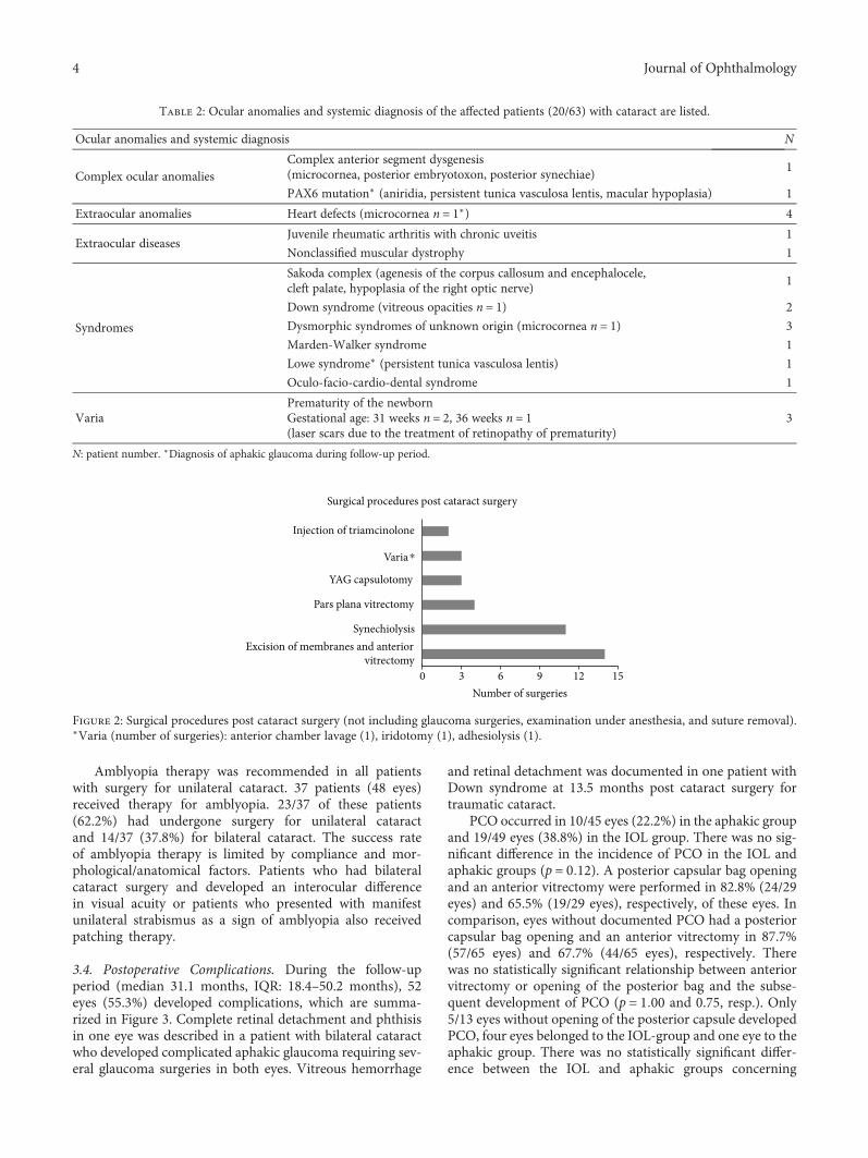
Table 2: Ocular anomalies and systemic diagnosis of the affected patients (20/63) with cataract are listed.
Ocular anomalies and systemic diagnosis N
Complex ocular anomaliesComplex anterior segment dysgenesis(microcornea, posterior embryotoxon, posterior synechiae)
1
PAX6 mutation∗ (aniridia, persistent tunica vasculosa lentis, macular hypoplasia) 1
Extraocular anomalies Heart defects (microcornea n = 1∗) 4
Extraocular diseasesJuvenile rheumatic arthritis with chronic uveitis 1
Nonclassified muscular dystrophy 1
Syndromes
Sakoda complex (agenesis of the corpus callosum and encephalocele,cleft palate, hypoplasia of the right optic nerve)
1
Down syndrome (vitreous opacities n = 1) 2
Dysmorphic syndromes of unknown origin (microcornea n = 1) 3
Marden-Walker syndrome 1
Lowe syndrome∗ (persistent tunica vasculosa lentis) 1
Oculo-facio-cardio-dental syndrome 1
VariaPrematurity of the newbornGestational age: 31 weeks n = 2, 36 weeks n = 1(laser scars due to the treatment of retinopathy of prematurity)
3
N: patient number. ∗Diagnosis of aphakic glaucoma during follow-up period.
Excision of membranes and anteriorvitrectomy
Synechiolysis
Pars plana vitrectomy
YAG capsulotomy
Varia⁎
Injection of triamcinolone
0 3 6 9 12 15Number of surgeries
Surgical procedures post cataract surgery
Figure 2: Surgical procedures post cataract surgery (not including glaucoma surgeries, examination under anesthesia, and suture removal).∗Varia (number of surgeries): anterior chamber lavage (1), iridotomy (1), adhesiolysis (1).
4 Journal of Ophthalmology
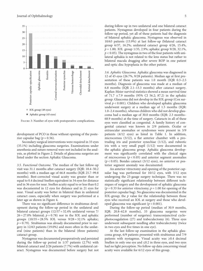
development of PCO in those without opening of the poste-rior capsular bag (p = 0 56).
Secondary surgical interventions were required in 33 eyes(35.1%) including glaucoma surgeries. Examinations underanesthesia and suture removal were not included in the anal-ysis, as plotted in Figure 2. Details of glaucoma surgeries arelisted under the section Aphakic Glaucoma.
3.5. Functional Outcome. The median of the last follow-upvisit was 31.1 months after cataract surgery (IQR: 18.4–50.2months) with a median age of 66.8 months (IQR 25.7–99.8months). Best-corrected visual acuity was greater than orequal to 0.4 decimal Snellen equivalent in 34 eyes for distanceand in 36 eyes for near. Snellen acuity equal to or less than 0.2was documented in 12 eyes for distance and in 21 eyes fornear. Visual acuity was better in bilateral than in unilateralcataract cases, especially when surgery was performed at alater age as shown in Figure 4.
There was no significant difference in strabismus devel-opment during the follow-up period in the unilateral andbilateral cataract groups (12/37= 32.4% unilateral versus 7/26=27.0% bilateral; p = 0 78) nor in the IOL and aphakicgroups (10/35 =28.5% IOL versus 9/28= 32.1% aphakic;p = 0 79). Strabismus was already documented prior to sur-gery in 12/63 patients (19.0%) and more often in the unilat-eral (nine patients) than in the bilateral (three patients)cataract group.
Nystagmus was documented preoperatively and persistedduring the follow-up period in 1/37 patients (2.7%) withbilateral cataract and 2/26 patients (7.7%) with unilateral cat-aract. Nystagmus was documented before surgery but not
during follow-up in two unilateral and one bilateral cataractpatients. Nystagmus developed in four patients during thefollow-up period, yet all of these patients had the diagnosisof bilateral aphakic glaucoma. Nystagmus was observed in10/63 patients (15.9%) at last follow-up (bilateral cataractgroup 6/37, 16.2%; unilateral cataract group 4/26, 15.4%,p = 1 00; IOL group 1/35, 2.9%; aphakic group 9/28, 32.1%,p < 0 05). The nystagmus in two of the four patients with uni-lateral aphakia is not related to the lens status but rather tobilateral macula dragging after severe ROP in one patientand optic disc hypoplasia in the other patient.
3.6. Aphakic Glaucoma. Aphakic glaucoma was diagnosed in12 of 45 eyes (26.7%, 9/28 patients). Median age at first pre-sentation of these patients was 1.0 month (IQR 0.3–2.5months). Diagnosis of glaucoma was made at a median of6.8 months (IQR 2.1–13.3 months) after cataract surgery.Kaplan-Meier survival statistics showed a mean survival timeof 71.7 ± 7.9 months (95% CI 56.2; 87.2) in the aphakicgroup. Glaucoma did not develop in the IOL group (Cox sur-vival p < 0 001). Children who developed aphakic glaucomaunderwent surgery at a median age of 1.5 months (IQR:1.0–2.4 months), whereas children who did not develop glau-coma had a median age of 30.0 months (IQR: 2.3 months–68.9 months) at the time of surgery. Cataracts in all of theseeyes were classified as congenital. A family history of con-genital cataract was known in 2/9 patients. Ocular orextraocular anomalies or syndromes were present in 3/9patients (4/12 eyes) as listed in Table 1. In addition,microcornea (3/12), a flat anterior chamber with a pro-truding iris and posterior synechiae (1/12), and rubeoticiris with a very small pupil (1/12) were documented inthe aphakic glaucoma group. Aphakic glaucoma develop-ment was significantly correlated with the clinical signsof microcornea (p < 0 05) and anterior segment anomalies(p < 0 05). Besides cataract (3/12 eyes), no anterior or pos-terior segment anomaly was documented.
An anterior vitrectomy and opening of the posterior cap-sular bag was performed for 10/12 eyes, with 3/12 eyesundergoing the 23-gauge surgery technique. There was nostatistically significant relationship between different tech-niques of surgery and the development of aphakic glaucoma(p = 0 33 for anterior vitrectomy; p = 1 00 for opening of theposterior capsular bag). No glaucoma was documented in theIOL-group; the p value for the relationship between thoseeyes who received an IOL at surgery and those who devel-oped glaucoma was significant (p < 0 001).
During the follow-up period (median of 30.9 months,IQR: 20.8–62.8 months), 33 glaucoma surgeries wereperformed (number of surgeries): transconjunctival cyclo-photocoagulation (27) and trabeculectomy (6). Three eyesunderwent subsequent needling after trabeculectomy (twicein two eyes and five times in one eye).
At the last follow-up examination in the aphakic glau-coma group, 6/9 patients presented with strabismus and 7/9patients with nystagmus. Visual acuity was ≥0.4 decimalSnellen in only one eye and ≤0.2 in three eyes, and two eyeshad no light perception. No follow-up data concerning visualacuity were available for 6/12 eyes of this group.
0
5
10
15
20
25
30
35
IOL group (49 eyes)
Aphakic group (45 eyes)
Poste
rior c
apsu
lar o
paci
ficat
ion
Apha
kic g
lauc
oma
Kera
titis
Cor
neal
neo
vasc
ular
izat
ion
Poste
rior s
ynec
hia
Vitre
ous h
emor
rhag
e
Chor
oida
l det
achm
ent
Retin
al d
etac
hmen
t
Figure 3: Number of eyes with postoperative complications.
5Journal of Ophthalmology

4. Discussion
There was no association between the surgical procedureemployed and the subsequent development of PCO, whichwas the most frequent postoperative complication. Our anal-ysis of the large cohort confirmed aphakic glaucoma as ahigh-risk complication after congenital cataract surgeryassociated with ocular or syndromic anomalies. Nystagmuswas diagnosed in a statistically significantly greater num-ber of patients at last follow-up in the aphakic than inthe IOL group.
4.1. Classification of Cataract. Congenital cataracts were themain reason for surgical intervention in our pediatric cata-ract group. The majority (68.3%) presented with isolated cat-aracts without associated anomalies, which is in accordancewith previous reports [3, 17]. The presentation of bilateralcataract in 72.3% is in agreement with previous reports fromdifferent geographical areas [3, 4, 18, 19].
4.2. First Presentation and Examination. Analysis of the per-sonal medical history revealed that 31.7% (20 patients) pre-sented with extraocular and ocular anomalies or diseases.Syndromal congenital cataracts were most often bilateral(7/9). We had no cases of cataract due to congenital infec-tions, contrary to other studies [4, 5]. This may be due tothe high vaccination coverage and low incidence of diseasessuch as congenital rubella in Switzerland. The Federal Officeof Public Health of Switzerland reported no cases ofmaterno-fetal rubella between 2010 and 2016 [20]. PFV waspresent in five eyes with congenital unilateral cataract andin none with congenital bilateral cataract, which is in agree-ment with the study by Tartarella et al. [5]. They reportedPFV in 8.2% of patients, and the majority of cases werediagnosed in patients with unilateral cataract [5].
4.3. Surgery. Median age at surgery was lower in the aphakicas compared to the IOL group. There is an ongoing contro-versy about the correct time of surgery due to the risk of
developing aphakic glaucoma and amblyopia. Ruddle et al.reported earlier surgery as the “key risk factor” for the devel-opment of aphakic glaucoma [21], but Birch et al. state that adelay of surgery beyond six weeks was associated with ahigher risk for strabismus and nystagmus [45]. A recentlypublished retrospective study from Germany reported astrong relationship between cataract surgery within the first14 weeks of life and the development of aphakic glaucoma[22]. They compared the outcome of bilateral congenital cat-aract treatment for those who underwent surgery within thefirst 10 weeks of life with those who underwent surgerybetween 10 weeks and 12 months of age [22]. The prevalenceof amblyopia was not statistically different between the twoage groups [22]. Nagamoto et al. concluded that glaucomadeveloped more frequently in eyes which required surgeryat one year of age or younger [19]. The age at surgery forthe aphakia group was 14± 24 months (mean± SD), andtheir prevalence of aphakic glaucoma was observed to be5.8% [19]. Chen et al. compared bilateral congenital cataractpatients and observed a better outcome for those who under-went surgery at six months of age than for those at threemonths of age [23]. In our study, we observed glaucomadevelopment in patients who underwent surgery at a veryyoung age (in the first 4.7 months of age). There is no clearconsensus in the literature regarding the best age for surgeryto avoid development of glaucoma and amblyopia, eventhough some reports describe a better outcome for cataractsurgery after the first three months of life [22, 23].
The microincision cataract surgery technique (23-gauge(23G)) was introduced in our institution in July 2013 andhas been used for 12 cataract surgeries through to December2014. According to Li et al., this technique has many advan-tages, particularly including fewer postoperative complica-tions [10]. Lim et al. report that they applied transcorneal23G-vitrectomy for routine congenital cataracts due to thesmall incision size, anterior chamber stability, and handpiecerigidity [24]. Given that our numbers and follow-up time forthose who underwent surgery with the 23G-vitrectomy
00
0.2
0.4
0.6
0.8
1
1.2
1.4
20 20 60 80 100 120
Visu
al ac
uity
at la
st fo
llow
-up
(dist
ance
)
Age at cataract surgery (months)
BilateralUnilateral cataract
(a)
0 20 20 60 80 100 120Age at cataract surgery (months)
1.2
1
0.8
0.6
0.4
0.2
0
Visu
al ac
uity
at la
st fo
llow
-up
(nea
r)
BilateralUnilateral cataract
(b)
Figure 4: Visual acuity at last follow-up versus age at cataract surgery. Distance (a) and near (b) Snellen visual acuity at last follow-up as afunction of age at cataract surgery is plotted for bilateral (blue) and unilateral (red) cataracts. Functional outcome of bilateral cataractappeared better than for unilateral cataract, especially at a later age at surgery.
6 Journal of Ophthalmology
continue to be significantly limited, it is not yet possible toanalyze data regarding functional outcomes.
4.4. Amblyopia Therapy. Amblyopia therapy was primarilyperformed in children with unilateral cataract. According tothe Infant Aphakia Treatment Study, the number of reportedhours of patching throughout the first four years of life isassociated with a better visual acuity in cases of unilateralcongenital cataracts [25]. Patching compliance was foundto be one of the most important determinants of visual out-come [26]. Unfortunately, there was a lack of informationwithin our reviewed medical charts regarding the daily hoursof patching and the patients’ compliance. Consequently, itwas not possible to perform a detailed analysis of the visualoutcome following amblyopia therapy.
4.5. Postoperative Complications. As shown in Figure 3, themost common postoperative complications were PCO (IOLgroup 38.8%, aphakic group 22.2%) and aphakic glaucoma,followed by contact lens-related corneal neovascularizationand keratitis (both exclusively in the aphakic group) andsynechiae (both groups). Bar-Sela et al. found a similar prev-alence of PCO development with 38% in their patient group[27]. They concluded that the management of the posteriorcapsule at cataract surgery influences the incidence and thetiming of PCO after IOL implantation [27]. We did not iden-tify an association between the opening of the posterior bagor the performing of an anterior vitrectomy and the develop-ment of PCO. Retinal detachment occurred in 3.2% of theeyes, which is in agreement with a large previous study (3%of 1043 eyes) [28]. The variability in our follow-up periodcould lead to an underestimation of postoperative complica-tions, especially in those patients with a short follow-up.
4.6. Aphakic Glaucoma. In accordance with previously pub-lished data from other tertiary centers, aphakic glaucoma isone of the main postoperative complications [7, 12, 29].The incidence of aphakic glaucoma after cataract surgery atour institution is 12.8% of eyes and was diagnosed at amedian time of 6.8 months after surgery. Our data is in agree-ment with other European studies, which reported a range of9.7% to 15.3% incidence of developing aphakic glaucomabetween 5.0 and 6.1 months after surgery [30, 31].
Aphakic glaucoma is often associated with abnormalocular development such as microcornea [12], anteriorsegment dysgenesis, and aniridia [32, 33]. Microcorneais known to be an independent risk factor for aphakicglaucoma [7]. We were able to confirm this finding inour group with a significant relationship between thepresence of microcornea and the development of glau-coma. There was an even stronger relationship betweenthe presence of ocular developmental abnormalities andaphakic glaucoma. None of the patients who had experi-enced ocular trauma with consecutive cataract subsequentlydeveloped glaucoma.
At our institution, the median age at surgery of theaphakic glaucoma group was 45.0 days (mean 60.9 days),compared to the nonglaucoma group, which was 2.5 years.Other studies reported a slightly later onset of aphakic
glaucoma in comparison to our group [29, 34]. A direct com-parison may be problematic due to differing IOP criteria usedto define glaucoma. According to Solebo et al., a tenfoldhigher age at the time of surgery decreases the develop-ment of glaucoma after cataract surgery by 60% and anolder age at surgery is interpreted as a protective factor[12]. Ruddle et al. confirmed an association between youn-ger age at time of surgery and a higher incidence of glau-coma [21]. This may explain the absence of glaucoma inthe IOL group, considering that the median for age at sur-gery was considerably higher in the IOL group than in theaphakia group. Furthermore, the youngest child with anIOL implantation was 21 months old. In a randomizedclinical control, Lambert et al. compared contact lens tointraocular lens correction of monocular aphakia in chil-dren who underwent surgery between one and six monthsof age. In their study, more children in the IOL groupdeveloped glaucoma by the age of one year [35].
Diagnosis of aphakic glaucoma was made at a median of6.8 months postoperative, with the earliest onset being 11.0days after surgery. These results are comparable to the BritishCongenital Cataract Study (median time 1.34 years; range of12 days to 6.73 years) [36]. Our follow-up period (median 2.6years, mean 3.4 years) is comparable to other European stud-ies from 2.5 to 9.4 years [30, 31, 37, 38]. At the last docu-mented follow-up examination, the majority of our patientspresented with strabismus, nystagmus, and a poor visual out-come, which is in agreement with other studies [38]. Theinterpretation of the visual outcome in this group was limitedby our retrospective study design with limited follow-up data.
The preferred treatment of congenital and early juvenileglaucoma at our department is trabeculotomy. We knowfrom the recent literature that 360° trabeculotomy is prefer-able over standard trabeculotomy, for example, with theHarms probe [39]. Currently, we perform 360° trabeculot-omy rather than standard trabeculotomy with the Harmsprobe. However, in some cases, the parents did not allowus to follow this standard treatment plan. In some cases,(1) examination under anesthesia (EUA) was performedin the children’s hospital where it was not possible to per-form ocular surgeries or (2) parents refused surgery onboth eyes at the same time or refused immediate surgeryafter the EUA. In some cases, cyclodestructive proceduresseem to be a valid option to lower the IOP until definitesurgery can be performed.
4.7. Functional Outcome. To evaluate the functional andmorphological outcome after cataract surgery, we consideredvisual acuity and the presence of strabismus and nystagmusat last follow-up.
4.7.1. Visual Outcome.Median time of the last follow-up visitwas 31.1 months postsurgery, which is comparable to otherinvestigations [19, 40–42]. Similar to the results of otherstudies, we found development of visual acuity at both farand near distances to be more favorable in the bilateralcataract group [21] and for those individuals who hadsurgery at an age older than 2 years.
7Journal of Ophthalmology

4.7.2. Nystagmus. All patients who developed nystagmus inthe follow-up period belonged to the aphakic glaucoma andbilateral cataract group (4/37 patients, 10.8%). We identifieda statistically significant greater number of patients withnystagmus at last follow-up in the aphakic than in the IOLgroup. Furthermore, nystagmus was diagnosed more oftenin the bilateral than in the unilateral group at last follow-up,as also documented in other studies [18, 43, 44]. However,the small number of patients does not allow us to confirm ahigher likelihood of nystagmus in children after bilateral thanin unilateral cataract surgery. Nystagmus, and particularlylatent nystagmus, may be underreported in the medicalrecords, and the retrospective study design does not allowfurther analysis of this.
Preoperative nystagmus was no longer visible during thefollow-up period in three patients, which was most likelycaused by improved visual function. In a prospectiveinvestigation, Lloyd et al. described cases which convertedpostoperatively from preoperative nystagmus to manifestlatent nystagmus (MLN) [6, 44]. Young et al. reported thatpreoperative nystagmus did not appear to be a risk factorfor a worse visual outcome [11].
4.7.3. Strabismus. There was no statistically significantdifference regarding the incidence of strabismus that devel-oped during the follow-up period between the unilateraland bilateral groups as well as between the IOL and aphakicgroups. 19.0% (12/63) of patients were diagnosed withstrabismus at first presentation. Tartarella et al. describe aneven higher prevalence (52.2%) of strabismus preoperatively,but they did not report the postoperative data [5]. A study byBirch et al. investigated risk factors for strabismus develop-ment. They concluded that laterality and age at onset weresignificant nonmodifiable risk factors for strabismus [45]. Areduced risk for nystagmus and strabismus was associatedwith deprivation of less than 6 weeks [45].
4.8. Limitations of the Study. Our study represents aretrospective analysis of medical charts and surgical reports.Visual acuity analysis is depending on age and tests used.Preferable, only one method of visual acuity testing is usedto compare data to avoid overestimating outcome data, espe-cially when preferential-looking charts are used. This couldnot be achieved in this retrospective study but would encour-age for further data collection in a prospective manner.
Data acquisition, especially of follow-up data, was par-tially restricted. Hence, there were no preestablished proto-cols regarding the timing and performance of surgicalprocedures and follow-up regimen. Furthermore, it is impor-tant to consider that differences in the age at surgery areinfluenced by the presentation and laterality of cataract. In2012, the care of a number of aphakic patients was trans-ferred to a private practice and therefore no clinical datawas available after the point of transfer for these patients.The only factor in the transfer of these particular patientswas parental desire to maintain continuity of care with theinitially treating physician, rather than other demographicfactors (e.g., age, outcome, and unilateral versus bilateralcataract), and so we agree that selection bias is highly unlikely
to be introduced by this transfer. This data would havestrengthened the statistical power of the follow-up dataanalysis, especially for visual development. Patients withmedically uncontrolled glaucoma are always transferred backto our institution. Therefore, we are confident that all caseswith severe aphakic glaucoma are included in our analysis,but cases with medically controlled aphakic glaucoma arelikely to be underreported.
5. Conclusions
Congenital cataract was the main reason for surgicalintervention in our pediatric cataract group, and most ofthe juvenile cataracts were classified as acquired. PCO wasthe most common postoperative complication and wasobserved significantly more often in the IOL group. Therewas no association between the surgical technique andthe development of PCO postoperatively. Children whounderwent surgery at an age older than two years andsuffered from bilateral cataract had a better visual outcome.Aphakic glaucoma is the secondmost common postoperativecomplication and is often associated with ocular develop-mental abnormalities such as microcornea.
Conflicts of Interest
The authors declare that they have no conflicts of interest.
Acknowledgments
James V. M. Hanson is partially funded by the ClinicalResearch Priority Programme of the University of Zurich.Special thanks are due to Natasha N. Toeteberg-Harms,CPA, MBA, for editorial comments and to Niels Hagenbuch,MD/MSc Statistics, for statistical advice.
References
[1] X. Wu, E. Long, H. Lin, and Y. Liu, “Prevalence and epidemi-ological characteristics of congenital cataract: a systematicreview and meta-analysis,” Scientific Reports, vol. 6, no. 1, arti-cle 28564, 2016.
[2] A. Sorsby and M. Sheridan, “The eye at birth: measurement ofthe principal diameters in forty-eight cadavers,” Journal ofAnatomy, vol. 94, pp. 192–197, 1960.
[3] Z. Song, D. Zhao, C. Lv, W. Pu, and W. Xiao, “Ten-yearetiologic review of Chinese children hospitalized for pediatriccataracts,” Eye Science, vol. 29, no. 3, pp. 138–142, 2014.
[4] S. Naz, S. Sharif, H. Badar, F. Rashid, A. Kaleem, andM. Iqtedar, “Incidence of environmental and genetic factorscausing congenital cataract in Children of Lahore,” TheJournal of the Pakistan Medical Association, vol. 66, no. 7,pp. 819–822, 2016.
[5] M. B. Tartarella, G. F. Britez-Colombi, S. Milhomem,M. C. Lopes, and J. B. Fortes Filho, “Pediatric cataracts:clinical aspects, frequency of strabismus and chronologi-cal, etiological, and morphological features,” ArquivosBrasileiros de Oftalmologia, vol. 77, no. 3, pp. 143–147,2014.
8 Journal of Ophthalmology

[6] I. C. Lloyd, J. Ashworth, S. Biswas, and R. V. Abadi, “Advancesin the management of congenital and infantile cataract,” Eye,vol. 21, no. 10, pp. 1301–1309, 2007.
[7] A. O. Khan and S. Al-Dahmash, “Age at the time of cataractsurgery and relative risk for aphakic glaucoma in nontrau-matic infantile cataract,” Journal of American Association forPediatric Ophthalmology and Strabismus, vol. 13, no. 2,pp. 166–169, 2009.
[8] K. Yi and T. C. Chen, “Aphakic glaucoma after congenitalcataract surgery,” International Ophthalmology Clinics,vol. 48, no. 2, pp. 87–94, 2008.
[9] S. M. Carden, A. A. Mathew, and W. V. Good, “Caughtbetween a rock and a hard place: what is the optimal timingfor infantile cataract surgery?,” Clinical & ExperimentalOphthalmology, vol. 41, no. 7, pp. 633-634, 2013.
[10] S. Y. Li, Z. P. Zhang, S. J. Ji, H. Y. Liu, M. Y. Si, and K. S. Fan,“Application of minimally invasive 23G vitrectomy via cornealapproach for the treatment of pediatric cataract,” EuropeanReview for Medical and Pharmacological Sciences, vol. 18,no. 17, pp. 2413–2418, 2014.
[11] M. P. Young, G. Heidary, and D. K. VanderVeen, “Rela-tionship between the timing of cataract surgery anddevelopment of nystagmus in patients with bilateral infan-tile cataracts,” Journal of American Association for PediatricOphthalmology and Strabismus, vol. 16, no. 6, pp. 554–557,2012.
[12] A. L. Solebo, J. Rahi, and F. Grehn, “Aphakic and pseudo-phakic glaucoma following pediatric cataract surgery,” DerOphthalmologe, vol. 109, no. 1, pp. 83–92, 2012.
[13] S. R. Lambert, “The timing of surgery for congenital cataracts:minimizing the risk of glaucoma following cataract surgerywhile optimizing the visual outcome,” Journal of AmericanAssociation for Pediatric Ophthalmology and Strabismus,vol. 20, no. 3, pp. 191-192, 2016.
[14] A. H. Weiss, B. G. Kousseff, E. A. Ross, and J. Longbottom,“Complex microphthalmos,” Archives of Ophthalmology,vol. 107, no. 11, pp. 1619–1624, 1989.
[15] A. H. Weiss, B. G. Kousseff, E. A. Ross, and J. Longbottom,“Simple microphthalmos,” Archives of Ophthalmology,vol. 107, no. 11, pp. 1625–1630, 1989.
[16] A. Beck, T. Chang, and S. Freedman, “Definition, classifica-tion, differential diagnosis,” in Childhood Glaucoma. The 9thConsensus Report of the Word Glaucoma Association, KuglerPublications, Amsterdam, Netherlands, 2013.
[17] X. Wu, E. Long, H. Lin, and Y. Liu, “Global prevalence andepidemiological characteristics of congenital cataract: asystematic review and meta-analysis,” The Lancet, vol. 388,Supplement 1, p. S55, 2016.
[18] Z. Rajavi, S. Mokhtari, H. Sabbaghi, and M. Yaseri, “Long-term visual outcome of congenital cataract at a TertiaryReferral Center from 2004 to 2014,” Journal of CurrentOphthalmology, vol. 27, no. 3-4, pp. 103–109, 2015.
[19] T. Nagamoto, T. Oshika, T. Fujikado et al., “Surgical outcomesof congenital and developmental cataracts in Japan,” JapaneseJournal of Ophthalmology, vol. 60, no. 3, pp. 127–134, 2016.
[20] Swiss Goverment, “Zahlen zu Infektionskrankheiten,” https://www.bag.admin.ch/bag/de/home/service/zahlen-fakten/zahlen-zu-infektionskrankheiten.exturl.html.
[21] J. B. Ruddle, S. E. Staffieri, J. G. Crowston, J. C. Sherwin, andD. A. Mackey, “Incidence and predictors of glaucoma follow-ing surgery for congenital cataract in the first year of life in
Victoria, Australia,” Clinical & Experimental Ophthalmology,vol. 41, no. 7, pp. 653–661, 2013.
[22] C. Kuhli-Hattenbach, M. Fronius, and T. Kohnen, “Impact oftiming of surgery on outcome in children with bilateralcongenital cataract,” Der Ophthalmologe, vol. 114, no. 3,pp. 252–258, 2017.
[23] W. Chen, E. Long, J. Chen et al., “Timing and approaches incongenital cataract surgery: a randomised controlled trial,”The Lancet, vol. 388, Supplement 1, p. S52, 2016.
[24] M. E. Lim, E. G. Buckley, and S. G. Prakalapakorn, “Update oncongenital cataract surgery management,” Current Opinion inOphthalmology, vol. 28, no. 1, pp. 87–92, 2017.
[25] C. Drews-Botsch, M. Celano, G. Cotsonis, E. E. Hartmann, andS. R. Lambert, “Association between occlusion therapy andoptotype visual acuity in children using data from the infantaphakia treatment study: a secondary analysis of a randomizedclinical trial,” JAMA Ophthalmology, vol. 134, no. 8, pp. 863–869, 2016.
[26] S. R. Lambert, E. G. Buckley, C. Drews-Botsch et al., “Theinfant aphakia treatment study: design and clinical measuresat enrollment,” Archives of Ophthalmology, vol. 128, no. 1,pp. 21–27, 2010.
[27] S. M. Bar-Sela, N. B. Har-Noy, and A. Spierer, “Secondarymembrane formation after cataract surgery with primaryintraocular lens implantation in children,” International Oph-thalmology, vol. 34, no. 4, pp. 767–772, 2014.
[28] B. Haargaard, E. W. Andersen, A. Oudin et al., “Risk of retinaldetachment after pediatric cataract surgery,” InvestigativeOphthalmology & Visual Science, vol. 55, no. 5, pp. 2947–2951, 2014.
[29] T. C. Chen, D. S. Walton, and L. S. Bhatia, “Aphakic glaucomaafter congenital cataract surgery,” Archives of Ophthalmology,vol. 122, no. 12, pp. 1819–1825, 2004.
[30] G. Magnusson, M. Abrahamsson, and J. Sjostrand, “Glaucomafollowing congenital cataract surgery: an 18-year longitudinalfollow-up,” Acta Ophthalmologica Scandinavica, vol. 78,no. 1, pp. 65–70, 2000.
[31] I. B. Wong, V. D. Sukthankar, M. Cortina-Borja, and K. K.Nischal, “Incidence of early-onset glaucoma after infantcataract extraction with and without intraocular lens implan-tation,” The British Journal of Ophthalmology, vol. 93, no. 9,pp. 1200–1203, 2009.
[32] P. Calvao-Pires, R. Santos-Silva, F. Falcao-Reis, andA. Rocha-Sousa, “Congenital aniridia: clinic, genetics, thera-peutics, and prognosis,” International Scholarly ResearchNotices, vol. 2014, Article ID 305350, 10 pages, 2014.
[33] L. Godavova, M. Godava, J. Sabova, G. Kolarova, andS. Mohlerova, “Clinical findings in family with aniridia duethe PAX6 mutation,” Ceská a Slovenská Oftalmologie, vol. 70,no. 4, pp. 138–144, 2014.
[34] S. Al-Dahmash and A. O. Khan, “Aphakic glaucoma aftercataract surgery for isolated nontraumatic pediatric cataract,”Eye & Contact Lens, vol. 36, no. 3, pp. 177–180, 2010.
[35] Infant Aphakia Treatment Study Group, S. R. Lambert, E. G.Buckley et al., “A randomized clinical trial comparing contactlens with intraocular lens correction of monocular aphakiaduring infancy: grating acuity and adverse events at age 1year,” Archives of Ophthalmology, vol. 128, no. 7, pp. 810–818, 2010.
[36] M. Chak, J. S. Rahi, and British Congenital Cataract InterestGroup, “Incidence of and factors associated with glaucoma
9Journal of Ophthalmology
after surgery for congenital cataract: findings from the BritishCongenital Cataract Study,” Ophthalmology, vol. 115, no. 6,pp. 1013–1018.e2, 2008.
[37] B. Urban and A. Bakunowicz-Lazarczyk, “Aphakic glaucomaafter congenital cataract surgery with and without intraocularlens implantation,” Klinika Oczna, vol. 112, no. 4-6, pp. 105–107, 2010.
[38] C. Kirwan, B. Lanigan, and M. O'Keefe, “Glaucoma in aphakicand pseudophakic eyes following surgery for congenital cata-ract in the first year of life,” Acta Ophthalmologica, vol. 88,no. 1, pp. 53–59, 2010.
[39] R. F. Neustein and A. D. Beck, “Circumferential trabeculotomyversus conventional angle surgery: comparing long-term sur-gical success and clinical outcomes in children with primarycongenital glaucoma,” American Journal of Ophthalmology,vol. 183, pp. 17–24, 2017.
[40] L. Zhang, X.Wu, D. Lin et al., “Visual outcome and related fac-tors in bilateral total congenital cataract patients: a prospectivecohort study,” Scientific Reports, vol. 6, no. 1, article 31307,2016.
[41] Z. Lim, S. Rubab, Y. H. Chan, and A. V. Levin, “Managementand outcomes of cataract in children: the Toronto experience,”Journal of American Association for Pediatric Ophthalmologyand Strabismus, vol. 16, no. 3, pp. 249–254, 2012.
[42] S. F. Freedman, M. J. Lynn, A. D. Beck, E. D. Bothun, F. H.Orge, and S. R. Lambert, “Glaucoma-related adverse eventsin the first 5 years after unilateral cataract removal in theInfant Aphakia Treatment Study,” JAMA Ophthalmology,vol. 133, no. 8, pp. 907–914, 2015.
[43] F. Ma, M. Ren, L. Wang, Q. Wang, and J. Guo, “Visual out-comes of dense pediatric cataract surgery in eastern China,”PLoS One, vol. 12, no. 7, article e0180166, 2017.
[44] S. J. Lee and W. S. Kim, “Factors associated with strabismusafter cataract extraction and primary intraocular lens implan-tation in congenital cataracts,” International Journal ofOphthalmology, vol. 7, no. 3, pp. 522–527, 2014.
[45] E. E. Birch, J. Wang, J. Felius, D. R. Stager Jr, and R. W. Hertle,“Fixation control and eye alignment in children treated fordense congenital or developmental cataracts,” Journal ofAmerican Association for Pediatric Ophthalmology andStrabismus, vol. 16, no. 2, pp. 156–160, 2012.
10 Journal of Ophthalmology

Stem Cells International
Hindawiwww.hindawi.com Volume 2018
Hindawiwww.hindawi.com Volume 2018
MEDIATORSINFLAMMATION
of
EndocrinologyInternational Journal of
Hindawiwww.hindawi.com Volume 2018
Hindawiwww.hindawi.com Volume 2018
Disease Markers
Hindawiwww.hindawi.com Volume 2018
BioMed Research International
OncologyJournal of
Hindawiwww.hindawi.com Volume 2013
Hindawiwww.hindawi.com Volume 2018
Oxidative Medicine and Cellular Longevity
Hindawiwww.hindawi.com Volume 2018
PPAR Research
Hindawi Publishing Corporation http://www.hindawi.com Volume 2013Hindawiwww.hindawi.com
The Scientific World Journal
Volume 2018
Immunology ResearchHindawiwww.hindawi.com Volume 2018
Journal of
ObesityJournal of
Hindawiwww.hindawi.com Volume 2018
Hindawiwww.hindawi.com Volume 2018
Computational and Mathematical Methods in Medicine
Hindawiwww.hindawi.com Volume 2018
Behavioural Neurology
OphthalmologyJournal of
Hindawiwww.hindawi.com Volume 2018
Diabetes ResearchJournal of
Hindawiwww.hindawi.com Volume 2018
Hindawiwww.hindawi.com Volume 2018
Research and TreatmentAIDS
Hindawiwww.hindawi.com Volume 2018
Gastroenterology Research and Practice
Hindawiwww.hindawi.com Volume 2018
Parkinson’s Disease
Evidence-Based Complementary andAlternative Medicine
Volume 2018Hindawiwww.hindawi.com
Submit your manuscripts atwww.hindawi.com