overview on the identification of different classes of ... · overview on the identification of...
TRANSCRIPT

Overview on the identification of different classes of
lipids by HPTLC (High Performance Thin Layer
Chromatography) and ITLC (Immuno Thin Layer
Chromatography)
Iuliana Popa 1, Marie-Jeanne David 2, Daniel Schmitt 1 and Jacques Portoukalian1
1-Laboratory of Dermatology, INSERM U.346, Edouard Herriot Hospital, Lyon 69437 Cx 03, France
2- Laboratory of Biochemistry B, Edouard Herriot Hospital, Lyon 69437 Cx 03, France

Analyses of different classes of lipides
Gangliosides isolated on copolymer styrene divinylbenzen
Popa et al., J. Lipid Res. 2002, 43:1335-1340
Separation of chlosterol, cholesterol-ester, fatty acids, ceramides,
triglicerides, sphingomyelin, neutral and acid phospholipides on
LC-NH2 aminopropyl-bonded silica gel columns (Bodennec et al., J. Lipid Res. 2000, 41:1524-1531)
Separation of neutral glycolipides (CMH, CDH, CTH...)
from free long chain bases (sphingosine, sphinganine…)
on LC-WCX columns
(Bodennec et al., Anal. Biochem. 2000, 279:245-248)

Chemical structures of some sphingoid molecules naturally occurring in mammalian cells
ceramide
oligosaccharide

Gangliosides isolated on copolymer styrene-divinylbenzene columnsPopa et al., J. Lipid Res. 2002, 43:1335-1340
Apply on the TLC plate 1 to 5 µg of ganglioside
Migration in Ch/M/CaCl 0.2% 60/35/8 (by volume)
Visualization in resorcinol-HCl reagent, 120°C,
10 min

Ganglioside Biosynthetic Pathway

Analyses of different classes of lipids
Gangliosides isolated on copolymer styrene divinylbenzene
Popa et al., J. Lipid Res. 2002, 43:1335-1340
Separation of cholesterol, cholesteryl-ester, fatty acids, ceramides,
triglycerides, sphingomyelin, neutral and acid phospholipids on
LC-NH2 aminopropyl-bonded silica gel columns (Bodennec et al., J. Lipid Res. 2000, 41:1524-1531)
Separation of neutral glycolipids (CMH, CDH, CTH...)
from free long chain bases (sphingosine, sphinganine…)
on LC-WCX columns
(Bodennec et al., Anal. Biochem. 2000, 279:245-248)

Analyses of different classes of lipidsSeparation of cholesterol, cholesteryl-ester, fatty acids, ceramides, triglycerides,
sphingomyelin, neutral and acid phospholipids on LC-NH2 aminopropyl-bonded
silica gel columns (Bodennec et al., J. Lipid Res. 2000, 41:1524-1531) Fractions eluted from LC-NH2 columns
Standard for TLC
Solvent system Visualization of the TLC
F1 Cholesterol (Ch), diglycerides, triglycerides, cholesterol-ester
Ch Hexane/diethylether/acetic acid 70:30:1 (v/v/v)
Cu acetate 3 % in H3PO4 8% reagent
F2 Free ceramides and monoglycerides
Cer III, IV chloroform/methanol 50:4 (v/v)
Cu acetate 3 % in H3PO4 8% reagent
F3 Free Fatty Acids (FFA) Free Hydroxy Fatty Acids
FFA Hexane/ diethyl ether/acetic acid 70:30:1 (v/v/v)
Cu acetate 3 % in H3PO4 8% reagent
F4 Neutral Glycolipids Free Sphingoid Bases (SO, SA and PhytoSO)
CMH, CDH SO, SA
chloroform/methanol/water 65:25:4 (v/v/v)
Orcinol –H2SO4 reagent Ninhydrin reagent
F5 Neutral Phospholipids PC, PE, LPC and SM
SM, PE chloroform/methanol/water 65:25:4 (v/v/v)
Dittmer and Lester reagent
F6 acidic phospholipids PI, PS and cardiolipin (CL)Gangliosides, sulphatides
PS CL
chloroform/methanol/water 65:25:4 (v/v/v)
Dittmer and Lester reagent Ninhydrin reagent

TG
ChSt F1 F1
cells
TG
Ch
DG
Lipids F3 standard melanome
CO
F1-cholesterol (Ch),diglycerides (DG), triglycerides(TG), cholesteryl-esters (CO)
F3- cholesterol,fatty acids, hydroxy fatty acids, cholesteryl-ester
Migration in Hexane/ diethyl ether/ acetic acid 70:30:1 (by volume)
Visualization with reagent Cu acetate 3 % in H3PO4 8%

F2 ceramides fraction:
Migration in chloroform/methanol 50:4(by volume)
Visualisation with Cu acetate 3 % in H3PO4 8%, at 120°C

Analysis of ceramides extracted from melanomatissues and from plasma membranes of cells
Analysis of ceramides extracted from cells and spent
medium of cells
Pretreated plates in 1% arsenite solution
Cer III cerIV ceramide
cells medium
migration of the plates in chloroform-methanol 50/4(by volume)visualised in Cu acetatereagent at 150°C, 10 min.

Analyses of different classes of lipids
Gangliosides isolated on copolymer styrene divinylbenzene
Popa et al., J. Lipid Res. 2002, 43:1335-1340
Separation of cholesterol, cholesteryl-ester, fatty acids, ceramides,
triglycerides, sphingomyelin, neutral and acid phospholipids on
LC-NH2 aminopropyl-bonded silica gel columns (Bodennec et al., J. Lipid Res. 2000, 41:1524-1531)
Separation of neutral glycolipids (CMH, CDH, CTH...)
from free long chain bases (sphingosine, sphinganine…)
on LC-WCX columns
(Bodennec et al., Anal. Biochem. 2000, 279:245-248)

Analysis of neutral glycolipids and free long-chain bases
CDH
CMH
CTH
Melanoma brain melanoma standards cells
Purification of neutral glycolipids and free-long chain bases on LC-NH2 columnsMigrated in chloroform/methanol/water 65/25/4 v/vVisualised in orcinol-H2SO4 reagent
Separation of free long chain bases (sphingosine, sphinganine…) from neutral glycolipids (CMH, CDH, CTH...) on LC-WCX columns

Analysis of neutral and acidic phospholipids
Sulphatides cardiolipin PS/PI human melanoma visualised in orcinol/ in Ditmer reagent
PE
PC
SM
PS S-1P Cer-1P PI
Phosphatidylcholine (PC)
Phosphatidylethanolamine (PE)
F5 Neutral Phospholipids PC, PE, LPC and SMF6 acidic phospholipids PI, PS and cardiolipin(CL), Gangliosides, sulphatidesMigrated in chloroform/methanol/water 65:25:4 (v/v/v)Visualised with Dittmer and Lester reagent or Ninhydrin reagent

TLC ImmunostainingSeparate samples on an HPTLC plate×2.
Wash the plate with 0.1%PBS 3min×5times.
Wash the plate with PBS several times.
Incubate with 4-Chloronaphtol solutionto visualise
Dry the plate with a hair drier.
Incubate the plate with 1st Ab diluted in PBS or with the sera at RT for 60 min.
Incubate the plate with biotinylated antibody diluted in PBS at RT for 60 min
Blocking with 1% BSA/PBS for 60 min at RT.
② Dip the plate in 0.4% PIM(polyisobutyl methacrylate) in hexane for 30 sec.
① Detection with Orcinol / H2SO4 reagent.
Incubate the plate with the complexe streptavidine-peroxidase at RT for 60 min
Wash the plate with PBS several times.

Examples of I-TLC versus TLC
I-TLC with specific antibody to de-N-AcetylGD3 compare toTLC of HPLC samples from a melanoma ganglioside sample

TLC immunostaining (14G2a 5µg/ml)(KM641 10µg/ml)
1st Ab: KM641 (10µg/ml) 1st Ab: 14G2a (5µg/ml)2nd Ab: α-mouse IgG-HRP 1/250 2nd Ab:α-mouse IgG-HRP 1/250
GD2 GD3 totalganglioside2µg 4µg
4µl
GD2 GD3 totalganglioside2µg 4µg
4µl
GD2 GD3 totalganglioside0.08µg 4µg
2µl

Autoradiography
← Glc-Cer
Autoradiography of lipids from rat liver subcellular fractions incubated with UDP-[14C]-glucose and exogenous ceramides.
Extracted lipids from subcellular fractions (150 µg protein / incubation)were resolved by TLC using solvent system II on silica gel plates pre-run in 1 % borate. Lipids were isolated from : (1) whole microsomes, (2) MAM, (3) Golgi. Identification of labeled compounds was performed by co-migration with unlabeled commercial Glc-Cer
Informations obtained from the upper left TLC visualisedby orcinol (left panel) comparatively with autoradiography of glycolipids (right panel), after exposure to a film for2 months. Method applied for monitoring of pathways of biosynthesis of lipids.
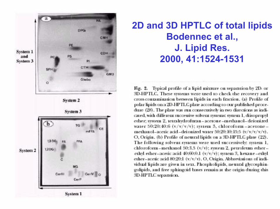
2D and 3D HPTLC of total lipidsBodennec et al.,
J. Lipid Res. 2000, 41:1524-1531

Densitometric analysis of the HPTLC platefollowing migration of gangliosides and visualisation
with resorcinol-HCl
1 2 Gg bovine brain
Sample 2
Sample 1
Wavelength set at 630 nmzig-zag scan (10 mm) with square slit 1/1mm
*length of scanning is equal to that of the plate
ChromatoScan CS-930, Shimadzu, Kyoto, Japan

Application:Use of HPTLC plates in qualitative and quantitative manner
for physical detection with iodine
for chemical detection with specific reagents ,
for immunochemical detection with antibodies,
for radiochemical detection of labeled spots (autoradiograms)
Conclusion:This method gives useful informations about the nature of the lipids
before going to more sophisticated analytical methods such as HPLC,
mass spectrometry, gas chromatography.