p2x7 antagonism using brilliant blue g - peerj · 2017-03-01 · submitted 17 september 2016...
TRANSCRIPT
Submitted 17 September 2016Accepted 4 February 2017Published 1 March 2017
Corresponding authorsRonald Sluyter, [email protected] J. Yerbury,[email protected]
Academic editorRenae Ryan
Additional Information andDeclarations can be found onpage 23
DOI 10.7717/peerj.3064
Copyright2017 Bartlett et al.
Distributed underCreative Commons CC-BY 4.0
OPEN ACCESS
P2X7 antagonism using Brilliant Blue Greduces body weight loss and prolongssurvival in female SOD1G93A amyotrophiclateral sclerosis miceRachael Bartlett, Vanessa Sluyter, Debbie Watson, Ronald Sluyter andJustin J. YerburyIllawarra Health and Medical Research Institute, Wollongong, NSW, AustraliaSchool of Biological Sciences, University of Wollongong, Wollongong, NSW, AustraliaCentre for Medical and Molecular Biosciences, Wollongong, NSW, Australia
ABSTRACTBackground. Amyotrophic lateral sclerosis (ALS) is a rapidly progressive neurode-generative disease characterised by the accumulation of aggregated proteins, microgliaactivation andmotor neuron loss. The mechanisms underlying neurodegeneration anddisease progression in ALS are unknown, but the ATP-gated P2X7 receptor channel isimplicated in this disease. Therefore, the current study aimed to examine P2X7 in thecontext of neurodegeneration, and investigate whether the P2X7 antagonist, BrilliantBlue G (BBG), could alter disease progression in a murine model of ALS.Methods. Human SOD1G93A transgenic mice, which normally develop ALS, wereinjected with BBG or saline, three times per week, from pre-onset of clinical disease(62–64 days of age) until end-stage. During the course of treatment mice were assessedfor weight, clinical score and survival, and motor coordination, which was assessed byrotarod performance. Various parameters from end-stagemice were assessed as follows.Motor neuron loss and microgliosis were assessed by immunohistochemistry. Relativeamounts of lumbar spinal cord SOD1 and P2X7 were quantified by immunoblotting.Serummonocyte chemoattractant protein-1 wasmeasured by ELISA. Splenic leukocytepopulations were assessed by flow cytometry. Relative expression of splenic and hepaticP2X7 mRNA was measured by quantitative real-time PCR. Lumbar spinal cord SOD1and P2X7 were also quantified by immunoblotting in untreated female SOD1G93A miceduring the course of disease.Results. BBG treatment reduced body weight loss in SOD1G93A mice of combined sex,but had no effect on clinical score, survival or motor coordination. BBG treatmentreduced body weight loss in female, but not male, SOD1G93A mice. BBG treatmentalso prolonged survival in female, but not male, SOD1G93A mice, extending the meansurvival time by 4.3% in female mice compared to female mice treated with saline.BBG treatment had no effect on clinical score or motor coordination in either sex. BBGtreatment had no major effect on any end-stage parameters. Total amounts of lumbarspinal cord SOD1 and P2X7 in untreated female SOD1G93A mice did not change overtime.Discussion. Collectively, this data suggests P2X7 may have a partial role in ALS pro-gression in mice, but additional research is required to fully elucidate the contributionof this receptor in this disease.
How to cite this article Bartlett et al. (2017), P2X7 antagonism using Brilliant Blue G reduces body weight loss and prolongs survival infemale SOD1G93A amyotrophic lateral sclerosis mice. PeerJ 5:e3064; DOI 10.7717/peerj.3064
Subjects Molecular Biology, NeuroscienceKeywords P2X7 receptor, SOD1, Amyotrophic lateral sclerosis, Protein aggregation,Motor neurone disease, Purinergic signalling
INTRODUCTIONAmyotrophic lateral sclerosis (ALS) is themost common form of adult-onset motor neurondisease, and is characterised by the degeneration and death of both upper and lower motorneurons (Boillee, Vande Velde & Cleveland, 2006). This leads to progressive spasticity,muscle weakness and muscle atrophy, culminating in paralysis and death, generally within3–5 years from clinical onset (Al-Chalabi et al., 2012). While most cases of ALS are sporadic(sporadic ALS, or sALS) and of unclear cause, the 5–10% of cases in which the disease is in-herited (familial ALS, or fALS) can be linked to specific geneticmutations.Mutations in oneormore of at least a dozen genes give rise to distinct disease neuropathological subtypes, eachof which is associated with the aggregation of a variety of proteins into inclusions with histo-logically distinct structures. The inclusions in ALS can also be categorized on the basis of thedominant protein involved, such as Cu, Zn superoxide dismutase 1 (SOD1), and the RNA-binding proteins, TDP-43 and FUS, which result in inclusions with varied characteristics(Farrawell et al., 2015). Protein aggregates associated with ALS pathology are thought todrive progression of disease, possibly through a prion-like action (Grad et al., 2014) or bychronically activating microglia (Roberts et al., 2013).
SOD1was the first gene inwhichmutationswere found to be associatedwithALS, and ac-count for approximately 20%of familial cases (2%of all cases) (Rosen et al., 1993). To inves-tigate mechanisms of disease and potential therapeutic strategies, several transgenic super-oxide dismutase 1 (SOD1)ALSmousemodels have been generated (Turner & Talbot, 2008),including the SOD1G93A mouse model (Gurney et al., 1994). SOD1G93A mice carry a highcopy number of a transgene encoding for the G93A variant of human SOD1, and developan ALS-like disease which is characterised by the loss of motor neurons leading to hindlimb weakness and paralysis (Gurney et al., 1994). These mice display similar pathologicalhallmarks to those seen in the human disease (Cheroni et al., 2005;Hall, Oostveen & Gurney,1998; Kawaguchi-Niida et al., 2013), and display a similar gender incidence and prevalenceof disease to human ALS (Cervetto et al., 2013;McCombe & Henderson, 2010; Veldink et al.,2003).
Neuroinflammation is emerging as a central component of ALS progression (Henkel etal., 2009). In the central nervous system (CNS), activation of the ATP-gated P2X7 receptorchannel is responsible for mediating a number of neuroinflammatory events, includingmicroglial activation and proliferation (Monif et al., 2009). P2X7 immunoreactivity isincreased in activated microglia of spinal cords from (post-mortem) humans with ALS andadvanced-stage transgenic SOD1G93A rats (Casanovas et al., 2008;Yiangou et al., 2006). Fur-thermore, P2X7 is up-regulated in primary microglia from SOD1G93A mice (D’Ambrosi etal., 2009). These cells produce increased amounts of pro-inflammatory factors and undergooxidative stress following ATP stimulation (Apolloni et al., 2013b; D’Ambrosi et al., 2009;Parisi et al., 2013; Parisi et al., 2016). These exacerbated pro-inflammatory responses lead
Bartlett et al. (2017), PeerJ, DOI 10.7717/peerj.3064 2/29
to the death of neuronal cell lines, and are abrogated by pharmacological blockade or genedeletion of microglial P2X7. Similarly, ATP-induced activation of P2X7 causes neurotoxicphenotypes in wild type (WT) microglia and WT or SOD1G93A astrocytes, leading to thedeath of co-cultured neurons (Gandelman et al., 2010; Skaper et al., 2006).
Despite the deleterious effects reported for P2X7 in an ALS context in vitro, current invivo evidence suggests amore complex dual role for this receptor. In SOD1G93A mice lackingthe P2RX7 gene, females, but not males, had an extended lifespan compared to SOD1G93A
mice encoding P2X7 (Apolloni et al., 2013a). However, these double transgenic mice alsohad anticipated onset, accelerated disease progression and increased astrocytosis,microglio-sis and motor neuron loss compared to mice carrying the SOD1G93A transgene alone. Thissuggests that constitutive deletion of P2X7 is detrimental in ALS, but that this receptormay be neuroprotective during the early stages of the disease. Supporting this hypothesis,administration of the P2X7 antagonist, Brilliant BlueG (BBG), beginning at late pre-onset ofclinical disease delayed disease onset, improved condition andmotor coordination, reducedmicrogliosis and inflammatory markers and enhanced motor neuron survival (Apolloni etal., 2014). Similarly, administration of BBG beginning at disease onset improved motorcoordination and slowed weight loss in male, but not female, SOD1G93A mice (Cervettoet al., 2013). However, BBG inhibition of P2X7 did not correspond with an increase insurvival in either study (Apolloni et al., 2014; Cervetto et al., 2013).
To further investigate P2X7 in ALS progression, this study aimed to investigate whetheradministration of BBG could alter disease progression in SOD1G93A mice, beginning at thepre-onset of clinical disease. Body weight, ALS score and motor coordination were moni-tored throughout. Furthermore, a number of pathological hallmarks of disease were anal-ysed at end-stage, including motor neuron counts, microgliosis, lumbar P2X7 and SOD1protein amounts and serummonocyte chemoattractant protein-1 (MCP-1) concentrations.
MATERIALS AND METHODSReagents and antibodiesBBG, ethidium bromide, ATP, paraformaldehyde (PFA), RNAlater and glycerol gelatinwere from Sigma-Aldrich (St. Louis, MO, USA). Sterile 0.9%NaCl was from Fresenius Kabi(BadHomburg, Germany). Agarose was fromBioline (Alexandria, Australia). Ammonium-chloride-potassium (ACK) lysing buffer, N-PERTM neuronal protein extraction reagent,100 × HaltTM protease inhibitor single-use cocktail, normal horse serum (NHS),DNase/RNase-free distilled water and SuperSignal West Pico Chemiluminescent Substratewere from ThermoFisher Scientific (Waltham, MA, USA). Foetal bovine serum (FBS)was from Lonza (Basel, Switzerland). Tissue-Tek R© optimal cutting temperature (OCT)compound was from Sakura (Flemingweg, Netherlands) and 22 mm glass coverslips werefromMenzel Glaser (Braunschweig, Germany). Diploma full-creammilk powder was fromFonterra (Mount Waverley, Australia). Bovine serum albumin (BSA) and all other reagentgrade chemicals and salts were from Amresco (Solon, OH, USA). The antibodies used arelisted in Table 1.
Bartlett et al. (2017), PeerJ, DOI 10.7717/peerj.3064 3/29
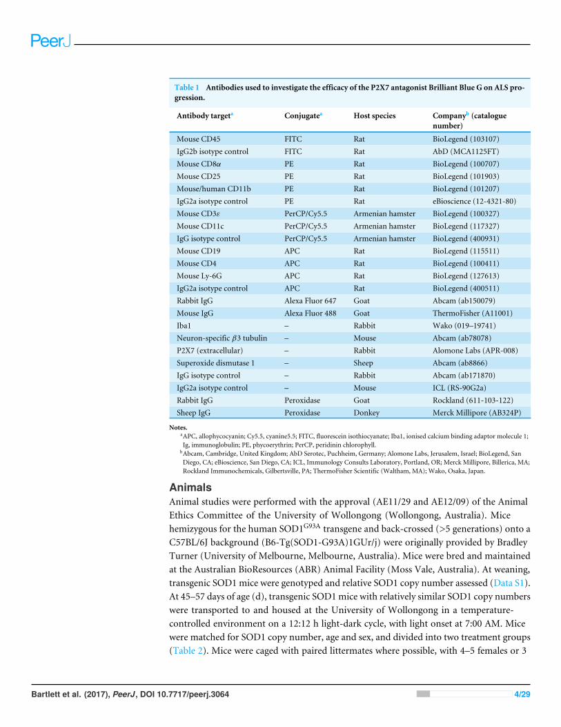
Table 1 Antibodies used to investigate the efficacy of the P2X7 antagonist Brilliant Blue G on ALS pro-gression.
Antibody targeta Conjugatea Host species Companyb (cataloguenumber)
Mouse CD45 FITC Rat BioLegend (103107)IgG2b isotype control FITC Rat AbD (MCA1125FT)Mouse CD8α PE Rat BioLegend (100707)Mouse CD25 PE Rat BioLegend (101903)Mouse/human CD11b PE Rat BioLegend (101207)IgG2a isotype control PE Rat eBioscience (12-4321-80)Mouse CD3ε PerCP/Cy5.5 Armenian hamster BioLegend (100327)Mouse CD11c PerCP/Cy5.5 Armenian hamster BioLegend (117327)IgG isotype control PerCP/Cy5.5 Armenian hamster BioLegend (400931)Mouse CD19 APC Rat BioLegend (115511)Mouse CD4 APC Rat BioLegend (100411)Mouse Ly-6G APC Rat BioLegend (127613)IgG2a isotype control APC Rat BioLegend (400511)Rabbit IgG Alexa Fluor 647 Goat Abcam (ab150079)Mouse IgG Alexa Fluor 488 Goat ThermoFisher (A11001)Iba1 – Rabbit Wako (019–19741)Neuron-specific β3 tubulin – Mouse Abcam (ab78078)P2X7 (extracellular) – Rabbit Alomone Labs (APR-008)Superoxide dismutase 1 – Sheep Abcam (ab8866)IgG isotype control – Rabbit Abcam (ab171870)IgG2a isotype control – Mouse ICL (RS-90G2a)Rabbit IgG Peroxidase Goat Rockland (611-103-122)Sheep IgG Peroxidase Donkey Merck Millipore (AB324P)
Notes.aAPC, allophycocyanin; Cy5.5, cyanine5.5; FITC, fluorescein isothiocyanate; Iba1, ionised calcium binding adaptor molecule 1;Ig, immunoglobulin; PE, phycoerythrin; PerCP, peridinin chlorophyll.
bAbcam, Cambridge, United Kingdom; AbD Serotec, Puchheim, Germany; Alomone Labs, Jerusalem, Israel; BioLegend, SanDiego, CA; eBioscience, San Diego, CA; ICL, Immunology Consults Laboratory, Portland, OR; Merck Millipore, Billerica, MA;Rockland Immunochemicals, Gilbertsville, PA; ThermoFisher Scientific (Waltham, MA); Wako, Osaka, Japan.
AnimalsAnimal studies were performed with the approval (AE11/29 and AE12/09) of the AnimalEthics Committee of the University of Wollongong (Wollongong, Australia). Micehemizygous for the human SOD1G93A transgene and back-crossed (>5 generations) onto aC57BL/6J background (B6-Tg(SOD1-G93A)1GUr/j) were originally provided by BradleyTurner (University of Melbourne, Melbourne, Australia). Mice were bred and maintainedat the Australian BioResources (ABR) Animal Facility (Moss Vale, Australia). At weaning,transgenic SOD1mice were genotyped and relative SOD1 copy number assessed (Data S1).At 45–57 days of age (d), transgenic SOD1mice with relatively similar SOD1 copy numberswere transported to and housed at the University of Wollongong in a temperature-controlled environment on a 12:12 h light-dark cycle, with light onset at 7:00 AM. Micewere matched for SOD1 copy number, age and sex, and divided into two treatment groups(Table 2). Mice were caged with paired littermates where possible, with 4–5 females or 3
Bartlett et al. (2017), PeerJ, DOI 10.7717/peerj.3064 4/29
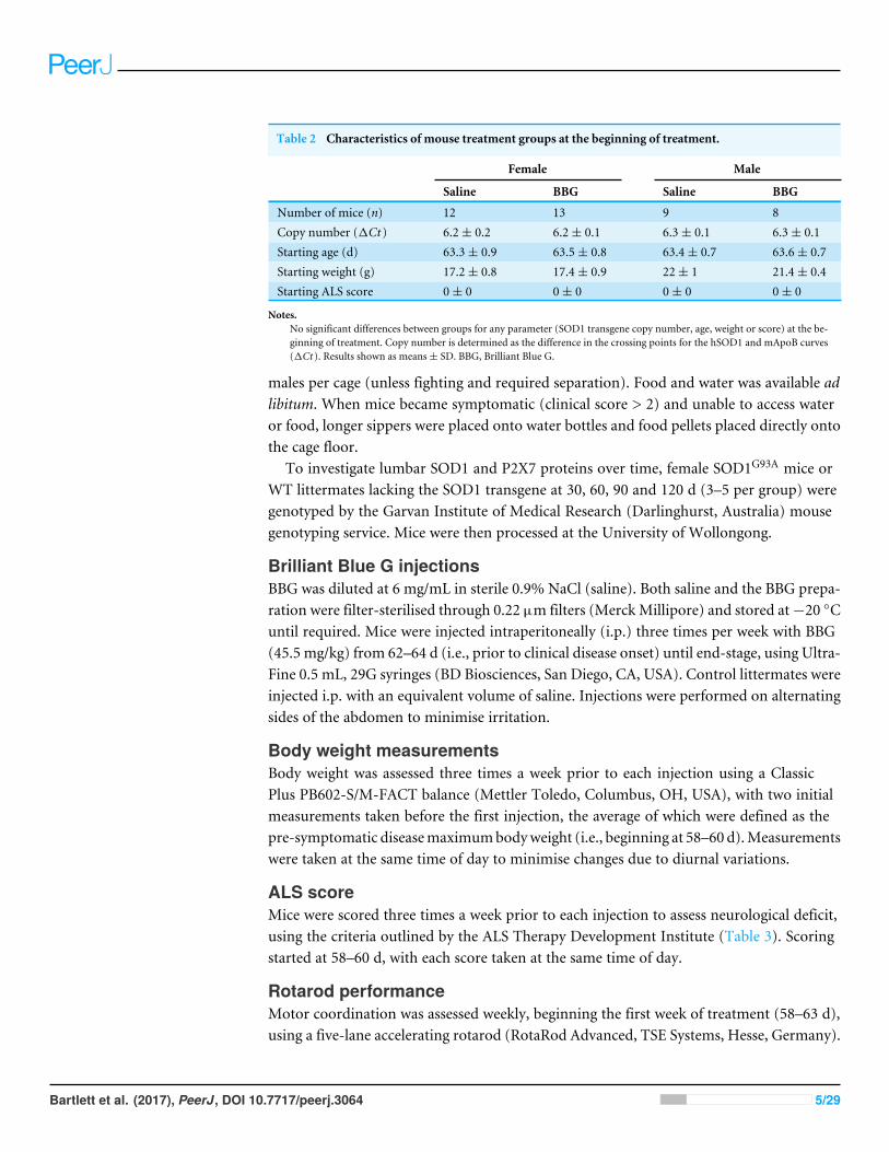
Table 2 Characteristics of mouse treatment groups at the beginning of treatment.
Female Male
Saline BBG Saline BBG
Number of mice (n) 12 13 9 8Copy number (1Ct ) 6.2± 0.2 6.2± 0.1 6.3± 0.1 6.3± 0.1Starting age (d) 63.3± 0.9 63.5± 0.8 63.4± 0.7 63.6± 0.7Starting weight (g) 17.2± 0.8 17.4± 0.9 22± 1 21.4± 0.4Starting ALS score 0± 0 0± 0 0± 0 0± 0
Notes.No significant differences between groups for any parameter (SOD1 transgene copy number, age, weight or score) at the be-ginning of treatment. Copy number is determined as the difference in the crossing points for the hSOD1 and mApoB curves(1Ct ). Results shown as means± SD. BBG, Brilliant Blue G.
males per cage (unless fighting and required separation). Food and water was available adlibitum. When mice became symptomatic (clinical score > 2) and unable to access wateror food, longer sippers were placed onto water bottles and food pellets placed directly ontothe cage floor.
To investigate lumbar SOD1 and P2X7 proteins over time, female SOD1G93A mice orWT littermates lacking the SOD1 transgene at 30, 60, 90 and 120 d (3–5 per group) weregenotyped by the Garvan Institute of Medical Research (Darlinghurst, Australia) mousegenotyping service. Mice were then processed at the University of Wollongong.
Brilliant Blue G injectionsBBG was diluted at 6 mg/mL in sterile 0.9% NaCl (saline). Both saline and the BBG prepa-ration were filter-sterilised through 0.22 µm filters (Merck Millipore) and stored at−20 ◦Cuntil required. Mice were injected intraperitoneally (i.p.) three times per week with BBG(45.5 mg/kg) from 62–64 d (i.e., prior to clinical disease onset) until end-stage, using Ultra-Fine 0.5 mL, 29G syringes (BD Biosciences, San Diego, CA, USA). Control littermates wereinjected i.p. with an equivalent volume of saline. Injections were performed on alternatingsides of the abdomen to minimise irritation.
Body weight measurementsBody weight was assessed three times a week prior to each injection using a ClassicPlus PB602-S/M-FACT balance (Mettler Toledo, Columbus, OH, USA), with two initialmeasurements taken before the first injection, the average of which were defined as thepre-symptomatic diseasemaximumbodyweight (i.e., beginning at 58–60 d).Measurementswere taken at the same time of day to minimise changes due to diurnal variations.
ALS scoreMice were scored three times a week prior to each injection to assess neurological deficit,using the criteria outlined by the ALS Therapy Development Institute (Table 3). Scoringstarted at 58–60 d, with each score taken at the same time of day.
Rotarod performanceMotor coordination was assessed weekly, beginning the first week of treatment (58–63 d),using a five-lane accelerating rotarod (RotaRod Advanced, TSE Systems, Hesse, Germany).
Bartlett et al. (2017), PeerJ, DOI 10.7717/peerj.3064 5/29
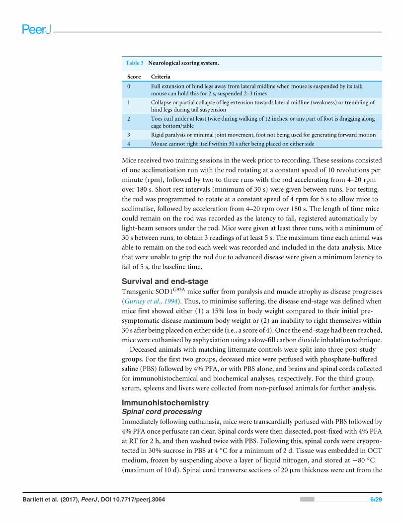
Table 3 Neurological scoring system.
Score Criteria
0 Full extension of hind legs away from lateral midline when mouse is suspended by its tail;mouse can hold this for 2 s, suspended 2–3 times
1 Collapse or partial collapse of leg extension towards lateral midline (weakness) or trembling ofhind legs during tail suspension
2 Toes curl under at least twice during walking of 12 inches, or any part of foot is dragging alongcage bottom/table
3 Rigid paralysis or minimal joint movement, foot not being used for generating forward motion4 Mouse cannot right itself within 30 s after being placed on either side
Mice received two training sessions in the week prior to recording. These sessions consistedof one acclimatisation run with the rod rotating at a constant speed of 10 revolutions perminute (rpm), followed by two to three runs with the rod accelerating from 4–20 rpmover 180 s. Short rest intervals (minimum of 30 s) were given between runs. For testing,the rod was programmed to rotate at a constant speed of 4 rpm for 5 s to allow mice toacclimatise, followed by acceleration from 4–20 rpm over 180 s. The length of time micecould remain on the rod was recorded as the latency to fall, registered automatically bylight-beam sensors under the rod. Mice were given at least three runs, with a minimum of30 s between runs, to obtain 3 readings of at least 5 s. The maximum time each animal wasable to remain on the rod each week was recorded and included in the data analysis. Micethat were unable to grip the rod due to advanced disease were given a minimum latency tofall of 5 s, the baseline time.
Survival and end-stageTransgenic SOD1G93A mice suffer from paralysis and muscle atrophy as disease progresses(Gurney et al., 1994). Thus, to minimise suffering, the disease end-stage was defined whenmice first showed either (1) a 15% loss in body weight compared to their initial pre-symptomatic disease maximum body weight or (2) an inability to right themselves within30 s after being placed on either side (i.e., a score of 4). Once the end-stage had been reached,mice were euthanised by asphyxiation using a slow-fill carbon dioxide inhalation technique.
Deceased animals with matching littermate controls were split into three post-studygroups. For the first two groups, deceased mice were perfused with phosphate-bufferedsaline (PBS) followed by 4% PFA, or with PBS alone, and brains and spinal cords collectedfor immunohistochemical and biochemical analyses, respectively. For the third group,serum, spleens and livers were collected from non-perfused animals for further analysis.
ImmunohistochemistrySpinal cord processingImmediately following euthanasia, mice were transcardially perfused with PBS followed by4% PFA once perfusate ran clear. Spinal cords were then dissected, post-fixed with 4% PFAat RT for 2 h, and then washed twice with PBS. Following this, spinal cords were cryopro-tected in 30% sucrose in PBS at 4 ◦C for a minimum of 2 d. Tissue was embedded in OCTmedium, frozen by suspending above a layer of liquid nitrogen, and stored at −80 ◦C(maximum of 10 d). Spinal cord transverse sections of 20 µm thickness were cut from the
Bartlett et al. (2017), PeerJ, DOI 10.7717/peerj.3064 6/29
lumbar regions of the spinal cords on a CM1950 cryostat (Leica, Mannheim, Germany),using spinal cord enlargement to identify the lumbar region. Sections were mounted ontoStarFrost R© advanced adhesive slides (Knittel Glaser, Braunschweig, Germany) and storedat −80 ◦C (maximum of four weeks).
Iba1 and β3-tubulin stainingMicroglia and motor neuron numbers were assessed by staining mounted spinal cords forIba1 and β3-tubulin, respectively. Firstly, a pap pen (Daido Sangyo, Tokyo, Japan) was usedto separate tissue sections on the same slide. All incubations were carried out in a humidifiedchamber. Sections were fixed with 4% PFA in PBS at room temperature (RT) for 15 min,and then washed three times with PBS over 30 min. Sections were then blocked with 20%NHS in PBS at RT for 20 min, and incubated at 4 ◦C overnight with rabbit anti-Iba1 poly-clonal antibody (pAb) (0.1µg/100µL) andmouse β3-tubulinmonoclonal antibody (mAb)(0.5 µg/100 µL), or rabbit IgG isotype control pAb and mouse IgG2a isotype control atcorresponding concentrations, all in PBS containing 1% BSA, 0.2% NHS and 0.05%NaN3.The following day, sections were washed as above, and incubated at RT for 1 h withAlexa Fluor 647-conjugated goat anti-rabbit IgG Ab (0.3 µg/100 µL) and Alexa Fluor 488-conjugated goat anti-mouse IgG Ab (0.3 µg/100 µL) in PBS containing 0.2% NHS. Cellswere washed as above and then coverslipsmounted onto tissue sections with 50% (v/v) glyc-erol gelatin in PBS. Coverslips were sealed with nail varnish. Sections were visualised usinga TCS SP5 II confocal imaging system and images of anterior horns captured using Leica Ap-plication Suite Advanced Fluorescence Lite software (version 2.6.3) (Leica) (excitation 633,emission collected at 655–695 nm for Iba1; excitation 488 nm, emission collected at 510–550nm for β3-tubulin). Images were taken of 6–12 anterior horns per animal, for a total of 12animals (6 per treatment group). Spinal cord anterior horns were defined as the regionsof gray matter on the ventral side of a horizontal line crossing through the central canal.
Motor neuron countsThe numbers of motor neurons in anterior horns were manually counted using images ofβ3-tubulin-stained spinal cords visualised in Leica Application Suite Advanced Fluores-cence Lite software. Motor neurons were defined as cells with cell body diameters of at least15 µm showing positive immunostaining for β3 tubulin. Counts from the right and leftanterior horns for each section were averaged, and then the average count from 3–6 sectionsused as a single measurement per animal for statistical analyses.
Microglia densityMicroglia density was assessed in anterior horns using images of Iba1-stained spinal cordsand ImageJ software (version 1.48) (National Institutes of Health, Bethesda, MD). Firstly,thresholds were adjusted into 16-bit black and white images to account for any differencesin the intensity of staining or in the background fluorescence between sections. Particlesbetween 10–1,000 pixel units were then counted, and the percent area showing positiveimmunostaining for Iba1 of the total anterior horn calculated. Percent areas from the rightand left anterior horns for each section were averaged, and then the average count from3–6 sections used as a single measurement per animal for statistical analyses.
Bartlett et al. (2017), PeerJ, DOI 10.7717/peerj.3064 7/29
ImmunoblottingSpinal cord protein extractionImmediately following euthanasia, mice were transcardially perfusedwith PBS. Spinal cordswere then dissected, and the lumbar portion excised using spinal cord enlargement to iden-tify the region. Lumbar spinal cord segments were snap-frozen in liquid nitrogen and storedat −80 ◦C until required. Detergent-soluble proteins were extracted from lumbar spinalcords by homogenising tissue in ice-cold neuronal protein extraction reagent containing 100×HaltTM protease inhibitor cocktail using amicropestle. A ratio of 10µL extraction reagentper 1 mg of tissue was utilised. Homogenates were incubated on ice for 10 min and cleared(20,000× g at 4 ◦C for 10 min). To extract detergent-insoluble proteins, the resulting pelletwas resuspended in 0.5 M Tris HCl (pH 6.8) containing 2% w/v SDS at the same volumeas utilised for soluble protein extraction (to maintain a consistent concentration with theoriginal solution), and incubated at RT for 10 min. Unsolubilised material was cleared(20,000 × g for 15 min). Soluble and insoluble protein samples were stored at −20 ◦C.
P2X7 and SOD1 protein detectionSoluble protein (50 µg) or an equivalent volume of insoluble protein were separated underreducing conditions (5% β-mercaptoethanol) using Any kD Mini-PROTEAN TGX Stain-Free Gels (Bio-Rad, Hercules, CA, USA). Before immunoblotting, equal protein loadingwas confirmed by visualising stain-free gels using a Bio-Rad Criterion Stain Free Imager andImage Lab software. Proteins were then transferred to nitrocellulose membranes (Bio-Rad)using a Bio-Rad Trans-Blot Turbo Transfer System. To allow for separate P2X7 and SOD1immunoblotting, membranes were cut horizontally between 25 and 37 kDa, using the pre-stained marker as a guide. Both halves were then blocked at RT for 1 h with Tris-bufferedsaline (250 mM NaCl and 50 mM Tris, pH 7.5) containing 0.2% Tween-20 and 5% milkpowder, and then incubated at 4 ◦C overnight with either an anti-P2X7 pAb (1:500) or anti-SOD1pAb (1:1000) inTris-buffered saline containing 0.2%Tween-20 and 5%milk powder.Membranes were washed three times over 30 min with Tris-buffered saline containing0.2% Tween-20. Membranes were then incubated at RT for 1 h with peroxidise-conjugatedanti-rabbit (1:1000) or anti-sheep (1:500) IgG Ab (for P2X7 or SOD1, respectively) inTris-buffered saline containing 0.2% Tween-20 and 5% milk powder. Membranes werewashed as above, and proteins visualised using chemiluminescent substrate and AmershamHyperfilm ECL (GE Healthcare, Little Chalfont, Buckinghamshire, UK). Films wereprocessed using GBX Developer and Replenisher, and GBX Fixer and Replenisher as perthe manufacturer’s instructions (Kodak Australasia, Collingwood, Australia). Images offilms were collected using a GS-800 Calibrated Densitometer (Bio-Rad). Relative P2X7and SOD1 was quantified from these images using ImageJ software, normalising to a singlesoluble protein sample included on all gels.
Serum monocyte chemoattractant protein-1 measurementsBlood was collected immediately following euthanasia via cardiac puncture using a 27GPrecisionGlide needle (BD Biosciences). Blood was incubated at RT for 45–90 min andthen centrifuged (1,700× g for 5min). Serumwas stored at−80 ◦C. The amount ofMCP-1
Bartlett et al. (2017), PeerJ, DOI 10.7717/peerj.3064 8/29
present in serum was measured using a mouse CCL2 (MCP-1) ELISA Ready-SET-Go! kit(eBioscience, San Diego, CA, USA), as per the manufacturer’s instructions.
Splenic leukocyte phenotypingSpleens from euthanisedmice were homogenised in PBS and filtered through Falcon 70µmnylon filters (BDBiosciences). Cells were washedwith PBS (300× g for 5min), resuspendedin lysing buffer (155 mM NH4Cl, 10 mM KHCO3 and 0.1 mM Na2EDTA, pH 7.4), andincubated at RT for 5 min. Cells were then washed as above and resuspended in cold PBScontaining 2%FBS.Cells (1×10⁶/tube)were incubated on ice for 20minwith combinationsof fluorochrome-conjugated anti-mouse mAbs or corresponding isotype control mAbs.Cells were washed as above, and events collected using a LSRFortessa X-20 Cell Analyzer(BD Biosciences) (excitation 488 nm, emission collected with 525/50 and 695/40 band-passfilters for FITC- and PerCP/Cy5.5-conjugated mAbs, respectively; excitation 561, emissioncollected with 585/15 band-pass filter for PE-conjugated mAbs; excitation 640, emissioncollected with 670/30 band-pass filter for APC-conjugated mAbs). Percentages of leukocytesubsets were determined using FlowJo software (version 8.7) (Tree Star, Ashland,OR,USA).
P2X7 expression by qPCRSections of livers and spleens, approximately 5 mm3 in size, were stored in RNAlaterat 4 ◦C. After 1–2 d, tissues were transferred to −20 ◦C. Tissues were removed fromRNAlater and total RNA isolated using TRIzol reagent (ThermoFisher Scientific), as per themanufacturer’s instructions. cDNAwas synthesised from isolated RNAusing qScript cDNASynthesis Kit (Quanta Biosciences, Gaithersburg, MD, USA), as per the manufacturer’s in-structions. qPCR amplification was performed in a 10 µL reaction volume, containing 5 µL2×TaqManUniversalMasterMix II (ThermoFisher Scientific), 2µL liver or spleen cDNA,0.5 µL of 20 × TaqMan Gene Expression Assay-Specific primers/probes (ThermoFisherScientific), and PCR-grade water. Standard primers for murine P2RX7 (FAM-labelled;Mm00440578_m1) and murine glyceraldehyde 3-phosphate dehydrogenase (GAPDH )(VIC-labelled; Mm99999915_g1) were used. Amplification was performed using an EcoReal-Time PCR System (Illumina, San Diego, CA, USA). Reactions were held at 50 ◦C for 2min and 95 ◦C for 10min, followed by 40 two-segment cycles of 95 ◦C for 15 s and 60 ◦C for1min. A single fluorescence acquisition was taken each cycle, using FAM (505–545 nm) andVIC (562–596 nm) filter combinations. No-template controls were included in each runand all samples were run in triplicate. cDNA obtained from the spleen of a BALB/c mouse(Watson et al., 2014) was also included in each run to compare relative P2X7 expression(given a value of 1). Relative gene expression was normalised to the murine GAPDHhousekeeping gene and determined using EcoStudy software (version 4.1.2.0) (Illumina).
Data presentation and statistical analysesData is presented as mean ± SD, unless otherwise indicated. Differences between multipletreatments were compared by ANOVA paired with Tukey’s HSD post-tests. For singlecomparisons, unpaired Student’s t -tests were performed. Differences in body weightbetween treatment groups were compared using an unpaired Student’s t -tests in two ways:first, for the mean percentage body weight for each mouse over the entire period of the
Bartlett et al. (2017), PeerJ, DOI 10.7717/peerj.3064 9/29
study; second, for the percentage body weight for each mouse at each recorded time-point.For ALS score, rotarod performance and survival data, differences in the values of BBG-and saline-treated mice were assessed using Kaplan–Meier analysis paired with log-ranktests, considering the time to reach a consistent score of 2, drop to a consistent run of lessthan 60 s or end-stage, respectively, as an event. To prevent decomposition of means due toanimals reaching end-stage, for weight, score and rotarod analyses, the last values prior toeuthanasia were carried forward until the last mouse in each group had reached end-stage.These values were used to compute means at the end of the study. The software packagePrism 5 for Windows (version 5.01) (GraphPad Software, San Diego, CA, USA) was usedfor all statistical analyses, with differences considered significant for P < 0.05.
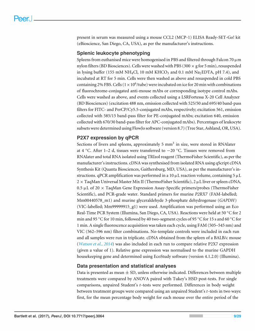
RESULTSBrilliant Blue G treatment reduces body weight loss in SOD1G93A mice,but has no effect on clinical score, motor coordination or survivalThe P2X7 antagonist, BBG, has been used in over 20 pre-clinical studies of neurological-related disease in rodents (Bartlett, Stokes & Sluyter, 2014). To investigate the therapeuticefficacy of BBG treatment on ALS progression, SOD1G93A mice were treated with saline orBBG from 62–64 d (i.e., prior to clinical disease onset) until end-stage, and body weight,ALS score and motor coordination recorded for the duration of the study.
Body weight of saline- and BBG-treated SOD1G93A mice was maintained or increaseduntil approximately 105 d, after which continuous body weight loss was observed (Fig.1A). Overall, BBG-treated mice had greater initial weight gain and a delayed decline inweight compared to saline-treated mice, in addition, BBG treated mice had a higher meanpercentage body weight (P = 0.014, n= 21), when calculated for eachmouse over the entireperiod of the study (Fig. 1A, insert). Furthermore, at 63, 66, 80, 82, 84, 87, 89, 91, 94, 96,98, 101, 103, 105, 108, 117, 119, 122 and 131 d, BBG-treated mice had significantly greaterpercentage body weights compared to saline-treated mice (P < 0.05, n= 21) (Fig. 1A).
To assess neurological deficit, mice were scored from 58-60 d using the criteria outlinedby the ALS Therapy Development Institute (Table 3). At 58–60 d, all saline- and BBG-treated mice had clinical scores of 0 (Fig. 1B). These scores increased throughout the study,plateauing at an approximate score of 2.5 at 145 and 150 d for saline- and BBG-treatedmice,respectively. Overall, considering the time to maintain a consistent score of 2 as anevent, there was no significant difference in score between saline- and BBG-treated mice(P = 0.349, n= 21) (Fig. 1B).
Motor coordination was assessed weekly using an accelerating rotarod, beginning at58–63 d.Motor coordination initially rose for both saline- andBBG-treatedmice, consistentwithmice continuing to learn how to use the rod, andwas thenmaintainedwith some varia-tion until 95 d (Fig. 1C). After 95 d, motor coordination rapidly declined for both treatmentgroups, with comparable rates of decline (P = 0.311, n= 21 considering the time to reacha latency to fall consistently less than 60 s) (Fig. 1C).
End-stage was considered reached once mice displayed either a 15% loss in body weightcompared to their initial pre-symptomatic disease maximum body weight, or an inability
Bartlett et al. (2017), PeerJ, DOI 10.7717/peerj.3064 10/29
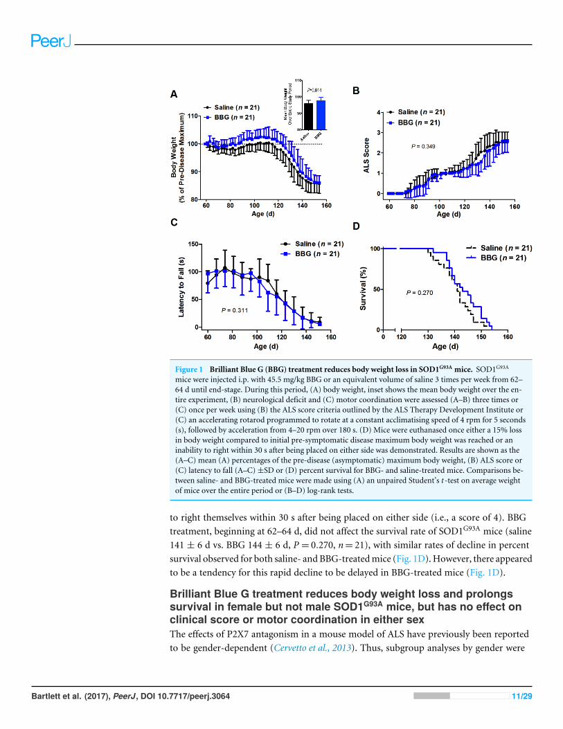
Figure 1 Brilliant Blue G (BBG) treatment reduces body weight loss in SOD1G93A mice. SOD1G93A
mice were injected i.p. with 45.5 mg/kg BBG or an equivalent volume of saline 3 times per week from 62–64 d until end-stage. During this period, (A) body weight, inset shows the mean body weight over the en-tire experiment, (B) neurological deficit and (C) motor coordination were assessed (A–B) three times or(C) once per week using (B) the ALS score criteria outlined by the ALS Therapy Development Institute or(C) an accelerating rotarod programmed to rotate at a constant acclimatising speed of 4 rpm for 5 seconds(s), followed by acceleration from 4–20 rpm over 180 s. (D) Mice were euthanased once either a 15% lossin body weight compared to initial pre-symptomatic disease maximum body weight was reached or aninability to right within 30 s after being placed on either side was demonstrated. Results are shown as the(A–C) mean (A) percentages of the pre-disease (asymptomatic) maximum body weight, (B) ALS score or(C) latency to fall (A–C)±SD or (D) percent survival for BBG- and saline-treated mice. Comparisons be-tween saline- and BBG-treated mice were made using (A) an unpaired Student’s t -test on average weightof mice over the entire period or (B–D) log-rank tests.
to right themselves within 30 s after being placed on either side (i.e., a score of 4). BBGtreatment, beginning at 62–64 d, did not affect the survival rate of SOD1G93A mice (saline141 ± 6 d vs. BBG 144 ± 6 d, P = 0.270, n= 21), with similar rates of decline in percentsurvival observed for both saline- and BBG-treatedmice (Fig. 1D).However, there appearedto be a tendency for this rapid decline to be delayed in BBG-treated mice (Fig. 1D).
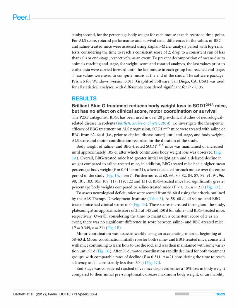
Brilliant Blue G treatment reduces body weight loss and prolongssurvival in female but not male SOD1G93A mice, but has no effect onclinical score or motor coordination in either sexThe effects of P2X7 antagonism in a mouse model of ALS have previously been reportedto be gender-dependent (Cervetto et al., 2013). Thus, subgroup analyses by gender were
Bartlett et al. (2017), PeerJ, DOI 10.7717/peerj.3064 11/29
performed to elucidate whether any differences in body weight, clinical score, motor coor-dination or survival could be observed following BBG treatment in either males or females.
Body weight loss occurred in a comparable manner for both saline- and BBG-treatedmale mice (P = 0.649, n= 8–9) (Fig. 2A and insert). Furthermore, there were no significantdifferences in male body weight as a percentage of the pre-symptomatic disease maximumson any weight measurement day. In contrast, BBG-treated female mice had significantlygreater initial weight gain and a delayed decline in weight compared to saline-treated femalemice over the entire period of the study (P = 0.006, n= 12–13) (Fig. 2A and insert). At 63,66, 73, 80, 82, 84, 87, 89, 91, 94, 96, 98, 101, 103, 105, 108, 117, 119, 122, 126 and 131 d,BBG-treated female mice had significantly greater percentage body weights compared tosaline-treated female mice (P < 0.05, n= 12–13) (Fig. 2A).
Score trajectories similar to those described above were observed for both male andfemale saline- and BBG-treatedmice (Fig. 2B). Overall, there were no significant differencesin score between saline- and BBG-treated male or saline- and BBG-treated female micewhen considering the time to maintain a consistent score of 2 as an event (male P = 0.647,n= 8–9; female P = 0.084, n= 12–13) (Fig. 2B).
Similar patterns ofmotor deficit to that described above were also observed for bothmaleand female saline- and BBG-treated mice (Fig. 2C), again with comparable rates of declinebetween those treated with saline and those with BBG (male P = 0.390, n= 8–9; female P =0.586, n= 12–13; considering the time to reach a latency to fall consistently less than 60 s).
When considering males alone, survival was not affected by BBG treatment (saline 143± 7 d vs. BBG 142 ± 6 d, P = 0.582, n= 8–9) (Fig. 2D). Conversely, for females, BBGtreatment extended survival (saline 139± 6 d vs. BBG 145± 6 d, P = 0.035, n= 12–13) (Fig.2D). Whilst similar rates of decline were observed for both female saline- and BBG-treatedmice, this decline was delayed following BBG treatment in females.
Brilliant Blue G treatment does not reduce motor neuron loss ormicrogliosis in SOD1G93A miceTo determine the effect of BBG-treatment on motor neuron loss and microgliosis, neuronsand microglia in the anterior horn of lumber spinal cords from end-stage saline- andBBG-treated mice were labelled and imaged by confocal microscopy. Motor neurons werecounted and the density of microglia assessed. The lumbar spinal cord was chosen as this isthe CNS tissue in which lower motor neurons reside and that is predominately damagedduring ALS (Apolloni et al., 2013a).
Representative images of stained anterior horn sections from saline and BBG-treatedSOD1G93A mice are shown (Fig. 3). A stained section derived from a WT littermate ofcorresponding age is included for comparison. The average number of motor neurons,identified as cells with cell body diameters of at least 15 µm, was similar between saline- andBBG-treated SOD1G93A mice (saline 19.3 ± 2.0 vs. 19.5 ± 1.4 motor neurons, P = 0.868,n= 6) (Fig. 3A). Similarly, there was no difference in the amount of microgliosis betweensaline- and BBG-treatedmice (saline 2.6± 0.3% vs. 2.5± 0.8%, P = 0.821, n= 6) (Fig. 3B).
Bartlett et al. (2017), PeerJ, DOI 10.7717/peerj.3064 12/29
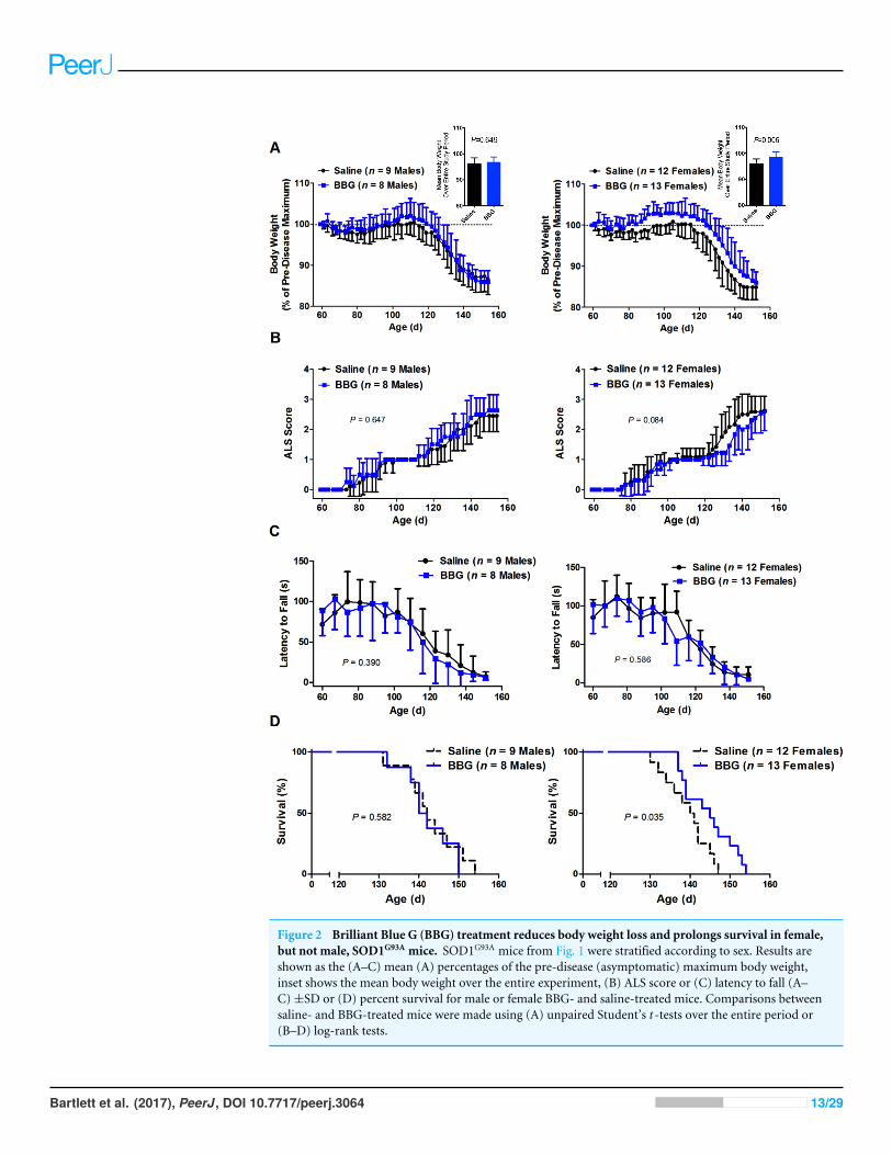
Figure 2 Brilliant Blue G (BBG) treatment reduces body weight loss and prolongs survival in female,but not male, SOD1G93A mice. SOD1G93A mice from Fig. 1 were stratified according to sex. Results areshown as the (A–C) mean (A) percentages of the pre-disease (asymptomatic) maximum body weight,inset shows the mean body weight over the entire experiment, (B) ALS score or (C) latency to fall (A–C)±SD or (D) percent survival for male or female BBG- and saline-treated mice. Comparisons betweensaline- and BBG-treated mice were made using (A) unpaired Student’s t -tests over the entire period or(B–D) log-rank tests.
Bartlett et al. (2017), PeerJ, DOI 10.7717/peerj.3064 13/29
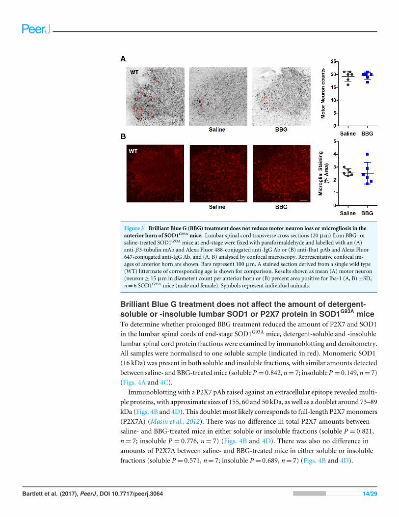
Figure 3 Brilliant Blue G (BBG) treatment does not reduce motor neuron loss or microgliosis in theanterior horn of SOD1G93A mice. Lumbar spinal cord transverse cross sections (20 µm) from BBG- orsaline-treated SOD1G93A mice at end-stage were fixed with paraformaldehyde and labelled with an (A)anti-β3-tubulin mAb and Alexa Fluor 488-conjugated anti-IgG Ab or (B) anti-Iba1 pAb and Alexa Fluor647-conjugated anti-IgG Ab, and (A, B) analysed by confocal microscopy. Representative confocal im-ages of anterior horn are shown. Bars represent 100 µm. A stained section derived from a single wild type(WT) littermate of corresponding age is shown for comparison. Results shown as mean (A) motor neuron(neuron ≥ 15 µm in diameter) count per anterior horn or (B) percent area positive for Iba-1 (A, B)±SD,n= 6 SOD1G93A mice (male and female). Symbols represent individual animals.
Brilliant Blue G treatment does not affect the amount of detergent-soluble or -insoluble lumbar SOD1 or P2X7 protein in SOD1G93A miceTo determine whether prolonged BBG treatment reduced the amount of P2X7 and SOD1in the lumbar spinal cords of end-stage SOD1G93A mice, detergent-soluble and -insolublelumbar spinal cord protein fractions were examined by immunoblotting and densitometry.All samples were normalised to one soluble sample (indicated in red). Monomeric SOD1(16 kDa) was present in both soluble and insoluble fractions, with similar amounts detectedbetween saline- andBBG-treatedmice (solubleP = 0.842, n= 7; insolubleP = 0.149, n= 7)(Figs. 4A and 4C).
Immunoblotting with a P2X7 pAb raised against an extracellular epitope revealed multi-ple proteins, with approximate sizes of 155, 60 and 50 kDa, as well as a doublet around 73–89kDa (Figs. 4B and 4D). This doublet most likely corresponds to full-length P2X7monomers(P2X7A) (Masin et al., 2012). There was no difference in total P2X7 amounts betweensaline- and BBG-treated mice in either soluble or insoluble fractions (soluble P = 0.821,n= 7; insoluble P = 0.776, n= 7) (Figs. 4B and 4D). There was also no difference inamounts of P2X7A between saline- and BBG-treated mice in either soluble or insolublefractions (soluble P = 0.571, n= 7; insoluble P = 0.689, n= 7) (Figs. 4B and 4D).
Bartlett et al. (2017), PeerJ, DOI 10.7717/peerj.3064 14/29
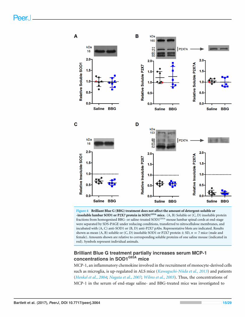
Figure 4 Brilliant Blue G (BBG) treatment does not affect the amount of detergent-soluble or-insoluble lumbar SOD1 or P2X7 protein in SOD1G93A mice. (A, B) Soluble or (C, D) insoluble proteinfractions from homogenised BBG- or saline-treated SOD1G93A mouse lumbar spinal cords at end-stagewere separated by SDS-PAGE under reducing conditions, transferred to nitrocellulose membranes, andincubated with (A, C) anti-SOD1 or (B, D) anti-P2X7 pAbs. Representative blots are indicated. Resultsshown as mean (A, B) soluble or (C, D) insoluble SOD1 or P2X7 protein± SD, n = 7 mice (male andfemale). Amounts shown are relative to corresponding soluble proteins of one saline mouse (indicated inred). Symbols represent individual animals.
Brilliant Blue G treatment partially increases serum MCP-1concentrations in SOD1G93A miceMCP-1, an inflammatory chemokine involved in the recruitment ofmonocyte-derived cellssuch as microglia, is up-regulated in ALS mice (Kawaguchi-Niida et al., 2013) and patients(Henkel et al., 2004; Nagata et al., 2007; Wilms et al., 2003). Thus, the concentrations ofMCP-1 in the serum of end-stage saline- and BBG-treated mice was investigated to
Bartlett et al. (2017), PeerJ, DOI 10.7717/peerj.3064 15/29
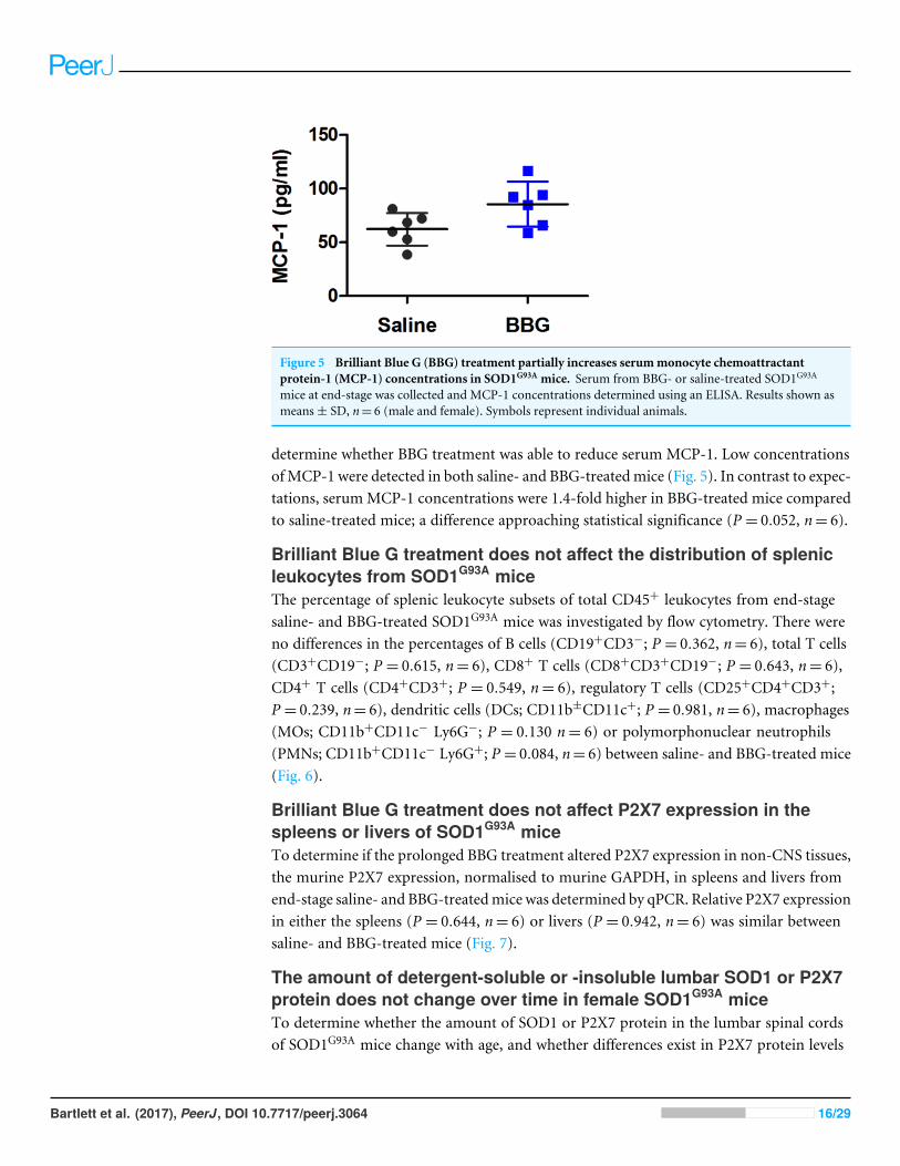
Figure 5 Brilliant Blue G (BBG) treatment partially increases serummonocyte chemoattractantprotein-1 (MCP-1) concentrations in SOD1G93A mice. Serum from BBG- or saline-treated SOD1G93A
mice at end-stage was collected and MCP-1 concentrations determined using an ELISA. Results shown asmeans± SD, n= 6 (male and female). Symbols represent individual animals.
determine whether BBG treatment was able to reduce serum MCP-1. Low concentrationsofMCP-1 were detected in both saline- and BBG-treatedmice (Fig. 5). In contrast to expec-tations, serumMCP-1 concentrations were 1.4-fold higher in BBG-treated mice comparedto saline-treated mice; a difference approaching statistical significance (P = 0.052, n= 6).
Brilliant Blue G treatment does not affect the distribution of splenicleukocytes from SOD1G93A miceThe percentage of splenic leukocyte subsets of total CD45+ leukocytes from end-stagesaline- and BBG-treated SOD1G93A mice was investigated by flow cytometry. There wereno differences in the percentages of B cells (CD19+CD3−; P = 0.362, n= 6), total T cells(CD3+CD19−; P = 0.615, n= 6), CD8+ T cells (CD8+CD3+CD19−; P = 0.643, n= 6),CD4+ T cells (CD4+CD3+; P = 0.549, n= 6), regulatory T cells (CD25+CD4+CD3+;P = 0.239, n= 6), dendritic cells (DCs; CD11b±CD11c+; P = 0.981, n= 6), macrophages(MOs; CD11b+CD11c− Ly6G−; P = 0.130 n= 6) or polymorphonuclear neutrophils(PMNs; CD11b+CD11c− Ly6G+; P = 0.084, n= 6) between saline- and BBG-treated mice(Fig. 6).
Brilliant Blue G treatment does not affect P2X7 expression in thespleens or livers of SOD1G93A miceTo determine if the prolonged BBG treatment altered P2X7 expression in non-CNS tissues,the murine P2X7 expression, normalised to murine GAPDH, in spleens and livers fromend-stage saline- and BBG-treatedmice was determined by qPCR. Relative P2X7 expressionin either the spleens (P = 0.644, n= 6) or livers (P = 0.942, n= 6) was similar betweensaline- and BBG-treated mice (Fig. 7).
The amount of detergent-soluble or -insoluble lumbar SOD1 or P2X7protein does not change over time in female SOD1G93A miceTo determine whether the amount of SOD1 or P2X7 protein in the lumbar spinal cordsof SOD1G93A mice change with age, and whether differences exist in P2X7 protein levels
Bartlett et al. (2017), PeerJ, DOI 10.7717/peerj.3064 16/29
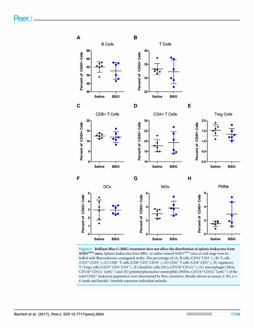
Figure 6 Brilliant Blue G (BBG) treatment does not affect the distribution of splenic leukocytes fromSOD1G93A mice. Splenic leukocytes from BBG- or saline-treated SOD1G93A mice at end-stage were la-belled with fluorochrome-conjugated mAbs. The percentage of (A) B cells (CD19+CD3−), (B) T cells(CD3+CD19−), (C) CD8+ T cells (CD8+CD3+CD19−), (D) CD4+ T cells (CD4+CD3+), (E) regulatoryT (Treg) cells (CD25+CD4+CD3+), (F) dendritic cells (DCs; CD11b±CD11c+), (G) macrophages (MOs;CD11b+CD11c−Ly6G−) and (H) polymorphonuclear neutrophils (PMNs; CD11b+CD11c−Ly6G+) of thetotal CD45+ leukocyte population were determined by flow cytometry. Results shown as means± SD, n=6 (male and female). Symbols represent individual animals.
Bartlett et al. (2017), PeerJ, DOI 10.7717/peerj.3064 17/29
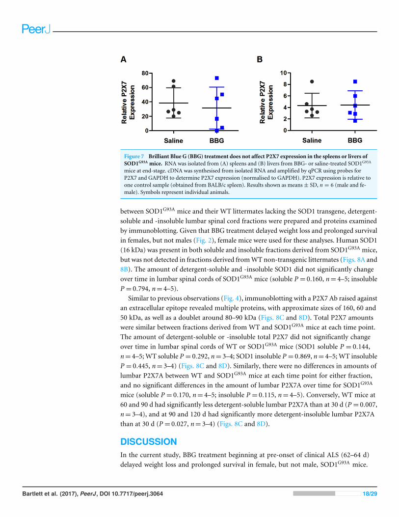
Figure 7 Brilliant Blue G (BBG) treatment does not affect P2X7 expression in the spleens or livers ofSOD1G93A mice. RNA was isolated from (A) spleens and (B) livers from BBG- or saline-treated SOD1G93A
mice at end-stage. cDNA was synthesised from isolated RNA and amplified by qPCR using probes forP2X7 and GAPDH to determine P2X7 expression (normalised to GAPDH). P2X7 expression is relative toone control sample (obtained from BALB/c spleen). Results shown as means± SD, n = 6 (male and fe-male). Symbols represent individual animals.
between SOD1G93A mice and their WT littermates lacking the SOD1 transgene, detergent-soluble and -insoluble lumbar spinal cord fractions were prepared and proteins examinedby immunoblotting. Given that BBG treatment delayed weight loss and prolonged survivalin females, but not males (Fig. 2), female mice were used for these analyses. Human SOD1(16 kDa) was present in both soluble and insoluble fractions derived from SOD1G93A mice,but was not detected in fractions derived fromWT non-transgenic littermates (Figs. 8A and8B). The amount of detergent-soluble and -insoluble SOD1 did not significantly changeover time in lumbar spinal cords of SOD1G93A mice (soluble P = 0.160, n= 4–5; insolubleP = 0.794, n= 4–5).
Similar to previous observations (Fig. 4), immunoblotting with a P2X7 Ab raised againstan extracellular epitope revealed multiple proteins, with approximate sizes of 160, 60 and50 kDa, as well as a doublet around 80–90 kDa (Figs. 8C and 8D). Total P2X7 amountswere similar between fractions derived from WT and SOD1G93A mice at each time point.The amount of detergent-soluble or -insoluble total P2X7 did not significantly changeover time in lumbar spinal cords of WT or SOD1G93A mice (SOD1 soluble P = 0.144,n= 4–5; WT soluble P = 0.292, n= 3–4; SOD1 insoluble P = 0.869, n= 4–5; WT insolubleP = 0.445, n= 3–4) (Figs. 8C and 8D). Similarly, there were no differences in amounts oflumbar P2X7A between WT and SOD1G93A mice at each time point for either fraction,and no significant differences in the amount of lumbar P2X7A over time for SOD1G93A
mice (soluble P = 0.170, n= 4–5; insoluble P = 0.115, n= 4–5). Conversely, WT mice at60 and 90 d had significantly less detergent-soluble lumbar P2X7A than at 30 d (P = 0.007,n= 3–4), and at 90 and 120 d had significantly more detergent-insoluble lumbar P2X7Athan at 30 d (P = 0.027, n= 3–4) (Figs. 8C and 8D).
DISCUSSIONIn the current study, BBG treatment beginning at pre-onset of clinical ALS (62–64 d)delayed weight loss and prolonged survival in female, but not male, SOD1G93A mice.
Bartlett et al. (2017), PeerJ, DOI 10.7717/peerj.3064 18/29
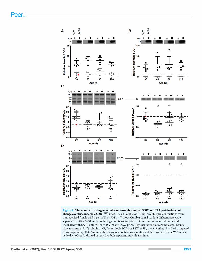
Figure 8 The amount of detergent-soluble or -insoluble lumbar SOD1 or P2X7 protein does notchange over time in female SOD1G93A mice. (A, C) Soluble or (B, D) insoluble protein fractions fromhomogenised female wild type (WT) or SOD1G93A mouse lumbar spinal cords at different ages wereseparated by SDS-PAGE under reducing conditions, transferred to nitrocellulose membranes, andincubated with (A, B) anti-SOD1 or (C, D) anti-P2X7 pAbs. Representative blots are indicated. Resultsshown as mean (A, C) soluble or (B, D) insoluble SOD1 or P2X7±SD, n= 3–5 mice; *P < 0.05 comparedto corresponding 30 d. Amounts shown are relative to corresponding soluble proteins of one WT mouseat 30 days of age (indicated in red). Symbols represent individual animals.
Bartlett et al. (2017), PeerJ, DOI 10.7717/peerj.3064 19/29
Treatment had no effect on ALS score or motor coordination in either sex. Furthermore,BBG treatment had no effect on motor neuron loss, microgliosis, lumbar SOD1 or P2X7protein amounts, splenic leukocyte immunophenotype, or P2X7 expression in the spleenor liver at end-stage. BBG treatment partially, but not significantly, increased serumMCP-1. Together, this suggests a limited efficacy of BBG on ALS progression as used in thecurrent study. Furthermore, there was no difference in the amount of lumbar P2X7 proteinobserved in SOD1G93A mice of different ages or between SOD1G93A andWTmice at any age.
The therapeutic effects of BBG in SOD1G93A murine models of ALS have been reportedin two other independent studies (Apolloni et al., 2014; Cervetto et al., 2013). In both thesestudies, treatment with BBG delayed disease onset, improved motor coordination, diseasescores and motor neuron survival, and reduced weight loss and microgliosis. Comparisonsof these studies with the current study highlight important parameters effecting drugefficacy in murine ALS models, which should be considered in future studies.
One important parameter is gender. In the current study, BBG treatment slowed weightloss and extended survival in female, but notmale,mice. This is consistent with the extendedlifespan of female heterozygous and homozygous P2X7 knock-out (P2X7KO)/SOD1G93A
mice compared to P2X7WT/SOD1G93A mice (Apolloni et al., 2013a). Gender also affectedP2X7 antagonism in another study investigating BBG efficacy in the SOD1G93A murinemodel of ALS. However, in contrast to the current study, BBG significantly delayed thetime taken for 10% weight loss and a decline in motor coordination to be observed in male,but not female, mice (Cervetto et al., 2013). Furthermore, this gender dependency was notobserved for any other reported outcomes, including weight or motor coordination,following BBG treatment in another previous study (Apolloni et al., 2013a). Gender-specificoutcomes may be impacted by differences in the genetic background of SOD1 mice(Heiman-Patterson et al., 2005) or differences in sex hormones such as oestrogens (Choi etal., 2008; Groeneveld et al., 2004; Trieu & Uckun, 1999).
Vehicle composition is another factor that may have influenced the efficacy of BBGobserved in the current study compared to previous studies. In the current study, dimethylsulfoxide (DMSO) was not included in the vehicle for BBG delivery, while DMSO waspresent in the vehicle solution in both previous studies investigating the therapeutic effectsof BBG (Apolloni et al., 2014; Cervetto et al., 2013). In the current study, DMSO was ex-cluded as BBGwas soluble in its absence and given that it is well known to be toxic, especiallywhen used chronically or at concentrations greater than 10% (Kloverpris et al., 2010). Dueto the fact that intraperitoneally delivered DMSO is able to cross the blood–brain barrier(Broadwell, Salcman & Kaplan, 1982), it is possible that the lack of DMSO in the currentstudy prevented adequate BBG blood–brain barrier penetrance. In a study of spinal cordinjury, intravenous administration of a similar concentration of BBG in the absence ofDMSO resulted in blue colouring in the injury area, but not surrounding tissue (Peng etal., 2009). This suggests that BBG entered the site of injury mainly via the disrupted spinalblood–brain barrier in this study, and that DMSO may be required in the drug vehicle foradequate BBG blood–brain barrier penetrance.
The limited efficacy of BBG in the current study may have been due to the dose used.BBG was used at 45.5 mg/kg due to the efficacy of this dose in murine models of other
Bartlett et al. (2017), PeerJ, DOI 10.7717/peerj.3064 20/29
neurodegenerative conditions, includingHuntington’s disease and spinal cord injury (Diaz-Hernandez et al., 2009; Peng et al., 2009). This dose also improved disease outcomes inC57BL/6J × SJL/J hybrid SOD1G93A mice (Cervetto et al., 2013). However, in the studyof Apolloni et al. (2014), 50 mg/kg BBG had no effect on disease outcomes in C57BL/6JSOD1G93A mice. When the BBG dose was increased to 250 mg/kg in this study, improve-ments in behavioural scores, motor performance and median disease onset were observed(Apolloni et al., 2014). This may indicate that the BBG dose utilised in the current studywas insufficient to fully antagonise P2X7 in the CNS of C57BL/6J SOD1G93A mice.
An important consideration when testing therapeutic intervention in ALSmousemodelsis the timing of treatment commencement. In the current study, BBG treatment was startedbefore the onset of symptoms. In comparison, Cervetto et al. (2013) began treatment atdisease onset, with a similar BBG dosage, number of treatments per week and deliveryroute utilised as in the current study. Thus, the differences observed between this and thecurrent study may be explained by the timing of treatment commencement. In the study byApolloni et al. (2014), BBG treatment was started at late pre-onset, resulting in a wide rangeof beneficial effects on disease progression. Whilst this study only observed these effectsby utilising a five-fold higher dose of BBG than that used in the current study, these highBBG doses were not effective when began during the asymptomatic or pre-onset phasesof disease. Collectively, this suggests a very tight window for therapeutic interventiontargeting P2X7. This is further supported by the exacerbated disease progression observedin SOD1G93A mice lacking P2X7 (Apolloni et al., 2013a), and suggests that P2X7 may playa dual role in ALS progression. In this regard, P2X7 activation may be neuroprotective inthe initial stages of disease, but neuroinflammatory or neurotoxic as the disease progresses.This is consistent with the trophic and toxic actions mediated by P2X7 in surveilling andactivated microglia, respectively (Bartlett, Yerbury & Sluyter, 2013; Gendron et al., 2003;Hide et al., 2000; Monif et al., 2009; Shieh et al., 2014).
Microgliosis and loss ofmotor neurons are twomajor hallmarks of disease in ALS.Whilstthese disease hallmarks were observed in the current study, there were no differences at end-stage between untreated and BBG-treatedmice. In contrast, BBG treatment reduced lumbarmicroglia activation and inflammatory microglial markers, enhanced neurotrophic factorsand improved motor neuron survival at end-stage in the study by Apolloni et al. (2014).This is consistent with the anti-inflammatory effects resulting fromP2X7 inhibition in vitro,whereby BBG treatment reduced the release of pro-inflammatory factors and subsequenttoxicity of SOD1G93A microglia towards neuronal cells (Apolloni et al., 2013b; D’Ambrosiet al., 2009). In the current study, motor neuron and microglial numbers were assessedwithout stratifying for gender due to the small n values of available samples. However, giventhe limited clinical efficacy of BBG, increasing the n value to allow for gender stratificationmay not be worthwhile. Furthermore, there is limited evidence that gender influencesmotor neuron loss (Veldink et al., 2003).
An accumulation of SOD1-containing aggregates is another major hallmark of diseasein ALS. In the current study, detergent-insoluble SOD1 was present in lumbar spinal cordsof SOD1G93A mice at end-stage. However, there were no differences in the amounts ofdetergent-insoluble SOD1 between saline- and BBG-treated SOD1G93A mice. Consistent
Bartlett et al. (2017), PeerJ, DOI 10.7717/peerj.3064 21/29
with this data, ablation of P2X7 does not modify the lumbar SOD1 content in SOD1G93A
mice (Apolloni et al., 2013a). BBG, but not other P2X7 antagonists, prevents protease-resistant prion protein accumulation in scrapie-infected microglial and neuronal cell lines,and in the brains of prion-infected mice (Iwamaru et al., 2012). This suggests that BBGhas anti-aggregation properties, independent of its P2X7 interactions. However, giventhe current data, the anti-aggregation properties of BBG may be restricted to anti-prionactivities, consistent with amolecular framework analogous to other anti-prion compounds(Iwamaru et al., 2012).
P2X7 protein was detected in the lumbar spinal cord of SOD1G93A mice treated witheither saline or BBG. Similar amounts were detected between treatments, consistent withthe lack of effect of BBG on lumbar spinal P2X7 amounts in other studies (Apolloni et al.,2014). In the current study, a number of bands were detected in addition to the expectedfull length P2X7A isoform (73–89 kDa). These proteins were approximately 160, 80, 60 and50 kDa in size, and may correspond to P2X7 dimers, non-glycosylated P2X7, P2X7 splicevariants or P2X7 breakdown products. C terminal truncated P2X7 variants have recentlybeen identified in mice (Masin et al., 2012). One of these, termed P2X713B, is detected inthe mouse CNS and has a molecular mass of approximately 60 kDa (Masin et al., 2012),similar to one of the proteins detected in this current and other studies (Barth et al., 2007).Interestingly, when expressed in human embryonic kidney (HEK)-293 cells, the majority ofP2X713B was retained in the endoplasmic reticulum and not efficiently trafficked to the cellsurface (Masin et al., 2012). Thismay explain differences in the amount of 60 kDa protein indetergent-soluble and insoluble fractions in the current study.However, furtherworkwouldbe required to identify whether this band does indeed correspond to a P2X7 splice variant.
In the current study, the amount of lumbar spinal cord P2X7 did not change significantlywith age in female SOD1G93A mice. It has been reported that there is no sexual dimorphismin P2X7 expression at any age (Crain, Nikodemova & Watters, 2009). However, whilemicroglial P2X7 expression was maintained at similar levels from 21 to 365 d in healthyC57BL/6mice in onemixed-sex study (Crain, Nikodemova & Watters, 2009), another foundthat P2X7 protein increased with age in healthy Sprague–Dawley rats (Lai et al., 2013).Furthermore, similar amounts of lumbar P2X7 protein were present in spinal cords fromWT and SOD1G93A mice at all investigated time points in the current study, while P2X7immunoreactvities have been reported to be increased in spinal cords from (post-mortem)humans with ALS and advanced-stage transgenic SOD1G93A rats (Casanovas et al., 2008;Yiangou et al., 2006).
In the current study, P2X7 was detected in both detergent-soluble and -insoluble frac-tions. The presence of detergent-insoluble P2X7 has been reported in mouse lung alveolarepithelial cells, HEK-293 cells transfected with WT P2X7, mouse peritoneal macrophagesand rat submandibular glands (Barth et al., 2007;Garcia-Marcos et al., 2006;Gonnord et al.,2009). In these studies, it was suggested that P2X7 partially localisedwith detergent-resistantmembranes, or lipid rafts, at the plasma membrane. This association was reported to bedependent on the post-translational modification of P2X7 by palmitic acid (Gonnordet al., 2009). In the current study, the amount of detergent-insoluble 75 kDa P2X7 wasrelatively low, suggesting that the majority of 75 kDa P2X7 was localised in the plasma
Bartlett et al. (2017), PeerJ, DOI 10.7717/peerj.3064 22/29
membrane. Determining whether the detergent-insoluble 75 kDa protein represented lipidraft-associated or intracellular P2X7 was beyond the scope of the current work.
BBG treatment increased serum MCP-1 in SOD1G93A mice by 1.4-fold compared tosaline treatment; a difference that approached statistical significance (P = 0.052). MCP-1 isup-regulated in ALS mice (Kawaguchi-Niida et al., 2013) and in patients with ALS (Henkelet al., 2004; Nagata et al., 2007; Wilms et al., 2003), where it is thought to drive diseaseprogression. Whilst in vitro studies have showed that pre-incubation with BBG can reduceP2X7-mediated accumulation of intracellular MCP-1 in rat microglia (Fang et al., 2011)and prevent P2X7-mediated MCP-1 release from murine microglia (Shieh et al., 2014).Thus, the current observation that serum MCP-1 was potentially increased in SOD1G93A
mice following BBG treatment was unexpected. Possible explanations for this finding is thatBBG treatment in vivo affects pathways other than the ATP-P2X7 signalling axis to promoteMCP-1 production or alters the circulatory system to extend the half-life of serumMCP-1.
CONCLUSIONSIn conclusion, BBG treatment reduced body weight loss and prolonged survival in femaleSOD1G93A mice. This represents the first report of an extension in survival following P2X7blockade in an ALS mouse model. Given that this result was seen only in females, the im-portance of considering sex in pre-clinical studies is clear, consistent with reports by others(Cervetto et al., 2013). Despite this, in the current study, BBG had no effect on body weightloss or survival inmale SOD1G93Amice, or onALS score,motor coordination,motor neuronloss, microgliosis, lumbar SOD1 or P2X7, serum MCP-1, splenic leukocyte immunophe-notype or P2X7 expression at end-stage in either gender. While the other two studiesinvestigating the efficacy of BBG in ALS found more beneficial effects of this compoundat molecular and phenotypic levels, these effects did not correlate with an extension inlife span in these studies (Apolloni et al., 2014; Cervetto et al., 2013). Together, the differentoutcomes obtained from these three studies highlight how drug regime may influencedisease outcomes in murine models of ALS.
ACKNOWLEDGEMENTSWe are grateful to Masoud Yousefi (University of British Columbia, Vancouver, Canada)for assistance with statistical analysis. We are grateful to Natalie Farrawell (University ofWollongong) for assistance with immunostaining. We are grateful to staff of the IllawarraHealth and Medical Research Institute (Wollongong, Australia) and Phillip Mullany(University of Wollongong) for technical assistance.
ADDITIONAL INFORMATION AND DECLARATIONS
FundingThis project was supported by the University of Wollongong, Wollongong, Australia.Rachael Bartlett was a recipient of a Global Challenges Scholarship, University ofWollongong. Justin J. Yerbury was supported by the Australian Research Council
Bartlett et al. (2017), PeerJ, DOI 10.7717/peerj.3064 23/29
(DE120102840). The funders had no role in study design, data collection and analysis,decision to publish, or preparation of the manuscript.
Grant DisclosuresThe following grant information was disclosed by the authors:University of Wollongong, Wollongong, Australia.Global Challenges Scholarship, University of Wollongong.Australian Research Council: DE120102840.
Competing InterestsThe authors declare there are no competing interests.
Author Contributions• Rachael Bartlett conceived and designed the experiments, performed the experiments,analyzed the data, wrote the paper, prepared figures and/or tables, reviewed drafts of thepaper.• Vanessa Sluyter performed the experiments, reviewed drafts of the paper.• Debbie Watson performed the experiments, analyzed the data, contributedreagents/materials/analysis tools, reviewed drafts of the paper.• Ronald Sluyter conceived and designed the experiments, performed the experiments,analyzed the data, contributed reagents/materials/analysis tools, wrote the paper,reviewed drafts of the paper.• Justin J. Yerbury conceived and designed the experiments, performed the experiments,analyzed the data, contributed reagents/materials/analysis tools, wrote the paper,prepared figures and/or tables, reviewed drafts of the paper.
Animal EthicsThe following information was supplied relating to ethical approvals (i.e., approving bodyand any reference numbers):
Animal Ethics Committee of the University of Wollongong.Approval numbers AE11/29 and AE12/09.
Data AvailabilityThe following information was supplied regarding data availability:
The raw data has been supplied as Data S1.
Supplemental InformationSupplemental information for this article can be found online at http://dx.doi.org/10.7717/peerj.3064#supplemental-information.
REFERENCESAl-Chalabi A, Jones A, Troakes C, King A, Al-Sarraj S, Van den Berg LH. 2012. The
genetics and neuropathology of amyotrophic lateral sclerosis. Acta Neuropathologica124(3):339–352 DOI 10.1007/s00401-012-1022-4.
Bartlett et al. (2017), PeerJ, DOI 10.7717/peerj.3064 24/29
Apolloni S, Amadio S, Montilli C, Volonte C, D’Ambrosi N. 2013a. Ablation of P2X7receptor exacerbates gliosis and motoneuron death in the SOD1-G93A mouse modelof amyotrophic lateral sclerosis. Human Molecular Genetics 22(20):4102–4116DOI 10.1093/hmg/ddt259.
Apolloni S, Amadio S, Parisi C, Matteucci A, Potenza RL, ArmidaM, Popoli P,D’Ambrosi N, Volonte C. 2014. Spinal cord pathology is ameliorated by P2X7antagonism in a SOD1-mutant mouse model of amyotrophic lateral sclerosis. DiseaseModels & Mechanisms 7:1101–1109 DOI 10.1242/dmm.017038.
Apolloni S, Parisi C, Pesaresi MG, Rossi S, Carri MT, CozzolinoM, Volonte C,D’Ambrosi N. 2013b. The NADPH oxidase pathway is dysregulated by the P2X7receptor in the SOD1-G93A microglia model of amyotrophic lateral sclerosis. Journalof Immunology 190(10):5187–5195 DOI 10.4049/jimmunol.1203262.
Barth K,Weinhold K, Guenther A, YoungMT, Schnittler H, Kasper M. 2007. Caveolin-1 influences P2X7 receptor expression and localization in mouse lung alveolarepithelial cells. FEBS Journal 274(12):3021–3033DOI 10.1111/j.1742-4658.2007.05830.x.
Bartlett R, Stokes L, Sluyter R. 2014. The P2X7 receptor channel: recent developmentsand the use of P2X7 antagonists in models of disease. Pharmacological Reviews66(3):638–675 DOI 10.1124/pr.113.008003.
Bartlett R, Yerbury JJ, Sluyter R. 2013. P2X7 receptor activation induces reactiveoxygen species formation and cell death in murine EOC13 microglia.Mediators ofInflammation 2013:271815 DOI 10.1155/2013/271813.
Boillee S, Vande Velde C, Cleveland DW. 2006. ALS: a disease of motor neurons andtheir nonneuronal neighbors. Neuron 52:39–59 DOI 10.1016/j.neuron.2006.09.018.
Broadwell RD, SalcmanM, Kaplan RS. 1982.Morphologic effect of dimethyl sulfoxideon the blood–brain barrier. Science 217(4555):164–166 DOI 10.1126/science.7089551.
Casanovas A, Hernandez S, Tarabal O, Rossello J, Esquerda JE. 2008. Strong P2X4purinergic receptor-like immunoreactivity is selectively associated with degeneratingneurons in transgenic rodent models of amyotrophic lateral sclerosis. Journal ofComparative Neurology 506(1):75–92 DOI 10.1002/cne.21527.
Cervetto C, Frattaroli D, Maura G, Marcoli M. 2013.Motor neuron dysfunction ina mouse model of ALS: gender-dependent effect of P2X7 antagonism. Toxicology311(1):69–77 DOI 10.1016/j.tox.2013.04.004.
Cheroni C, Peviani M, Cascio P, Debiasi S, Monti C, Bendotti C. 2005. Accumulationof human SOD1 and ubiquitinated deposits in the spinal cord of SOD1G93A miceduring motor neuron disease progression correlates with a decrease of proteasome.Neurobiology of Disease 18(3):509–522 DOI 10.1016/j.nbd.2004.12.007.
Choi CI, Lee YD, Gwag BJ, Cho SI, Kim SS, Suh-KimH. 2008. Effects of estrogen onlifespan and motor functions in female hSOD1 G93A transgenic mice. Journal ofNeurological Sciences 268(1–2):40–47 DOI 10.1016/j.jns.2007.10.024.
Crain JM, NikodemovaM,Watters JJ. 2009. Expression of P2 nucleotide receptors varieswith age and sex in murine brain microglia. Journal of Neuroinflammation 6:Article24 DOI 10.1186/1742-2094-6-24.
Bartlett et al. (2017), PeerJ, DOI 10.7717/peerj.3064 25/29
D’Ambrosi N, Finocchi P, Apolloni S, CozzolinoM, Ferri A, Padovano V, Pietrini G,Carri MT, Volonte C. 2009. The proinflammatory action of microglial P2 receptorsis enhanced in SOD1 models for amyotrophic lateral sclerosis. Journal of Immunology183(7):4648–4656 DOI 10.4049/jimmunol.0901212.
Diaz-HernandezM, Diez-Zaera M, Sanchez-Nogueiro J, Gomez-Villafuertes R, CanalsJM, Alberch J, Miras-Portugal MT, Lucas JJ. 2009. Altered P2X7-receptor leveland function in mouse models of Huntington’s disease and therapeutic efficacy ofantagonist administration. FASEB Journal 23:1893–1906 DOI 10.1096/fj.08-122275.
Fang KM,Wang YL, HuangMC, Sun SH, Cheng H, Tzeng SF. 2011. Expression ofmacrophage inflammatory protein-1 α and monocyte chemoattractant protein-1in glioma-infiltrating microglia: involvement of ATP and P2X7 receptor. Journal ofNeuroscience Research 89(2):199–211 DOI 10.1002/jnr.22538.
Farrawell NE, Lambert-Smith IA,Warraich ST, Blair IP, Saunders DN, Hatters DM,Yerbury JJ. 2015. Distinct partitioning of ALS associated TDP-43, FUS and SOD1mutants into cellular inclusions. Scientific Reports 5:13416 DOI 10.1038/srep13416.
GandelmanM, Peluffo H, Beckman JS, Cassina P, Barbeito L. 2010. Extracellular ATPand the P2X7 receptor in astrocyte-mediated motor neuron death: implications foramyotrophic lateral sclerosis. Journal of Neuroinflammation 7:Article 33DOI 10.1186/1742-2094-7-33.
Garcia-Marcos M, Perez-Andres E, Tandel S, Fontanils U, Kumps A, Kabre E, Gomez-Munoz A, Marino A, Dehaye JP, Pochet S. 2006. Coupling of two pools of P2X7receptors to distinct intracellular signaling pathways in rat submandibular gland.Journal of Lipid Research 47:705–714 DOI 10.1194/jlr.M500408-JLR200.
Gendron FP, ChalimoniukM, Strosznajder J, Shen S, Gonzalez FA,Weisman GA, SunGY. 2003. P2X7 nucleotide receptor activation enhances IFN gamma-induced typeII nitric oxide synthase activity in BV-2 microglial cells. Journal of Neurochemistry87(2):344–352 DOI 10.1046/j.1471-4159.2003.01995.x.
Gonnord P, Delarasse C, Auger R, Benihoud K, Prigent M, Cuif MH, Lamaze C,Kanellopoulos JM. 2009. Palmitoylation of the P2X7 receptor, an ATP-gatedchannel, controls its expression and association with lipid rafts. FASEB Journal23:795–805 DOI 10.1096/fj.08-114637.
Grad LI, Yerbury JJ, Turner BJ, GuestWC, Pokrishevsky E, O’Neill MA, Yanai A,Silverman JM, Zeineddine R, Corcoran L, Kumita JR, Luheshi LM, Yousefi M,Coleman BM, Hill AF, Plotkin SS, Mackenzie IR, Cashman NR. 2014. Intercellularpropagated misfolding of wild-type Cu/Zn superoxide dismutase occurs viaexosome-dependent and -independent mechanisms. Proceedings of the NationalAcademy of Sciences of the United States of America 111:3620–3625DOI 10.1073/pnas.1312245111.
Groeneveld GJ, VanMuiswinkel FL, De leeuw vanWeenen J, BlauwH, Veldink JH,Wokke JH, Van den Berg LH, Bar PR. 2004. CGP 3466B has no effect on diseasecourse of (G93A) mSOD1 transgenic mice. Amyotrophic Lateral Sclerosis and OtherMotor Neuron Disorders 5(4):220–225 DOI 10.1080/14660820410019530.
Bartlett et al. (2017), PeerJ, DOI 10.7717/peerj.3064 26/29
GurneyME, Pu H, Chiu AY, Dal CantoMC, Polchow CY, Alexander DD, CaliendoJ, Hentati A, Kwon YW, Deng HX, ChenW, Zhai P, Sufit RL, Siddique T. 1994.Motor neuron degeneration in mice that express a human Cu,Zn superoxidedismutase mutation. Science 264(5166):1772–1775 DOI 10.1126/science.8209258.
Hall ED, Oostveen JA, GurneyME. 1998. Relationship of microglial and astrocyticactivation to disease onset and progression in a transgenic model of familial ALS.Glia 23(3):249–256.
Heiman-Patterson TD, Deitch JS, Blankenhorn EP, Erwin KL, Perreault MJ, AlexanderBK, Byers N, Toman I, Alexander GM. 2005. Background and gender effects onsurvival in the TgN(SOD1-G93A)1Gur mouse model of ALS. Journal of NeurologicalSciences 236(1–2):1–7 DOI 10.1016/j.jns.2005.02.006.
Henkel JS, Beers DR, ZhaoW, Appel SH. 2009.Microglia in ALS: the good, thebad, and the resting. Journal of Neuroimmune Pharmacology 4(4):389–398DOI 10.1007/s11481-009-9171-5.
Henkel JS, Engelhardt JI, Siklos L, Simpson EP, Kim SH, Pan T, Goodman JC, SiddiqueT, Beers DR, Appel SH. 2004. Presence of dendritic cells, MCP-1, and activatedmicroglia/macrophages in amyotrophic lateral sclerosis spinal cord tissue. Annalsof Neurology 55(2):221–235 DOI 10.1002/ana.10805.
Hide I, TanakaM, Inoue A, Nakajima K, Kohsaka S, Inoue K, Nakata Y. 2000. Extracel-lular ATP triggers tumor necrosis factor-alpha release from rat microglia. Journal ofNeurochemistry 75(3):965–972 DOI 10.1046/j.1471-4159.2000.0750965.x.
Iwamaru Y, Takenouchi T, Murayama Y, Okada H, ImamuraM, Shimizu Y,HashimotoM,Mohri S, Yokoyama T, Kitani H. 2012. Anti-prion activity ofBrilliant Blue G. PLOS ONE 7(5):e37896 DOI 10.1371/journal.pone.0037896.
Kawaguchi-Niida M, Yamamoto T, Kato Y, Inose Y, Shibata N. 2013.MCP-1/CCR2signaling-mediated astrocytosis is accelerated in a transgenic mouse model of SOD1-mutated familial ALS. Acta Neuropathologica Communications 1:Article 21DOI 10.1186/2051-5960-1-21.
Kloverpris H, Fomsgaard A, Handley A, Ackland J, SullivanM, Goulder P. 2010.Dimethyl sulfoxide (DMSO) exposure to human peripheral blood mononuclearcells (PBMCs) abolish T cell responses only in high concentrations and followingcoincubation for more than two hours. Journal of Immunological Methods 356(1–2):70–78DOI 10.1016/j.jim.2010.01.014.
Lai AY, Dibal CD, Armitage GA,Winship IR, Todd KG. 2013. Distinct activationprofiles in microglia of different ages: a systematic study in isolated embryonic toaged microglial cultures. Neuroscience 254:185–195DOI 10.1016/j.neuroscience.2013.09.010.
MasinM, Young C, Lim K, Barnes SJ, Xu XJ, Marschall V, BrutkowskiW,Mooney ER,Gorecki DC, Murrell-Lagnado R. 2012. Expression, assembly and function of novelC-terminal truncated variants of the mouse P2X7 receptor: re-evaluation of P2X7knockouts. British Journal of Pharmacology 165(4):978–993DOI 10.1111/j.1476-5381.2011.01624.x.
Bartlett et al. (2017), PeerJ, DOI 10.7717/peerj.3064 27/29
McCombe PA, Henderson RD. 2010. Effects of gender in amyotrophic lateral sclerosis.Gender Medicine 7(6):557–570 DOI 10.1016/j.genm.2010.11.010.
Monif M, Reid CA, Powell KL, Smart ML,Williams DA. 2009. The P2X7 receptor drivesmicroglial activation and proliferation: a trophic role for P2X7R pore. Journal ofNeuroscience 29(12):3781–3791 DOI 10.1523/JNEUROSCI.5512-08.2009.
Nagata T, Nagano I, Shiote M, Narai H, Murakami T, Hayashi T, Shoji M, Abe K. 2007.Elevation of MCP-1 and MCP-1/VEGF ratio in cerebrospinal fluid of amyotrophiclateral sclerosis patients. Neurological Research 29(8):772–776DOI 10.1179/016164107X229795.
Parisi C, Arisi I, D’Ambrosi N, Storti AE, Brandi R, D’Onofrio M, Volonte C. 2013.Dysregulated microRNAs in amyotrophic lateral sclerosis microglia modulate geneslinked to neuroinflammation. Cell Death & Disease 4:e959DOI 10.1038/cddis.2013.491.
Parisi C, Napoli G, Amadio S, Spalloni A, Apolloni S, Longone P, Volonte C. 2016.MicroRNA-125b regulates microglia activation and motor neuron death in ALS. CellDeath & Differentiation 23:531–541 DOI 10.1038/cdd.2015.153.
PengW, CotrinaML, Han X, Yu H, Bekar L, Blum L, Takano T, Tian GF, GoldmanSA, NedergaardM. 2009. Systemic administration of an antagonist of the ATP-sensitive receptor P2X7 improves recovery after spinal cord injury. Proceedings of theNational Academy of Sciences of the United States of America 106(30):12489–12493DOI 10.1073/pnas.0902531106.
Roberts K, Zeineddine R, Corcoran L, LiW, Campbell IL, Yerbury JJ. 2013. Extracel-lular aggregated Cu/Zn superoxide dismutase activates microglia to give a cytotoxicphenotype. Glia 61(3):409–419 DOI 10.1002/glia.22444.
Rosen DR, Siddique T, Patterson D, Figlewicz DA, Sapp P, Hentati A, DonaldsonD, Goto J, O’Regan JP, Deng HX, Rahmani Z, Krizus A, Mckenna-Yasek D,Cayabyab A, Gaston SM, Berger R, Tanzi RE, Halperin JJ, Herzfeldt B, Van DenBergh R, HungW, Bird T, Deng G, Mulder DW, Smyth C, Laing NG, Soriano E,Pericak-VanceMA, Haines J, Rouleau GA, Gusella JS, Horvitz HR, Brown Jr RH.1993.Mutations in Cu/Zn superoxide dismutase gene are associated with familialamyotrophic lateral sclerosis. Nature 362:59–62 DOI 10.1038/362059a0.
Shieh CH, Heinrich A, Serchov T, Van Calker D, Biber K. 2014. P2X7-dependent, butdifferentially regulated release of IL-6, CCL2, and TNF-alpha in cultured mousemicroglia. Glia 62(4):592–607 DOI 10.1002/glia.22628.
Skaper SD, Facci L, Culbert AA, Evans NA, Chessell I, Davis JB, Richardson JC. 2006.P2X 7, receptors on microglial cells mediate injury to cortical neurons in vitro. Glia54(3):234–242 DOI 10.1002/glia.20379.
Trieu VN, Uckun FM. 1999. Genistein is neuroprotective in murine models of familialamyotrophic lateral sclerosis and stroke. Biochemical and Biophysical ResearchCommunications 258(3):685–688 DOI 10.1006/bbrc.1999.0577.
Turner BJ, Talbot K. 2008. Transgenics, toxicity and therapeutics in rodent modelsof mutant SOD1-mediated familial ALS. Progress in Neurobiology 85(1):94–134DOI 10.1016/j.pneurobio.2008.01.001.
Bartlett et al. (2017), PeerJ, DOI 10.7717/peerj.3064 28/29
Veldink JH, Bar PR, Joosten EA, OttenM,Wokke JH, Van den Berg LH. 2003. Sexualdifferences in onset of disease and response to exercise in a transgenic model of ALS.Neuromuscular Disorders 13(9):737–743 DOI 10.1016/S0960-8966(03)00104-4.
Watson D, Zhang GY, HuM,Wang YM, Fletcher J, Sartor M, Alexander SI. 2014.Transforming growth factor beta (TGFbeta) plays a crucial role in prolonging allo-graft survival in an allodepletion (‘‘pruning’’) skin transplant model. TransplantationImmunology 30(4):168–177 DOI 10.1016/j.trim.2014.03.002.
Wilms H, Sievers J, Dengler R, Bufler J, Deuschl G, Lucius R. 2003. Intrathecal synthesisof monocyte chemoattractant protein-1 (MCP-1) in amyotrophic lateral sclerosis:further evidence for microglial activation in neurodegeneration. Journal of Neuroim-munology 144:139–142 DOI 10.1016/j.jneuroim.2003.08.042.
Yiangou Y, Facer P, Durrenberger P, Chessell IP, Naylor A, Bountra C, Banati RR,Anand P. 2006. COX-2, CB2 and P2X7-immunoreactivities are increased inactivated microglial cells/macrophages of multiple sclerosis and amyotrophic lateralsclerosis spinal cord. BMC Neurology 6:12 DOI 10.1186/1471-2377-6-12.
Bartlett et al. (2017), PeerJ, DOI 10.7717/peerj.3064 29/29