p62/sqstm1bindsdirectlytoatg8/lc3tofacilitate ... · pdf...
TRANSCRIPT

p62/SQSTM1 Binds Directly to Atg8/LC3 to FacilitateDegradation of Ubiquitinated Protein Aggregatesby Autophagy*□S
Received for publication, April 3, 2007, and in revised form, May 18, 2007 Published, JBC Papers in Press, June 19, 2007, DOI 10.1074/jbc.M702824200
Serhiy Pankiv‡, Terje Høyvarde Clausen‡, Trond Lamark‡, Andreas Brech§1, Jack-Ansgar Bruun‡, Heidi Outzen‡,Aud Øvervatn‡, Geir Bjørkøy‡, and Terje Johansen‡2
From the ‡Biochemistry Department, Institute of Medical Biology, University of Tromsø, 9037 Tromsø and the §Department ofBiochemistry, Institute for Cancer Research, The Norwegian Radium Hospital, Montebello N-0310, Oslo, Norway
Protein degradation by basal constitutive autophagy is impor-tant toavoidaccumulationofpolyubiquitinatedproteinaggregatesand development of neurodegenerative diseases. The polyubiq-uitin-bindingproteinp62/SQSTM1 isdegradedby autophagy. It isfound in cellular inclusion bodies together with polyubiquitinatedproteinsand incytosolicproteinaggregates thataccumulate invar-iouschronic, toxic,anddegenerativediseases.Hereweshowfor thefirst time a direct interaction between p62 and the autophagiceffector proteins LC3A and -B and the related �-aminobutyratereceptor-associatedprotein and�-aminobutyrate receptor-associ-ated-like proteins. The binding is mediated by a 22-residuesequence of p62 containing an evolutionarily conserved motif. Tomonitor the autophagic sequestration of p62- and LC3-positivebodies, we developed a novel pH-sensitive fluorescent tag consist-ing of a tandem fusionof the red, acid-insensitivemCherry and theacid-sensitive green fluorescent proteins. This approach revealedthat p62- and LC3-positive bodies are degraded in autolysosomes.Strikingly, even rather large p62-positive inclusion bodies (2 �mdiameter)becomedegradedbyautophagy.Thespecific interactionbetween p62 and LC3, requiring the motif we have mapped, isinstrumental inmediating autophagic degradation of the p62-pos-itive bodies. We also demonstrate that the previously reportedaggresome-like induced structures containing ubiquitinated pro-teins in cytosolic bodies are dependent on p62 for their formation.In fact, p62 bodies and these structures are indistinguishable.Takentogether,ourresultsclearlysuggest thatp62isrequiredbothfor the formation and thedegradationof polyubiquitin-containingbodies by autophagy.
All eukaryotic cells use the following two systems for proteindegradation: the ubiquitin-proteasome system and the lyso-
some. The proteasome is used for selective degradation of shortlived and abnormal/misfolded proteins following labeling withLys-48-linked polyubiquitin chains (1). The lysosome degradesextracellular and plasma membrane proteins brought there byendocytosis and cytoplasmic components delivered by autoph-agy. Various categories of autophagy have been defined differ-ing in the delivery route of cytoplasmic material. These includemacroautophagy (hereafter called autophagy), microautoph-agy, and chaperone-mediated autophagy (2–4). Macroautoph-agy is regarded as the main pathway. This process involves thesequestration of a region of the cytoplasm within a double ormultiple membrane-bounded autophagosome. Autophago-somes then undergo a maturation process, including fusionevents with endosomes and/or lysosomes forming structurescalled amphisomes and autolysosomes, respectively (3–5).Autophagy is thought to be mainly a nonselective, bulk degrada-tion pathway responsible for degradation of the majority of longlived proteins and some organelles. Two evolutionarily conservedprotein conjugation systems are necessary for the formationof theautophagosome, the Atg12-Atg5- and the Atg8-phosphati-dylethanolamine conjugation systems (6). The best character-ized mammalian Atg8 homologue is light chain 3 (LC3).3 Aftersynthesis pro-LC3 is cleaved by Atg4B to expose a C-terminalglycine residue (7, 8). This represents the cytosolic LC3-I form.Conjugation of phosphatidylethanolamine to the C terminus ofLC3-I defines the LC3-II form that is tightly associated with theautophagosomal membrane (7, 9). The LC3-II form is involvedduring the late steps of autophagy after the isolation membranehas formed (3). In humans, three LC3 isoforms (LC3A, -B, and -C)and four additional humanAtg8 homologues have been identified(GABARAP, GEC1/GABARAPL1, GATE16/GABARAPL2, andGABARAPL3) (10, 11). The role(s) of the GABARAP isoforms inautophagy is not known.A number of studies have identified autophagy as a crucial
cellular process to avoid accumulation of abnormal proteins indifferent neurodegenerative diseases (reviewed in Ref. 12).Mice carrying neuron-specific knock-outs of Atg5 or Atg7 dis-
* This work was supported in part by grants from the FUGE and “Top ResearchProgramme” of the Norwegian Research Council, the Norwegian CancerSociety, the Aakre Foundation, Simon Fougner Hartmanns Familiefond,and the Blix Foundation (to T. J.). The costs of publication of this articlewere defrayed in part by the payment of page charges. This article musttherefore be hereby marked “advertisement” in accordance with 18 U.S.C.Section 1734 solely to indicate this fact.
□S The on-line version of this article (available at http://www.jbc.org) containssupplemental Figs. 1–5.
1 Recipient of a career fellowship from the FUGE programme of the Norwe-gian Research Council.
2 To whom correspondence should be addressed: Dept. of Biochemistry,Institute of Medical Biology, University of Tromsø, 9037 Tromsø, Norway.Tel.: 47-776-44720; Fax: 47-776-45350; E-mail: [email protected].
3 The abbreviations used are: LC3, light chain 3; ALIS, aggresome-like inducedstructures; GFP, green fluorescent protein; EGFP, enhanced GFP; mCherry,monomeric red fluorescent protein; GABARAP, �-aminobutyrate receptor-associated protein; siRNA, small interfering RNA; GST, glutathione S-trans-ferase; MBP, maltose-binding protein; LIR, LC3-interacting region; MAP,microtubule-associated protein; MEF, murine embryo fibroblasts; Ab, anti-body; mAb, monoclonal antibody.
THE JOURNAL OF BIOLOGICAL CHEMISTRY VOL. 282, NO. 33, pp. 24131–24145, August 17, 2007© 2007 by The American Society for Biochemistry and Molecular Biology, Inc. Printed in the U.S.A.
AUGUST 17, 2007 • VOLUME 282 • NUMBER 33 JOURNAL OF BIOLOGICAL CHEMISTRY 24131
at Universitaet H
eidelberg on January 24, 2008 w
ww
.jbc.orgD
ownloaded from
http://www.jbc.org/cgi/content/full/M702824200/DC1
Supplemental Material can be found at:
play intracellular accumulation of ubiquitin-positive proteinaggregates in the neural cells and show clear symptoms of pro-gressive neurodegeneration (13, 14). By generating mice with aconditional liver-specific knock-out of Atg7, it was also dem-onstrated that loss of autophagy causes liver dysfunctionaccompanied by intracellular accumulation of ubiquitinatedprotein aggregates (15). These results suggest that it is the basalconstitutive autophagy that is needed in order to avoid accu-mulation of ubiquitinated protein aggregates (13, 14, 16).Recently, the termALIS (aggresome-like induced structures)
was used to describe ubiquitin-containing bodies induced inresponse to various stressors, including amino acid starvation,oxidative stress, and puromycin (17). ALIS refers to DALIS(dendritic cell aggresome-like induced structures) originallydescribed in lipopolysaccharide-stimulated dendritic cells asstorage compartments for polyubiquitinated proteins prior totheir degradation (18). ALIS or DALIS are inclusion bodieswhere newly synthesized ubiquitinated proteins transientlyaccumulate, many of which are defective ribosomal products(17–19). However, also long lived proteins are targeted to ALIS(17). Puromycin increases the formation of defective ribosomalproducts and is an efficient inducer of ALIS. ALIS are distinctfrom aggresomes that are aggregates that form at the pericent-riolar area by microtubule-dependent conglomeration ofsmaller aggregates (20).The p62 protein, also called sequestosome 1(SQSTM1), is
commonly found in inclusion bodies containing polyubiquiti-nated protein aggregates. In neurodegenerative diseases p62 isdetected in ubiquitinated protein aggregates, including Lewybodies in Parkinson disease, neurofibrillary tangles in Alzhei-mer disease, and Huntingtin aggregates in Huntington disease(21–24). In protein aggregate diseases of the liver, largeamounts of p62 are found in Mallory bodies of alcoholic andnonalcoholic steatohepatitis, hyaline bodies in hepatocellularcarcinoma, and in �1-antitrypsin aggregates (24). The p62 pro-tein is able to polymerize via the N-terminal Phox and Bem1p(PB1) domain (25, 26). It binds ubiquitin and polyubiquitin viaits C-terminal UBA domain (27, 28).Here we report that p62 binds directly to the autophagic
effector proteins LC3A and -B and to the related GABARAPand GABARAP-like proteins. A short 22-amino acid regionlocatedN-terminally to theUBAdomain in p62was found to berequired for this interaction. We developed a novel, pH-sensi-tive, fluorescent tandem tag, which we use to show that thisinteraction isnecessary forautophagicdegradationofp62-positivecytoplasmic inclusion bodies containing ubiquitinated proteins.We also demonstrate that ALIS are indistinguishable from p62inclusion bodies and that p62 is required for their formation.
EXPERIMENTAL PROCEDURES
Antibodies and Reagents—The following antibodies wereused: anti-p62 monoclonal antibody (BD Transduction Labo-ratories); anti-p62 C-terminal guinea pig polyclonal antibody(Progen Biotechnik); FK2 monoclonal antibody to mono- andpolyubiquitinated proteins (Biomol International); anti-GFPantibody (Ab290, AbcamLtd.); anti-GFP IRDye800-conjugatedpolyclonal antibody (Rockland Immunochemicals); anti-LC3monoclonal antibody (NanoTools Antikorpertechnik); anti-
actin and anti-LAMP1 monoclonal antibodies (Sigma); andhorseradish peroxidase-conjugated anti-mouse and anti-rabbit polyclonal antibody (Pharmingen). The following flu-orescent secondary antibodies were used: goat anti-mouseIgG; AlexaFluor 488, AlexaFluor 568, and AlexaFluor 680;goat anti-rabbit IgG AlexaFluor 488; and goat anti-guineapig AlexaFluor 568 and AlexaFluor 633 (all from Invitrogen).LysoTracker Green and Red and AlexaFluor 647 dextran (Mr10,000) were obtained from Invitrogen. Bafilomycin A1 andpuromycin were purchased from Sigma. Redivue Pro-mix[35S]methionine was obtained from GE Healthcare.Plasmids—Plasmids used in this work are listed in Table 1.
Details on their construction are available upon request.Point mutants were made using the QuickChange site-di-rectedmutagenesis kit (Stratagene). Gateway LR recombina-tion reactions were done as described in the Gateway cloningtechnology instruction manual (Invitrogen). Oligonucleo-tides for mutagenesis, PCR, and DNA sequencing reactionswere obtained from Operon. All plasmid constructs wereverified by restriction digestion and/or DNA sequencing(BigDye; Applied Biosystems).Cell Transfections—Subconfluent HeLa cells and mouse
embryonic fibroblasts (a generous gift from Noboru Mishi-zuma) were transfected using Lipofectamine PLUS (Invitro-gen). The p62 siRNA SMARTpool oligonucleotide mixture(catalogue numberM-010230-00,Dharmacon) or nontargetingsiRNAs controls (catalogue number 4635, Ambion; and cata-logue number D-001210, Dharmacon) were routinely trans-fected twice with a 24-h interval at a 20 nM final concentrationusing Lipofectamine 2000. The specific human p62 siRNA oli-gonucleotide sequence from the SMARTpool, 5�-GCATT-GAAGTTGATATCGAT-3�, was used for most experiments(see supplemental Fig. 1).Immunoprecipitations and Immunoblots—For immunopre-
cipitation experiments, cells were lysed 24 or 48 h after trans-fection in HA buffer (50 mM Tris-HCl, pH 7.5, 150 mM NaCl, 2mM EDTA, 1 mM EGTA, 1% Triton X-100) with phosphataseinhibitor mixture set II (Calbiochem) and Complete Mini,EDTA-free protease inhibitor mixture (Roche Applied Sci-ence). Immunoprecipitationswere performed as described pre-viously (29). In Fig. 1 the membrane was stained with PonceauS, destained, and developed with anti-p62 (BD TransductionLaboratories) followed by AlexaFluor 680-conjugated anti-mouse (Invitrogen) and IRDye800-conjugated anti-GFP(Rockland Immunochemicals) antibodies. The membranewas imaged on an Odyssey infrared imaging system (LI-CORBiosciences).Mass Spectrometry—Gel bands were excised and subjected
to in-gel reduction, alkylation, and tryptic digestion using 2–10ng/�l trypsin (V511A, Promega) (30). Peptide mixtures con-taining 0.1% formic acid were loaded onto a nanoAcquityTMUltra Performance LC (Waters), containing a 3-�m Symme-try� C18 Trap column (180 �m � 22 mm) (Waters) in front ofa 3-�mAtlantisTM C18 analytical column (100 �m � 100mm)(Waters). Peptides were separated with a gradient of 5–95%acetonitrile, 0.1% formic acid, with a flow of 0.4 �l/min elutedto a Q-TOF Ultima Global mass spectrometer (Micromass/Waters) and subjected to data-dependent tandem mass spec-
p62 Links Ubiquitinated Protein Bodies to LC3
24132 JOURNAL OF BIOLOGICAL CHEMISTRY VOLUME 282 • NUMBER 33 • AUGUST 17, 2007
at Universitaet H
eidelberg on January 24, 2008 w
ww
.jbc.orgD
ownloaded from

TABLE 1Plasmids used in this study
p62 Links Ubiquitinated Protein Bodies to LC3
AUGUST 17, 2007 • VOLUME 282 • NUMBER 33 JOURNAL OF BIOLOGICAL CHEMISTRY 24133
at Universitaet H
eidelberg on January 24, 2008 w
ww
.jbc.orgD
ownloaded from

trometry analysis. Peak lists were generated by the ProteinLynxGlobal server software (version 2.1). The resulting pkl files weresearched against the Swiss-Prot 51.6 protein sequence databases using an in-house Mascot server (Matrix Sciences, Lon-don UK). Peptide mass tolerances used in the search were 50ppm, and fragment mass tolerance was 0.1 Da.GST- andMBP Pulldown Assays—All GST- and His6-tagged
proteins were expressed in Escherichia coli BL21(DE3)pLysS.GST fusion proteins were purified on glutathione-Sepharose 4Fast Flow beads (Amersham Biosciences). His6 fusion proteinswere purified on Ni2�-nitrilotriacetic acid-agarose columns(Qiagen) and eluted with 0.2 M imidazole, 0.3 M NaCl in phos-phate-buffered saline, pH 7.5. MBP fusion proteins were puri-fied on amylose resin (New England Biolabs). 35S-LabeledGFP-tagged proteins were co-transcribed/translated in vitro usingthe TNT T7 coupled reticulocyte lysate system (Promega). ForGST pulldowns with His6-tagged p62 constructs, 2–4 �g ofGST-LC3B was incubated with 0.3–0.5 �g of His6-tagged pro-teins in 800 �l of NETN-E buffer (50 mM Tris, pH 8.0, 100 mMNaCl, 6 mM EDTA, 6 mM EGTA, 0.5% Nonidet P-40, 1 mMdithiothreitol supplemented with Complete Mini EDTA-freeprotease inhibitor cocktail (Roche Applied Science)) for 1 h at4 °C and then washed five times with 1 ml of NETN-E buffer.For GST pulldowns with 35S-labeled GFP-tagged proteins invitro, translation reaction products from0.5�g of plasmidwereincubated with 1–2 �g of GST-LC3 or LC3 homologues in 300�l of NETN-E buffer for 1 h at 4 °C, washed six times with 1 mlof NETN-E buffer, boiled with 2� SDS gel loading buffer, andsubjected to SDS-PAGE. For GST-pulldowns with 35S-labeledproteins, gels were stained with Coomassie Blue and vacuum-dried. 35S-Labeled proteins were detected on a Fujifilm bio-imaging analyzer BAS-5000 (Fuji). For GST pulldowns withHis6-tagged protein, SDS-PAGE-resolved proteins were trans-ferred to nitrocellulose membrane (Amersham Biosciences)and detected by staining with Ponceau S or immunoblottingwith anti-p62 antibody (BD Transduction Laboratories). ForMBP pulldowns, 1 �g of MBP or MBP-p62 proteins bound toamylose resin (New England Biolabs) were mixed with 1 �g ofGST-LC3B in 200 �l of NETN-E buffer, incubated for 1 h at4 °C on a rotating wheel, washed six times with 1ml of NETN-Ebuffer, boiled with 15 �l of 2� SDS gel loading buffer, andsubjected to SDS-PAGE andWestern blotting. The nitrocellu-losemembranewas stainedwith Ponceau S, followed by immu-noblotting with anti-GST antibody.Confocal Microscopy Analyses—The cell cultures were
directly examined under the microscope or fixed in 4%paraformaldehyde and stained as described previously (25).Live cells were placed inHanks’mediumwith or without aminoacids and serum at 37 °C and imaged for up to 1 h. Images werecollected using a Zeiss Axiovert 200 microscope with a �40,1.2W C-Apochroma objective, equipped with an LSM510-META confocal module using the LSM 5 software version 3.2(Carl Zeiss Inc.), or a Leica TCS SP5 confocal microscope, 60�,1.2W objective, equipped with incubation chamber with CO2and temperature control. Images were processed using Canvasversion 9 (ACD Systems).Electron Microscopy—Cells were fixed and embedded as
described previously (31). Small blocks were cut and infused
FIGURE 1. p62 binds directly to LC3B. A, substantial amount of endogenousp62 co-immunoprecipitates with GFP-LC3B from HeLa cell extracts. GFP orGFP-LC3B were immunoprecipitated (IP) from total cellular extracts after tran-siently transfecting the indicated constructs. Co-purified proteins weredetected by Ponceau S staining (left panel). Using the Odyssey infrared imag-ing system, endogenous p62 (red) was visualized by immunoblotting withanti-p62 antibody, and GFP-LC3B (green) was detected using anti-GFP anti-body (right panel). WB, Western blot. The most prominent co-purified proteinband was identified as p62 by mass spectrometry (open arrowhead). Theasterisk indicates another prominent band identified by mass spectrometryas MAP1B. B, His-tagged p62 constructs used in GST pulldown assays withfull-length LC3B. C, full-length GST-LC3B binds directly to the central region ofrecombinant p62. GST-LC3B, purified from E. coli and immobilized on gluta-thione-Sepharose beads, was incubated 60 min with purified, full-length, PB1or UBA deletion mutants of p62 fused to an N-terminal His6 tag. After washingthe beads five times, bound proteins were eluted by boiling, subjected toSDS-PAGE, and immunoblotted with anti-p62 antibody (upper panels) orstained with Ponceau S (lower panels). D, full-length recombinant p62 fusedto MBP binds directly to GST-LC3B. MBP-p62 purified from E. coli and immo-bilized on amylose-resin was incubated with GST-LC3B for 1 h at 4 °C, washedsix times, and subjected to SDS-PAGE and Western blotting. The nitrocellu-lose membrane was stained with Ponceau S followed by immunoblottingwith anti-GST antibody.
p62 Links Ubiquitinated Protein Bodies to LC3
24134 JOURNAL OF BIOLOGICAL CHEMISTRY VOLUME 282 • NUMBER 33 • AUGUST 17, 2007
at Universitaet H
eidelberg on January 24, 2008 w
ww
.jbc.orgD
ownloaded from
with 2.3 M sucrose for 1 h, mounted on silver pins, and frozen inliquid nitrogen. Ultrathin cryosections were cut at �110 °C ona Leica Ultracut and collected with a 1:1 mixture of 2% methylcellulose and 2.3 M sucrose. Sections were transferred to Form-var/carbon-coated grids and labeled with primary antibodiesfollowed by protein A-gold conjugates essentially as described(32). After embedding in 2% methyl cellulose, 0.4% uranyl ace-tate, we observed sections at 60–80 kV in a JEOL 1230 electronmicroscope. Micrographs were recorded with a Morada digitalcamera using iTEM (SIS) software. Further image processingwas performed using Adobe Photoshop software.
RESULTS
p62/SQSTM1 Binds Directly to LC3—We have shown previ-ously that both endogenous and overexpressed p62 co-immu-noprecipitated with GFP-LC3 from HeLa cell extracts (29).However, it is not established if this is because of an indirect ordirect protein-protein interaction. It is also not knownwhetherp62 is a major interaction partner for LC3 or not. To beginevaluating the significance of this interaction, we immunopre-
cipitated GFP-LC3B or GFP fromHeLa cells and detected co-immu-noprecipitated proteins by stainingthe membrane with Ponceau S aftergel electrophoresis and blotting(Fig. 1A, left panel). Two bands co-purified specifically withGFP-LC3Bas follows: a major 60-kDa band andanother larger than the 175-kDaband. The p62 protein was identi-fied in the 60-kDa protein bandusing a p62 antibody (Fig. 1A, rightpanel). To evaluate if the PonceauS-stained 60-kDa protein bandcould be a mixture of several pro-teins, we immunoprecipitated tripleFLAG-tagged LC3B, Coomassie-stained the gel after electrophoresis,and excised the 60-kDa band. Thegel piece was trypsinized, and elutedpeptides were identified by tandemmass spectrometry. Interestingly,the only cellularly derived proteinidentified in the 60-kDa proteinband was p62 (supplemental Fig. 1).To identify the �175-kDa protein,we passed HeLa cell extract over aGST-LC3B column. Bound proteinswere eluted and separated by elec-trophoresis, and the�175-kDa pro-tein band was cut out of the gel andidentified as MAP-1B (supplemen-tal Fig. 1). This approach also iden-tified p62 as the major endogenousLC3B-binding protein in the HeLacell extract. To test whether there isa direct or indirect associationbetween p62 and LC3, we per-
formed pulldown assays with recombinant proteins purifiedfrom Escherichia coli (Fig. 1, B–D). We found a strong bindingbetween GST-LC3B bound to beads and His-tagged p62 (Fig.1C). This was also the case in the reverse experiment whereMBP-p62 bound to beads pulled down GST-LC3B (Fig. 1D).From the first set of pulldowns the region of p62 binding toLC3B was found to be located somewhere between the N-ter-minal PB1 domain and the C-terminal UBA domain (Fig. 1C).To avoid polymerization of p62 via the PB1 domain, whichcomplicates direct comparison of the binding of full-length,polymeric, and truncated monomeric forms of p62, we used aK7A/D69Amutant of p62 compromising both interaction sur-faces of the PB1 domain (25).Mapping of the LC3 Interacting Region (LIR) of p62—To fur-
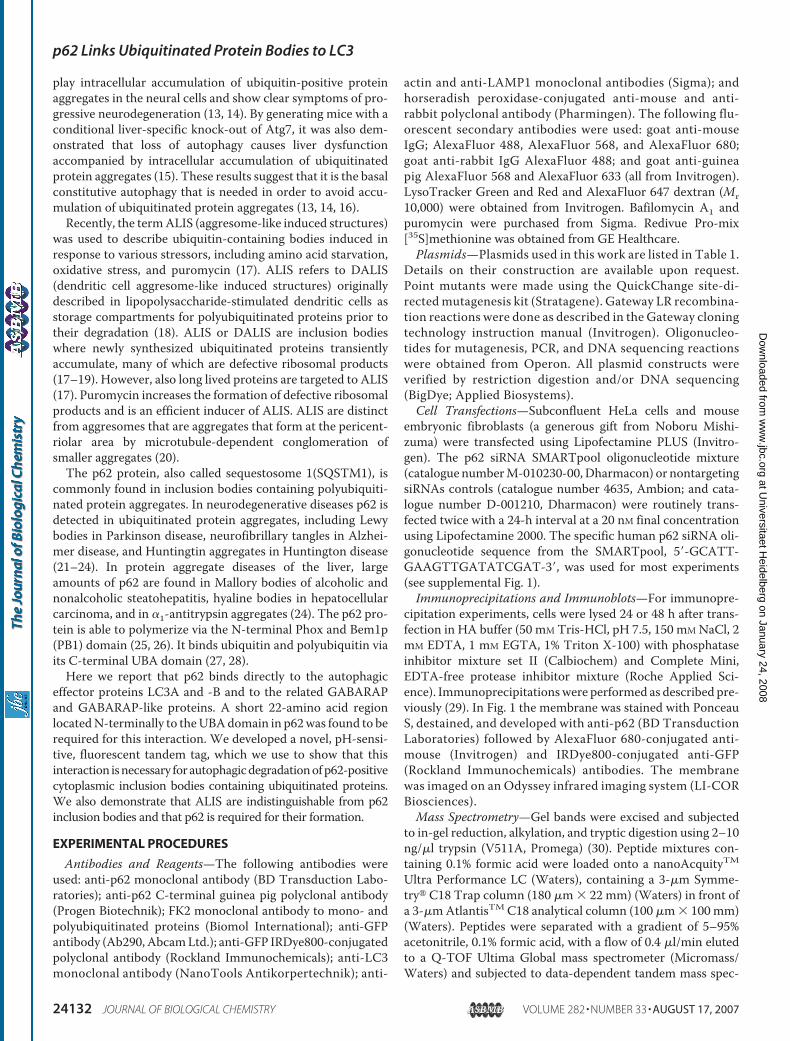
ther map the LC3 interaction region, we made a series of dele-tion constructs of GFP-tagged p62 that were translated in vitroin the presence of [35S]methionine and subjected to GST pull-down experiments with GST-LC3B bound to glutathione-Sepharose beads (Fig. 2). This enabled us to define the regionencompassing amino acids 321–342 of human p62 as an LIR.
FIGURE 2. The region spanning amino acids 321–342 of p62 is sufficient for interaction with LC3B.A, constructs used for GST pulldown between full-length LC3B, fused to GST, and deletion mutants of p62.B and C, mapping of the minimal LIR of p62 by GST pulldown assays between full-length GST-LC3B and deletionmutants of GFP-p62 (or myc-GFP-p62-(256 –370)) produced by coupled in vitro transcription and translationreaction in the presence of [35S]methionine. Twenty percent of input of proteins translated in vitro were run onthe same gel. The upper panels show the autoradiographs of the gels and the lower panels the same gels stainedwith Coomassie Blue (Coom. Blue).
p62 Links Ubiquitinated Protein Bodies to LC3
AUGUST 17, 2007 • VOLUME 282 • NUMBER 33 JOURNAL OF BIOLOGICAL CHEMISTRY 24135
at Universitaet H
eidelberg on January 24, 2008 w
ww
.jbc.orgD
ownloaded from

We could confirm this result bypulldown assays with internal dele-tions of p62. Deleting amino acids303–349 in the context of full-length p62 abolished binding toGST-LC3B, whereas deleting aminoacids 303–320 did not affect bindingof p62 lacking the N-terminal PB1domain (Fig. 3, A and B).The 22-amino acid long LIR is an
acidic peptide sequence containingthree glutamate and four aspartateresidues. We therefore asked if thebinding to LC3 could be dependenton electrostatic interactions be-tween acidic residues in LIR and abasic surface of LC3. Because thereare acidic residues both in the N-and C-terminal half of LIR, wedecided tomutate both clusters sep-arately. Thus, we mutated two con-secutive glutamate residues in theN-terminal cluster to alanines(E323A/E324A) and three consecu-tive aspartate residues in the C-ter-minal half to alanines (D335A/D336A/D337A) (see Fig. 3A). Asshown in Fig. 3,C andD, the bindingto GST-LC3B is reduced by 75%upon mutating the C-terminalaspartate residues, whereas it isunaffected by mutating the gluta-mate residues. As seen from thealignment of p62 sequences frommammals, opossum, chicken, frogs,fishes, sea urchin, and honeybee, itis apparent that the most conservedmotif is D(D/E) (D/E)WT at theC-terminal end of LIR (Fig. 3A).This is consistent with our findingthat mutation of the DDD motifstrongly affects binding to LC3. Wealso found that a single alanine sub-stitution of the absolutely conservedTrp-338 residue had the same dra-matic effect as mutating the DDDmotif, whereas similar substitutionsof the conserved Thr-339 residueand the serines at 332 and 342 hadno effect (Fig. 3A and data notshown).Both a crystal structure of form I
of rat LC3B and a solution structureof human form I LC3B have beenreported (33, 34). The LC3-I struc-ture consists of an N-terminal sub-domain (residues 1–29) with two�-helices and a C-terminal subdo-
p62 Links Ubiquitinated Protein Bodies to LC3
24136 JOURNAL OF BIOLOGICAL CHEMISTRY VOLUME 282 • NUMBER 33 • AUGUST 17, 2007
at Universitaet H
eidelberg on January 24, 2008 w
ww
.jbc.orgD
ownloaded from

main (residues 30–120) that adopt a ubiquitin fold. Surpris-ingly, neither the N-terminal subdomain (residues 1–28) northe C-terminal subdomain of human LC3B interacted with p62in GST pulldown assays, whereas full-length LC3B boundstrongly to p62 (data not shown). Consistent with the resultsfrom the pulldown experiments, only full-length LC3B inter-acted with the central part of p62 in the yeast two-hybrid sys-tem (data not shown).p62 Binds Both to LC3A and -B and the Related GABARAP
Family Proteins—LC3B is only one of several homologues ofyeast Atg8 found in mammals, including the GABARAP familyof proteins (35). We therefore wanted to determine whether
p62 could bind to other Atg8 homo-logues. To this end we transientlyexpressed GFP-tagged LC3A,LC3B, GABARAP, GABARAPL1,and GABARAPL2 in HeLa cells andsubjected cell extracts to immuno-precipitation using an anti-GFPantibody. Co-immunoprecipitationof endogenous p62 was thenassessed by immunoblotting with amonoclonal anti-p62 antibody. Asshown in Fig. 4A, endogenous p62was co-immunoprecipitated at thesame efficiency with GFP-taggedLC3A, GABARAP, GABARAPL1,and GABARAPL2 as with LC3B.Next, we performed GST pulldownassays where we incubated GST-tagged LC3- and GABARAP familyproteins, purified from E. coli andimmobilized on glutathione-Sepha-rose beads, with three different invitro translated, 35S-labeled GFP-p62 constructs. As shown in the toppanel of Fig. 4B, full-length p62 asrepresented by GFP-p62K7A/D69Abound strongly to all LC3 andGABARAP proteins tested. If theLIR found to bind to LC3B isdeleted, as in the GFP-p62K7A/D69A(�303–349) construct testedin the middle panel of Fig. 4B, theinteractions were abolished. How-ever, the LIR alone is sufficient forbinding to all GST-tagged LC3- andGABARAP family proteins (Fig. 4B,lower panel).Our finding of a direct interaction
between p62 and the human Atg8homologues tested here prompted
the question whether these proteins would localize to p62 bod-ies upon transient overexpression in cells. To test this HeLacells were transfected with the different GFP-tagged LC3 andGABARAP proteins, and the cells were fixed and stained forendogenous p62. Confocal fluorescence microscopy demon-strated a striking co-localization of theseGFP fusion proteins top62 bodies (Fig. 4C). Taken together, our results show that p62is able to bind in a similar manner to all the five tested Atg8human homologues and that these proteins can localize to p62bodies in cells. Using an antibody raised against an N-terminalpeptide of LC3B, we were also for the first time able to clearly
FIGURE 3. Characterization of the LIR of p62 by deletion mapping and point mutations. A, summary of GST pulldown assays between full-length LC3Bfused to GST and deletion mutants of p62 (upper panel). The lower panel shows an alignment of the LIR of human p62 to the corresponding sequences ofrepresentatives of mammals, birds, frogs, fishes, sea urchins, and insect species. A triple alanine substitution of the DDD motif and a single alanine substitutionof the conserved Trp residue strongly inhibited binding as indicated by filled circles above the alignment. Open circles indicate alanine substitutions withouteffect. B, interaction between GST-LC3B and in vitro translated, 35S-labeled deletion mutants of GFP-p62 analyzed by GST pulldown assays. C, p62 residuesAsp-335 to Asp-337 are required for efficient interaction with LC3B. D, quantitation of the GST pulldown assays as shown in C. The data are the mean � S.D. fromthree independent experiments. Coom. Blue, Coomassie Blue.
FIGURE 4. p62 interacts with other MAP1LC3 family proteins. A, endogenous p62 co-immunoprecipitateswith GFP fusions of LC3A, LC3B, GABARAP, GABARAPL1, and GABARAPL2 from HeLa cell extracts. GFP or GFPfusion constructs of LC3 family proteins were immunoprecipitated (IP) from total cellular extract of transfectedHeLa cells and subjected to SDS-PAGE. Co-purified p62 was detected by immunoblotting with anti-p62 anti-body (upper panel). WB, Western blot. Immunoprecipitated GFP fusion proteins were detected with an anti-GFPantibody (lower panel). B, direct interaction between p62 and LC3 family members was assessed using GSTpulldown assays as described in the legend to Fig. 2B. Coom. Blue, Coomassie Blue. C, GFP fusion proteins ofLC3A, LC3B, GABARAP, GABARAPL1, and GABARAPL2 co-localize with endogenous p62 (visualized with anti-p62 antibody staining) after transient transfection in HeLa cells. D, endogenous LC3 and p62 co-localize incytoplasmic bodies. HeLa cells were fixed and immunostained with p62 Ab and LC3 mAb. Scale bars are 5 �m.
p62 Links Ubiquitinated Protein Bodies to LC3
AUGUST 17, 2007 • VOLUME 282 • NUMBER 33 JOURNAL OF BIOLOGICAL CHEMISTRY 24137
at Universitaet H
eidelberg on January 24, 2008 w
ww
.jbc.orgD
ownloaded from
demonstrate co-localization of endogenous LC3 with p62 incytoplasmic bodies (Fig. 4D).A Novel Double Tag Strategy Makes It Possible to Visualize
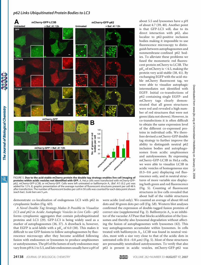
LC3 and p62 in Acidic Autophagic Vesicles in Live Cells—p62forms cytoplasmic aggregates that contain polyubiquitinatedproteins and LC3 (29). GFP-LC3 is being widely used as amarker of autophagosomes (36, 37). A drawback is, however,that EGFP is acid-labile with a pKa of 6.0 (38). This makes itdifficult to use GFP fusions to follow autophagosomes by fluo-rescence microscopy after they become acidified followingfusion with endosomes or lysosomes to produce amphisomesor autolysosomes. The pHof the lumenof early endosomesmayvary frompH6.1 to 5.5, and late endosomes usually have a pHof
about 5.5 and lysosomes have a pHof about 4.7 (39, 40). Another pointis that GFP-LC3 will, due to itsdirect interaction with p62, alsolocalize to p62-positive inclusionbodies making it impossible to usefluorescence microscopy to distin-guish between autophagosomes andnonmembrane-confined p62 bod-ies. To alleviate these problems wefused the monomeric red fluores-cent protein mCherry to LC3B. ThepKa of mCherry is �4.5, making theprotein very acid-stable (38, 41). Byexchanging EGFP with the acid-sta-ble mCherry fluorescent tag, wewere able to visualize autophagicintermediates not identified withEGFP. Initial co-transfections ofp62 containing single EGFP- andmCherry tags clearly demon-strated that all green structureswere red and revealed a high num-ber of red structures that were notgreen (data not shown).However, inco-transfections it is often difficultto obtain the same expression levelof the different co-expressed pro-teins in individual cells. We there-fore devised amCherry-GFP doubletag strategy to further improve theability to distinguish neutral p62inclusion bodies and autophago-somes from acidic amphisomesand autolysosomes. By expressingmCherry-GFP-LC3B in HeLa cells,we were able to visualize LC3B inacidic vesicles of homogeneous size(0.3–0.6 �m) displaying red fluo-rescence only, and in neutral struc-tures of more variable size display-ing both green and red fluorescence(Fig. 5). Counting of fluorescentstructures in live cells revealed thatabout half of the visible structures
were acidic (red only). We counted an average of about 60 reddots and 30 green dots per cell (Fig. 5B). Western blot analysesconfirmed the expression of double-tagged fusion proteins ofcorrect size (supplemental Fig. 2). Bafilomycin A1 is an inhibi-tor of the vacuolar ATPase that blocks acidification of the lyso-somes and thereby also lysosomal degradation without affect-ing the fusion of autophagosomes with lysosomes (42). Thisway autophagosomes accumulate within lysosomes. In cellstreated with bafilomycin A1, LC3B was found in neutral vesi-cles, most with a size very close to that of the acidic dots inuntreated cells (0.4–0.8 �m) (Fig. 5). Most of these structuresare presumably neutralized autolysosomes. To verify that alsop62 is present in acidic vesicles, mCherry-GFP-p62 was
FIGURE 5. Due to the acid-stable mCherry protein the double tag strategy enables live cell imaging ofproteins within acidic vesicles not identified with GFP. A, HeLa cells were transfected with mCherry-GFP-p62, mCherry-GFP-LC3B, or mCherry-GFP. Cells were left untreated or bafilomycin A1 (Baf. A1) (0.2 �M) wasadded for 12 h. B, graphic presentation of the average number of fluorescent structures present per cell 48 hafter transfection. The number of fluorescent bodies per cell in 50 cells was counted for each data point shown(each bar). Scale bars are 5 �m.
p62 Links Ubiquitinated Protein Bodies to LC3
24138 JOURNAL OF BIOLOGICAL CHEMISTRY VOLUME 282 • NUMBER 33 • AUGUST 17, 2007
at Universitaet H
eidelberg on January 24, 2008 w
ww
.jbc.orgD
ownloaded from
expressed in HeLa cells. As expected, p62 was found in bothacidic and neutral structures. The acidic vesicles, amounting tohalf (about 40 per cell) of the fluorescent structures, generallyhad a size very similar to those formed by LC3B (around 0.5�m).However, larger acidic structureswere also seen. The neu-tral (yellow) structures vary in size and mobility and repre-sent a mixture of p62-positive inclusion bodies and autopha-gosomes (29) (Fig. 5). Treatment with bafilomycin A1strongly increased the number of neutral structures, verysimilar to what was observed with mCherry-GFP-LC3B (Fig.5). To compare the results obtained with p62 and LC3B withthose of a randomly degraded protein, we expressed the dou-ble tag itself. When expressed in HeLa cells, relatively fewweak signals of mCherry-GFP could be detected in acidicvesicles of untreated cells and in neutral structures aftertreatment of cells with bafilomycin A1 (Fig. 5). However, thedifference between the double tag alone and the double tagfused to either LC3B or p62 is striking and strongly supportsthe notion that LC3B and p62 are specifically recruited intoautophagosomal structures.To verify that the acidic structures visualized with mCherry-
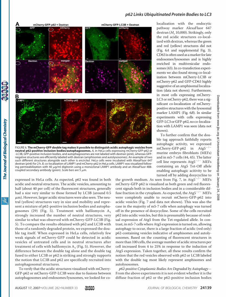
GFP-p62 or mCherry-GFP-LC3B were due to fusions betweenautophagosomes and endosomes/lysosomes, we looked for co-
localization with the endocyticpathway marker AlexaFluor 647dextran (Mr 10,000). Strikingly, onlythe red acidic structures co-local-izedwith dextran,whereas the greenand red (yellow) structures did not(Fig. 6A and supplemental Fig. 3).CD63 isoftenusedasamarker for lateendosomes/lysosomes and is highlyenriched in multivesicular endo-somes (43). In co-transfection experi-ments we also found strong co-local-ization between mCherry-LC3B ormCherry-p62 and GFP-CD63 highlysuggestive of an amphisomal localiza-tion (data not shown). Furthermore,in most cells expressing mCherry-LC3 ormCherry-p62, therewas a sig-nificant co-localization of mCherry-positive structureswith the lysosomalmarker LAMP1 (Fig. 6B). In similarexperiments with cells expressingGFP-LC3orGFP-p62,noco-localiza-tion with LAMP1 was seen (data notshown).To further confirm that the dou-
ble tag approach faithfully reportsautophagic activity, we expressedmCherry-GFP-p62 in Atg5�/�
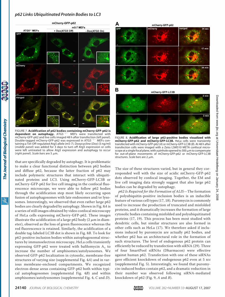
murine embryo fibroblasts (MEFs)and inm5-7 cells (44, 45). The lattercell line represents Atg5�/� MEFscontaining an Atg5 Tet-Off alleleenabling autophagic acitivity to beturned off by adding doxycycline to
the growth medium. As seen from Fig. 7, in Atg5�/� MEFsmCherry-GFP-p62 is visualized as both green and red fluores-cent signals both in inclusion bodies and in a considerable dif-fuse fraction in the cytoplasm. As expected, the Atg5�/� MEFswere completely unable to recruit mCherry-GFP-p62 intoacidic vesicles (Fig. 7 and data not shown). This was also thecase in the majority of m5-7 cells when autophagy was turnedoff in the presence of doxycycline. Some of the cells recruitedp62 into acidic vesicles, but this is presumably because of resid-ual expression of Atg5 from the Tet-regulated allele. In con-trast, inm5-7 cellswhereAtg5 expression is turned on, allowingautophagy to occur, there is a large fraction of acidic (red only)p62-containing vesicles indicative of amphisomes and autoly-sosomes. Based on the counting of fluorescent structures inmore than 100 cells, the average number of acidic structures percell increased from 4 to 25% in response to the induction ofAtg5 expression. Taken together, all these results confirm thenotion that the red vesicles observed with p62 or LC3B labeledwith the double tag most likely represent amphisomes andautolysosomes.p62-positive Cytoplasmic Bodies Are Degraded by Autophagy—
From the above experiments it is not evident whether it is thediffuse fraction of p62 or the p62-positive inclusion bodies
FIGURE 6. The mCherry-GFP double tag makes it possible to distinguish acidic autophagic vesicles fromneutral p62-positive inclusion bodies/autophagosomes. A, in HeLa cells expressing mCherry-GFP-p62 or-LC3B, GFP-positive inclusion bodies, and autophagosomes are not labeled with dextran (pink), whereas GFP-negative structures are efficiently labeled with dextran (amphisomes and autolysosomes). An example of twosuch different structures alongside each other is encircled. HeLa cells were incubated with AlexaFluor 647dextran (pink) for 2 h. B, co-localization of LAMP1 and mCherry-p62 in HeLa cells. LAMP1 was visualized follow-ing permeabilization with 40 �g/ml digitonin using a monoclonal LAMP1 antibody and an AlexaFluor 488-coupled secondary antibody (green). Scale bars are 5 �m.
p62 Links Ubiquitinated Protein Bodies to LC3
AUGUST 17, 2007 • VOLUME 282 • NUMBER 33 JOURNAL OF BIOLOGICAL CHEMISTRY 24139
at Universitaet H
eidelberg on January 24, 2008 w
ww
.jbc.orgD
ownloaded from
that are specifically degraded by autophagy. It is problematicto make a clear functional distinction between p62 bodiesand diffuse p62, because the latter fraction of p62 mayinclude polymeric structures that interact with ubiquiti-nated proteins and LC3. Using mCherry-GFP-LC3B ormCherry-GFP-p62 for live cell imaging in the confocal fluo-rescence microscope, we were able to follow p62 bodiesthrough the acidification step most likely occurring uponfusion of autophagosomes with late endosomes and/or lyso-somes. Interestingly, we observed that even rather large p62bodies are clearly degraded by autophagy. Shown in Fig. 8A isa series of still images obtained by video confocal microscopyof HeLa cells expressing mCherry-GFP-p62. These imagesillustrate the acidification of a large p62 body (2 �m in diam-eter), observed as the loss of green fluorescence whereas thered fluorescence is retained. Similarly, the acidification of adouble tag-labeled LC3B dot is shown in Fig. 8B. To look forp62-positive inclusion bodies within autophagosomal struc-tures by immunoelectron microscopy, HeLa cells transientlyexpressing GFP-p62 were treated with bafilomycin A1 toincrease the number of amphisomes/autolysosomes. Weobserved GFP-p62 localization in cytosolic, membrane-freestructures of varying size (supplemental Fig. 4A) and in var-ious membrane-enclosed compartments. We recognizedelectron-dense areas containing GFP-p62 both within typi-cal autophagosomes (supplemental Fig. 4B) and withinamphisomes/autolysosomes (supplemental Fig. 4, C and D).
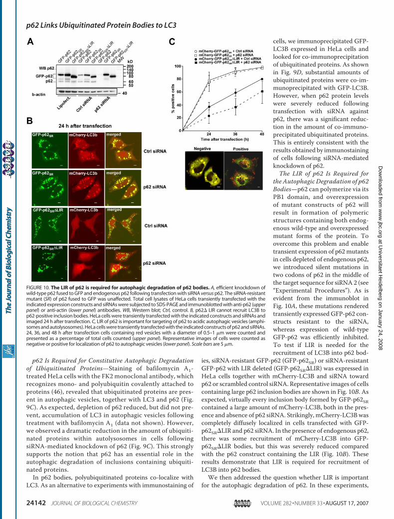
The size of these structures varied, but in general they cor-responded well with the size of acidic mCherry-GFP-p62dots observed by confocal imaging. Together, the EM andlive cell imaging data strongly suggest that also large p62bodies can be degraded by autophagy.p62 Is Required for the Formation of ALIS—The formation
of polyubiquitin-positive inclusion bodies is an induciblefeature of various cell types (17, 18). Puromycin is commonlyused to increase the production of truncated and misfoldedproteins, and it dramatically increases the formation of largecytosolic bodies containing misfolded and polyubiquitinatedproteins (17, 19). This process has been most studied withdendritic cells, but similar structures are also formed inother cells such as HeLa (17). We therefore asked if inclu-sions induced by puromycin are actually p62 bodies, andwhether p62 has an architectural role in the formation ofsuch structures. The level of endogenous p62 protein canefficiently be reduced by transfection with siRNA (29). Threeof four SmartPool siRNAs (Dharmacon) were effectiveagainst human p62. Transfection with one of these siRNAsgave efficient knockdown of endogenous p62 even at 5 nM
(supplemental Fig. 5). Interestingly, we found that puromy-cin-induced bodies contain p62, and a dramatic reduction intheir number was observed following siRNA-mediatedknockdown of p62 (Fig. 9, A and B).
FIGURE 7. Acidification of p62 bodies containing mCherry-GFP-p62 isdependent on autophagy. ATG5�/� MEFs were transfected withmCherry-GFP-p62 and live cells imaged 48 h after transfection (left panel).Double-tagged mCherry-GFP-p62 was expressed in ATG5�/� MEFs con-taining a Tet-Off-regulated Atg5 allele (m5-7). Doxycycline (Dox) (5 ng/ml)(middle panel) was added for 3 days to turn off Atg5 expression or cellswere left untreated to allow Atg5 expression and autophagy to occur(right panel). Scale bars are 5 �m.
FIGURE 8. Acidification of large p62-positive bodies visualized withmCherry-GFP-p62 and mCherry-GFP-LC3B. HeLa cells were transientlytransfected with mCherry-GFP-p62 (A) or mCherry-GFP-LC3B (B). At 48 h aftertransfection cells were imaged with a Zeiss LSM510 META confocal micro-scope at a single focal plane, with a pinhole opened to 300 �m to compensatefor out-of-plane movements of mCherry-GFP-p62 or mCherry-GFP-LC3Bstructures. Scale bars are 2 �m.
p62 Links Ubiquitinated Protein Bodies to LC3
24140 JOURNAL OF BIOLOGICAL CHEMISTRY VOLUME 282 • NUMBER 33 • AUGUST 17, 2007
at Universitaet H
eidelberg on January 24, 2008 w
ww
.jbc.orgD
ownloaded from

FIGURE 9. ALIS are indistinguishable from p62 inclusion bodies and dependent on p62 for their formation. A and B, formation of cytoplasmicpolyubiquitin-positive bodies (ALIS) is dependent on p62. HeLa cells were transfected with control (Ctrl) siRNA or p62 siRNA as indicated. Cell cultureswere stressed with puromycin (5 �g/ml) as indicated to induce formation of ALIS and then fixed and stained with p62 Ab and FK2 mAb. A, number of cellscontaining ubiquitinated (Ub) protein bodies increased with time after addition of puromycin reaching a maximum after 8 h (right panel). The inductionof ubiquitinated protein bodies (aggregates) following puromycin treatment or amino acid starvation is strongly inhibited by siRNA-mediated deple-tion of p62 (left panel). Results from representative experiments are shown. From 300 to 600 cells were scored for the presence or absence ofFK2-positive round bodies for each data point shown (each bar). Only cells that did not stain positive for p62 following transfection with p62 siRNA wereincluded in these quantitations. B, representative confocal images of cells from experiments used to perform the quantitations shown in A. Note thatknockdown of p62 results in loss of FK2 staining in cytoplasmic bodies. C, p62 links ubiquitinated proteins to LC3 in cytoplasmic bodies that becomedegraded by autophagy. Endogenous LC3, ubiquitinated proteins, and p62 co-localize in autolysosomes in bafilomycin A1-treated cells (upper panels).Depletion of p62 blocks the recruitment of ubiquitinated proteins to autolysosomes (lower panel). HeLa cells were incubated with bafilomycin A1 (0.2�M) for 12 h, fixed, and immunostained with LC3 mAb, FK2 mAb, and p62 Ab. D, siRNA-mediated knockdown of p62 reduces the interaction of transientlyexpressed GFP-LC3B with ubiquitinated proteins. GFP or GFP-LC3B was immunoprecipitated from total cellular extracts of HeLa cells that had beentreated with siRNA to p62 or control siRNA. Co-purified ubiquitinated proteins were detected using the FK2 mAb. Scale bars: 5 �m in B and C, upperpanels, and 10 �m in C, lower panel. WB, Western blot.
p62 Links Ubiquitinated Protein Bodies to LC3
AUGUST 17, 2007 • VOLUME 282 • NUMBER 33 JOURNAL OF BIOLOGICAL CHEMISTRY 24141
at Universitaet H
eidelberg on January 24, 2008 w
ww
.jbc.orgD
ownloaded from
p62 Is Required for Constitutive Autophagic Degradationof Ubiquitinated Proteins—Staining of bafilomycin A1-treated HeLa cells with the FK2 monoclonal antibody, whichrecognizes mono- and polyubiquitin covalently attached toproteins (46), revealed that ubiquitinated proteins are pres-ent in autophagic vesicles, together with LC3 and p62 (Fig.9C). As expected, depletion of p62 reduced, but did not pre-vent, accumulation of LC3 in autophagic vesicles followingtreatment with bafilomycin A1 (data not shown). However,we observed a dramatic reduction in the amount of ubiquiti-nated proteins within autolysosomes in cells followingsiRNA-mediated knockdown of p62 (Fig. 9C). This stronglysupports the notion that p62 has an essential role in theautophagic degradation of inclusions containing ubiquiti-nated proteins.In p62 bodies, polyubiquitinated proteins co-localize with
LC3. As an alternative to experiments with immunostaining of
cells, we immunoprecipitated GFP-LC3B expressed in HeLa cells andlooked for co-immunoprecipitationof ubiquitinated proteins. As shownin Fig. 9D, substantial amounts ofubiquitinated proteins were co-im-munoprecipitated with GFP-LC3B.However, when p62 protein levelswere severely reduced followingtransfection with siRNA againstp62, there was a significant reduc-tion in the amount of co-immuno-precipitated ubiquitinated proteins.This is entirely consistent with theresults obtained by immunostainingof cells following siRNA-mediatedknockdown of p62.The LIR of p62 Is Required for
the Autophagic Degradation of p62Bodies—p62 can polymerize via itsPB1 domain, and overexpressionof mutant constructs of p62 willresult in formation of polymericstructures containing both endog-enous wild-type and overexpressedmutant forms of the protein. Toovercome this problem and enabletransient expression of p62mutantsin cells depleted of endogenous p62,we introduced silent mutations intwo codons of p62 in the middle ofthe target sequence for siRNA 2 (see“Experimental Procedures”). As isevident from the immunoblot inFig. 10A, these mutations renderedtransiently expressed GFP-p62 con-structs resistant to the siRNA,whereas expression of wild-typeGFP-p62 was efficiently inhibited.To test if LIR is needed for therecruitment of LC3B into p62 bod-
ies, siRNA-resistant GFP-p62 (GFP-p62SR) or siRNA-resistantGFP-p62 with LIR deleted (GFP-p62SR�LIR) was expressed inHeLa cells together with mCherry-LC3B and siRNA towardp62 or scrambled control siRNA. Representative images of cellscontaining large p62 inclusion bodies are shown in Fig. 10B. Asexpected, virtually every inclusion body formed by GFP-p62SRcontained a large amount of mCherry-LC3B, both in the pres-ence and absence of p62 siRNA. Strikingly, mCherry-LC3Bwascompletely diffusely localized in cells transfected with GFP-p62SR�LIR and p62 siRNA. In the presence of endogenous p62,there was some recruitment of mCherry-LC3B into GFP-p62SR�LIR bodies, but this was severely reduced comparedwith the p62 construct containing the LIR (Fig. 10B). Theseresults demonstrate that LIR is required for recruitment ofLC3B into p62 bodies.We then addressed the question whether LIR is important
for the autophagic degradation of p62. In these experiments,
FIGURE 10. The LIR of p62 is required for autophagic degradation of p62 bodies. A, efficient knockdown ofwild-type p62 fused to GFP and endogenous p62 following transfection with siRNA versus p62. The siRNA-resistantmutant (SR) of p62 fused to GFP was unaffected. Total cell lysates of HeLa cells transiently transfected with theindicated expression constructs and siRNAs were subjected to SDS-PAGE and immunoblotted with anti-p62 (upperpanel) or anti-actin (lower panel) antibodies. WB, Western blot; Ctrl, control. B, p62� LIR cannot recruit LC3B top62-positive inclusion bodies. HeLa cells were transiently transfected with the indicated constructs and siRNAs andimaged 24 h after transfection. C, LIR of p62 is important for targeting of p62 to acidic autophagic vesicles (amphi-somes and autolysosomes). HeLa cells were transiently transfected with the indicated constructs of p62 and siRNAs.24, 36, and 48 h after transfection cells containing red vesicles with a diameter of 0.5–1 �m were counted andpresented as a percentage of total cells counted (upper panel). Representative images of cells were counted asnegative or positive for localization of p62 to autophagic vesicles (lower panel). Scale bars are 5 �m.
p62 Links Ubiquitinated Protein Bodies to LC3
24142 JOURNAL OF BIOLOGICAL CHEMISTRY VOLUME 282 • NUMBER 33 • AUGUST 17, 2007
at Universitaet H
eidelberg on January 24, 2008 w
ww
.jbc.orgD
ownloaded from

mCherry-GFP-p62SRormCherry-GFP-p62SR�LIRwasexpressedin HeLa cells together with siRNAs. For each experiment, wecounted the fraction of cells that contained p62 in acidic vesi-cles. Cells with more than five acidic p62 dots were counted aspositive (Fig. 10C). As expected, most cells expressingmCherry-GFP-p62SR recruited the protein into acidic vesicles,whereas deletion of LIR strongly reduced the ability of p62 to berecruited into acidic vesicles, both when co-expressed withsiRNA toward p62 or scrambled control siRNA (Fig. 10C). Thisfits well with the observation above that LC3B was poorlyrecruited into p62 bodies formed by p62SR�LIR, even in cellsexpressing a normal level of endogenous p62 (Fig. 10B). How-ever, it should be noted that at 48 h after transfectionp62SR�LIR also became gradually recruited into autophago-somes. Because siRNA depletion of p62 is not complete, weattribute that effect to residual endogenous p62 that helpsrecruit LC3 into structures containing p62SR�LIR. In conclu-sion, our results suggest that recruitment of LC3 via the LIR ofp62 is essential for autophagic degradation of p62 and p62-positive bodies.
DISCUSSION
In this study we show that p62 binds directly to LC3A and -Band other human Atg8 homologues such as GABARAP,GABARAPL1, andGABARAPL2. In fact, p62 is likely themajorLC3-interacting protein inHeLa cells (Fig. 1A). The interactionbetween p62 and Atg8 homologues is mediated by a 22-aminoacid acidic peptide motif (LIR) in p62 and requires both the N-and C-terminal subdomains of LC3B. Interestingly, one recentlarge scale yeast two-hybrid screen suggested an interactionbetween p62 and LC3B (47) and another an interactionbetween p62 and GABARAPL1 and -2 (48). We use a noveldouble tag strategy to demonstrate that the interaction betweenp62 and LC3 is necessary for degradation of p62-positive bodiescontaining polyubiquitinated proteins, by autophagy. By bind-ing polyubiquitinated proteins via the UBA domain, polymer-izing via its PB1 domain, and binding to LC3 via the LIR motif,p62 forms protein bodies containing LC3 that are degraded byautophagy.LC3belongs to the family ofmicrotubule-associated proteins
(MAPs) and is known to interact with both MAP1A and -B.MAP1B binds to both LC3-I and -II, and overexpression ofMAP1B results in reduced levels of LC3-II and reduced num-bers of GFP-LC3-labeled autophagosomes (49). Interestingly,we identified MAP1B by mass spectrometry as a prominentband co-immunoprecipitating with GFP-LC3 from a HeLa celllysate (Fig. 1A).GFP-LC3 has been used extensively as a marker for auto-
phagy (7, 37). However, because GFP-LC3 is also recruited toinclusions, the use of this marker may not always give reli-able information about the autophagic process. The pHlability of GFP makes it impossible to follow GFP-LC3 afterthe short lived autophagosomes fuse with lysosomes. Fusionwith late endosomes to create amphisomes may also lead toan environment where the fluorescence from GFP isquenched due to low pH. We therefore fused LC3 to theacid-stable fluorescent protein mCherry, and mCherry-LC3could easily be followed into amphisomes and autolyso-
somes. By combining these two tags, in mCherry-GFP-LC3and mCherry-GFP-p62, we were able to distinguish inclu-sions and autophagosomes (green and red) from amphi-somes/autolysosomes (red only). Use of AlexaFluor 647 dex-tran and LAMP1 antibodies confirmed that the redfluorescence of mCherry remains intact, whereas virtually allGFP fluorescence is lost in amphisomes and autolysosomes.The double tag can be used for live cell imaging and is strik-ingly informative about the autophagic process. When usedto study p62 bodies, green GFP-positive structures consti-tute p62 inclusions and autophagosomes, whereas bodiesthat are red only represent acidic amphisomes/autolyso-somes. The double tag approach is not limited to autophagicproteins such as LC3 and p62. The tag should therefore serveas a valuable tool to study internalization and lysosomal deg-radation of plasma membrane receptors by live cell imaging.Because p62 itself is degraded by autophagy, both we and
others have found a general correlation between inhibition ofautophagy and increased levels of p62 (16, 29, 49). Clearly, p62may also be used as an autophagicmarker. p62 has a less diffuselocalization pattern than LC3, making it easier to identify thesmall autophagic vesicles using this marker. However, it isimportant to keep in mind that by transiently overexpressingp62, the formation of inclusion bodies is also increased.Using fusion proteins containing the mCherry-GFP tandem
tag, we demonstrate that cytoplasmic bodies containing p62,LC3, and ubiquitinated proteins are degraded by autophagy.Both large (more than 1 �m in diameter) and small (less than0.5 �m in diameter) protein bodies could be engulfed by auto-phagy. Small p62 bodies changed fromneutral (yellow) to acidic(red) within aminute, whereas larger structures needed consid-erablymore time to become acidified.However, we observed anaccumulation of p62 in autophagic structures also in cells thatdid not contain large p62 bodies. Our current hypothesis is thatp62 bodies are degraded by basal constitutive autophagy evenbefore they grow to sizes detectable by light microscopy butthat also large structures are degraded. In Atg5�/� MEFs,which are completely deficient in autophagy (44, 45), the acidicp62-containing vesicleswere absent. UsingAtg5�/�MEFswithinducible expression of an Atg5 minigene (cDNA), we couldshow that mCherry-GFP-p62 could enter the autophagic path-way when Atg5 was expressed. Our results suggest the follow-ing: (i) p62 plays an architectural role in the formation of inclu-sion bodies and (ii) p62 also links these structures to theautophagic machinery via direct interaction with LC3s and/orother mammalian Atg8 homologues. The p62 LC3 interactingregion, LIR, was found indispensable for LC3 recruitment intop62-positive inclusion bodies.Our studieswith purified recom-binant proteins show that p62 binds to both the pro-form andthe processed form I of LC3 (Fig. 1 and data not shown). Pre-sumably the isolation membrane is recruited to p62-positivebodies concomitant with or following lipidation of LC3. It willbe important to identify proteins that recognize LC3 or p62 (orboth) and simultaneously are bound to the forming isolationmembrane, either directly or indirectly.Our data clearly show that puromycin-induced ALIS and
p62-positive inclusion bodies are the same structures and thattheir formation depends on the presence of p62. In fact, similar
p62 Links Ubiquitinated Protein Bodies to LC3
AUGUST 17, 2007 • VOLUME 282 • NUMBER 33 JOURNAL OF BIOLOGICAL CHEMISTRY 24143
at Universitaet H
eidelberg on January 24, 2008 w
ww
.jbc.orgD
ownloaded from

bodies can also be induced by overexpression of p62 or protea-somal inhibition. The latter very efficiently induces p62 bodies(29).Mutations in the p62/SQSTM1 gene at 5q35 are a common
cause of classical, adult onset Paget disease of the bone.Between 30 and 50% of the familial cases are due to dominantacting mutations leading to loss of function of polyubiquitinbinding by either deletion of the UBA domain or point muta-tions within this domain (27, 50). Genetic inactivation of p62 inmice leads to impaired osteoclastogenesis and mature onsetobesity with insulin resistance (51, 52). It will be important toelucidate how the role p62 has in protein degradation by auto-phagy is connected to the complex phenotypic consequencesobserved upon knocking out p62. Atg5�/� or Atg7�/�mice diesoon after birth, whereas the p62 knock-out mice show noextensive lethality at this stage. p62 is clearly a stress-inducedprotein increasing after oxygen radical stress, inhibition of pro-teasomal activity (53), and in response to expression of mutantHuntingtin (23). Hence, p62 may make an important contribu-tion to autophagy under conditions of oxidative stress and inthe aging organism. It will be interesting to learn if p62 knock-out mice show a late onset neurodegenerative phenotype.Genetic models of neurodegenerative diseases where aggrega-tion-prone mutant proteins are expressed in a p62-deficientbackground will clearly also yield important insights.
Acknowledgments—We are very grateful to Noboru Mizushima forthe generous gift ofMEF cell lines.We are also indebted to R. Tsien forthe kind gift of pRSET-B-mCherry and H. Berglund for pDEST-TH1.We acknowledge Anne Simonsen for help with MEF cells. We aregrateful to Harald Stenmark for the gift of polyclonal mCherry anti-body and for critical reading of the manuscript.
REFERENCES1. Goldberg, A. L. (2003) Nature 426, 895–8992. Cuervo, A. M. (2004) Trends Cell Biol. 14, 70–773. Klionsky, D. J. (2005) J. Cell Sci. 118, 7–184. Yoshimori, T. (2004) Biochem. Biophys. Res. Commun. 313, 453–4585. Eskelinen, E. L. (2005) Autophagy 1, 1–106. Ohsumi, Y. (2001) Nat. Rev. Mol. Cell Biol. 2, 211–2167. Kabeya, Y., Mizushima, N., Yamamoto, A., Oshitani-Okamoto, S., Oh-
sumi, Y., and Yoshimori, T. (2004) J. Cell Sci. 117, 2805–28128. Tanida, I., Ueno, T., and Kominami, E. (2004) J. Biol. Chem. 279,
47704–477109. Sou, Y. S., Tanida, I., Komatsu, M., Ueno, T., and Kominami, E. (2006)
J. Biol. Chem. 281, 3017–302410. He, H., Dang, Y., Dai, F., Guo, Z.,Wu, J., She, X., Pei, Y., Chen, Y., Ling,W.,
Wu, C., Zhao, S., Liu, J. O., and Yu, L. (2003) J. Biol. Chem. 278,29278–29287
11. Xin, Y., Yu, L., Chen, Z., Zheng, L., Fu, Q., Jiang, J., Zhang, P., Gong, R., andZhao, S. (2001) Genomics 74, 408–413
12. Nixon, R. A. (2006) Trends Neurosci. 29, 528–53513. Hara, T., Nakamura, K., Matsui, M., Yamamoto, A., Nakahara, Y., Suzuki-
Migishima, R., Yokoyama, M., Mishima, K., Saito, I., Okano, H., and Mi-zushima, N. (2006) Nature 441, 885–889
14. Komatsu, M., Waguri, S., Chiba, T., Murata, S., Iwata, J., Tanida, I., Ueno,T., Koike, M., Uchiyama, Y., Kominami, E., and Tanaka, K. (2006)Nature441, 880–884
15. Komatsu,M.,Waguri, S., Ueno, T., Iwata, J.,Murata, S., Tanida, I., Ezaki, J.,Mizushima, N., Ohsumi, Y., Uchiyama, Y., Kominami, E., Tanaka, K., andChiba, T. (2005) J. Cell Biol. 169, 425–434
16. Mizushima, N., and Hara, T. (2006) Autophagy 2, 302–30417. Szeto, J., Kaniuk, N. A., Canadien, V., Nisman, R.,Mizushima, N., Yoshimori,
T., Bazett-Jones, D. P., and Brumell, J. H. (2006)Autophagy 2, 189–19918. Lelouard,H., Gatti, E., Cappello, F., Gresser, O., Camosseto, V., and Pierre,
P. (2002) Nature 417, 177–18219. Lelouard, H., Ferrand, V., Marguet, D., Bania, J., Camosseto, V., David, A.,
Gatti, E., and Pierre, P. (2004) J. Cell Biol. 164, 667–67520. Kopito, R. R. (2000) Trends Cell Biol. 10, 524–53021. Kuusisto, E., Salminen, A., and Alafuzoff, I. (2001) Neuroreport 12,
2085–209022. Kuusisto, E., Salminen, A., and Alafuzoff, I. (2002) Neuropathol. Appl.
Neurobiol. 28, 228–23723. Nagaoka, U., Kim, K., Jana, N. R., Doi, H., Maruyama, M., Mitsui, K.,
Oyama, F., and Nukina, N. (2004) J. Neurochem. 91, 57–6824. Zatloukal, K., Stumptner, C., Fuchsbichler, A., Heid, H., Schnoelzer, M.,
Kenner, L., Kleinert, R., Prinz, M., Aguzzi, A., and Denk, H. (2002) Am. J.Pathol. 160, 255–263
25. Lamark, T., Perander, M., Outzen, H., Kristiansen, K., Øvervatn, A.,Michaelsen, E., Bjørkøy, G., and Johansen, T. (2003) J. Biol. Chem. 278,34568–34581
26. Wilson, M. I., Gill, D. J., Perisic, O., Quinn, M. T., and Williams, R. L.(2003)Mol. Cell 12, 39–50
27. Cavey, J. R., Ralston, S. H., Hocking, L. J., Sheppard, P.W., Ciani, B., Searle,M. S., and Layfield, R. (2005) J. Bone Miner. Res. 20, 619–624
28. Vadlamudi, R. K., Joung, I., Strominger, J. L., and Shin, J. (1996) J. Biol.Chem. 271, 20235–20237
29. Bjørkøy, G., Lamark, T., Brech, A., Outzen, H., Perander,M., Øvervatn, A.,Stenmark, H., and Johansen, T. (2005) J. Cell Biol. 171, 603–614
30. Shevchenko, A., Wilm, M., Vorm, O., and Mann, M. (1996) Anal. Chem.68, 850–858
31. Peters, P. J., Neefjes, J. J., Oorschot, V., Ploegh, H. L., and Geuze, H. J.(1991) Nature 349, 669–676
32. Slot, J. W., Geuze, H. J., Gigengack, S., Lienhard, G. E., and James, D. E.(1991) J. Cell Biol. 113, 123–135
33. Kouno, T., Mizuguchi, M., Tanida, I., Ueno, T., Kanematsu, T., Mori, Y.,Shinoda, H., Hirata, M., Kominami, E., and Kawano, K. (2005) J. Biol.Chem. 280, 24610–24617
34. Sugawara, K., Suzuki, N. N., Fujioka, Y., Mizushima, N., Ohsumi, Y., andInagaki, F. (2004) Genes Cells 9, 611–618
35. Tanida, I., Ueno, T., and Kominami, E. (2004) Int. J. Biochem. Cell Biol. 36,2503–2518
36. Kabeya, Y., Mizushima, N., Ueno, T., Yamamoto, A., Kirisako, T., Noda,T., Kominami, E., Ohsumi, Y., and Yoshimori, T. (2000) EMBO J. 19,5720–5728
37. Mizushima, N. (2004) Int. J. Biochem. Cell Biol. 36, 2491–250238. Shaner, N. C., Steinbach, P. A., and Tsien, R. Y. (2005) Nat. Meth. 2,
905–90939. Kielian, M. C., and Cohn, Z. A. (1982) J. Cell Biol. 93, 875–88240. Zen, K., Biwersi, J., Periasamy, N., and Verkman, A. S. (1992) J. Cell Biol.
119, 99–11041. Shaner, N. C., Campbell, R. E., Steinbach, P. A., Giepmans, B. N., Palmer,
A. E., and Tsien, R. Y. (2004) Nat. Biotechnol. 22, 1567–157242. Fass, E., Shvets, E., Degani, I., Hirschberg, K., and Elazar, Z. (2006) J. Biol.
Chem. 281, 36303–3631643. Escola, J. M., Kleijmeer, M. J., Stoorvogel, W., Griffith, J. M., Yoshie, O.,
and Geuze, H. J. (1998) J. Biol. Chem. 273, 20121–2012744. Hosokawa, N., Hara, Y., and Mizushima, N. (2006) FEBS Lett. 580,
2623–262945. Kuma, A., Hatano,M.,Matsui, M., Yamamoto, A., Nakaya, H., Yoshimori,
T., Ohsumi, Y., Tokuhisa, T., and Mizushima, N. (2004) Nature 432,1032–1036
46. Fujimuro, M., Sawada, H., and Yokosawa, H. (1994) FEBS Lett. 349,173–180
47. Stelzl, U., Worm, U., Lalowski, M., Haenig, C., Brembeck, F. H., Goehler,H., Stroedicke, M., Zenkner, M., Schoenherr, A., Koeppen, S., Timm, J.,Mintzlaff, S., Abraham, C., Bock, N., Kietzmann, S., Goedde, A., Toksoz,E., Droege, A., Krobitsch, S., Korn, B., Birchmeier, W., Lehrach, H., andWanker, E. E. (2005) Cell 122, 957–968
p62 Links Ubiquitinated Protein Bodies to LC3
24144 JOURNAL OF BIOLOGICAL CHEMISTRY VOLUME 282 • NUMBER 33 • AUGUST 17, 2007
at Universitaet H
eidelberg on January 24, 2008 w
ww
.jbc.orgD
ownloaded from

48. Rual, J. F., Venkatesan, K., Hao, T., Hirozane-Kishikawa, T., Dricot, A., Li, N.,Berriz,G. F.,Gibbons, F.D.,Dreze,M.,Ayivi-Guedehoussou,N., Klitgord,N.,Simon,C.,Boxem,M.,Milstein, S., Rosenberg, J.,Goldberg,D. S.,Zhang,L.V.,Wong,S.L., Franklin,G.,Li, S.,Albala, J. S., Lim, J., Fraughton,C.,Llamosas,E.,Cevik, S., Bex, C., Lamesch, P., Sikorski, R. S., Vandenhaute, J., Zoghbi, H. Y.,Smolyar, A., Bosak, S., Sequerra, R., Doucette-Stamm, L., Cusick, M. E., Hill,D. E., Roth, F. P., and Vidal, M. (2005)Nature 437, 1173–1178
49. Wang, Q. J., Ding, Y., Kohtz, D. S., Mizushima, N., Cristea, I. M., Rout,M. P., Chait, B. T., Zhong, Y., Heintz, N., and Yue, Z. (2006) J. Neurosci. 26,8057–8068
50. Daroszewska, A., and Ralston, S. H. (2006)Nat. Clin. Pract. Rheumatol. 2,270–277
51. Duran, A., Serrano, M., Leitges, M., Flores, J. M., Picard, S., Brown, J. P.,Moscat, J., and Diaz-Meco, M. T. (2004) Dev. Cell 6, 303–309
52. Rodriguez, A., Duran, A., Selloum, M., Champy, M. F., Diez-Guerra, F. J.,Flores, J. M., Serrano, M., Auwerx, J., Diaz-Meco, M. T., and Moscat, J.(2006) Cell Metab. 3, 211–222
53. Ishii, T., Yanagawa, T., Yuki, K., Kawane, T., Yoshida, H., and Bannai, S.(1997) Biochem. Biophys. Res. Commun. 232, 33–37
54. Hammarstrom,M.,Hellgren,N., vanDenBerg, S., Berglund,H., andHard,T. (2002) Protein Sci. 11, 313–321
55. Simonsen, A., Birkeland, H. C., Gillooly, D. J., Mizushima, N., Kuma, A.,Yoshimori, T., Slagsvold, T., Brech, A., and Stenmark, H. (2004) J. Cell Sci.117, 4239–4251
p62 Links Ubiquitinated Protein Bodies to LC3
AUGUST 17, 2007 • VOLUME 282 • NUMBER 33 JOURNAL OF BIOLOGICAL CHEMISTRY 24145
at Universitaet H
eidelberg on January 24, 2008 w
ww
.jbc.orgD
ownloaded from