panax quinquefolius) attenuates leptin-induced cardiac...
TRANSCRIPT

JPET #182600
1
Title Page
Ginseng (Panax quinquefolius) attenuates leptin-induced cardiac
hypertrophy through inhibition of p115RhoGEF-RhoA/ROCK-
dependent MAPK pathway activation
Melissa Moey, Venkatesh Rajapurohitam, Asad Zeidan and Morris Karmazyn
Department of Physiology and Pharmacology, Schulich School of Medicine and Dentistry,
University of Western Ontario (M.M, V.R, and M.K), London, Ontario N6A 5C1, Canada and
Department of Anatomy, Cell Biology and Physiological Sciences, American University of
Beirut (A.Z), Beirut, Lebanon.
JPET Fast Forward. Published on August 29, 2011 as DOI:10.1124/jpet.111.182600
Copyright 2011 by the American Society for Pharmacology and Experimental Therapeutics.
This article has not been copyedited and formatted. The final version may differ from this version.JPET Fast Forward. Published on August 29, 2011 as DOI: 10.1124/jpet.111.182600
at ASPE
T Journals on Septem
ber 21, 2018jpet.aspetjournals.org
Dow
nloaded from

JPET #182600
2
Running Title Page
Running Title: Ginseng Inhibits Leptin-induced Cardiac Hypertrophy
Number of Text Pages: 32
Number of Tables: 1
Number of Figures: 8
Number of References: 40
Number of Words in Abstract: 249
Number of Words in Introduction: 627
Number of Words in Discussion: 1411
Nonstandard abbreviations: cardiovascular disease (CVD); fetal bovine serum (FBS); α-skeletal
actin (α-SA); myosin heavy chain (MHC); proliferating cell nuclear antigen (PCNA); guanine
nucleotide exchange factor (GEF); Ras homolog gene family, member A (RhoA); Rho-
associated, coiled-coil containing protein kinase (ROCK); LIM domain kinase (LIMK);
filamentous actin (F actin); globular actin (G actin); extracellular-signal-related kinase 1/2
(ERK1/2 ); mitogen activated protein kinase (MAPK); leptin receptor isoform b (Ob-Rb)
Recommended Section Assignment: cardiovascular
Corresponding Author:
Morris Karmazyn PhD
Department of Physiology and Pharmacology
The University of Western Ontario
London, Ontario, Canada N6A 5C1
This article has not been copyedited and formatted. The final version may differ from this version.JPET Fast Forward. Published on August 29, 2011 as DOI: 10.1124/jpet.111.182600
at ASPE
T Journals on Septem
ber 21, 2018jpet.aspetjournals.org
Dow
nloaded from

JPET #182600
3
Abstract
Leptin is a 16 kDa peptide primarily derived from white adipocytes and is typically elevated in
plasma of obese individuals. Although leptin plays a critical role in appetite regulation, leptin
receptors have been identified in numerous tissues including the heart and have been shown to
directly mediate cardiac hypertrophy through RhoA/ROCK-dependent p38 MAPK activation;
however the basis for RhoA stimulation is unknown. Rho GEFS (guanine nucleotide exchange
factors) catalyze the exchange of GDP for GTP resulting in Rho activation and may be the
potential upstream factors mediating leptin-induced RhoA activation and therefore a potential
target for inhibition. We investigated the effects of North American ginseng (Panax
quinquefolius), reported to reduce cardiac hypertrophy, on RhoA/ROCK and MAPK activation
in ventricular cardiomyocytes exposed to leptin (50 ng/ml) and the possible role of p115RhoGEF
and p63RhoGEF in these responses. Leptin produced a robust hypertrophic response which was
associated with RhoA/ROCK activation resulting in a significant increase in cofilin-2
phosphorylation and actin polymerization, the latter evidenced by a reduction in the G/F actin
ratio. These effects were prevented by ginseng (10 µg/ml). The stimulation of RhoA/ROCK by
leptin was associated with significantly increased p115RhoGEF gene and protein expression and
exchange activity, all of which were significantly inhibited by ginseng. The ability of ginseng to
prevent leptin-induced activation of RhoA/ROCK was further associated with diminished p38
MAPK activation and nuclear translocation. These results demonstrate a potent inhibitory effect
of ginseng against leptin-induced cardiac hypertrophy, an effect associated with prevention of
p115RhoGEF-RhoA/ROCK-dependent p38 MAPK activation.
This article has not been copyedited and formatted. The final version may differ from this version.JPET Fast Forward. Published on August 29, 2011 as DOI: 10.1124/jpet.111.182600
at ASPE
T Journals on Septem
ber 21, 2018jpet.aspetjournals.org
Dow
nloaded from

JPET #182600
4
Introduction
The prevalence of obesity in North America has significantly increased over the past 5 years
(Luo et al., 2007) along with the detrimental accompanying risks for the development of
cardiovascular disease (Bui et al., 2011). The underlying mechanisms for obesity-associated
cardiovascular disease are not well understood although emerging evidence implicates a
potential role of leptin, a member of the family of peptides known as adipokines which is
produced by the ob (obesity) gene (Zhang et al., 1994) and which has been reported by a number
of investigators to produce a direct hypertrophic effect on the heart (reviewed in Karmazyn et al.,
2008). The primary source of leptin is white adipose tissue and plasma levels of the peptide have
been shown to be closely correlated to the degree of adiposity (Maffei et al., 1995). We have
previously shown that the rat heart produces leptin within the cardiomyocyte and also expresses
leptin receptors suggesting that the heart is a target for leptin’s effects, potentially in a
paracrine/autocrine manner (Purdham et al., 2004). Interestingly, plasma levels of leptin are
elevated in patients with heart failure independently of obesity (Schulze et al., 2003).
Several molecular signalling pathways upregulated by leptin in cardiac hypertrophy have
been identified namely the RhoA/ROCK (Ras homolog gene family, member A/Rho-associated,
coiled-coil containing protein kinase) and p38 and ERK1/2 (extracellular regulated kinase 1/2)
MAPK (mitogen-activated protein kinases) pathways (Fruhbeck, 2006; Zeidan et al., 2006;
Zeidan et al., 2008), all of which can be activated by leptin. A particular importance of the
RhoA/ROCK pathway is that its activation, and subsequent changes in actin dynamics is critical
for selective p38 MAPK translocation into nuclei thus initiating the hypertrophic process (Zeidan
et al., 2006; Zeidan et al., 2008). Although we have previously demonstrated the potent
stimulation of the RhoA/ROCK pathway by leptin, the specific mechanism of activation
This article has not been copyedited and formatted. The final version may differ from this version.JPET Fast Forward. Published on August 29, 2011 as DOI: 10.1124/jpet.111.182600
at ASPE
T Journals on Septem
ber 21, 2018jpet.aspetjournals.org
Dow
nloaded from

JPET #182600
5
however has not yet been defined. Regulators of small G-proteins such as RhoA include GTP-
ase activating proteins, which hydrolyze GTP to GDP thereby deactivating RhoA, guanine
nucleotide dissociation inhibitors which sequester GDP-bound small G-proteins in the cytoplasm
and guanine nucleotide exchange factors (GEFs) which catalyze the exchange of GDP to GTP
resulting in activation of RhoA (Schmidt and Hall, 2002; Rossman et al., 2005; Bos et al., 2007).
RhoA GEFs (RhoGEFs), specifically p115RhoGEF and p63RhoGEF, have been shown to be
involved in the upregulation of RhoA/ROCK in cardiac hypertrophy in response to G-protein
coupled receptor-linked hypertrophic agonists such as endothelin-1 (Porchia et al., 2008) and
angiotensin II (Guilluy et al., 2010).
Ginseng (genus Panax) is a medicinal herb which has been used widely in Asia for more
than 2000 years (Goldstein, 1975; Lu et al., 2009). Its principal bioactive components are
ginsenosides, which are triterpene saponins and are considered the main constituents responsible
for ginseng’s medicinal effects (Attele et al., 1999). Emerging understanding of the chemistry of
ginseng and its potential therapeutic use has resulted in increasing interest in western countries
for the use of ginseng as a pharmacological agent for the treatment of a number of diseases.
With respect to the cardiovascular system, ginsenosides have been shown to inhibit the
development of atherosclerosis (Li et al., 2011), hypertension (Jeon et al., 2000) and cardiac
hypertrophy, the latter effect being seen in a number of experimental models (Jiang et al., 2007;
Qin et al., 2008; Deng et al., 2010; Guo et al., 2011). Whether ginseng affects leptin-induced
hypertrophy has not been demonstrated. Accordingly, in this study we determined the effect of
North American ginseng (P. quinquefolius) on leptin-induced cardiomyocyte hypertrophy and
studied the potential underlying mechanisms for these effects. Our study centered on the
possible modulatory effect of ginseng on the RhoA/ROCK pathway following leptin addition
This article has not been copyedited and formatted. The final version may differ from this version.JPET Fast Forward. Published on August 29, 2011 as DOI: 10.1124/jpet.111.182600
at ASPE
T Journals on Septem
ber 21, 2018jpet.aspetjournals.org
Dow
nloaded from

JPET #182600
6
and the role of GEFs. In addition, we studied the relationship between RhoA and MAPK
pathway activation.
This article has not been copyedited and formatted. The final version may differ from this version.JPET Fast Forward. Published on August 29, 2011 as DOI: 10.1124/jpet.111.182600
at ASPE
T Journals on Septem
ber 21, 2018jpet.aspetjournals.org
Dow
nloaded from

JPET #182600
7
Methods
Treatment and Experimental Groups
Neonatal ventricular cardiomoycytes were isolated and cultured from one to three day old
Sprague Dawley rats as previously described (Rajapurohitam et al., 2006). Cells were grown in
fetal bovine serum (FBS) medium for up to 48 hours following 24 hour serum starvation. For
cell size and Western blotting time-course experiments, cardiomyocytes were pre-treated with
the alcoholic extract of North American ginseng (Panax quinquefoilus). Extracts were prepared
by Naturex (South Hackensack, NJ) using ginseng roots supplied from 5 different farms in
Ontario, Canada as previously described (Guo et al., 2011) and were studied at concentrations of
0.1, 1, 10 and 100 μg/ml for up to 24 hours in the presence or absence of 3.1 nM (50 ng/ml)
leptin (Sigma-Aldrich, Oakville, Ontario, Canada) a concentration representative of plasma
levels in obese individuals (Maffei et al., 1995). For all subsequent experiments, cells were pre-
treated with a ginseng concentration of 10 μg/ml for 1 hour in the presence or absence of leptin
for up to 24 hours. Treatment durations reflected the period of peak activation of the parameter
under study. The protocols for the use of animals were approved by the University of Western
Ontario Animal Care and Use Committee and conformed to guidelines in the Guide for the Care
and Use of Laboratory Animals published by the US National Institutes of Health and the
Canadian Council of Animal Care (Ottawa, Ontario, Canada).
Cell Surface Area Measurement
Cardiomyocyte images were taken with a Leica microscope (Leica, Westzlar, Germany)
equipped with an Infinity 1 camera at 100x magnification. The surface area of a minimum of 50
cells per treatment group was measured using SigmaScan Pro 5 software (Systat, Richmond,
CA) and averaged.
This article has not been copyedited and formatted. The final version may differ from this version.JPET Fast Forward. Published on August 29, 2011 as DOI: 10.1124/jpet.111.182600
at ASPE
T Journals on Septem
ber 21, 2018jpet.aspetjournals.org
Dow
nloaded from
JPET #182600
8
RNA Isolation, Reverse Transcription (RT) and Real-time Polymerase Chain Reaction
(PCR)
RNA was collected from cultured and treated neonatal ventricular cardiomyocytes using Trizol
Reagent (Invitrogen, Carlsbad, CA) as per the manufacturer’s instructions and reverse
transcribed to complementary DNA (cDNA) for real-time polymerase chain reaction analysis of
α-skeletal actin, myosin heavy chain α, p115RhoGEF and p63RhoGEF. Briefly, cDNA was
synthesized from 4 μg of total RNA using random primers (Invitrogen, Carlsbad, CA) and M-
MLV Reverse Transcriptase (Invitrogen) following the manufacturer’s protocol. The reaction
was performed with a SYBR Green Master Mix (Applied Biosystems, Foster City, CA) and the
gene products quantified with a DNA Engine Opticon 2 thermal cycler (MJ Research, Waltham,
MA). Primer sequences (Invitrogen) for the genes of interest are listed in Table 1. PCR cycle
conditions involved 40 cycles of denaturation at 95°C for 30s, followed by annealing at 60°C for
30s and finally by elongation at 72°C for 30s. The housekeeping gene, 18S, was measured and
quantified to normalize cDNA levels.
[3H] Leucine Incorporation Measurement
Leucine incorporation was performed as previously described (Zeidan et al., 2006) to analyze
protein synthesis under different experimental conditions. Cardiomyocytes were cultured in 24-
well Primaria culture plates for 48 hours in serum media following by 24 hour serum starvation.
Leptin was administered with or without ginseng pre-treatment in the presence of 2 μCi of [3H]-
leucine for 24 hours. Myocytes were washed the following day with cold PBS and proteins were
precipitated with 5% trichloroacetic acid (TCA) for 30 minutes on ice. Protein precipitates were
This article has not been copyedited and formatted. The final version may differ from this version.JPET Fast Forward. Published on August 29, 2011 as DOI: 10.1124/jpet.111.182600
at ASPE
T Journals on Septem
ber 21, 2018jpet.aspetjournals.org
Dow
nloaded from

JPET #182600
9
dissolved with 0.5N NaOH following with two washes of cold 5% TCA. 0.5N HCl was used to
neutralize the precipitates and the total radioactivity was measured by liquid scintillation.
Isolation of Cytosolic-Enriched and Membrane Fractions
Cytosolic-enriched and membrane fractions from treated cell lysates were prepared using
differential centrifugation as previously described (Zeidan et al., 2008). Briefly, cell lysates were
collected and homogenized in a cold buffer containing 20 mM Tris-HCl, 2 mM EDTA, 137 mM
NaCl, 1 mM sodium orthovanadate, 2 mM sodium pyrophosphate, 10% glycerol, 1mM 4-(2-
aminorthyl)-benzenesulfonyl fluoride and 10 mg/ml leupeptin (buffer A). After clarification of
the homogenate by centrifugation at 750g for 20 minutes at 4°C, the collected lysate was further
centrifuged at 10,000 g for 20 minutes at 4°C and the cytosolic-enriched fraction (supernatant)
was obtained. The remaining pellet was resuspended in a second cold buffer B (buffer A with
2% SDS) and kept on ice to be used as the nuclear-containing membrane fraction.
Western Blotting
Total cellular lysates were collected using a lysis buffer and protease cocktail inhibitor mixture
as previously described (Zeidan et al., 2006; Zeidan et al., 2008) for the measurement of proteins
of interest. Proteins were loaded equally on either 7.5%, 10% or 15% SDS gels as appropriate
after protein quantification via BioRad Reagent (BioRad, Hercules, CA) per the manufacturer’s
instructions. For time course experiments, ventricular cardiomyocytes were treated for 5, 10, 15,
30 or 60 minutes with leptin in the presence or absence of ginseng (10 µg/ml). For the
quantification of cofilin-2 phosphorylation, cells were pre-treated with ginseng for 1 hour
followed by administration of leptin for 10 minutes. For all additional protein measurements,
This article has not been copyedited and formatted. The final version may differ from this version.JPET Fast Forward. Published on August 29, 2011 as DOI: 10.1124/jpet.111.182600
at ASPE
T Journals on Septem
ber 21, 2018jpet.aspetjournals.org
Dow
nloaded from

JPET #182600
10
cells were pre-treated with ginseng for 1 hour in the presence or absence of leptin for 24 hours.
The primary antibodies and respective dilutions used in this study include total (1:1000 dilution,
Santa Cruz Biotechnology Inc., Santa Cruz, CA, USA) and phosphorylated p38 (Thr180/Tyr182)
forms (1:1000 dilution, Cell Signaling, Danvers, MA), total (1:1000 dilution, Santa Cruz) and
phosphorylated ERK1/2 (Thr202/Tyr204) forms (1:1000 dilution, Cell Signaling), actin (1:1000
dilution, Cytoskeleton Inc., Denver, CO), p115RhoGEF (1:250 dilution, Santa Cruz),
p63RhoGEF (1:200 dilution, Santa Cruz) and phosphorylated (1:1000 dilution, Santa Cruz) and
total cofilin-2 (1:1000 dilution, Millipore, Billerica, MA). Goat-anti-rabbit IgG and goat-anti-
mouse IgG HRP conjugate (BioRad) were used at 1:5000 dilution and donkey-anti-goat IgG
HRP conjugate (Santa Cruz) was used at 1:10,000 dilution as appropriate. β-actin (1:1000
dilution, Cytoskeleton) and PCNA (1:1000 dilution) were used for cytosolic and nuclear loading
controls, respectively. Spot densitometry using FluorChem (Alpha Innotech Corporation, Santa
Clara, CA) software was performed to quantify protein.
GST-RhoG17A Bead Preparation
GST-RhoG17A beads were prepared as previously described (Garcia-Mata et al., 2006;
Kakiashvili et al., 2009). The nucleotide free RhoG17A cDNA construct mutant was generously
provided by Dr. Katalin Szászi (St. Michael’s Hospital, Toronto, ON, Canada) and Dr. Keith
Burridge (University of North Carolina, Chapel Hill, NC).
Co-immunoprecipitation of p115RhoGEF
Active p115RhoGEF from cells treated with leptin in the presence or absence of leptin were
immunoprecipitated with the nucleotide-free (does not bind to GTP or GDP) GST-RhoG17A
This article has not been copyedited and formatted. The final version may differ from this version.JPET Fast Forward. Published on August 29, 2011 as DOI: 10.1124/jpet.111.182600
at ASPE
T Journals on Septem
ber 21, 2018jpet.aspetjournals.org
Dow
nloaded from
JPET #182600
11
prepared beads, which has a high affinity for active RhoGEFs, including p115RhoGEF, as
previously described (Garcia-Mata et al., 2006; Kakiashvili et al., 2009).
Immunofluorescence
Cells were prepared for immunofluorescence on collagen coated (3 µl of collagen/1 ml of PBS
A) glass cover slips and incubated at 37°C for a minimum of 30 minutes. Cells were allowed to
attach to prepared cover slips in serum media for 24 hours followed by serum-free media
starvation for an additional 24 hours before appropriate treatment. Immunofluorescence
measurements from cells administered leptin for 3 hours were performed for total p38 and total
ERK1/2, 10 minutes for p115RhoGEF and RhoA and 24 hours for G/F actin with or without
ginseng pre-treatment.
Total p38, total ERK1/2, p115RhoGEF and RhoA immunofluorescence. Cells were fixed with
2:5 acetone-methanol for 1 hour at 4°C followed by permeabilization of cells for 15 minutes with
0.2% (v/v) Triton X-100 and blocking with blocking solution (1% BSA, 0.1% Triton X-100) for
1 hour. Cells were incubated with the primary antibody of interest (1:100 dilution) in 2% BSA in
PBS A overnight at 4°C. Cells were subsequently probed with the appropriate secondary
antibody, IgG anti-mouse AlexFluor-488 (Invitrogen) or IgG anti-rabbit AlexFluor-596
(Invitrogen) (1:250 dilution) in 2% BSA in PBS A for 1 hour at room temperature under light-
free conditions. For detection of the nucleus, cells were incubated with Hoechst dye for 30
minutes prior to mounting on microscope slides (VWR, West Chester, PA) for image capture
using a Zeiss (Oberkochen, Germany) inverted fluorescence microscope at 630X magnification.
G and F actin immunofluorescence. Cells were prepared as previously described (Albinsson et
al., 2004). Cells were fixed with 3.7% (w/v) paraformaldehyde in PBS A for 1 hour followed by
This article has not been copyedited and formatted. The final version may differ from this version.JPET Fast Forward. Published on August 29, 2011 as DOI: 10.1124/jpet.111.182600
at ASPE
T Journals on Septem
ber 21, 2018jpet.aspetjournals.org
Dow
nloaded from

JPET #182600
12
a similar protocol for permeabilization and blocking as indicated above. To detect G and F actin,
cells were incubated with 1 µg/ml Phalloidin-FITC and 10 µg/ml deoxyribonuclease I (DNase I),
Texas Red conjugate in 2% BSA with 0.1% Triton X-100 in PBS A for 1 hour at room
temperature under light-free conditions. Similarly, Hoechst dye was used to detect the nucleus
and glass cover slips were subsequently mounted onto microscope slides (VWR) for
visualization of G/F actin. All immunofluorescence images shown in results are representative of
a minimum of 3 independent experiments.
Measurement of p115RhoGEF and RhoA Co-localization
Co-localization of p115RhoGEF and RhoA was measured from merged immunofluorescence
images detecting p115RhoGEF and RhoA under different experimental conditions using a built-
in co-localization plug-in (Li et al., 2004) of ImageJ (National Institute of Mental Health,
Betheseda, MD), which is quantitatively represented by Pearson’s correlation coefficient (Rr).
Measurement of RhoA Activity
RhoA activity, measured by RhoA-GTP levels, was quantified using the RhoA G-LISA Activity
Biochem Assay Kit as per the manufacturer’s protocol (Cytoskeleton). Measurements were
performed using a SpectraMax M5 (Molecular Devices, Sunnyvale, CA) plate reader at an
absorbance of 490 nm.
p115RhoGEF Activity Assay
Measurement of immunoprecipitated p115RhoGEF activity was measured as per the
manufacturer’s protocol using the RhoGEF Biochem Exchange Assay (Cytoskeleton Inc).
This article has not been copyedited and formatted. The final version may differ from this version.JPET Fast Forward. Published on August 29, 2011 as DOI: 10.1124/jpet.111.182600
at ASPE
T Journals on Septem
ber 21, 2018jpet.aspetjournals.org
Dow
nloaded from

JPET #182600
13
Fluorescence was measured using a SpectraMax M5 (Molecular Devices) plate reader at
excitation of 360 nm and emission of 440 nm.
G/F Actin Measurement
Globlular (G) and filamentous (F) actin were isolated using ultracentrifugation as previously
described (Albinsson et al., 2004). Briefly cell lysates were collected and homogenized at 37°C
in a lysis and F-actin stabilizing buffer (50mM PIPES, 50mM NaCl, 5mM MgCl2, 5mM EGTA,
1mM ATP, 5% glycerol, 0.1% Nonidet-P40, 0.1% Triton X-100, 0.1% Tween 20, 0.1% β-
mercaptoethanol, 1:100 protease inhibitor cocktail and 0.0001% anti-foam). Cell lysates were
ultracentrifuged at 100,000g at 30°C for 1 hour and the supernatant (soluble, G-actin) was
collected at diluted 1:2 in Laemmli buffer. The remaining pellet (F-actin) was resupended in cold
distilled water with 1 µM cytochalasin D. The resuspended pellet was kept on ice for 45 to 60
minutes to dissociate the F-actin and was then diluted 1:4 with Laemmli buffer. Protein
concentrations were then quantified, equalized and loaded onto 12.5% bis-acrylamide gels for
Western blotting. Membranes were probed with anti-actin antibody (Cytoskeleton) and
quantified using spot densitometry.
Statistics
Data were analyzed with one-way ANOVA followed by a post-hoc Student’s t-test. P values of <
0.05 were considered statistically significant.
This article has not been copyedited and formatted. The final version may differ from this version.JPET Fast Forward. Published on August 29, 2011 as DOI: 10.1124/jpet.111.182600
at ASPE
T Journals on Septem
ber 21, 2018jpet.aspetjournals.org
Dow
nloaded from

JPET #182600
14
Results
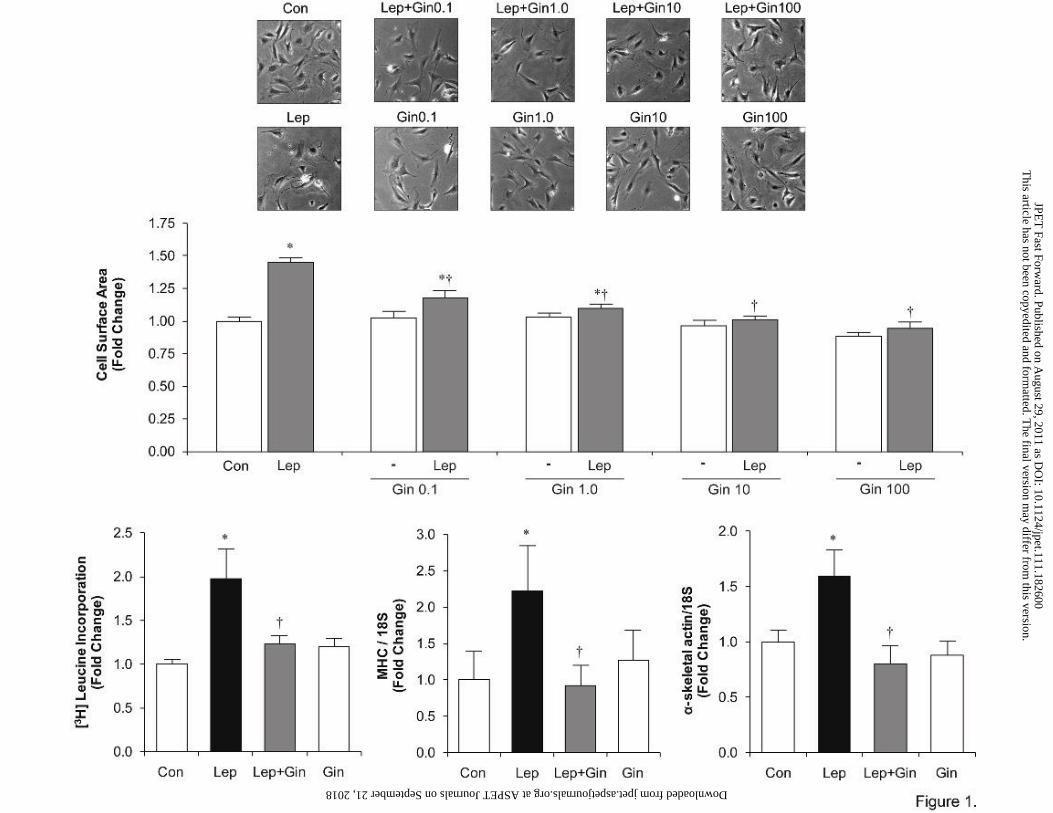
Ginseng inhibits leptin-induced myocyte hypertrophy To first determine an appropriate
concentration for studying the effects of ginseng on leptin-induced cardiac hypertrophy,
cardiomyocytes were subjected to increasing concentrations of ginseng (0.1, 1, 10, and 100
µg/ml) for 1 hour prior to the addition of leptin for a total incubation time of 24 hours (Figure
1A). As shown in Figure 1A, leptin induced a significant increase (p<0.05) in cell size which
was attenuated by ginseng in a concentration-dependent manner. A ginseng concentration of 10
µg/ml of ginseng was used for all subsequent experiments as this represented the lowest
concentration which completely abrogated the hypertrophic response to leptin (Figure 1A).
An increase in cell surface area in leptin treated cells after 24 hours was additionally
associated with a significant increase (p<0.05) in protein synthesis as indicated by increased [3H]
leucine incorporation as well as (Figure 1B) myosin heavy chain α (Figure 1C) and α-skeletal
actin (α-SA) (Figure 1D) gene expression, as quantified by real-time PCR. As shown in Figure 1
ginseng alone had no affect any parameter.
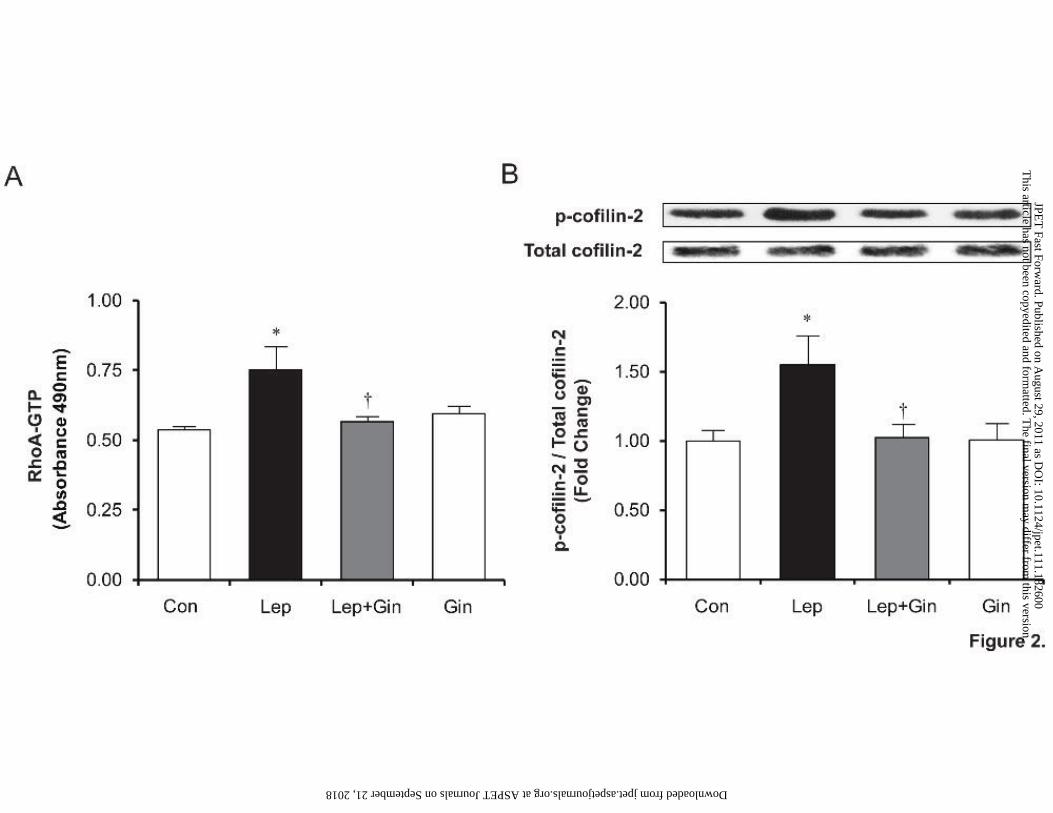
Ginseng inhibits leptin-induced RhoA activation, cofilin-2 phosphorylation (inactivation)
and the decrease in G/F actin ratio. RhoA activation was measured after 10 minutes of leptin
administration in the presence or absence of ginseng. As demonstrated in Figure 2A, ginseng
significantly inhibited (p<0.05) leptin-induced RhoA activation, returning RhoA-GTP levels to
control values, while ginseng alone had no effect on its own. Activation of the RhoA/ROCK
pathway was additionally indirectly measured by quantifying phosphorylated cofilin-2
(inactivated form) by Western blotting. Cofilin-2 is an ubiquitous enzyme responsible for
depolymerising filamentous (F) to globular (G) actin thereby regulating cellular actin dynamics.
This article has not been copyedited and formatted. The final version may differ from this version.JPET Fast Forward. Published on August 29, 2011 as DOI: 10.1124/jpet.111.182600
at ASPE
T Journals on Septem
ber 21, 2018jpet.aspetjournals.org
Dow
nloaded from
JPET #182600
15
A change in this ratio favouring a higher F to G actin content as a result of RhoA activation and
subsequent cofilin-2 phosphorylation (inactivation) has been previously demonstrated to
represent a key mechanism underlying the hypertrophic effects of leptin (Zeidan et al., 2006).
The results, as shown in Figure 2B, reveal a significant (25%) increase in p-cofilin-2 in cells
administered leptin (p<0.05), which was attenuated and returned to control levels in the presence
of ginseng although ginseng alone had no significant effect on p-cofilin-2 or RhoA-GTP levels.
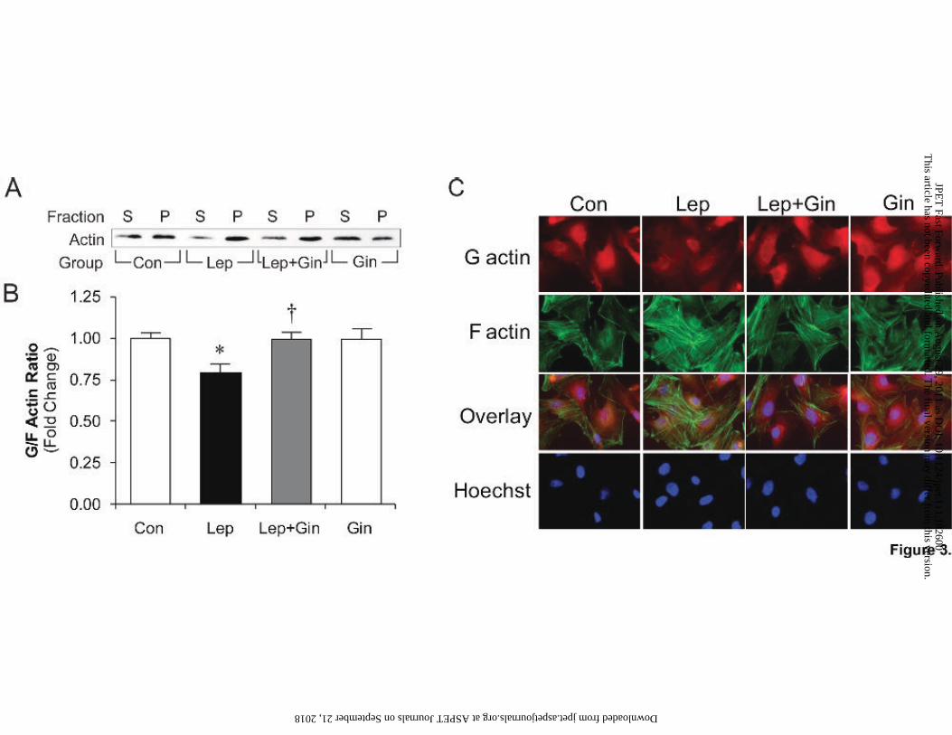
To further characterize the effects of ginseng on leptin-induced RhoA/ROCK pathway
activation the G/F actin ratio was assessed by Western blotting of isolated G and F actin fractions
(Figure 3A) as well as visualization using immunofluorescence (Figure 3C) after 24 hours. In
leptin-treated cells, the G/F actin ratio was decreased as measured by quantification of G actin
(S; supernatant) and F actin (P; pellet) using Western blotting (Figure 3A), while pre-treatment
with ginseng restored this ratio to control values (Figure 3B). This was similarly observed in the
representative immunofluorescence images (Figure 3C, 2nd column) of leptin-treated cells as
depicted by a lighter red staining of G actin and intensified green staining of F actin, which was
returned to control conditions by pre-treatment with ginseng (Figure 3C, 3rd column). Treatment
with ginseng alone had no direct effect on the G/F actin dynamics.
Ginseng inhibits leptin-induced p38 and ERK1/2 MAPK phosphorylation. Leptin
significantly induced both p38 (Figure 4A) and ERK1/2 (Figure 5A) phosphorylation as early as
5 minutes after addition with maximum activation seen at 15 minutes followed by values
returning to control by 30 minutes. Pre-treatment with ginseng inhibited leptin-induced p38
(Figure 4A) and ERK1/2 activation (Figure 5A) at all time points although ginseng had no direct
effect on its own.
This article has not been copyedited and formatted. The final version may differ from this version.JPET Fast Forward. Published on August 29, 2011 as DOI: 10.1124/jpet.111.182600
at ASPE
T Journals on Septem
ber 21, 2018jpet.aspetjournals.org
Dow
nloaded from

JPET #182600
16
Ginseng inhibits leptin-induced p38 nuclear translocation. Leptin induced a significant
increase (p<0.05) in p38 expression in the nuclear-containing membrane fraction (Figure 4C),
which was complemented by a significant decrease (p<0.05) in cytosolic p38 levels, indicative of
nuclear translocation in p38 in leptin-treated cells (Figure 4B). Nuclear translocation of p38 was
further visualized by immunofluorescence (Figure 4D) where total p38, indicated by red
fluorescence was much more centralized in the nuclear region of leptin-treated cells. The ability
of leptin to induce p38 translocation was significantly inhibited by ginseng. As summarized in
Figure 5 (panels B-D), leptin had no effect on ERK1/2 nuclear translocation.
Ginseng inhibits leptin-induced increase in p115RhoGEF protein and gene expression. The
effects of ginseng on leptin-induced RhoGEF activation was first determined by measurement of
p115RhoGEF and p63RhoGEF gene expression through real-time PCR and protein expression
using Western blotting (Figure 6). A 6-fold increase (p<0.05) in p115RhoGEF gene expression
after 24 hours was observed in leptin-treated cells, which was abolished in the presence of
ginseng (Figure 6A). Similarly, pre-treatment with ginseng significantly inhibited leptin-induced
increase in p115RhoGEF protein expression (Figure 6B), while ginseng alone had no direct
effect on either parameter. In comparison to p115RhoGEF, neither leptin nor ginseng exerted
any effect on p63RhoGEF gene (Figure 6C) or protein (Figure 6D) expression.
Ginseng inhibits leptin-induced p115RhoGEF membrane translocation with RhoA co-
localization and p115RhoGEF activity. Several studies have shown membrane translocation
with RhoA co-localization as a component of p115RhoGEF activation (Kozasa et al., 1998;
This article has not been copyedited and formatted. The final version may differ from this version.JPET Fast Forward. Published on August 29, 2011 as DOI: 10.1124/jpet.111.182600
at ASPE
T Journals on Septem
ber 21, 2018jpet.aspetjournals.org
Dow
nloaded from

JPET #182600
17
Rossman et al., 2005; Aittaleb et al., 2009). Initial time course experiments with leptin treatment
revealed translocation and RhoA co-localization as early as 5 minutes after leptin addition (data
not shown). Previous studies from our laboratory have correspondingly indicated activation of
RhoA as early as 10 minutes (Zeidan et al., 2006; Zeidan et al., 2008). Consequently,
cardiomyocytes were treated with ginseng prior to the administration of leptin for 10 minutes and
prepared for immunofluorescence. As depicted in Figure 7A (row c), p115RhoGEF membrane
translocation with RhoA co-localization was observed in leptin-treated cells as indicated by
evident yellow fluorescence at the cardiomyocyte border. Isolation of the co-localized pixels
(Figure 7A, row d) additionally revealed co-localization of p115RhoGEF and RhoA in leptin-
treated cells, which was inhibited by pre-treatment with ginseng. Treatment with ginseng alone
was without any effect (Figure 7A, 4th column).
Using the same treatment protocol, cell lysates were collected at 10 minutes after leptin
administration and incubated with GST-RhoG17A, a nucleotide-free RhoA mutant that has a
high affinity for active RhoGEFs (Garcia-Mata et al., 2006), to immunoprecipitate p115RhoGEF.
Measurement of activated p115RhoGEF from leptin-treated cells in the presence or absence of
ginseng to facilitate guanine nucleotide exchange activity by purified small GTP-ase RhoA was
then performed. p115RhoGEF activation was increased 2-fold in leptin-treated cells in
comparison to control (Figure 7B). Particularly of interest, this observed increase in guanine
nucleotide exchange activity was significantly abolished in cells pre-treated with ginseng.
p115RhoGEF activity was unaffected by ginseng alone.
This article has not been copyedited and formatted. The final version may differ from this version.JPET Fast Forward. Published on August 29, 2011 as DOI: 10.1124/jpet.111.182600
at ASPE
T Journals on Septem
ber 21, 2018jpet.aspetjournals.org
Dow
nloaded from

JPET #182600
18
Discussion
Increasing evidence from a number of laboratories has demonstrated that leptin exerts a
direct hypertrophic effect on cardiomyocytes (Rajapurohitam et al., 2003; Xu et al., 2004;
Madani et al., 2006; Hou et al., 2010) as well as intact myocardium in vivo (Abe et al., 2007). In
addition, blocking leptin receptors attenuates remodeling and heart failure in the post-infarcted
rat heart (Purdham et al., 2008). Although the precise mechanism of action of leptin accounting
for its hypertrophic effect are not completely understood, we have previously suggested that
activation of the RhoA/ROCK pathway plays a critical role in mediating leptin-induced cardiac
hypertrophy, likely through activation and subsequent nuclear translocation of p38 MAPK, the
latter effect dependent on alterations in actin dynamics (Zeidan et al., 2006; Zeidan et al., 2008).
In the present study, we assessed the effects of North American Ginseng (P. quinquefolius) on
leptin-induced cardiac hypertrophy based on emerging evidence that ginseng exerts
antihypertrophic effects in a varied number of experimental models (Jiang et al., 2007; Qin et al.,
2008; Deng et al., 2010) and also reduces the severity of heart failure in rats subjected to chronic
coronary artery ligation (Guo et al., 2011). We hypothesized that ginseng would attenuate leptin-
induced cardiac hypertrophy by attenuating RhoA/ROCK activation following leptin
administration. Our study shows for the first time that ginseng is a potent inhibitor of leptin-
induced hypertrophy and indeed this occurs through a mechanism associated with the abrogation
of RhoA/ROCK activation. Moreover, we identified a potential key role of p115RhoGEF in
facilitating RhoA/ROCK-dependent p38 and ERK1/2 MAPK pathway activation in leptin-
induced cardiac hypertrophy and critically, the ability of ginseng to target p115RhoGEF as a
mechanism for its ability to prevent RhoA/ROCK activation, thus preventing cardiomyocyte
hypertrophy.
This article has not been copyedited and formatted. The final version may differ from this version.JPET Fast Forward. Published on August 29, 2011 as DOI: 10.1124/jpet.111.182600
at ASPE
T Journals on Septem
ber 21, 2018jpet.aspetjournals.org
Dow
nloaded from

JPET #182600
19
Leptin-induced hypertrophy was manifested by an increase in cell surface area, a 2-fold
increase in [3H] leucine incorporation, as well as increased expression of two molecular
hypertrophic gene markers, α-SA and MHC, all of which were significantly attenuated by
ginseng. Moreover, the hypertrophic effect of leptin was associated with an activation of the
RhoA/ROCK pathway as exhibited by an increase in RhoA-GTP levels, in support of our
previous findings (Zeidan et al., 2006; Zeidan et al., 2008). RhoA/ROCK activation was further
demonstrated by increased phosphorylation (inactivation) of cofilin-2, an ubiquitous enzyme
downstream of RhoA which depolymerizes actin, resulting in a decrease in the G/F actin ratio.
We have previously reported that leptin-induced RhoA/ROCK activation was critical for p38
MAPK, but not ERK1/2 MAPK nuclear translocation and the subsequent hypertrophic response,
a response likely dependent on the changes in actin dynamics (Zeidan et al., 2008). When taken
together, the ability of ginseng to completely prevent activation of the RhoA/ROCK pathway,
p38 translocation and the associated hypertrophic response strongly suggests that inhibition of
RhoA/ROCK represents a key mechanism for the antihypertrophic effect of ginseng as seen in
our study.
We next assessed the potential target mediating the ability of ginseng to inhibit
RhoA/ROCK activation. Our study centered primarily on the potential role of RhoGEFs which
are critical for downstream RhoA activation (Rossman et al., 2005). However, the role of
RhoGEFs in the cardiac hypertrophic program has not been extensively studied. Although a
number of RhoGEFs have been identified, p115RhoGEF and p63RhoGEF were considered of
particular interest since their expression have been demonstrated in cardiovascular tissues
including the heart (Souchet et al., 2002; Porchia et al., 2008; Wuertz et al., 2010) and their
activation has been shown in response to various hypertrophic agonists including angiotensin II
This article has not been copyedited and formatted. The final version may differ from this version.JPET Fast Forward. Published on August 29, 2011 as DOI: 10.1124/jpet.111.182600
at ASPE
T Journals on Septem
ber 21, 2018jpet.aspetjournals.org
Dow
nloaded from

JPET #182600
20
(Guilluy et al., 2010), and endothelin-1 (Porchia et al., 2008). Our study shows for the first time
that leptin induced a significant increase in p115RhoGEF gene and protein expression without
affecting p63RhoGEF. We next studied whether increased expression of p115RhoGEF is also
associated with increased GEF activity. Activation of p115RhoGEF involves receptor-mediated
membrane translocation and co-localization with RhoA (Kozasa et al., 1998; Rossman et al.,
2005; Aittaleb et al., 2009). Our results revealed membrane translocation of p115RhoGEF with
RhoA co-localization after 10 minutes of leptin stimulation and also demonstrate an increase
p115RhoGEF activity.
The ability of ginseng to inhibit p115RhoGEF activation suggest this as a target for the
ability of ginseng to inhibit leptin-induced cardiomyocyte hypertrophy and it reasonable to
assume that p115RhoGEF activation by leptin and inhibition by ginseng represent the main
regulatory sites influencing subsequent p38 and ERK1/2 phosphorylation or translocation of the
former. However, a limitation of our study is that it does not establish a direct causal relationship
between p115RhoGEF activation by leptin and stimulation of the RhoA pathway. Further
studies are required to confirm this particularly by determining the effect of p115RhoGEF
downregulation on the ability of leptin to activate RhoA.
As previously reported (Zeidan et., 2008) leptin induces phosphorylation of both p38 and
ERK1/2 although only the former is translocated into nuclei thus suggesting that phosphorylation
is not a precondition for nuclear transport. Although the precise mechanisms for selective p38
translocation into nuclei are not known, the phenomenon is likely mediated by changes in actin
dynamics since the effect is prevented by latrunculin B which prevents actin polymerization as a
result of RhoA activation (Zeidan et al., 2008). The selective translocation of p38 into nuclei
following leptin addition also helps to explain our initial finding that pharmacological inhibition
This article has not been copyedited and formatted. The final version may differ from this version.JPET Fast Forward. Published on August 29, 2011 as DOI: 10.1124/jpet.111.182600
at ASPE
T Journals on Septem
ber 21, 2018jpet.aspetjournals.org
Dow
nloaded from

JPET #182600
21
of p38, but not ERK1/2, prevents leptin-induced hypertrophy (Rajapurohitam et al., 2003). The
specific mechanism of p115RhoGEF-RhoA/ROCK-dependent p38 and ERK1/2 MAPK
inhibition by ginseng however still remains unclear. The chemical structures of ginsenosides,
which are triterpine saponins and which are considered the primary active constituents
contributing to the medicinal effects of ginseng (Attele et al., 1999), have been compared to
steroidal structures such as estrogen. Recently, Leung and colleagues alluded to the competitive
binding of the specific ginsenoside, Rb1 (the predominant ginsenoside of North American
ginseng), selective to the estrogen receptor β where it was theorized to be engulfed with the
bound ginsenoside through endocytosis into the cytoplasm in which it translocates into the
nucleus to bind to transcription factors eliciting its effects as an anti-angiogenic factor (Leung et
al., 2007). Indeed, we have previously shown that estrogen (as 17β estradiol) exerts a
prohypertrophic effect on cultured ventricular myocytes at very low (1 pM) concentrations but
has antihypertrophic actions at nanomolar concentrations (Kilic et al., 2009). The observation of
the ability of ginsenoside Rb1 to bind to estrogen receptors is intriguing and raises the question
of potential gender specific effects of ginseng. Although it appears that estrogen receptors are
expressed in ventricular myocytes of both male and female rats, nonetheless potential gender-
dependent effects of ginseng are deserving of further study. In the present study an alcoholic
ginseng extract containing a large number of ginsenosides was used and consequently the
specific ginsenoside(s) responsible for the observed inhibition of leptin-induced effects cannot be
currently identified.
Another potential mechanism for the observed inhibition of these pathways by ginseng
may occur extracellularly at the level of the leptin-receptor long isoform (Ob-Rb), considered the
principle receptor mediating the biological effects of leptin (reviewed in Villanueva and Myers,
This article has not been copyedited and formatted. The final version may differ from this version.JPET Fast Forward. Published on August 29, 2011 as DOI: 10.1124/jpet.111.182600
at ASPE
T Journals on Septem
ber 21, 2018jpet.aspetjournals.org
Dow
nloaded from
JPET #182600
22
2008), potentially as a result of early binding and antogonism of Ob-Rb, consequently down-
regulating further leptin signalling (Figure 8). Although it was not an aim of the current study,
competitive binding studies between leptin and ginseng at the Ob-Rb are deserving of further
investigation.
In conclusion, our results show for the first time that ginseng markedly attenuates the
direct hypertrophic effect of leptin. Moreover, our results are strongly supportive of the concept
that this effect of ginseng against leptin-induced hypertrophy occurs via inhibition of
p115RhoGEF expression and activity, thus abrogating RhoA/ROCK activation. The latter results
in diminished p38 MAPK phosphorylation and translocation into nuclei thus attenuating
transcription and reducing the hypertrophic response. As previously noted, further work is
necessary to demonstrate a precise causal relationship between leptin-induced p115RhoGEF
activation and subsequent activation of downstream pathways. Also, since ginseng exerts
numerous and diverse effects on the heart (reviewed in Karmazyn et al., 2011), the contribution
of other pathways as targets for the antihypertrophic effects of ginseng cannot be excluded.
Moreover, it is important to point out that the present study was carried out using neonatal
ventricular myocytes and therefore extrapolation of these results to the adult myocardium,
particularly under in vivo conditions should be done cautiously. Although the role of leptin in
cardiac pathology still remains to be fully determined, our overall results suggest that ginseng
could be an effective therapeutic approach aimed at mitigating potential deleterious
cardiovascular complications associated with hyperleptinemia, particularly those involving a
cardiac hypertrophic phenotype.
This article has not been copyedited and formatted. The final version may differ from this version.JPET Fast Forward. Published on August 29, 2011 as DOI: 10.1124/jpet.111.182600
at ASPE
T Journals on Septem
ber 21, 2018jpet.aspetjournals.org
Dow
nloaded from

JPET #182600
23
Acknowledgements
We would like to thank Dr. Katalin Szászi (St. Michael’s Hospital, University of Toronto) for
providing the RhoG17A construct and reference to appropriate protocols, Dr. Keith Burridge
(University of North Carolina at Chapel Hill) for permission to use the construct and the
laboratory of Dr. Peter Chidiac (University of Western Ontario) for providing the expertise,
reagents and equipment to perform these assays. M.M is funded by OGIRC. M.K holds a Tier 1
Canada Research Chair in Experimental Cardiology.
Authorship Contributions
Participated in research design: Moey, Rajapurohitam, Zeidan and Karmazyn
Conducted experiments: Moey, Rajapurohitam, Zeidan
Contributed new reagents or analytical tools: Moey
Performed data analysis: Moey and Rajapurohitam
Wrote or contributed to the writing of the manuscript: Moey, Zeidan and Karmazyn
Other:
This article has not been copyedited and formatted. The final version may differ from this version.JPET Fast Forward. Published on August 29, 2011 as DOI: 10.1124/jpet.111.182600
at ASPE
T Journals on Septem
ber 21, 2018jpet.aspetjournals.org
Dow
nloaded from

JPET #182600
24
References
Abe Y, Ono K, Kawamura T, Wada H, Kita T, Shimatsu A and Hasegawa K (2007) Leptin induces elongation of cardiac myocytes and causes eccentric left ventricular dilatation with compensation. Am J Physiol Heart Circ Physiol 292:H2387-2396.
Aittaleb M, Boguth CA and Tesmer JJ (2009) Structure and function of heterotrimeric G protein-regulated Rho guanine nucleotide exchange factors. Mol Pharmacol 77:111-125.
Albinsson S, Nordstrom I and Hellstrand P (2004) Stretch of the vascular wall induces smooth muscle differentiation by promoting actin polymerization. J Biol Chem 279:34849-34855.
Attele AS, Wu JA and Yuan CS (1999) Ginseng pharmacology: multiple constituents and multiple actions. Biochem Pharmacol 58:1685-1693.
Bos JL, Rehmann H and Wittinghofer A (2007) GEFs and GAPs: critical elements in the control of small G proteins. Cell 129:865-877.
Bui AL, Horwich TB and Fonarow GC (2011) Epidemiology and risk profile of heart failure. Nat Rev Cardiol 8: 30-41.
Deng J, Wang YW, Chen WM, Wu Q and Huang XN (2010) Role of nitric oxide in ginsenoside Rg(1)-induced protection against left ventricular hypertrophy produced by abdominal aorta coarctation in rats. Biol Pharm Bull 33:631-635.
Fruhbeck G (2006) Intracellular signalling pathways activated by leptin. Biochem J 393:7-20. Garcia-Mata R, Wennerberg K, Arthur WT, Noren NK, Ellerbroek SM and Burridge K (2006)
Analysis of activated GAPs and GEFs in cell lysates. Methods Enzymol 406:425-437. Goldstein B (1975) Ginseng: its history, dispersion, and folk tradition. Am J Chin Med (Gard
City N Y) 3:223-234. Guilluy C, Bregeon J, Toumaniantz G, Rolli-Derkinderen M, Retailleau K, Loufrani L, Henrion
D, Scalbert E, Bril A, Torres RM, Offermanns S, Pacaud P and Loirand G (2010) The Rho exchange factor Arhgef1 mediates the effects of angiotensin II on vascular tone and blood pressure. Nat Med 16:183-190.
Guo J, Gan XT, Haist JV, Rajapurohitam V, Zeidan A, Faruq NS and Karmazyn M (2011) Ginseng inhibits cardiomyocyte hypertrophy and heart failure via NHE-1 inhibition and attenuation of calcineurin activation. Circ Heart Fail 4:79-88.
Hou N, Luo MS, Liu SM, Zhang HN, Xiao Q, Sun P, Zhang GS, Luo JD and Chen MS (2010) Leptin induces hypertrophy through activating the peroxisome proliferator-activated receptor alpha pathway in cultured neonatal rat cardiomyocytes. Clin Exp Pharmacol Physiol 37:1087-1095.
Jeon BH, Kim CS, Park KS, Lee JW, Park JB, Kim KJ, Kim SH, Chang SJ and Nam KY (2000) Effect of Korea red ginseng on the blood pressure in conscious hypertensive rats. Gen Pharmacol 35:135-141.
Jiang QS, Huang XN, Dai ZK, Yang GZ, Zhou QX, Shi JS and Wu Q (2007) Inhibitory effect of ginsenoside Rb1 on cardiac hypertrophy induced by monocrotaline in rat. JEthnopharmacol 111:567-572.
Kakiashvili E, Speight P, Waheed F, Seth R, Lodyga M, Tanimura S, Kohno M, Rotstein OD, Kapus A and Szaszi K (2009) GEF-H1 mediates tumor necrosis factor-alpha-induced Rho activation and myosin phosphorylation: role in the regulation of tubular paracellular permeability. J Biol Chem 284:11454-11466.
This article has not been copyedited and formatted. The final version may differ from this version.JPET Fast Forward. Published on August 29, 2011 as DOI: 10.1124/jpet.111.182600
at ASPE
T Journals on Septem
ber 21, 2018jpet.aspetjournals.org
Dow
nloaded from

JPET #182600
25
Karmazyn M, Purdham DM, Rajapurohitam V and Zeidan A (2008) Leptin Signaling in the Cardiovascular System, in Signal Transduction in the Cardiovascular System in Health and Disease (Srivastava AK and Anand-Srivastava MB eds) pp 377-395, Springer US.
Karmazyn M, Moey M, Gan XT. Therapeutic potential of ginseng in the management of cardiovascular disorders. Drugs, 2011 in press.
Kilic A, Javadov S and Karmazyn M (2009) Estrogen exerts concentration-dependent pro-and anti-hypertrophic effects on adult cultured ventricular myocytes. Role of NHE-1 in estrogen-induced hypertrophy. J Mol Cell Cardiol 46:360-369.
Kozasa T, Jiang X, Hart MJ, Sternweis PM, Singer WD, Gilman AG, Bollag G and Sternweis PC (1998) p115 RhoGEF, a GTPase activating protein for Galpha12 and Galpha13. Science 280:2109-2111.
Leung KW, Cheung LW, Pon YL, Wong RN, Mak NK, Fan TP, Au SC, Tombran-Tink J and Wong AS (2007) Ginsenoside Rb1 inhibits tube-like structure formation of endothelial cells by regulating pigment epithelium-derived factor through the oestrogen beta receptor. Br J Pharmacol 152:207-215.
Li J, Xie ZZ, Tang YB, Zhou JG and Guan YY (2011) Ginsenoside-Rd, a purified component from panax notoginseng saponins, prevents atherosclerosis in apoE knockout mice. Eur J Pharmacol 652:104-110.
Li Q, Lau A, Morris TJ, Guo L, Fordyce CB and Stanley EF (2004) A syntaxin 1, Galpha(o), and N-type calcium channel complex at a presynaptic nerve terminal: analysis by quantitative immunocolocalization. J Neurosci 24:4070-4081.
Lu JM, Yao Q and Chen C (2009) Ginseng compounds: an update on their molecular mechanisms and medical applications. Curr Vasc Pharmacol 7:293-302.
Luo W, Morrison H, de Groh M, Waters C, DesMeules M, Jones-McLean E, Ugnat AM, Desjardins S, Lim M and Mao Y (2007) The burden of adult obesity in Canada. Chronic Dis Can 27:135-144.
Madani S, De Girolamo S, Munoz DM, Li RK and Sweeney G (2006) Direct effects of leptin on size and extracellular matrix components of human pediatric ventricular myocytes. Cardiovasc Res 69:716-725.
Maffei M, Halaas J, Ravussin E, Pratley RE, Lee GH, Zhang Y, Fei H, Kim S, Lallone R, Ranganathan S and et al. (1995) Leptin levels in human and rodent: measurement of plasma leptin and ob RNA in obese and weight-reduced subjects. Nat Med 1:1155-1161.
Porchia F, Papucci M, Gargini C, Asta A, De Marco G, Agretti P, Tonacchera M and Mazzoni MR (2008) Endothelin-1 up-regulates p115RhoGEF in embryonic rat cardiomyocytes during the hypertrophic response. J Recept Signal Transduct Res 28:265-283.
Purdham DM, Rajapurohitam V, Zeidan A, Huang C, Gross GJ and Karmazyn M (2008) A neutralizing leptin receptor antibody mitigates hypertrophy and hemodynamic dysfunction in the postinfarcted rat heart. Am J Physiol Heart Circ Physiol 295:H441-446.
Purdham DM, Zou MX, Rajapurohitam V and Karmazyn M (2004) Rat heart is a site of leptin production and action. Am J Physiol Heart Circ Physiol 287:H2877-2884.
Qin N, Gong QH, Wei LW, Wu Q and Huang XN (2008) Total ginsenosides inhibit the right ventricular hypertrophy induced by monocrotaline in rats. Biol Pharm Bull 31:1530-1535.
This article has not been copyedited and formatted. The final version may differ from this version.JPET Fast Forward. Published on August 29, 2011 as DOI: 10.1124/jpet.111.182600
at ASPE
T Journals on Septem
ber 21, 2018jpet.aspetjournals.org
Dow
nloaded from

JPET #182600
26
Rajapurohitam V, Gan XT, Kirshenbaum LA and Karmazyn M (2003) The obesity-associated peptide leptin induces hypertrophy in neonatal rat ventricular myocytes. Circ Res 93:277-279.
Rajapurohitam V, Javadov S, Purdham DM, Kirshenbaum LA and Karmazyn M (2006) An autocrine role for leptin in mediating the cardiomyocyte hypertrophic effects of angiotensin II and endothelin-1. J Mol Cell Cardiol 41:265-274.
Rossman KL, Der CJ and Sondek J (2005) GEF means go: turning on RHO GTPases with guanine nucleotide-exchange factors. Nat Rev Mol Cell Biol 6:167-180.
Schmidt A and Hall A (2002) Guanine nucleotide exchange factors for Rho GTPases: turning on the switch. Genes Dev 16:1587-1609.
Schulze PC, Kratzsch J, Linke A, Schoene N, Adams V, Gielen S, Erbs S, Moebius-Winkler S and Schuler G (2003) Elevated serum levels of leptin and soluble leptin receptor in patients with advanced chronic heart failure. Eur J Heart Fail 5:33-40.
Villanueva EC and Myers MG, Jr. (2008) Leptin receptor signaling and the regulation of mammalian physiology. Int J Obes (Lond) 32 Suppl 7:S8-12.
Zeidan A, Javadov S, Chakrabarti S and Karmazyn M (2008) Leptin-induced cardiomyocyte hypertrophy involves selective caveolae and RhoA/ROCK-dependent p38 MAPK translocation to nuclei. Cardiovasc Res 77:64-72.
Zeidan A, Javadov S and Karmazyn M (2006) Essential role of Rho/ROCK-dependent processes and actin dynamics in mediating leptin-induced hypertrophy in rat neonatal ventricular myocytes. Cardiovasc Res 72:101-111.
Zhang Y, Proenca R, Maffei M, Barone M, Leopold L and Friedman JM (1994) Positional cloning of the mouse obese gene and its human homologue. Nature 372:425-432.
Footnotes
This project was funded by the Canadian Institutes of Health Research [MOP 62764] and the
Ontario Ginseng Innovation and Research Consortium (OGIRC).
This article has not been copyedited and formatted. The final version may differ from this version.JPET Fast Forward. Published on August 29, 2011 as DOI: 10.1124/jpet.111.182600
at ASPE
T Journals on Septem
ber 21, 2018jpet.aspetjournals.org
Dow
nloaded from

JPET #182600
27
Legends for Figures
Fig 1. Ginseng inhibits leptin-induced increase in cell surface area, [3H] leucine
incorporation and expression of the gene markers of cardiac hypertrophy, α-skeletal actin
and myosin heavy chain. Micrographs show representative images of neonatal ventricular
cardiomyocytes with or without pre-treatment with increasing ginseng (Gin) concentrations (0.1,
1.0, 10 and 100 μg/ml, respectively) in the absence (top row) or presence (bottom row) of leptin
(3.1 nM) pretreatment. Panel A shows surface area whereas panel B shows [3H] leucine
incorporation and B and C represent expression of myosin heavy chain (MHC) and α-skeletal
actin (α-SA), respectively, with different treatments. Data represent means ± S.E.M. N=8-10 for
surface area, N=6 for leucine incorporation and N=6-8 for molecular markers of hypertrophy.
*p<0.05 vs. control; †p <0.05 vs. leptin. Con, control; Lep, leptin; Gin, ginseng.
Fig 2. Ginseng inhibits leptin-induced RhoA activation and phosphorylation (inactivation)
of cofilin-2. Panel A shows RhoA-GTP (activated RhoA) levels whereas panel B demonstrates
Western blots and densitometric values for phosphorylated cofilin-2. Data represent means ±
S.E.M. N=6. *p<0.05 vs. control; †p <0.05 vs. leptin. Con, control; Lep, leptin; Gin, ginseng.
Fig 3. Ginseng inhibits leptin-induced decrease in G/F actin. Panel A shows Western blots for
actin dynamics with respect to G actin in supernatant (S) and F actin in pellet fraction (P) with
different treatments. Densitometric values are shown in panel B. Representative fluorescence
images of cardiomyocytes are shown in panel C. For these studies cells were fixed on collagen-
coated glass cover slips and G actin (1st row) and F actin (2nd row) were visualized with DNAse-I
Texas Red Conjugate and Phalloidin-FITC, respectively. Hoechst staining was used to detect
This article has not been copyedited and formatted. The final version may differ from this version.JPET Fast Forward. Published on August 29, 2011 as DOI: 10.1124/jpet.111.182600
at ASPE
T Journals on Septem
ber 21, 2018jpet.aspetjournals.org
Dow
nloaded from

JPET #182600
28
nuclei whereas overlay depicts all three stains merged. For panel B, data represent means ±
S.E.M. N=6-9. *p<0.05 vs. control; †p <0.05 vs. leptin. Con, control; Lep, leptin; Gin, ginseng.
Fig 4. Ginseng inhibits leptin-induced phosphorylation and nuclear translocation of p38
Panel A shows Western blots and corresponding densitometric values for phosphorylated p38
whereas panels B and C show Western blots and corresponding densitometric values for p38
MAPK content in cytoplasmic-enriched and nuclear-containing membrane fractions, respectively.
Panel D shows representative immunofluorescence images measuring total p38 indicated by red
staining (IgG AlexFluor-596) in the left column, Hoechst nuclear staining in the middle column
and merged staining in the right column. Data in panels A-C represent means ± S.E.M. N=5-8.
*p<0.05 vs. control; †p <0.05 vs. leptin. Con, control; Lep, leptin; Gin, ginseng. Minutes
indicate time after leptin addition.
Fig 5. Ginseng inhibits leptin-induced ERK1/2 phosphorylation with no effect on nuclear
translocation. Panel A shows Western blots and corresponding densitometric values for
phosphorylated ERK1/2 whereas panels B and C show Western blots and corresponding
densitometric values for ERK1/2 MAPK content in cytoplasmic-enriched and nuclear-containing
membrane fractions, respectively. Panel D shows representative immunofluorescence images
measuring total ERK1/2 indicated by red staining (IgG AlexFluor-596) in the left column,
Hoechst nuclear staining in the middle column and merged staining in the right column. Data in
panels A-C represent means ± S.E.M. N=5-8. *p<0.05 vs. control; †p <0.05 vs. leptin. Con,
control; Lep, leptin; Gin, ginseng. Minutes indicate time after leptin addition.
This article has not been copyedited and formatted. The final version may differ from this version.JPET Fast Forward. Published on August 29, 2011 as DOI: 10.1124/jpet.111.182600
at ASPE
T Journals on Septem
ber 21, 2018jpet.aspetjournals.org
Dow
nloaded from
JPET #182600
29
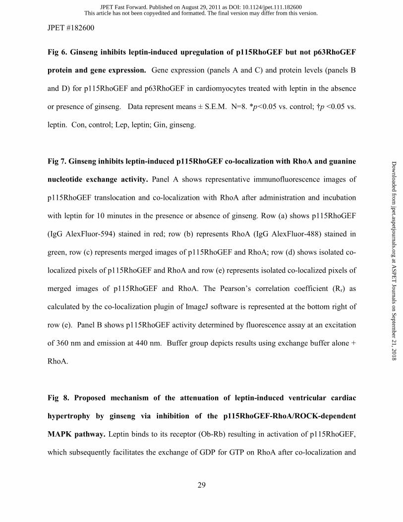
Fig 6. Ginseng inhibits leptin-induced upregulation of p115RhoGEF but not p63RhoGEF
protein and gene expression. Gene expression (panels A and C) and protein levels (panels B
and D) for p115RhoGEF and p63RhoGEF in cardiomyocytes treated with leptin in the absence
or presence of ginseng. Data represent means ± S.E.M. N=8. *p<0.05 vs. control; †p <0.05 vs.
leptin. Con, control; Lep, leptin; Gin, ginseng.
Fig 7. Ginseng inhibits leptin-induced p115RhoGEF co-localization with RhoA and guanine
nucleotide exchange activity. Panel A shows representative immunofluorescence images of
p115RhoGEF translocation and co-localization with RhoA after administration and incubation
with leptin for 10 minutes in the presence or absence of ginseng. Row (a) shows p115RhoGEF
(IgG AlexFluor-594) stained in red; row (b) represents RhoA (IgG AlexFluor-488) stained in
green, row (c) represents merged images of p115RhoGEF and RhoA; row (d) shows isolated co-
localized pixels of p115RhoGEF and RhoA and row (e) represents isolated co-localized pixels of
merged images of p115RhoGEF and RhoA. The Pearson’s correlation coefficient (Rr) as
calculated by the co-localization plugin of ImageJ software is represented at the bottom right of
row (e). Panel B shows p115RhoGEF activity determined by fluorescence assay at an excitation
of 360 nm and emission at 440 nm. Buffer group depicts results using exchange buffer alone +
RhoA.
Fig 8. Proposed mechanism of the attenuation of leptin-induced ventricular cardiac
hypertrophy by ginseng via inhibition of the p115RhoGEF-RhoA/ROCK-dependent
MAPK pathway. Leptin binds to its receptor (Ob-Rb) resulting in activation of p115RhoGEF,
which subsequently facilitates the exchange of GDP for GTP on RhoA after co-localization and
This article has not been copyedited and formatted. The final version may differ from this version.JPET Fast Forward. Published on August 29, 2011 as DOI: 10.1124/jpet.111.182600
at ASPE
T Journals on Septem
ber 21, 2018jpet.aspetjournals.org
Dow
nloaded from

JPET #182600
30
translocation to the membrane. Activated RhoA (RhoA-GTP) results in the activation of ROCK
and LIMK, which consequently phosphorylates (inactivates) cofilin-2 resulting in an increase in
the F to G actin ratio. Changes in actin dynamics results in activation of p38 and ERK1/2 via
phosphorylation and nuclear translocation of the former thus leading to an increase in
transcriptional growth factors of hypertrophy. Pre-treatment with ginseng attenuates the leptin-
induced ventricular cardiac hypertrophy by inhibiting the p115RhoGEF-RhoA/ROCK-dependent
MAPK pathways potentially through down regulating leptin signalling by competitive binding at
the receptor or by additional mechanisms, currently undefined, via direct entry into the cell.
This article has not been copyedited and formatted. The final version may differ from this version.JPET Fast Forward. Published on August 29, 2011 as DOI: 10.1124/jpet.111.182600
at ASPE
T Journals on Septem
ber 21, 2018jpet.aspetjournals.org
Dow
nloaded from

JPET #182600
31
Legend for Table
Table 1. Gene-specific primer sequences of analyzed genes of interest.
Gene Forward Primer Sequence Reverse Primer Sequence α-skeletal actin 5’-CACGGCATTATCACCAACTG-3’ 5’-CCGGAGGCATAGAGAGACAG-3’
myosin heavy chain
5’-CATCACCGGAGAATCCGGAGC-3’ 5’-CTATTGAGGCCACAGTCGTC-3’
p115RhoGEF 5’-TAGAGGACTTCCGCTCCAAA-3’ 5’-CAGTGACCACAGCAGCACTT-3’
p63RhoGEF 5’-TATGTGGACGACTTGGGACA-3’ 5’-TGATGAACAGCTGAGCCAAC-3’
18S 5’-GTAACCCGTTGAACCCCATT-3’ 5’-CCATCCAATCGGTAGTAGCG-3’
This article has not been copyedited and formatted. The final version may differ from this version.JPET Fast Forward. Published on August 29, 2011 as DOI: 10.1124/jpet.111.182600
at ASPE
T Journals on Septem
ber 21, 2018jpet.aspetjournals.org
Dow
nloaded from
This article has not been copyedited and form
atted. The final version m
ay differ from this version.
JPET
Fast Forward. Published on A
ugust 29, 2011 as DO
I: 10.1124/jpet.111.182600 at ASPET Journals on September 21, 2018 jpet.aspetjournals.org Downloaded from
This article has not been copyedited and form
atted. The final version m
ay differ from this version.
JPET
Fast Forward. Published on A
ugust 29, 2011 as DO
I: 10.1124/jpet.111.182600 at ASPET Journals on September 21, 2018 jpet.aspetjournals.org Downloaded from
This article has not been copyedited and form
atted. The final version m
ay differ from this version.
JPET
Fast Forward. Published on A
ugust 29, 2011 as DO
I: 10.1124/jpet.111.182600 at ASPET Journals on September 21, 2018 jpet.aspetjournals.org Downloaded from

This article has not been copyedited and formatted. The final version may differ from this version.JPET Fast Forward. Published on August 29, 2011 as DOI: 10.1124/jpet.111.182600
at ASPE
T Journals on Septem
ber 21, 2018jpet.aspetjournals.org
Dow
nloaded from

This article has not been copyedited and formatted. The final version may differ from this version.JPET Fast Forward. Published on August 29, 2011 as DOI: 10.1124/jpet.111.182600
at ASPE
T Journals on Septem
ber 21, 2018jpet.aspetjournals.org
Dow
nloaded from

This article has not been copyedited and form
atted. The final version m
ay differ from this version.
JPET
Fast Forward. Published on A
ugust 29, 2011 as DO
I: 10.1124/jpet.111.182600 at ASPET Journals on September 21, 2018 jpet.aspetjournals.org Downloaded from

This article has not been copyedited and formatted. The final version may differ from this version.JPET Fast Forward. Published on August 29, 2011 as DOI: 10.1124/jpet.111.182600
at ASPE
T Journals on Septem
ber 21, 2018jpet.aspetjournals.org
Dow
nloaded from

This article has not been copyedited and form
atted. The final version m
ay differ from this version.
JPET
Fast Forward. Published on A
ugust 29, 2011 as DO
I: 10.1124/jpet.111.182600 at ASPET Journals on September 21, 2018 jpet.aspetjournals.org Downloaded from