pantoea ananatis utilizes a type vi secretion system for
TRANSCRIPT
Pantoea ananatis utilizes a type VI secretion system for pathogenesis and bacterial
competition
Divine Y. Shyntum,1,2
Jacques Theron,1 Stephanus N. Venter,
1,2 Lucy N. Moleleki,
1,2 Ian K.
Toth,2,3
and Teresa A. Coutinho1,2
1Department of Microbiology and Plant Pathology, Faculty of Natural and Agricultural
Sciences, University of Pretoria, Pretoria 0002, South Africa.
2Forestry and Agricultural Biotechnology Institute (FABI), University of Pretoria, Pretoria
0002, South Africa.
3James Hutton Research Institute, Invergowrie, Dundee DD2 5DA, Scotland, United
Kingdom.
Corresponding Author: Prof Teresa A. Coutinho
E-mail: [email protected]
Tel: +27 12 420-3934
Fax: +27 12 420-3960
1
ABSTRACT
Type VI secretion systems (T6SSs) are a class of macromolecular machines that are
recognized as an important virulence mechanism in several Gram-negative bacteria. The
genome of Pantoea ananatis LMG 2665T, a pathogen of pineapple fruit and onion plants,
carries two gene clusters whose predicted products have homology with T6SS-associated
gene products from other bacteria. Nothing is known regarding the role of these T6SS-1 and
T6SS-3 gene clusters in the biology of P. ananatis. Here, we present evidence that T6SS-1
plays an important role in the pathogenicity of P. ananatis LMG 2665T in onion plants, while
a strain lacking T6SS-3 remains as pathogenic as the wild-type strain. We also investigated
the role of the T6SS-1 system in bacterial competition, the results of which indicated that
several bacteria compete less efficiently against wild-type LMG 2665T than a strain lacking
T6SS-1. Additionally, we demonstrated that these phenotypes of strain LMG 2665T were
reliant on the core T6SS products TssA and TssD (Hcp), thus indicating that the T6SS-1 gene
cluster encodes a functioning T6SS. Collectively, our data provides the first evidence
demonstrating that the T6SS-1 system is a virulence determinant of P. ananatis LMG 2665T
and plays a role in bacterial competition.
INTRODUCTION
Secretion of proteins such as extracellular proteases and toxins can provide selective
advantages to bacteria in various environmental niches, and many of the proteins secreted by
pathogenic bacteria are important colonization and virulence factors. To date, six types of
protein secretion systems (type I through type VI secretion system [T1SS through T6SS])
have been described in Gram-negative bacteria (Economou et al. 2006; Holland 2010). These
2
secretion systems are distinguished by the conserved structural components that define them,
as well as the characteristics of their substrates and the molecular mechanisms underlying the
export process. The most recently described T6SS has emerged as having a role in bacterial
pathogenesis and host interactions (Coulthurst 2013; Kapitein and Mogk 2013). Data from
structural studies, functional assays and protein localization studies suggest that the T6SS
consists of a membrane-associated assembly platform and a cell surface-exposed needle
structure that transports effector molecules into bacteria or eukaryotic cells (Filloux et al.
2008; Silverman et al. 2012).
Whole-genome analyses have predicted T6SS gene clusters to be widely distributed in Gram-
negative bacterial species (Bingle et al. 2008; Boyer et al. 2009). Although the T6SS gene
clusters differ between bacterial species in terms of gene order and composition, they are
comprised of at least 13 core genes (tss, nomenclature proposed by Shalom et al. [2007]) and
a variable number of non-conserved accessory elements that encode the T6SS “injectisome”
(Bingle et al. 2008; Cascales 2008). A number of the T6SS core proteins are evolutionary and
structurally related to bacteriophage proteins (Kanamura et al. 2009; Leiman et al. 2009).
Examples are the baseplate gp25-like protein TssE, the tail sheath-like proteins TssB and
TssC, the tail subunit-like hemolysin co-regulated protein (Hcp; TssD) that polymerizes into
the T6SS needle structure, and the valine-glycine repeat protein G (VgrG; TssI) that forms
the spike of the TssD nanotube (Ballister et al. 2008; Cascales and Cambillau 2012; Lossi et
al. 2011; Lossi et al. 2013; Pukatzki et al. 2007). Contraction and extension of the TssB-TssC
tubular sheath of the T6SS of Vibrio cholerae have been visualized in vivo, suggesting that
the T6SS sheath is a dynamic contractile structure that projects the T6SS spike into the target
cell analogous to the bacteriophage infection process (Basler et al. 2012). Disassembly of the
contracted sheath requires the ClpV (TssH) AAA+ ATPase, which binds specifically to the
3
contracted TssB-TssC sheath for its disassembly and cycling (Böneman et al. 2009; Kapitein
et al. 2013). Another group of T6SS building blocks (TssM-L) appears to be related to
proteins of the T4SS (Durand et al. 2012; Felisberto-Rodrigues et al. 2011) and may be
involved in the recruitment of TssD (Hcp) to the T6SS inner membrane assembly platform
(Ma et al. 2012).
The T6SSs have been implicated in a variety of functions ranging from biofilm formation to
host-cell invasion, cytotoxicity and survival in macrophages (Aschtgen et al. 2008; Cascales
2008; Schwarz et al. 2010a). However, most studies of the T6SS have focused on its role in
pathogenesis and host interactions. The T6SS has been implicated as a virulence factor in
several human or animal pathogens, including Vibrio cholerae (Pukatzki et al. 2006),
Pseudomonas aeruginosa (Mougous et al. 2006), Burkholderia mallei (Schell et al. 2007),
Aeromonas hydrophila (Suarez et al. 2008), Edwardsiella tarda (Zheng and Leung 2007),
Salmonella enterica serovar Gallinarum (Blondel et al. 2010), and avian pathogenic
Escherichia coli (de Pace et al. 2010). It has subsequently been revealed that some T6SSs are
used to target other bacteria, efficiently killing or inhibiting the growth of competitors, as
reported for T6SSs of Serratia marcescens (Murdoch et al. 2011), P. aeruginosa (Hood et al.
2010), Burkholderia thailandensis (Schwarz et al. 2010b), and V. cholerae (MacIntyre et al.
2010). In contrast to animal and human pathogens, the role of T6SSs in plant bacterial
pathogens is still largely unknown (Records, 2011). Nevertheless, T6SS functionality has
been demonstrated for a few plant-associated bacteria, including Agrobacterium tumefaciens
(Lin et al. 2013; Wu et al. 2008), Pectobacterium atrosepticum (Liu et al. 2008) and
Pseudomonas syringae (Haapalainen et al. 2012), and it was recently reported that the T6SS
of Pseudomonas fluorescens plays an important role in bacterial competition (Decoin et al.
2014).
4

Pantoea ananatis is a Gram-negative bacterial pathogen of plants. It causes disease in a wide
variety of economically important plants such as Eucalyptus spp., Sudangrass, cotton, rice,
corn, onion, melon, cantaloupe fruit, and pineapple (Coutinho and Venter 2009). Diseases
caused by P. ananatis in onion, for example, result in reductions in crop yield, thus leading to
substantial economic losses (Gitaitis and Gay 1997; Goszczynska et al. 2007; Walcott et al.
2002). To date, there are a limited number of reports that focus on the mechanisms by which
P. ananatis causes disease (Morohoshi et al. 2007; Morohoshi et al. 2011a; Morohoshi et al.
2011b; Sessitsch et al. 2004). Consequently, the pathogenesis of P. ananatis is still poorly
understood and the potential virulence determinants and mechanisms employed by P.
ananatis have yet to be defined. Comparative genomic analysis of Pantoea species have
demonstrated the presence of up to three gene clusters, designated T6SS-1 through T6SS-3,
encoding components of the T6SS (De Maayer et al. 2011). Subsequent comparative
genomics of sequenced P. ananatis strains indicated that the T6SS-1 and T6SS-3 gene
clusters are present in all strains analyzed, whereas the T6SS-2 gene cluster is present in
some but not all of these strains (Shyntum et al. 2014). It is currently not known whether
these gene clusters are functionally redundant or are required for a specific activity.
In this study, we made use of a targeted mutagenesis strategy to evaluate the T6SS-1 and
T6SS-3 gene clusters, present in the genome of P. ananatis LMG 2665T, for a role in
pathogenesis and competitiveness. The results indicate that T6SS-1 is an important virulence
determinant of P. ananatis LMG 2665T, and plays a role in intra- and interspecies bacterial
competition.
5

RESULTS
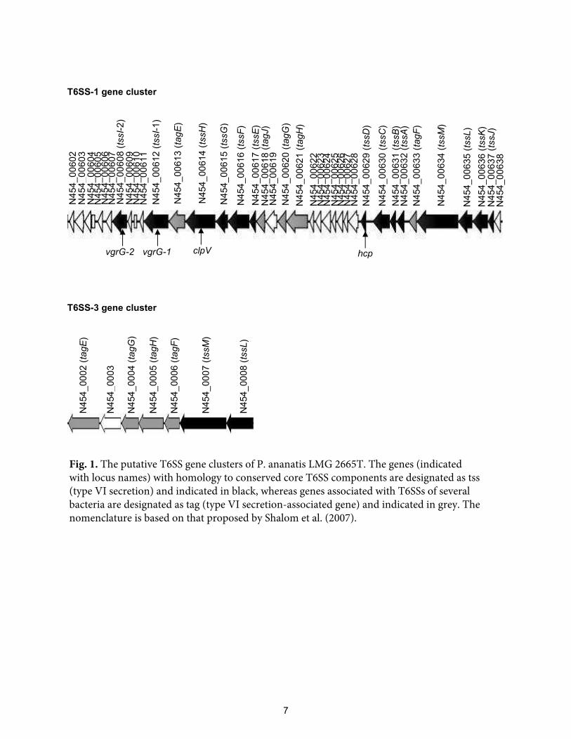
Construction of T6SS gene cluster deletions in P. ananatis LMG 2665T.
Previous sequence analyses demonstrated the presence of two gene clusters in the genome of
P. ananatis LMG 2665T that contain genes homologous to those present in T6SSs (Shyntum
et al. 2014). The 40.6-kb T6SS-1 gene cluster contains genes that are predicted to encode the
13 core T6SS proteins (TssA-M), five proteins associated with T6SSs in other bacteria (Tag)
and 18 proteins that are present in very few or no other systems. In contrast, the 8.4-kb T6SS-
3 gene cluster encodes two proteins (TssM and TssL) that are conserved in T6SSs and four
accessory proteins (Fig. 1). No genetic analysis of these loci has been performed previously,
and the function of the proteins encoded in these loci has also not yet been explored. Thus,
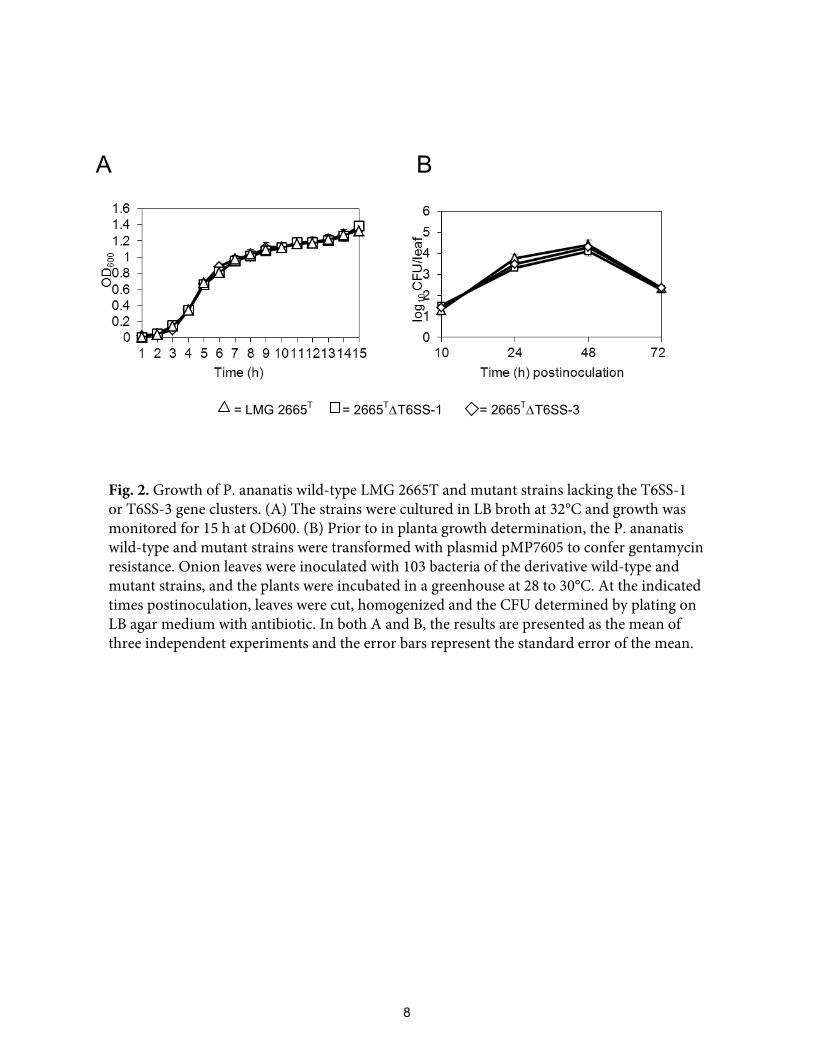
we began by deleting the individual putative T6SS loci of P. ananatis LMG 2665T. In this
study, we used the lambda Red-recombineering technique (Datsenko and Wanner 2000) to
delete the gene clusters and replace them with a kanamycin resistance cassette, yielding
strains 2665T∆T6SS-1 and 2665
T∆T6SS-3, respectively. The growth curves of the wild-type
LMG 2665T strain and the mutant strains in LB broth and in planta were similar (Fig. 2),
indicating that deletion of the respective gene clusters did not alter growth kinetics.
The T6SS-1 of P. ananatis LMG 2665T is required for pathogenesis in onion plants.
To determine whether the T6SS gene clusters play a role in the biology of P. ananatis, we
assessed the ability of mutants lacking the T6SS-1 or T6SS-3 gene clusters to cause disease
by conducting pathogenicity tests on susceptible onion plants. The wild-type strain LMG
2665T and its mutants were inoculated into onion leaves and the development of disease
symptoms was monitored. Although the 2665T∆T6SS-1 mutant strain did not induce any
disease symptoms, onion leaves infected with the wild-type LMG 2665T strain or the
6
T6SS-1 gene cluster
T6SS-3 gene cluster
N45
4_00
604
N45
4_00
602
N45
4_00
605
N45
4_00
606
N45
4_00
607
N45
4_00
609
N45
4_00
608
(tssI-2
)
N45
4_00
612
(tssI-1
)
N45
4_00
610
N45
4_00
631
(tssB
)
N45
4_00
611
N45
4_00
613
(tagE
)
N45
4_00
614
(tssH
)
N45
4_00
615
(tssG
)
N45
4_00
616
(tssF
) N
454_
0061
7 (tssE
)
N45
4_00
620
(tagG
) N
454_
0062
1 (tagH
)
N45
4_00
629
(tssD
)
N45
4_00
630
(tssC
)
N45
4_00
632
(tssA
) N
454_
0063
3 (tagF
)
N45
4_00
634
(tssM
)
N45
4_00
636
(tssK
) N
454_
0063
5 (tssL)
N45
4_00
637
(tssJ)
N45
4_00
638
N45
4_00
603
N45
4_00
622
N45
4_00
623
N45
4_00
624
N45
4_00
625
N45
4_00
626
N45
4_00
619
N45
4_00
628
vgrG-2 vgrG-1 clpV hcp
N45
4_00
618
(tagJ)
N45
4_00
627
N45
4_00
02 (tagE
)
N45
4_00
03
N45
4_00
04 (tagG
)
N45
4_00
05 (tagH
)
N45
4_00
06 (tagF
)
N45
4_00
07 (tssM
)
N45
4_00
08 (tssL)
Fig. 1. The putative T6SS gene clusters of P. ananatis LMG 2665T. The genes (indicatedwith locus names) with homology to conserved core T6SS components are designated as tss(type VI secretion) and indicated in black, whereas genes associated with T6SSs of severalbacteria are designated as tag (type VI secretion-associated gene) and indicated in grey. Thenomenclature is based on that proposed by Shalom et al. (2007).
7
A B
= LMG 2665T = 2665T∆T6SS-1 = 2665T∆T6SS-3
Fig. 2. Growth of P. ananatis wild-type LMG 2665T and mutant strains lacking the T6SS-1or T6SS-3 gene clusters. (A) The strains were cultured in LB broth at 32°C and growth wasmonitored for 15 h at OD600. (B) Prior to in planta growth determination, the P. ananatiswild-type and mutant strains were transformed with plasmid pMP7605 to confer gentamycinresistance. Onion leaves were inoculated with 103 bacteria of the derivative wild-type andmutant strains, and the plants were incubated in a greenhouse at 28 to 30°C. At the indicatedtimes postinoculation, leaves were cut, homogenized and the CFU determined by plating onLB agar medium with antibiotic. In both A and B, the results are presented as the mean ofthree independent experiments and the error bars represent the standard error of the mean.
8
2665T∆T6SS-3 mutant strain developed symptoms typical of disease caused by P. ananatis in
onion plants. Initially, the onion leaves infected with strain LMG 2665T or 2665
T∆T6SS-3
developed water-soaked spots on the sites of inoculation, which was followed by complete
collapse of the infected leaves, necrosis, wilting and then death (Fig. 3A). At 3 days
postinoculation, the number of wilted (dead) leaves per plant for each strain was recorded and
the average percentage of dead leaves calculated. On average, the mutant 2665T∆T6SS-1
strain was significantly (P < 0.05) reduced in virulence compared to the wild-type LMG
2665T strain, whereas the mutant 2665
T∆T6SS-3 strain retained virulence levels similar to
those of the wild-type strain (Fig. 3B). These data demonstrate that the pathogenicity of P.
ananatis LMG 2665T is dependent on the presence of the T6SS-1 but not the T6SS-3 gene
cluster.
The T6SS-1 of P. ananatis LMG 2665T is used to compete against different Gram-
negative bacteria.
An increasing number of T6SSs have been linked to interbacterial killing of Gram-negative
bacteria through the delivery of different toxins that target the peptidoglycan of susceptible
bacterial species (Carruthers et al. 2013; English et al. 2012; Hood et al. 2010; Russell et al.
2011). To determine whether P. ananatis LMG 2665T displays antibacterial activity and also
to explore the scope of the potential antibacterial activity, an in vitro competition assay was
performed. The wild-type LMG 2665T strain was initially tested against a panel of 30 Gram-
negative bacteria, including E. coli, which has previously been shown to be susceptible to
T6SS killing (MacIntyre et al. 2010; Weber et al. 2013; Zheng et al. 2011). Gram-positive
bacteria (Bacillus spp. and B. cereus) were included in the assay as controls (Table S2). The
wild-type LMG 2665T strain was virulent towards various Gram-negative bacteria, but did
not display antimicrobial activity towards any of the Gram-positive bacteria tested (data not
9

A LMG 2665T 2665T∆T6SSٞ 1 2665T∆T6SSٞ 3 Sterile dH2O
B
0102030405060708090
100
Perc
enta
ge o
f dea
d on
ion
leav
es
Bacterial strains
**
Fig. 3. Pathogenicity of P. ananatis wild-type LMG 2665T and mutant strains lacking the T6SS-1 or T6SS-3 gene clusters. The leaves of six-week-old onion plants were inoculated with 103 bacteria at one site per leaf and the plants were incubated in a greenhouse for 3 days. In these assays, plants inoculated with sterile distilled water (dH2O) were included as a negative control. Three individual experiments, each containing at least 20 plants pertreatment, were performed. (A) Disease symptoms of onion plants inoculated with the P. ananatis strains. The pictures were taken at 3 days postinoculation and indicate representative results. (B) At 3 days postinoculation, the number of inoculated wilted (dead) leaves per onion plant was recorded and the percentage of dead leaves was calculated. Data represent the mean percentage of dead leaves from the three biological repeats and the error bars represent the standard error of the mean. Statistically significant differences between P.ananatis LMG 2665T and the respective mutant strains was determined by an unpaired, twotailed Student’s t-test, and are indicated by asterisks.
10
shown). The antibacterial activity of the LMG 2665T strain was limited to E. coli DH5α, P.
carotovorum subsp. carotovorum LMG 2404T, Salmonella enterica serovar Typhimurium,
Pantoea stewartii subsp. indologenes, and two strains of P. ananatis (LMG 2669 and LMG
2664). These Gram-negative bacteria were used in all subsequent competition experiments.
To test the contribution of the T6SS gene clusters to the antibacterial properties of P.
ananatis LMG 2665T, we examined whether the mutants lacking the T6SS-1 or T6SS-3 gene
clusters could reduce the numbers of the above bacteria when grown in competition on agar
plates. When each competitor strain was cocultured with wild-type LMG 2665T or the
2665T∆T6SS-3 mutant strain, there was a significant (P < 0.05) drop in the number of viable
cells recovered (10- to 100-fold) compared with results for the no-treatment controls.
However, when the coculture was with 2665T∆T6SS-1, the survival of competitor bacteria
was increased up to 100-fold compared to that of wild-type LMG 2665T or 2665
T∆T6SS-3
(Fig. 4). Overall, the results suggest that the T6SS-1 gene cluster provides P. ananatis LMG
2665T with a competitive advantage towards selected Gram-negative bacteria.
Construction of tssA and tssD (hcp) mutants of P. ananatis LMG 2665T.
To determine whether P. ananatis LMG 2665T produces a functioning T6SS, we selected the
tssA and tssD (hcp) genes present in the T6SS-1 gene cluster for mutational analyses. TssD
(Hcp), a “hallmark” of T6SSs (Bingle et al. 2008), forms hexameric rings that polymerize
into tubules. It is believed that these nanotubes are extruded following contraction of the
surrounding TssB-TssC sheath, thereby facilitating transport of T6SS-dependent effector
proteins across membranes of target cells (Ballister et al. 2008; Basler et al. 2012; Jobichen et
al. 2010). TssA is predicted to be a cytoplasmic protein and contains an ImpA-like domain of
unknown function (Cascales and Cambillou 2012). It has been speculated that TssA plays a
11

0
1
2
3
4
a x c d e g
*
*
*
* * *
*
**
**
*Surv
ivin
g co
mpe
titor
(1010
CFU
/ml)
Competitor bacteria
= no-treatment control = LMG 2665T = 2665T∆T6SS-1 = 2665T∆T6SS-3
Fig. 4. The T6SS-1 of P. ananatis wild-type LMG 2665T is used for antibacterial activity.The P. ananatis wild-type, 2665TKT6SS-1 or 2665TKT6SS-3 mutant strains were mixed witha gentamycin-resistant bacterium at a ratio of 1:1, spotted onto LB agar and after overnightincubation, spots were recovered and survivor competitor bacterial cells were assessed byspreading dilutions on LB agar with antibiotic and CFU determination. Data represent themean CFU/ml from three independent experiments and error bars represent the standard errorof the mean. The CFU/ml of competitor bacteria was significantly reduced in competitionassays with strains LMG 2665T and 2665TKT6SS-3 (P < 0.05; unpaired, two-tailed Student’st-test), but not with strain 2665TKT6SS-1, when compared to the no-treatment controls.Statistically significant differences are denoted with asterisks.
12

regulatory role or is associated with proteins destined for secretion (Shrivastava and Monde
2008). The P. ananatis LMG 2665T mutant strains were constructed by replacement of the
selected genes with a kanamycin resistant cassette, yielding strains 2665T∆tssA and
2665T∆tssD, respectively. As expected from deletion of the entire T6SS-1 cluster (see
above), deletion of the tssA or tssD genes did not have any detectable impact on growth in
vitro and in planta (data not shown).
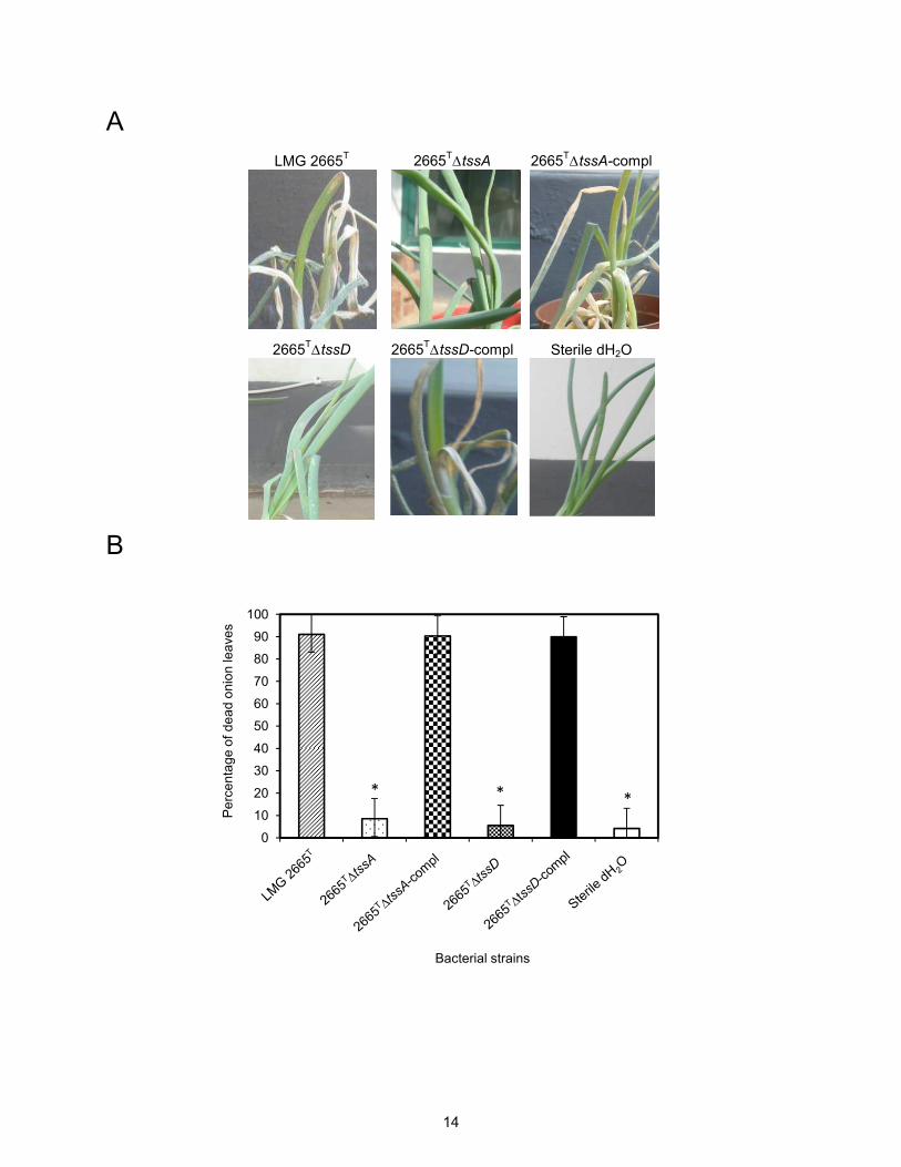
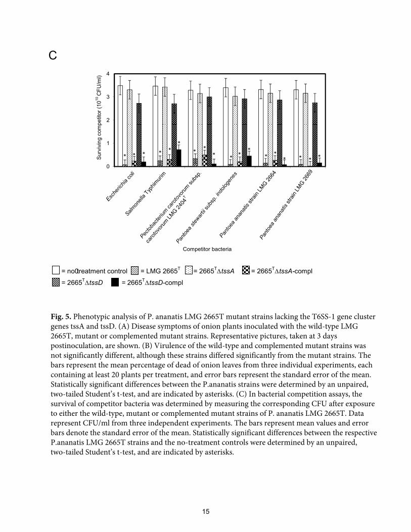
The T6SS-1 of P. ananatis LMG 2665T encodes a functioning T6SS.
To determine the effects of the tssA and tssD gene deletions on T6SS-1 activity, we examined
each mutant strain for their contributions to pathogenicity in onion plants and antibacterial
activity. The pathogenicity of the 2665T∆tssA and 2665
T∆tssD mutant strains was compared
to the wild-type LMG 2665T strain by conducting pathogenicity tests on onion leaves, as
described above. The results indicated that in contrast to the wild-type strain, neither of the
mutant strains induced disease (Figs. 5A and 5B). We also repeated the bacterial competition
assay with the Gram-negative bacterial strains previously shown to be susceptible to T6SS-1-
dependent antibacterial activity. After competition with the wild-type strain LMG 2665T a
10- to 100-fold reduction (P < 0.05) in bacteria was observed after coculture, while surviving
bacterial populations from competitions with 2665T∆tssA and 2665
T∆tssD were equivalent to
no-treatment controls (Fig. 5C). To directly link the phenotypes observed for the 2665T∆tssA
and 2665T∆tssD mutant strains to T6SS-1 functionality, the respective mutant strains were
transformed with a plasmid harboring a wild-type copy of the tssA or tssD genes. As shown
in Fig. 5, introduction of the tssA and tssD genes in trans restored the activity of the
2665T∆tssA and 2665
T∆tssD strains to cause disease in onion plants and to compete with
bacteria, thus demonstrating that the mutations are not polar and that these genes are required
to produce a functional T6SS apparatus in P. ananatis LMG 2665T.
13
A LMG 2665T 2665T∆tssA 2665T∆tssA-compl
2665T∆tssD 2665T∆tssD-compl Sterile dH2O
B
0
10
20
30
40
50
60
70
80
90
100
Per
cent
age
of d
ead
onio
n le
aves
Bacterial strains
* **
14
C
0
1
2
3
4
* * * **
*
**
**
**
**
* **
*Surv
ivin
g co
mpe
titor
(1010
CFU
/ml)
Competitor bacteria
= no0treatment control = LMG 2665T = 2665T∆tssA = 2665T∆tssA-compl
= 2665T∆tssD = 2665T∆tssD-compl
Fig. 5. Phenotypic analysis of P. ananatis LMG 2665T mutant strains lacking the T6SS-1 gene cluster genes tssA and tssD. (A) Disease symptoms of onion plants inoculated with the wild-type LMG 2665T, mutant or complemented mutant strains. Representative pictures, taken at 3 days postinoculation, are shown. (B) Virulence of the wild-type and complemented mutant strains was not significantly different, although these strains differed significantly from the mutant strains. The bars represent the mean percentage of dead of onion leaves from three individual experiments, each containing at least 20 plants per treatment, and error bars represent the standard error of the mean. Statistically significant differences between the P.ananatis strains were determined by an unpaired, two-tailed Student’s t-test, and are indicated by asterisks. (C) In bacterial competition assays, the survival of competitor bacteria was determined by measuring the corresponding CFU after exposure to either the wild-type, mutant or complemented mutant strains of P. ananatis LMG 2665T. Data represent CFU/ml from three independent experiments. The bars represent mean values and error bars denote the standard error of the mean. Statistically significant differences between the respective P.ananatis LMG 2665T strains and the no-treatment controls were determined by an unpaired,two-tailed Student’s t-test, and are indicated by asterisks.
15

DISCUSSION
Protein secretion systems are often critical to the virulence and host-interaction processes of
Gram-negative bacterial pathogens (Gerlach and Hensel 2007). Amongst the different
secretion systems, the T2SS secretes proteins from the bacteria to the exterior to degrade host
cell components (Cianciotto 2005; Sandkvist 2001), whereas the T3SS and T4SS transfer
effectors directly from the bacteria into host cells and, consequently, manipulate the host
response for their own benefit (Backert and Meyer 2006; Cornelis 2006; Hueck 1998).
Despite its ability to cause disease in a wide variety of economically important crops, P.
ananatis lacks genes homologous to the above-mentioned secretion systems (De Maayer et
al. 2011). Thus, the strategy and mechanism(s) that contribute to infection and disease
development are poorly understood in this plant pathogen. Notably, T6SS-associated genes
have been identified in Pantoea spp. (De Maayer et al. 2011) and P. ananatis (Shyntum et al.
2014) specifically. In the case of P. ananatis LMG 2665T, the T6SS-associated genes are
located in two clusters, named as T6SS-1 and T6SS-3, respectively. Considering that T6SSs
have been implicated in promoting virulence and cytotoxicity in eukaryotic and bacterial
hosts (Kapitein and Mogk 2013; Schwartz et al. 2010a), this has generated several questions
as to whether the respective gene clusters in P. ananatis encode a functional T6SS and
whether these T6SSs may play similar roles in the biology of P. ananatis LMG 2665T.
Pathogenicity assays in onion plants, performed with P. ananatis LMG 2665T mutant strains
lacking the T6SS-1 or T6SS-3 gene clusters, showed that mutant strain 2665T∆T6SS-3 was as
pathogenic as the wild-type LMG 2665T strain, while mutant strain 2665
T∆T6SS-1 was not
pathogenic. These results indicate that the T6SS-1 gene cluster likely encodes a functional
T6SS that has an essential role in pathogenicity. Although the truncated T6SS-3 gene cluster,
16
which lacks 11 of the core T6SS genes, appears not to play a role in either pathogenicity or
antibacterial competition, and therefore may not encode a functional T6SS, it is intriguing to
understand why this cluster is maintained. Its 100% prevalence amongst P. ananatis strains
(Shyntum et al. 2014) suggests that this seemingly stable gene cluster may be advantageous
to the bacteria for an as yet unknown function.
It is interesting to note that the T6SS-3 gene cluster is predicted to encode homologues of Fha
(TagH), PpkA (TagE) and PppA (TagG), which have been implicated in the regulation of
T6SS activity by a posttranslational protein phosphorylation mechanism. In P. aeruginosa,
Fha is phosphorylated by the serine-threonine kinase PpkA and dephosphorylated by the
phosphatase PppA, and the phosphorylation of Fha regulates the activity of the T6SS
(Mougous et al. 2007). Considering that these posttranslational regulatory components are
also encoded by the T6SS-1 gene cluster of P. ananatis LMG 2665T, the implications of this
potential redundancy are intriguing. Not only is it tempting to speculate that activation of the
T6SS-1 of strain LMG2665T depends on a similar posttranslational mechanism, but also that
the products of the T6SS-3 gene cluster may contribute to differentially regulating T6SS-1
activity under different culture conditions. This may explain why the P. ananatis strains
retain the truncated T6SS-3 gene cluster. Further work, however, will be required to clarify
this hypothesis and to identify environmental signals that may be responsible for triggering
the expression of the respective gene clusters.
Very little information is available about potential effector proteins that are secreted in a
T6SS-dependent manner into eukaryotic cells (Miyata et al. 2011; Zheng and Leung 2007).
In some cases, VgrG proteins can exert effector functions on eukaryotic cells. For these so-
called evolved VgrG proteins, this function is typically associated with the presence of an
17
additional C-terminal effector domain. Activities of these evolved VgrGs include cross-
linking or ADP-ribosylation of actin in eukaryotic cells, thereby promoting host cell toxicity
(Ma and Mekalanos 2010; Pukatzki et al. 2007; Suarez et al. 2010). As shown in Fig. 1, the
T6SS-1 gene cluster of P. ananatis LMG 2665T contains two VgrG (TssI) homologues. The
putative VgrG proteins lack C-terminal effector domains and are thus likely not essential for
pathogenesis in onion plants. Indeed, in the case of P. fluorescens pv. tomato, which also
contains two VgrG homologues lacking recognizable evolved C-terminal domains, it was
reported that the VgrG-1 and VgrG-2 deletion mutant strains had no effect on disease
development in tomato or in Nicotiana benthamiana (Sarris et al. 2012). A recent study,
however, suggested that adaptor proteins may be widely utilized to facilitate the recruitment
of effectors to VgrG proteins via binding at the VgrG C terminus (Schneider et al. 2013). It is
therefore conceivable that one or more such proteins may bind to the C terminus of the P.
ananatis LMG 2665T VgrG proteins and recruit effectors. In this way, each complex, VgrG-1
or VgrG-2, together with their cognate effectors, might sit independently or alternatively at
the tip of the TssD (Hcp) nanotube structures. Following contraction of the sheath, the T6SS-
1 may thus be capable of delivering a multifunctional cargo or a multiple effector VgrG spike
into the host cell in a single molecular translocation event. The effector proteins that may be
secreted by the T6SS-1 of P. ananatis LMG 2665T remains to be elucidated. Nevertheless,
several genes of unknown function and limited or no conservation with other T6SSs are
present in the P. ananatis LMG 2665T T6SS-1 gene cluster. These genes may present
candidates for system-specific effectors and will be studied in future.
P. ananatis LMG 2665T was also found to exhibit antibacterial activity. On the basis of the
33 species tested in antibacterial competition assays, the host range was found to be restricted
to selected Gram-negative bacteria. We subsequently investigated whether the T6SS-1 and
18

T6SS-3 contribute to the antibacterial activity of strain LMG 2665T. The results indicate that
strain LMG 2665T does indeed require T6SS-1 but not T6SS-3 to inhibit competitor bacteria,
since coculture with the wild-type or mutant 2665T∆T6SS-3 strains resulted in lower
recovery of viable bacteria compared to the mutant 2665T∆T6SS-1 strain. As the antibacterial
activity of the P. ananatis LMG 2665T T6SS-1 is seen during coculture on solid agar medium
surfaces, it may be that the T6SS-1 system acts through cell-cell contact (Dong et al. 2013;
MacIntyre et al. 2010). We hypothesize that the antibacterial activity of P. ananatis LMG
2665T could be mediated by T6SS-1-directed intoxication of other bacteria with protein
effectors as part of a toxin-immunity system. By implication, strain LMG 2665T must
therefore itself possess a cognate immunity protein to prevent self-intoxication, whereas
strains lacking cognate immunity proteins are inhibited. A functional link between T6SSs and
toxin-immunity systems has been established in P. aeruginosa (Hood et al. 2010), B.
thailandensis (Russell et al. 2012), S. marcescens (Chou et al. 2012; English et al. 2012) and
V. cholerae (Dong et al. 2013). Bioinformatic analysis indicated that there are no obvious
homologues to these toxins or immunity proteins in the genome of P. ananatis LMG 2665T,
thus supporting the notion that this bacterium may use a unique set(s) of effector and
immunity proteins.
Recently, Rhs-family proteins of the soft-rot pathogen Dickeya dadantii were reported to
mediate interbacterial competition (Koskinieni et al. 2013). Rhs proteins are characterized by
sequence-diverse C-terminal regions and vary considerably between different strains of the
same species. All rhs genes are closely linked to small downstream open reading frames that
encode RhsI immunity proteins. These immunity proteins are also sequence-diverse and only
protect against their cognate Rhs toxins (Koskinieni et al. 2013; Zhang et al. 2012). The Rhs
proteins are secreted in a T6SS-dependent manner in both S. marcescens (Fritsch et al. 2013)
19

and D. dadantii (Koskinieni et al. 2013), suggesting that these proteins might constitute a
new family of T6SS effectors. Interestingly, the genome of P. ananatis LMG 2665T harbors
two rhs/rhsI loci that are both located immediately adjacent to the T6SS-1 gene cluster only.
Notably, one of the Rhs proteins, designated RhsD-1 (N454_00601), contains a conserved
DUF4237 domain of unknown function at the C terminus. Given that Rhs toxins are encoded
adjacent to theT6SS-1 gene cluster and that they appear to be secreted in a T6SS-dependent
manner, it seems likely that one or both of the RhsD proteins may also play a role in P.
ananatis LMG 2665T pathogenesis. Indeed, RhsT from P. aeruginosa was shown to be
translocated into phagocytic cells, where it induces inflammasome-mediated cell death (Kung
et al. 2012). Moreover, Salmonella Typhimurium mutants lacking rhs were completely
attenuated in pig and cattle models of infection (Chaudhuri et al. 2013). Taking all of the
above into consideration, it is tempting to speculate that P. ananatis LMG 2665T Rhs proteins
are secreted in a T6SS-1-dependent manner and used as virulence factors against plant host
cells, in addition to prokaryotes. Whether the Rhs-family proteins are indeed T6SS-1 effector
proteins remains to be experimentally determined.
With the exception of Salmonella Typhimurium, it is noteworthy that the Gram-negative
bacteria inhibited by P. ananatis LMG 2665T are plant pathogens. P. carotovorum subsp.
carotovorum is a pathogen of numerous vegetables, including, cucumber, onion, potato and
cabbage (Toth et al. 2003), while P. stewartii subsp. indologenes has been isolated from
symptomatic millet, pineapple and onion (Mergaert et al. 1993), and the P. ananatis strains
LMG 2669 and LMG 2664 were both isolated from symptomatic pineapples displaying
brown and grey rot (Spiegelber, 1958 – unpublished data). Notably, Salmonella
Typhimurium, albeit not generally considered a plant pathogen, has been isolated from
cantaloupe fruit (Gallegos-Robles et al. 2000) and is capable of internalizing tomato plants
20
(Gu et al. 2011). Considering that both these plants are hosts of P. ananatis (Countinho and
Venter 2009), Salmonella Typhimurium may thus constitute a plausible competitor for P.
ananatis. Based on the finding that P. ananatis LMG 2665T uses its T6SS-1 not only for
competition with other bacterial species, but also for competition within its own species, it is
likely that killing of these bacteria in such a selective manner would be highly relevant to the
ability of P. ananatis LMG 2665T to mount a successful infection. The antibacterial T6SS-1
of strain LMG 2665T could be used as a means of competitive exclusion, thereby creating a
niche that is favorable for infection.
To determine whether the observed phenotypes are dependent on a functioning T6SS-1, we
generated tssA and tssD (hcp) deletion mutants in the T6SS-1 gene cluster. These mutations
led to attenuated pathogenesis in onion plants and antibacterial activity, while expression of
TssA and TssD from a plasmid restored the wild-type strain LMG 2665T phenotypes. The
results indicate that the observed phenotypes were caused by T6SS-1 activity. As other
studies have shown that TssD forms a nanotube that acts as a conduit to allow transport of
T6SS-dependent effector proteins or protein complexes (Ballister et al. 2008; Leiman et al.
2009; Schneider et al. 2013), it is likely that deletion of tssD in P. ananatis LMG 2665T
causes defects in the assembly of the secretion apparatus or in the secretion of T6SS-1-
dependent effectors, either of which would likely compromise T6SS-1 functionality.
Although the function of the cytoplasmic TssA protein is not known (Cascales and Cambillau
2012), it has been proposed that TssA may play a regulatory role (Shrivastava and Monde
2008). More recently, TssA was shown to interact with TssK, a cytoplasmic protein that is
responsible for TssB-TssC sheath polymerization (Zoued et al. 2013). TssA may thus
indirectly regulate the assembly of TssB-TssC tubules through its interaction with TssK, but
this requires further investigation.
21

In conclusion, we report the presence of a functional T6SS (T6SS-1) in P. ananatis LMG
2665T and provide evidence assigning functions to this T6SS in pathogenesis and bacterial
competition. Further studies are needed to identify genes involved in the assembly and the
mechanism of secretion of the T6SS-1, including the detection of TssD (Hcp) secretion, as
well as the nature of any effectors or toxins. This study has added to our understanding of
how P. ananatis causes disease, and provides a new potential target for control of diseases
caused by this plant pathogen.
MATERIALS AND METHODS
Bacterial strains, plasmids and growth conditions.
The bacterial strains and plasmids used in this study are listed in Table 1. All P. ananatis
strains in this study were derived from wild-type strain LMG 2665T. Bacterial strains were
grown on LB agar or in LB broth at 32°C (P. ananatis) or 37°C (E. coli) with shaking at 250
rpm. For plasmid DNA selection and maintenance, the growth medium was supplemented
with kanamycin (50 µg/ml), gentamycin (20 µg/ml) or chloramphenicol (50 µg/ml). The
growth of P. ananatis wild-type and mutant strains was compared in LB broth. To prepare
inoculum for plant inoculations, P. ananatis cultures grown overnight in LB broth were
diluted 100-fold in fresh LB broth and the cultures were incubated until they reached an
optical density at 600 nm (OD600) of 0.4. The cells were harvested by centrifugation (7000 ×
g, 3 min, 4°C) and suspended in sterile distilled water (dH2O) until an OD600 of 0.1
(approximately 6.2 × 10⁶ CFU/ml as determined by dilution plating).
22

In planta growth curve assays.
The growth of P. ananatis wild-type and mutant strains was compared in onion (Allium cepa
cv. Texas grando), which in previous studies have been shown to be an excellent
experimental host for P. ananatis (Goszczynska et al. 2006; Morohoshi et al. 2007). To
enable enumeration of the bacteria, the respective strains were electroporated with plasmid
pMP7605 to confer gentamycin resistance prior to the preparation of inoculum as described
above. Leaves of six-week-old onion seedlings were used for inoculation. Each leaf was
inoculated with 3 µl of a standardized bacterial suspension (6.2 × 10⁶ CFU/ml) and the plants
were incubated in a greenhouse at 28°C. Leaves were collected at 10 to 72 h postinoculation,
macerated in 2 ml of TE buffer (10 mM Tris-Cl, 1 mM EDTA; pH 8.0), serially diluted in
sterile dH2O and then plated in duplicate on LB agar supplemented with gentamycin.
Recombinant DNA techniques.
Molecular cloning techniques used in the construction of recombinant plasmids were carried
out using standard procedures (Sambrook and Russell 2001). Restriction enzymes, calf
intestine alkaline phosphatase, Klenow fragment of E. coli DNA polymerase I and T4 DNA
ligase (Roche Diagnostics, Mannheim, Germany) were used according to the manufacturer’s
protocols. Plasmid DNA was extracted from E. coli with a Zyppy Plasmid Miniprep kit,
genomic DNA was isolated from P. ananatis strains with a Quick gDNA isolation kit, and
restriction DNA fragments were purified from agarose gels by use of a Zymoclean Gel DNA
Recovery kit (all kits obtained from Zymo Research Corp., Orange, CA, U.S.A.). Plasmid
constructions were first established in E. coli DH5α and then transferred to P. ananatis
strains. Electro-competent cells were prepared and transformed according to published
procedures for E. coli (Cohen et al. 1972) and P. ananatis (Katashkina et al. 2009). PCR
assays were performed with SuperTherm DNA polymerase (Whitehead Scientific, Cape
23
Town, South Africa) and PCR amplicons were purified with the DNA Clean and
Concentrator kit (Zymo Research Corp.). Primers used in this study were designed from the
P. ananatis LMG 2665T genome sequence and obtained from Inqaba Biotechnical Industries
(Pretoria, South Africa). Southern blot hybridization was performed using the DIG-High
Prime DNA labeling and detection starter kit (Roche Diagnostics). Nucleotide sequencing
was performed with the ABI PRISM BigDye terminator v.3.1 cycle sequencing ready
reaction kit (Applied Biosystems, Foster City, CA, U.S.A.), followed by resolution on an ABI
PRISM 3100 Genetic Analyzer (Applied Biosystems), in accordance with the manufacturer’s
instructions. All plasmid constructs were verified by restriction endonuclease digestion and
by nucleotide sequencing.
Generation of P. ananatis LMG 2665T mutant strains.
Mutant strains with deletions of T6SS-1 (N454_00602 to N454_00638), T6SS-3 (N454_0002
to N454_0008), tssA (N454_00632) or tssD (N454_00629; hcp) were constructed with the
lambda Red-recombineering method as described previously (Datsenko and Wanner 2000;
Katashkina et al. 2009). Gene cluster and single gene disruption cassettes were first
constructed by an overlap-extension PCR protocol (Shevchuk et al. 2004). Briefly, 400- to
900-bp DNA fragments contiguous to the 5’ and 3’ ends of targeted genes were PCR
amplified by using LMG 2665T chromosomal DNA template and the appropriate Fup/Rup-kan
and Fdown-kan/Rdown primers (Table S1, Fig. S1). Each of the Rup-kan and Fdown-kan primers
contained 20 nucleotides that are homologous to the 5’ and 3’ termini of a kanamycin
resistance gene, respectively. Plasmid pkD13 was used as template to amplify the kanamycin
resistance gene with flanking regions homologous to the target gene using the appropriate F-
Kan and R-Kan primers (Table S1). In a second PCR, 20 ng of each of the purified amplified
24

DNA fragments were mixed and the fused DNA fragments were obtained by overlap-
extension PCR with the appropriate Fup/Rdown primers.
To enable lambda Red-dependent integration of the fused DNA fragments, the purified
disruption cassettes were introduced into electro-competent P. ananatis LMG 2665T carrying
the plasmid pRSFRedTER, which expresses the lambda-Red recombinase system and
encodes a sacB counter-selection gene (Katashkina et al. 2009). An overnight culture of P.
ananatis LMG 2665T(pRSFRedTER) was diluted 100-fold in LB broth supplemented with
chloramphenicol and 1.5 mM IPTG (isopropyl-β-D-thiogalactopyranoside) to induce
expression of the lambda recombinase system. The culture was grown to an OD600 of 0.5 at
32°C. Bacteria were then made electro-competent and transformed with 300-500 ng of the
corresponding purified PCR-generated disruption cassettes. The resulting strains were
selected by kanamycin resistance on LB agar. Selected strains were cured from the
pRSFRedTER plasmid DNA by streaking on LB agar containing 10% (w/v) sucrose and loss
of the plasmid DNA was confirmed by streaking the strains on agar containing
chloramphenicol. Allelic replacement in mutant strains that were chloramphenicol-sensitive
but kanamycin-resistant was confirmed by PCR with primers flanking the deletion sites (Fup-
out/Rdown-out) and sequencing of the amplicons, as well as by Southern blot hybridization
(data not shown).
Complementation of P. ananatis LMG 2665T ∆tssA and ∆tssD knockout mutants.
To complement the tssA and tssD genes, the full-length tssA (1.023 kb) and tssD (483 bp)
genes, together with upstream regions to include putative promoters (up to 540 bp), were
PCR amplified using P. ananatis LMG 2665T genomic DNA as template and the appropriate
primers (Table S1). The amplicons were treated with Klenow polymerase, blunt-end cloned
25
into the promoterless broad-host-range cloning vector pBRRMCS-5 and then transformed
into E. coli DH5α. The complementation plasmids pBRR-tssA and pBRR-tssD were extracted
from E. coli, electroporated into the corresponding P. ananatis mutant strains and
complemented strains were selected by gentamycin resistance on LB agar.
Pathogenicity assays.
Pathogenicity of P. ananatis wild-type and mutant strains was determined in onion plants as
described previously (Goszczynska et al. 2006; Morohoshi et al. 2007). In each test, at least
four leaves of six-week-old onion plants (Allium cepa cv. Texas grando) were inoculated with
3 µl of the inoculum (6.2 × 10⁶ CFU/ml) under the epidermis of the leaf. Sterile dH2O was
included in the assay as a negative control. On average, 20 onion plants were inoculated per
strain and each experiment was repeated three independent times. The plants were maintained
in the greenhouse during the evaluation period at a temperature of 25 to 28°C, and natural
day and night cycles. Plants were visually inspected daily for development of disease
symptoms. At three days postinoculation, the number of inoculated wilted (dead) leaves per
plant for each strain was recorded and the average percentage of dead leaves in all three
biological repeats was calculated.
Bacterial competition assays.
Bacterial strains used for the competition studies are provided in Table S2. The competition
assays were carried out with a slightly modified version of a protocol described by MacIntyre
et al. (2010). To ensure selection of competitor cells for counting, each of the bacterial strains
was transformed with plasmid pMP7605 to confer gentamycin resistance. For competition
assays, each strain was grown overnight in LB broth supplemented with antibiotic, and then
collected and washed twice in sterile LB broth (7000 × g, 2 min). The cell suspensions were
26
normalized to an OD600 of 0.1 and mixed at a ratio of 1:1 with either the P. ananatis wild-
type or mutant strains. Twenty µl of the mixture was spotted on LB agar and incubated
overnight at 32°C for all targeted bacteria analyzed, excepting Pectobacterium spp., which
were cocultured with P. ananatis at 28oC. After incubation, the bacteria were recovered from
the agar plates and suspended in 1 ml of sterile LB broth, and serial dilutions were plated in
duplicate on LB agar supplemented with gentamycin.
Statistical analyses.
Statistical significance of the data obtained in bacterial competition assays was evaluated by
making use of an unpaired, two-tailed Student’s t-test using JMP software (v.5; SAS Institute
Inc., Cary, NC, U.S.A.). P values of less than 0.05 were considered to be significant.
ACKNOWLEDGEMENTS
This study was supported by the University of Pretoria, the National Research Foundation
(NRF), the Forestry and Agricultural Biotechnology Institute (FABI), the Tree Protection Co-
operative Programme (TPCP), the NRF/Department of Science and Technology Centre of
Excellence in Tree Health Biotechnology (CTHB), and the THRIP support program of the
Department of Trade and Industry, South Africa. IKT is supported by the Scottish
Government Rural and Environment Research and Analysis Directorate (RERAD).
27

AUTHOR CONTRIBUTIONS
All of the authors participated in conceiving and designing the experiments, analysis and
interpretation of the data, and drafting of the manuscript. The experiments were performed by
DYS.
LITERATURE CITED
Aschtgen, M. S., Bernard, C. S., De Bentzmann, S., Lloubes, R., and Cascales, E. 2008. SciN
is an outer membrane lipoprotein required for type VI secretion in enteroaggregative
Escherichia coli. J. Bacteriol. 190:7523-7531.
Backert, S., and Meyer, F. M. 2006. Type IV secretion systems and their effectors in bacterial
pathogenesis. Curr. Opin. Microbiol. 9:207-217.
Ballister, E. R., Lai, A. H., Zuckermann, R. N., Cheng, Y., and Mougous, J. D. 2008. In vitro
self-assembly of tailorable nanotubes from a simple protein building block. Proc. Natl.
Acad. Sci. U.S.A. 105:3733-3738.
Basler, M., Pilhofer, M., Henderson, G. P., Jensen, G. J., and Mekalanos, J. J. 2012. Type VI
secretion requires a dynamic contractile phage tail-like structure. Nature 483:182-186.
Bingle, L. E. H., Bailey, C. E., and Pallen, M. J. 2008. Type VI secretion: A beginner’s
guide. Curr. Opin. Microbiol. 11:3-8.
Blondel, C. J., Yang, H-J., Castro, B., Chiang, S., Toro, C. S., Zaldivar, M., Contreras, I.,
Andrews-Polymenis, H. L., and Santiviago, C. A. 2010. Contribution of the type VI
secretion system encoded in SPI-19 to chicken colonization by Salmonella enterica
serotypes Gallinarum and Enteritidis. PLoS ONE 5:e11724.
28

Bönemann, G., Pietrosiuk, A., Diemand, A., Zentgraf, H., and Mogk, A. 2009. Remodelling
of VipA/VipB tubules by ClpV-mediated threading is crucial for type VI protein secretion.
EMBO Journal 28:315-325.
Boyer, F. G., Berthod, F. J., Vandenbrouck, Y., and Attree, I. 2009. Dissecting the bacterial
type VI secretion system by a genome wide in silico analysis: What can be learned from
available microbial genomic resources? BMC Genomics 10:104.
Carruthers, M. D., Nicholson, P. A., Tracy, E. N., and Munson, R. S. 2013. Acinetobacter
baumannii utilizes a type VI secretion system for bacterial competition. PLoS ONE
8:e59388.
Cascales, E. 2008. The type VI secretion toolkit. EMBO Rep. 9:735-741.
Cascales, E., and Cambillau, C. 2012. Structural biology of type VI secretion systems. Phil.
Trans. R. Soc. B 367:1102-1111.
Chaudhuri, R. R., Morgan, E., Peters, S. E., Pleasance, S. J., Hudson, D. L., Davies, H. M.,
Wang, J., van Diemen, P. M., Buckley, A. M., Bowen, A. J., Pullinger, G. D., Turner, D.
J., Langridge, G. C., Turner, A. K., Parkhill, J., Charles, I. G., Maskell, D. J., and Stevens,
M. P. 2013. Comprehensive assignment of roles for Salmonella typhimurium genes in
intestinal colonization of food-producing animals. PLoS Genet. 9:e1003456.
Chou, S., Bui, N. K., Russell, A. B., Lexa, K. W., Gardiner, T. E., LeRoux, M., Vollmer, W.,
and Mougous, J. D. 2012. Structure of a peptidoglycan amidase effector targeted to Gram-
negative bacteria by the type VI secretion system. Cell Rep. 1:656-664.
Cianciotto, N. P. 2005. Type II secretion: A protein secretion system for all seasons. Trends
Microbiol. 13:581-588.
Cohen, S. N., Chang, A. C. Y., and Hsu, L. 1972. Nonchromosomal antibiotic resistance in
bacteria: Genetic transformation of Escherichia coli by R-factor DNA. Proc. Natl. Acad.
Sci. U.S.A. 69:2110-2114.
29

Cornelis, G. R. 2006. The type III secretion injectisome. Nat. Rev. Microbiol. 4:811-825.
Coulthurst, S. J. 2013. The Type VI secretion system - a widespread and versatile cell
targeting system. Res. Microbiol. 164:640-654.
Coutinho, T. A., and Venter, S. N. 2009. Pantoea ananatis: an unconventional plant
pathogen. Mol. Plant Pathol. 10:325-335.
Datsenko, K. A., and Wanner, B. L. 2009. One-step inactivation of chromosomal genes in
Escherichia coli K-12 using PCR products. Proc. Natl. Acad. Sci. U.S.A. 97:6640-6645.
De Maayer, P., Venter, S. N., Kamber, T., Duffy, B., Coutinho, T. A., and Smits, T. H. 2011.
Comparative genomics of the type VI secretion systems of Pantoea and Erwinia species
reveals the presence of putative effector islands that may be translocated by the VgrG and
Hcp proteins. BMC Genomics 12:576.
de Pace, F., Nakazato, G., Pacheco, A., de Paiva, J. B., Sperandio, V., and da Silveira, W. D.
2010. The type VI secretion system plays a role in type 1 fimbria expression and
pathogenesis of an avian pathogenic Escherichia coli strain. Infect. Immun. 78:4990-4998.
Decoin, V., Barbey, C., Bergeau, D., Latour, X., Feuilloley, M. G. J., Orange, N., and
Merieau, A. 2014. A type VI secretion system is involved in Pseudomonas fluorescens
bacterial competition. PLoS ONE 9:e89411.
Dong, T. G., Ho, B. T., Yoder-Himes, D. R., and Mekalanos, J. J. 2013. Identification of
T6SS-dependent effector and immunity proteins by Tn-seq in Vibrio cholerae. Proc. Natl.
Acad. Sci. U.S.A. 110:2623-2628.
Durand, E., Zoued, A., Spinelli, S., Watson, P. J., Aschtgen, M. S., Journet, L., Cambillau,
C., and Cascales, E. 2012. Structural characterization and oligomerization of the TssL
protein, a component shared by bacterial type VI and type IVb secretion systems. J. Biol.
Chem. 287:14157-14168.
30
Economou, A., Christie, R. C., Christie, P. J., Fernandez, R. C., Palmer, T., and Pugsley, A.
P. 2006. Secretion by numbers: Protein traffic in prokaryotes. Mol. Microbiol. 62:308-319.
English, G., Trunk, K., Rao, V. A., Srikannathasan, V., Hunter, W. N. and Coulthurst, S. J.
2012. New secreted toxin and immunity proteins encoded within the type VI secretion
system gene cluster of Serratia marcescens. Mol. Microbiol. 86:921-936.
Felisberto-Rodrigues, C., Durand, E., Aschtgen, M. S., Blangy, S., Ortiz-Lombardia, M.,
Douzi, B., Cambillau, C., and Cascales, E. 2011. Towards a structural comprehension of
bacterial type VI secretion systems: Characterization of the TssJ-TssM complex of an
Escherichia coli pathovar. PLoS Pathog. 7:e1002386.
Filloux, A., Hachani, A., and Bleves, S. 2008. The bacterial type VI secretion machine: yet
another player for protein transport across membranes. Microbiology 154:1570-1583.
Fritsch, M. J., Trunk, K., Diniz, J. A., Guo, M., Trost, M., and Coulthurst, S. J. 2013.
Proteomic identification of novel secreted antibacterial toxins of the Serratia marcescens
type VI secretion system. Mol. Cell Proteomics. 12:2735-2749.
Gallegos-Robles, M. A., Morales-Loredo, A., Alvarez-Ojeda, G., Vega, P. A., Chew, M. Y.,
Velarde, S., and Fratamico P. 2008. Identification of Salmonella serotypes isolated from
cantaloupe and chile pepper production systems in Mexico by PCR-restriction fragment
length polymorphism. J. Food Prot. 71:2217-2222.
Gerlach, R. G., and Hensel, M. 2007. Protein secretion systems and adhesins: The molecular
armory of Gram-negative pathogens. Int. J. Med. Microbiol. 297:401-415.
Gitaitis, R. D., and Gay, J. D. 1997. First report of a leaf blight seed stalk rot, and bulb decay
of onion by Pantoea ananas in Georgia. Plant Dis. 81:1096.
Goszczynska, T., Botha, W. J., Venter, S. N., and Coutinho, T. A. 2007. Isolation and
identification of the causal agent of brown stalk rot, a new disease of maize in South
Africa. Plant Dis. 91:711-718.
31

Goszczynska, T., Moloto, V. M., Venter, S. N., and Coutinho, T. A. 2006. Isolation and
identification of Pantoea ananatis from onion seed in South Africa. Seed Science
Technol. 34:655-668.
Gu, G., Hu, J., Cevallos-Cevallos, J. M., Richardson, S. M., Bartz, J. A., and van Bruggen, A.
H. C. 2011. Internal colonization of Salmonella enterica serovar Typhimurium in tomato
plants. PLoS ONE 6:e27340.
Haapalainen, M., Mosorin, H., Dorati, F., Wu, R-F., Roine, E., Taira, S., Nissinen, R.,
Mattinen, L., Jackson, R., Pirhonen, M., and Lind, N-C. 2012. Hcp2, a secreted protein of
the phytopathogen Pseudomonas syringae pv. tomato DC3000, is required for fitness and
competition against bacteria and yeasts. J. Bacteriol. 194:4810-4822.
Holland, I. B. 2010. The extraordinary diversity of bacterial protein secretion mechanisms.
Methods Mol. Biol. 619:1-20.
Hood, R. D., Singh, P., Hsu, F., Guvener, T., Carl, M. A., Trinidad, R. R., Silverman, J. M.,
Ohlson, B. B., Hick, K. G., Plemel, R. L., Li, M., Schwarz, S., Wang, W. Y., Merz, A. J.,
Goodlett, D. R., and Mougous, J. D. 2010. A type VI secretion system of Pseudomonas
aeruginosa targets a toxin to bacteria. Cell Host Microbe 7:25-37.
Hueck, C. J. 1998. Type III protein secretion systems in bacterial pathogens of animals and
plants. Microbiol. Mol. Biol. Rev. 62:379-433.
Jobichen, C., Chakraborty, S., Li, M., Zheng, J., Joseph, L., Mok, Y-K., Leung, K. Y., and
Sivaraman, J. 2010. Structural basis for the secretion of EvpC: A key type VI secretion
system protein from Edwardsiella tarda. PLoS ONE 5:e12910.
Kanamaru, S. 2009. Structural similarity of tailed phages and pathogenic bacterial secretion
systems. Proc. Natl. Acad. Sci. U.S.A. 106: 4067-4068.
Kapitein, N., and Mogk, A. 2013. Deadly syringes: Type VI secretion system activities in
pathogenicity and interbacterial competition. Curr. Opin. Microbiol. 16:52-58.
32
Kapitein, N., Bonemann, G., Pietrosiuk, A., Seyffer, F., Hausser, I., Locker, J. K., and Mogk,
A. 2013. ClpV recycles VipA/VipB tubules and prevents non-productive tubule formation
to ensure efficient type VI protein secretion. Mol. Microbiol. 87:1013-1028.
Katashkina, J. I., Hara, Y., Lyubov, I., Golubeva, L. I., Andreeva, I. G., Kuvaeva, T. M., and
Mashko, S. V. 2009. Use of the λ Red-recombineering method for genetic engineering of
Pantoea ananatis. BMC Mol. Biol. 10:34.
Koskiniemi, S., Lamoureux, J. G., Nikolakakis, K. C., t’Kint de Roodenbeke, C., Kaplan, M.
D., Low, D. A., and Hayes, C. S. 2013. Rhs proteins from diverse bacteria mediate
intercellular competition. Proc. Natl. Acad. Sci. U.S.A. 110:7032-7037.
Kovach, M. E., Elzer, P. H., Hill, D. S., Robertson, G. T., Farris, M. A., Roop, R. M., and
Peterson, K. M. 1995. Four new derivatives of the broad host- range cloning vector
pBBR1MCS, carrying different antibiotic-resistance cassettes. Gene 166:175-176.
Kung, V. L., Khare, S., Stehlik, C., Bacon, E. M., Hughes, A. J., and Hauser, A. J. 2012. An
rhs gene of Pseudomonas aeruginosa encodes a virulence protein that activates the
inflammasome. Proc. Natl. Acad. Sci. U.S.A. 109:1275-1280.
Lagendijk, E. L., Validov, S., Lamers, G. E. M., de Weert, S., and Bloemberg, G. V. 2010.
Genetic tools for tagging Gram-negative bacteria with mCherry for visualization in vitro
and in natural habitats, biofilm and pathogenicity studies. FEMS Microbiol. Lett. 305:81-
90.
Leiman, P. G., Basler, M., Ramagopal, U. A., Bonanno, J. B., Sauder, J. M., Pukatzki, S.,
Burley, S. K., Almo, S. C., and Mekalanos, J. J. 2009. Type VI secretion apparatus and
phage tail-associated protein complexes share a common evolutionary origin. Proc. Natl.
Acad. Sci. U.S.A. 106:4154-4159.
33

Lin, J-S., Ma, L-S., and Lai, E-M. 2013. Systematic dissection of the Agrobacterium type VI
secretion system reveals machinery and secreted components for subcomplex formation.
PLoS ONE 8:e67647.
Liu, H., Coulhurst, S. J., Pritchard, L., Hedley, P. E., Ravensdale, M., Humphris, S., Burr, T.,
Takle, G., Brurberg, M. B., Birch, P. R., Salmond, G. P. C., and Toth, I. K. 2008. Quorum
sensing coordinates brute force and stealth modes of infection in the plant pathogen
Pectobacterium atrosepticum. PLoS Pathog. 4:e1000093.
Lossi, N. S., Dajani, R., Freemont, P., and Filloux, A. 2011. Structure-function analysis of
HsiF, a gp25-like component of the type VI secretion system, in Pseudomonas
aeruginosa. Microbiology 157:3292-3305.
Lossi, N. S., Manoli, E., Forster, A., Dajani, R., Pape, T., Freemont, P., and Filloux, A. 2013.
The HsiB1C1 (TssB-TssC) complex of the Pseudomonas aeruginosa type VI secretion
system forms a bacteriophage tail sheath-like structure. J. Biol. Chem. 288:7536-7548.
Ma, A. T., and Mekalanos, J. J. 2010. In vivo actin cross-linking induced by Vibrio cholerae
type VI secretion system is associated with intestinal inflammation. Proc. Natl. Acad. Sci.
U.S.A. 107:4365-4370.
Ma, L. S., Narberhaus, F., and Lai, E. M. 2012. IcmF family protein TssM exhibits ATPase
activity and energizes type VI secretion. J. Biol. Chem. 287:15610-15621.
MacIntyre, D. L., Miyata, S. T., Kitaoka, M., and Pukatzki, S. 2010. The Vibrio cholerae
type VI secretion system displays antimicrobial properties. Proc. Natl. Acad. Sci. U.S.A.
107:19520e19524.
Mergaert, J., Verdonck, L., and Kersters, K. 1993. Transfer of Erwinia ananas (synonym,
Erwinia uredovora) and Erwinia stewartii to the genus Pantoea emend. as Pantoea
ananas (Serrano 1928) comb. nov., and Pantoea stewartii (Smith 1898) comb. nov.,
34
respectively, and description of Pantoea stewartii subsp. indologenes subsp. nov. Int. J.
Syst. Bacteriol. 43:162-173.
Miyata, S. T., Kitaoka, M., Brooks, T. M., McAuley, S. B., and Pukatzki, S. 2011. Vibrio
cholerae requires the type VI secretion system virulence factor VasX to kill Dictyostelium
discoideum. Infect. Immun. 79:2941-2949.
Morohoshi, T., Nakamura, Y., Yamazaki, G., Ishida, A., Kato, N. and Ikeda, T. 2007. The
plant pathogen Pantoea ananatis produces N-acylhomoserine lactone and causes center rot
disease of onion by quorum sensing. J. Bacteriol. 189, 8333-8338.
Morohoshi, T., Ogata, Y., and Ikeda, T. 2011a. Cell aggregation is negatively regulated by N-
acylhomoserine lactone-mediated quorum sensing in Pantoea ananatis SK-1. J. Biosci.
Bioeng. 112:566-569.
Morohoshi, T., Oseki, K., and Ikeda, T. 2011b. Exopolysaccharide production is influenced
by sugars, N-acylhomoserine lactone, and transcriptional regulators RcsA and RcsB, but
does not affect pathogenicity in the plant pathogen Pantoea ananatis. Biosci. Biotechnol.
Biochem. 75:997-999.
Mougous, J. D., Cuff, M. E., Raunser, S., Shen, A., Zhou, M., Gifford, C. A., Goodman, A.
L., Joachimiak, G., Ordoñez, C. L., Lory, S., Walz, T., Joachimiak, A., and Mekalanos, J.
J. 2006. A virulence locus of Pseudomonas aeruginosa encodes a protein secretion
apparatus. Science 312:1526-1530.
Mougous, J. D., Gifford, C. A., Ramsdell, T. L., and Mekalanos, J. J. 2007. Threonine
phosphorylation post-translationally regulates protein secretion in Pseudomonas
aeruginosa. Nat. Cell Biol. 9:797-803.
Murdoch, S. L., Trunk, K., English, G., Fritsch, M. J., Pourkarimi, E., and Coulthurst, S. J.
2011. The opportunistic pathogen Serratia marcescens utilizes type VI secretion to target
bacterial competitors. J. Bacteriol. 193:6057-6069.
35
Pukatzki, S., Ma, A. T., Revel, A. T., Sturtevant, D., and Mekalanos, J. J. 2007. Type VI
secretion system translocates a phage tail spike-like protein into target cells where it cross-
links actin. Proc. Natl. Acad. Sci. U.S.A. 104:15508-15513.
Pukatzki, S., Ma, A. T., Sturtevant, D., Krastins, B., Sarracino, D., Nelson, W. C.,
Heidelberg, J. F., and Mekalanos, J. J. 2006. Identification of a conserved bacterial protein
secretion system in Vibrio cholerae using the Dictyostelium host model system. Proc. Natl.
Acad. Sci. U.S.A. 103:1528-1533.
Records, A. R. 2011. The type VI secretion system: A multipurpose delivery system with a
phage-like machinery. Mol. Plant-Microbe Interact. 24:751-757.
Russell, A. B., Hood, R. D., Bui, N. K., LeRoux, M., Vollmer, W., and Mougous, J. D. 2011.
Type VI secretion delivers bacteriolytic effectors to target cells. Nature 475:343-347.
Russell, A. B., Singh, P., Brittnacher, M., Bui, N. K., Hood, R. D., Carl, M. A., Agnello, D.
M. K., Schwarz, S., Goodlett, D. R., Vollmer, W., and Mougous, J. D. 2012. A widespread
bacterial type VI secretion effector superfamily identified using a heuristic approach. Cell
Host Microbe. 11:538-549.
Sambrook, J., and Russell, D.W. 2001. Molecular Cloning: A Laboratory Manual. Cold
Spring Harbor Laboratory Press, Cold Spring Harbor, NY, U.S.A.
Sandkvist, M. 2001. Biology of type II secretion. Mol. Microbiol. 40:271-283.
Sarris, P. F., Trantas, E. A., Skandalis, N., Tampakaki, A. P., Kapanidou, M., Kokkinidis, M.,
and Panopoulos, N. J. (2012). Phytobacterial type VI secretion system - Gene distribution,
phylogeny, structure and biological Functions. In: Plant Pathology, Cumagun, C. J. (Ed.).
Plant Pathol: InTech.
Schell, M. A., Ulrich, R. L., Ribot, W. J., Brueggemann, E. E., Hines, H. B., Chen, D.,
Lipscomb, L., Kim, H. S., Mrazek, J., Nierman, W. C., and Deshazer, D. 2007. Type VI
36
secretion is a major virulence determinant in Burkholderia mallei. Mol. Microbiol.
64:1466e1485.
Schneider, M. M., Buth, S. A., Ho, B. T., Basler, M., Mekalanos, J. J., and Leiman, P. G.
2013. PAAR-repeat proteins sharpen and diversify the type VI secretion system spike.
Nature 500:350-353.
Schwarz, S., Hood, R. D., and Mougous, J. D. 2010a. What is type VI secretion doing in all
those bugs? Trends Microbiol. 18:531-537.
Schwarz, S., West, T. E., Boyer, F., Chiang, W. C., Carl, M. A., Hood, R. D., Rohmer, L.,
Tolker-Nielson, T., Skerrett, S. J., and Mougous, J. D. 2010b. Burkholderia type VI
secretion systems have distinct roles in eukaryotic and bacterial cell interactions. PLoS
Pathog. 6:e1001068.
Serrano, F. B. 1928. Bacterial fruitlet brown-rot of pineapple in the Philippines. Philippine J.
Sci. 36:271-305.
Sessitsch, A., Reiter, B., and Berg, G. 2004. Endophytic bacterial communities of field-
grown potato plants and their plant-growth-promoting and antagonistic abilities. Can. J.
Microbiol. 50:239-249.
Shalom, G., Shaw, J. G., and Thomas, M. S. 2007. In vivo expression technology identifies a
type VI secretion system locus in Burkholderia pseudomallei that is induced upon
invasion of macrophages. Microbiology 153:2689-2699.
Shevchuk, N. A., Bryksin, A. V., Nusinovich, Y. A., Cabello, F. C., Sutherland, M., and
Ladisch, S. 2004. Construction of long DNA molecules using long PCR-based fusion of
several fragments simultaneously. Nucl. Acids Res. 32:2e19.
Shrivastava, S., and Mande, S. S. 2008. Identification and functional characterization of gene
components of type VI secretion system in bacterial genomes. PLoS One 3:e2955.
37

Shyntum, D. Y., Venter, S. N., Moleleki, L. N., Toth, I., and Coutinho, T. A. 2014.
Comparative genomics of type VI secretion systems in strains of Pantoea ananatis from
different environments. BMC Genomics 15:163.
Silverman, J. M., Brunet, Y. R., Cascales, E., and Mougous, J. D. 2012. Structure and
regulation of the type VI secretion system. Annu. Rev. Microbiol. 66:453-472.
Suarez, G., Sierra, J. C., Erova, T. E., Sha, J., Horneman, A. J., and Chopra, A. K. 2010. A
type VI secretion system effector protein VgrG1 from Aeromonas hydrophila that induces
host cell toxicity by ADP-ribosylation of actin. J. Bacteriol. 192:155-168.
Suarez, G., Sierra, J. C., Sha, J., Wang, S., Erova, T. E., Fadl, A. A., Foltz, S. M., Horneman,
A. J., and Chopra, A. K. 2008. Molecular characterization of a functional type VI secretion
system from a clinical isolate of Aeromonas hydrophila. Microb. Pathog. 44:344e361.
Toth, I. K., Bell, K. S., Holeva, M. C., and Birch, P. R. J. 2003. Soft rot erwiniae: From genes
to genomes. Mol. Plant Pathol. 4:17-30.
Walcott, R. R., Gitaitis, R. D., Castro, A. C., Sanders, F. H., and Diaz-Perez, J. C. 2002.
Natural infestation of onion seed by Pantoea ananatis, causal agent of center rot. Plant
Dis. 86:106-111.
Weber, B. S., Miyata, S. T., Iwashkiw, J. A., Mortensen, B. L., Skaar, E. P., Pukatzki, S., and
Feldman, M. F. 2013. Genomic and functional analysis of the type VI secretion system in
Acinetobacter. PLoS ONE 8:e55142.
Wu, H. Y., Chung, P. C., Shih, H. W., Wen, S. Y., and Lai, E. M. 2008. Secretome analysis
uncovers an Hcp-family protein secreted via a type VI secretion system in Agrobacterium
tumefaciens. J. Bacteriol. 190:2841-2850.
Zhang, D., de Souza, R. F., Anantharaman, V., Iyer, L. M., and Aravind, L. 2012.
Polymorphic toxin systems: Comprehensive characterization of trafficking modes,
38

processing, mechanisms of action, immunity and ecology using comparative genomics.
Biology Direct, 7:18.
Zheng, J., and Leung, K. Y. 2007. Dissection of a type VI secretion system in Edwardsiella
tarda. Mol. Microbiol. 66:1192-1206.
Zheng, J., Ho, B., and Mekalanos, J. J. 2011. Genetic analysis of anti-amoebae and anti-
bacterial activities of the type VI secretion system in Vibrio cholerae. PLoS ONE
6:e23876.
Zoued, A., Durand, E., Bebeacua, C., Brunet, Y. R., Douzi, B., Cambillau, C., Cascales, E.,
and Journet, L. 2013. TssK is a trimeric cytoplasmic protein interacting with components
of both phage-like and membrane anchoring complexes of the type VI secretion system. J.
Biol. Chem. 288:27031-27041.
39
Table S1. Primers used in this study
Primer Sequence (5’ to 3’) Use
T6SS-1 primers
C1-Fup CCAGATATTGCGGTGCGCTGTG Gene disruption cassette
C1-Rup-kan AGCTCCAGCCTACACAATCGCAGATGGGCCATATTCAGCAG Gene disruption cassette
C1-Fdown-kan GGTCCGACGGATCCCCGGAATTGCTCACCATTGTGTCATCAG Gene disruption cassette
C1-Rdown GACTTAATTACGATCTCAGAC Gene disruption cassette
C1-F-Kan CTGCTGAATATGGCCCATCTGCGATTGTGTAGGCTGGAGCT Amplification of Kmr gene
C1-R-Kan CTGATGACACAATGGTGAGCAATTCCGGGGATCCGTCGACC Amplification of Kmr gene
C1-Fup-out AGCATCAAGATCATAATGCATG Verification of deletion
C1-Rdown-out GCATGTTCCTCGACTGGACAG Verification of deletion
T6SS-3 primers
C3-Fup TGAATGTTGAACGTCACAGAGC Gene disruption cassette
C3-Rup-kan AGCTCCAGCTACACAATCGCCGTAACGACGAGCAGAATAAC Gene disruption cassette
C3-Fdown-kan GGTCGACGGATCCCCGGAATTTGTCAGCAGGAATAATCACC Gene disruption cassette
C3-Rdown ACACCCTGGTCACCAGTGATGTG Gene disruption cassette
C3-F-Kan GTTATTCTGCTCGTCGTTACGGCGATTGTGTAGGCTGGAGCT Amplification of Kmr gene
C3-R-Kan GGTGATTATTCCTGCTGACAAATTCCGGGGATCCGTCGACC Amplification of Kmr gene
C3-Fup-out AGCGAAGCGATCTTCCGGAGC Verification of deletion
C3-Rdown-out AAGTCTTAGGTAGACTGAGCG Verification of deletion
tssA primers
tssA-Fup a AAGCAGGCTGTTTAGTGGATCG Gene disruption cassette
tssA-Rup-kan AGCTCCAGCCTACACAATCGCGACTAAGCTAACAGTCAAG Gene disruption cassette
tssA-Fdown-kan GGTCGACGGATCCCCGGAATCATCCGCTTCGCCTCAGG Gene disruption cassette
tssA-Rdown a TGGTACCACCGATGTTGGGTAC Gene disruption cassette
tssA-F-Kan CTTGACTGTTAGCTTAGTCGCGATTGTGTAGGCTGGAGCT Amplification of Kmr gene
tssA-R-Kan CCTGAGGCGAAGCGGATGATTCCGGGGATCCGTCGACC Amplification of Kmr gene
tssA-Fup-out TGGTTGTTGAGAAGTCTGGTTC Verification of deletion
tssA-Rdown-out TCCAGCAGGCGCAGTCGATTCG Verification of deletion
tssD primers
tssD-Fupb ATTAAGTAGAACTTCTAATTCATTG Gene disruption cassette
tssD-Rup-kan AGCTCCAGCCTACACAATCGCCTGGCTGGAATATCAAAGAG Gene disruption cassette
tssD-Fdown-kan GGTCGACGGATCCCCGGAATCCTTCAAATACATATCAATAGC Gene disruption cassette
tssD-Rdownb TGCGAGCAACTGCACTAAAGCATG Gene disruption cassette
tssD-F-Kan CTCTTTGATATTCCAGCCAGGCGATTGTGTAGGCTGGAGCT Amplification of Kmr gene
tssA-R-Kan CAAATACATATCAATAGCATTCCGGGGATCCGTCGACC Amplification of Kmr gene
tssD-Fup-out CTTATCCATGATTAAGTCTACAGC Verification of deletion
tssD-Rdown-out CTGCGGAGTACCACGATGCTGAC Verification of deletion
Other primers
Km1 CAGTCATAGCCGAATAGCCT Verification of mutant
Km2 CGGTGCCCTGAATGAACTGC Verification of mutant
a Primers used to amplify the tssA gene for complementation plasmid construction.
b Primers used to amplify the tssD gene for complementation plasmid construction.
KmR = kanamycin resistance gene.
Supplementary material
40

Table S2. Bacterial strains used as competitors in bacterial competition assays
a Each bacterial strain was transformed with plasmid pMP7605, which confers gentamycin resistance, prior to use in
competition assays with Pantoea ananatis LMG 2665T and derived isogenic mutant strains.
b FABI, Forestry and Agricultural Biotechnology Institute, University of Pretoria, South Africa.
Bacterial strainsa Relevant characteristics
or host of isolation
Source
Gram-positive
Bacillus cereus Mn106-2a2c Isolated from termite FABIb
Bacillus subtilis A Environmental isolate FABI
B. subtilis B
Environmental isolate FABI
Gram-negative
Escherichia coli DH5α Derivative of E. coli K12 Invitrogen
Pantoea ananatis LMG 2669 Pathogen of pineapple FABI
P. ananatis LMG 2664 Pathogen of pineapple FABI
P. ananatis AJ13355 Isolated from soil in Japan FABI
P. ananatis BD442 Pathogen of maize FABI
P. ananatis PA-4 Pathogen of onion FABI
P. ananatis ICMP 10132 Pathogen of sugarcane FABI
P. ananatis ATCC 35400 Pathogen of honeydew melon FABI
P. ananatis LMG 2678 Pathogen of wheat FABI
P. ananatis LMG 2101 Pathogen of rice FABI
P. ananatis LMG 20104 Pathogen of Eucalyptus sp. FABI
P. ananatis Uruguay 40 Pathogen of Eucalyptus sp. FABI
P. ananatis BD 301 Pathogen of onion FABI
P. ananatis BD 622 Pathogen of maize FABI
P. ananatis Mmir 9 Isolated from Mirridiae sp. FABI
P. vagans BCC006 Isolated from Eucalyptus grandis FABI
P. eucalypti LMG 24197T Isolated from Eucalyptus grandis FABI
P. stewartii subsp. indologens Pathogen of maize FABI
Pectobacterium atrosepticum LMG 6687 Pathogen of tomato FABI
Pectobacterium betavasculorum LMG 2398 Pathogen of potato FABI
Pectobacterium carotovorum subsp.
carotovorum LMG 2404T
Pathogen of Irish potato FABI
Pectobacterium carotovorum subsp.
brasiliensis 1692
Pathogen of potato FABI
Brenneria nigrifluens LMG 2696 Pathogen of walnut FABI
Brenneria quercina LMG 5952 Pathogen of oak FABI
Salmonella enterica serovar Typhimurium Clinical isolate FABI
Klebsiella pneumonia TMA5 Isolated from Eucalyptus sp. in Thailand FABI
Serratia marcescens LMG 2792T Isolated from pond water FABI
Burkolderia sp. P19 Isolated from palm tree FABI
Pseudomonas putida WRB111 Isolated from Eucalyptus sp. FABI
Enterobacter sakazakii M658 Isolated from milk powder in the UK FABI
41
A
Fup
Rdown-kan
F-Kan
3’ flanking region 5’ flanking region Plasmid pKD13 carrying the kanamycin resistance gene
3’Kmr
Kmr
PCR amplification of the upstream and downstream flanking regions, and the kanamycin resistance gene (Kmr )
Overlap-extension PCR using Fup and Rdown primers
3’
Kmr3’ 5’
1)
2)
5’
Fup-Kan
RdownR-Kan
5’
B
Kmr
km1
km2Fup-out
Rdown-out
Fig. S1. Summary of the PCR reactions required for (A) construction of a gene disruption
cassette and (B) verification of newly introduced deletions. The first step is to PCR amplify the
upstream and downstream sequences flanking the targeted gene cluster or individual genes, and
the kanamycin resistance gene that is used as a selectable marker. The PCR products are then
fused in an overlap-extension PCR to generate the desired gene disruption cassette. Deletion of
the targeted gene clusters or individual genes in the constructed mutant strains were screened for
by making use of the primer pairs indicated in figure B.
42