the cell density-dependent expression of stewartan exopolysaccharide in pantoea stewartii ssp
TRANSCRIPT

University of ConnecticutDigitalCommons@UConn
Plant Science Articles Department of Plant Science
April 2005
The Cell Density-Dependent Expression ofStewartan Exopolysaccharide in Pantoea stewartiissp. stewartii is a Function of EsaR-MediatedRepression of the rcsA Gene.Timothy D. MinogueUniversity of Connecticut, Storrs CT
Aurelien L. CarlierUniversity of Connecticut, Storrs CT
Maria D. KoutsoudisUniversity of Connectictut, Storrs CT
Susanne B. von BodmanUniversity of Connecticut, [email protected]
Follow this and additional works at: http://digitalcommons.uconn.edu/plsc_articles
Recommended CitationMinogue, Timothy D.; Carlier, Aurelien L.; Koutsoudis, Maria D.; and von Bodman, Susanne B., "The Cell Density-DependentExpression of Stewartan Exopolysaccharide in Pantoea stewartii ssp. stewartii is a Function of EsaR-Mediated Repression of the rcsAGene." (2005). Plant Science Articles. 17.http://digitalcommons.uconn.edu/plsc_articles/17

Molecular Microbiology (2005)
56
(1), 189–203 doi:10.1111/j.1365-2958.2005.04529.x
© 2005 Blackwell Publishing Ltd
Blackwell Science, LtdOxford, UKMMIMolecular Microbiology0950-382XBlackwell Publishing Ltd, 2005
? 2005
56
1189203
Original Article
Quorum sensing control of stewartan EPST. D. Minogue, A. L. Carlier, M. D. Koutsoudis and S. B. von Bodman
Accepted 13 December, 2004. *For correspondence. [email protected]; Tel. (
+
1) 860 486 4408; Fax (
+
1)860 486 0534.
†
These authors contributed equally to this work.
The cell density-dependent expression of stewartan exopolysaccharide in
Pantoea stewartii
ssp.
stewartii
is a function of EsaR-mediated repression of the
rcsA
gene
Timothy D. Minogue,
1†
Aurelien L. Carlier,
1†
Maria D. Koutsoudis
2
and Susanne B. von Bodman
1,2
*
1
Departments of Plant Science and
2
Molecular and Cellular Biology, University of Connecticut, Storrs, CT 06269, USA.
Summary
The LuxR-type quorum-sensing transcription factorEsaR functions as a repressor of exopolysaccharide(EPS) synthesis in the phytopathogenic bacterium
Pantoea stewartii
ssp.
stewartii
. The cell density-dependent expression of EPS is critical for Stewart’swilt disease development. Strains deficient in the syn-thesis of a diffusible acyl-homoserine lactone inducerremain repressed for EPS synthesis and are conse-quently avirulent. In contrast, disruption of the
esaR
gene leads to hypermucoidy and attenuated diseasedevelopment. Ligand-free EsaR functions as a nega-tive autoregulator of the
esaR
gene and responds toexogenous acyl-homoserine lactone for derepres-sion. The focus of this study was to define the mech-anism by which EsaR governs the expression of the
cps
locus, which encodes functions required forstewartan EPS synthesis and membrane transloca-tion. Genetic and biochemical studies show that EsaRdirectly represses the transcription of the
rcsA
gene.RcsA encodes an essential coactivator for RcsA/RcsB-mediated transcriptional activation of
cps
genes.
In vitro
assays identify an EsaR DNA bindingsite within the
rcsA
promoter that is reasonably wellconserved with the previously described
esaR
box.We also describe that RcsA positively controls itsown expression. Interestingly, promoter proximalgenes within the
cps
cluster are significantly moreacyl-homoserine lactone responsive than geneslocated towards the middle or 3
¢¢¢¢
end of the gene clus-ter. We will discuss a possible role of EsaR-mediatedquorum sensing in the differential expression of the
cps
operon.
Introduction
Bacterial populations communicate and gauge their ownpopulation densities through production and perception ofself-produced membrane diffusible or secreted autoin-ducer (AI) molecules in a process known as quorum sens-ing (QS) (Kaplan and Greenberg, 1985; Fuqua
et al
.,1996; Pearson
et al
., 1999). As a result, bacterial commu-nities can co-ordinate and adjust the expression of spe-cialized target genes in response to external AIconcentrations. The paradigm for intraspecies-specific QSin Gram-negative bacteria is the LuxI/LuxR regulatorysystem that controls bioluminescence in the marine bac-terium
Vibrio fischeri
(reviewed in Fuqua
et al
., 1994;2001; Miller and Bassler, 2001). LuxI is a
N
-acyl-homoserine lactone (AHL) synthase. LuxR is an AHL-dependent transcriptional activator with affinity for a 20base pair (bp) palindromic sequence, termed the
lux
box(Engebrecht and Silverman, 1987; Stevens and Green-berg, 1997). In contrast, intraspecies-specific QS in Gram-positive bacteria typically utilizes secreted oligopeptideAIs and cognate two-component transduction systems(Dunny and Leonard, 1997; Lazazzera
et al
., 1997; Kleer-ebezem and Quadri, 2001; Sturme
et al
., 2002). Bothbacterial groups commonly also express a second type ofQS system characterized by a LuxS signal synthase forproduction of furanone-based AI-2 signals and a LuxP/LuxQ two-component signal transduction system (Millerand Bassler, 2001; Chen
et al
., 2002; Henke and Bassler,2004). AI-2 QS systems are thought to play a role ininterspecies communication among mixed, natural bacte-rial communities (Federle and Bassler, 2003). The broadspectrum of physiological processes governed by variousQS regulatory systems underscores their biological signif-icance in supporting bacterial colonization of diverseniches including animal and plant hosts (Davies
et al
.,1998; Williams
et al
., 2000; Withers
et al
., 2001).
Pantoea stewartii
ssp.
stewartii
(
P. stewartii
) is the aeti-ological agent of Stewart’s wilt disease in maize. Thebacterium colonizes the xylem of the plant host and pro-duces large amounts of stewartan exopolysaccharide(EPS), a major factor in the cause of Stewart’s vascularwilt (Leigh and Coplin, 1992). Mutants deficient in EPSsynthesis are avirulent. Stewartan EPS is an acidic, highmolecular weight polymer of heptameric oligosaccharide

190
T. D. Minogue, A. L. Carlier, M. D. Koutsoudis and S. B. von Bodman
© 2005 Blackwell Publishing Ltd,
Molecular Microbiology
,
56
, 189–203
repeat units that are composed of glucose, galactose, andglucuronic acid in a 3:3:1 proportion (Nimtz
et al
., 1996).The synthesis and translocation of EPS is encoded by a
~
18 kilobase (kb), 14 gene
cps/galF/galE
DNA region,which is linked to the
rfb/his
chromosomal genetic locusanalogous to other group I
cps
gene systems includingthe colanic acid biosynthetic operon in
Escherichia coli
(
E. coli
) (Coplin
et al
., 1992; Leigh and Coplin, 1992). Thenomenclature of individual
cps
genes has been changedto conform to the proposed
wce
designation according toReeves
et al
. (1996) (Fig. 1). The genetic conservation ofthese systems has allowed the putative assignment of
cps
-encoded functions in
P. stewartii
(Bernhard
et al
.,1993; Whitfield and Roberts, 1999; Nesper
et al
., 2003).Stewartan EPS is classified as a group 1 polysaccharide,in part, because polymerization initiation is undecaprenol-lipid carrier dependent, and the
cps
gene system isregulated by an RcsC/YojN/RcsB/A multicomponent phos-phorelay signal transduction system (Gottesman andStout, 1991; Kelm
et al
., 1997; Whitfield and Roberts,1999; Takeda
et al
., 2001). RcsC is a transmembranesensor kinase that responds to an unknown signal, pos-sibly desiccation, changes in osmolarity and/or other
membrane perturbations (Parker
et al
., 1992; Ophir andGutnick, 1994; Sledjeski and Gottesman, 1996). YojN isan inner membrane protein that is thought to shuttle phos-phoryl groups from the RcsC sensor kinase to the RcsBregulator (Takeda
et al
., 2001; Rogov
et al
., 2004). RcsBforms an activation complex with RcsA for the cooperativeactivation of promoters containing an RcsAB-specificbinding sequence (Wehland
et al
., 1999). The expressionof the
E. coli
RcsA coactivator is negatively regulated byH-NS, a transcriptional silencer, and positively by
DsrA
, asmall RNA molecule that acts as an antisilencer (Sledjeskiand Gottesman, 1995). Also, the RcsA protein is highlyunstable in presence of a functional Lon protease (Stout
et al
., 1991).Several previous studies confirmed that EPS synthesis
in
P. stewartii
is Rcs-dependent (Torres-Cabassa
et al
.,1987; Bernhard
et al
., 1990; Wehland
et al
., 1999). How-ever, in
P. stewartii,
QS regulation involving the EsaI signalsynthase and AHL-responsive EsaR transcription factor isdominant to RcsAB-mediated activation of
cps
(Beck vonBodman and Farrand, 1995). Disruption of the
esaI
geneblocks the synthesis of AHL and EPS even in presenceof a functional Rcs system. In contrast, a mutation in the
Fig. 1.
Gene organization of the EsaR controlled gene systems
, cps
and
rcsA
. Class I mutants are distributed throughout the EPS biosynthetic locus,
cps
; Class II mutants localized to the promoter or coding region of the
rcsA
gene (insertions are indicated by black arrows). The gene organization and putative functions of the
cps
gene cluster (Bernhard
et al
., 1993; Dolph
et al
., 1998) are shown with genes highlighted in grey encoding putative functions for precursor biosynthesis; genes indicated in black required for translocation and higher order polymerization. A putative stem-loop is shown immediately following the
wzc
gene. Heptameric repeat unit (3:3:1, glucose : galactose : glucuronic acid) synthesis initiates with the transfer of galactose-1-phosphate from UDP-galactose to an undecaprenyl phosphate lipid carrier catalysed by the membrane-localized WceG protein (grey). Additional
wce
-encoded glycosyltransferases (grey) complete heptamer polymerization through sequential addition of appropriate hexose constituents. A
wzx
-encoded function (black) ‘flips’ the lipid-linked heptamers across the plasma membrane into the periplasm. Further translocation and higher order polymerization requires the membrane-associated functions encoded by
wzc
and
wzb
(black). Export across the outer membrane and EPS surface assembly requires the
wza-
encoded
trans
-membrane protein complex (for detailed discussion of this process see Bernhard
et al
., 1993; Nesper
et al
., 2003). Class II mutants localize to the promoter or coding region of the
rcsA
gene, which encodes the RcsA transcription factor. The
rcsA
gene is located next to a gene with homology to
fliO
. Underlying brackets represent the primary
cps
promoter (indicated by the number 1) and potential internal
cps
promoters (numbers 2–7). The promoter region of the
rcsA
gene is indicated by the number 8.

Quorum sensing control of stewartan EPS
191
© 2005 Blackwell Publishing Ltd,
Molecular Microbiology
,
56
, 189–203
esaR
gene, or a double mutation in
esaI
and
esaR
, leadsto maximal synthesis of EPS (von Bodman
et al
., 1998).These findings suggested that EsaR-mediated QS regu-lation functions by gene repression in a mechanismfundamentally different from the paradigm QS model ofAHL-dependent gene activation (Beck von Bodman andFarrand, 1995).
Studies related to the autoregulatory role of EsaR pro-vided experimental proof for a QS repressor mechanism.The promoter of the
esaR
gene features a well-conserved
lux
box-like palindrome, the
esaR
box, which spans thepredicted
-
10 region of a
s
70
promoter consensussequence (Beck von Bodman and Farrand, 1995).Genetic and biochemical evaluation of EsaR function atthe
esaR
promoter differentiates EsaR from the LuxRparadigm in three fundamental aspects. First, EsaRdimerizes and becomes DNA binding competent inabsence of the cognate AHL signal (Qin
et al
., 2000).Second, EsaR exhibits reduced affinity for the
esaR
boxDNA target in presence of AHL ligand. Third, EsaRrepresses an
esaR
reporter gene fusion, and exogenousaddition of AHL promotes dose-dependent derepression(von Bodman
et al., 1998; Qin et al., 2000; Minogue et al.,2002).
This study focused on defining the mechanism by whichEsaR governs EPS synthesis by gene repression. Weutilized an unbiased random transposon mutagenesisapproach to consider all potential EsaR regulatory sce-narios. This approach yielded two classes of transposoninsertion mutations, one that localized to genes within thecps biosynthetic locus, and the other to the rcsA regula-tory gene. Genetic experiments and DNA binding studiesdetailed here allow us to conclude that EsaR functions asa transcriptional repressor of the rcsA gene by binding toan imperfect palindromic DNA sequence located in thercsA promoter. We also show that RcsA is positively auto-regulated and that maximal expression of the rcsA generequires AHL inducing conditions.
Results
Insertional mutagenesis of ESDIR
We reported previously that EsaR, the QS regulator ofP. stewartii, governs the autoregulation of its own gene,esaR, and the cell density-dependent synthesis of EPSby transcriptional repression and AHL-dependent dere-pression (von Bodman et al., 1998; Minogue et al., 2002).These studies did not resolve whether repression of EPSsynthesis was by direct EsaR control of the cps genecluster, by indirect control through the Rcs phosphorelaysystem or by other potential intermediary or alternateregulatory pathways. None of the cps and rcs promotersrevealed obvious conserved esaR box-like DNA
sequences, even though EsaR genetically controlled dif-ferent rcsA and cps reporter gene fusions. We thereforemutagenized the esaI–, esaR– double mutant, hypermu-coid ESDIR strain with the Tn5gfp-km transposon (Tanget al., 1999) to locate EsaR controlled genes with a rolein EPS synthesis. A screen of approximately 40 000 kan-amycin resistant transconjugants yielded nearly 300 EPSdeficient mutants that actively expressed the transposon-encoded promoterless green fluorescent protein (GFP)gene. Of these, 11 mutants showed a significant reductionin GFP fluorescence after coexpression of a functionalesaR gene from plasmid pSVB60. These 11 EPS defi-cient, GFP positive, EsaR responsive mutants wereselected for further study.
EPS deficient strains carry insertions primarily in the cps locus and the rcsA gene
Genomic DNA, separately isolated from the 11 mutantstrains, was subcloned into pBluescript II SK+ andexpressed in E. coli DH10B. DNA isolated from kanamy-cin resistant, GFP positive transformants was sequencedwith a set of transposon-specific primers (Table 1) todetermine the flanking sequences of each insertion. NCBIBLAST searches revealed two classes of mutants desig-nated class I and II. Class I mutants localized to the cpsgene cluster, while class II mutants carried allelic inser-tions in the rcsA gene (Fig. 1). All of the class I mutantswere readily complemented with the pES2144 plasmidthat carries the entire cps gene system plus galF andgalE. All of our class II mutants were complemented withplasmid pES4507 that carries a wild type copy of the rcsAgene. These data are consistent with our original modelthat EsaR governs the negative control of cps genesdirectly and/or indirectly through control of rcsA.
Relative expression of rcsA and cps genes under AHL-limiting and AHL-inducing conditions
The location of the transposon insertions (Fig. 1) sug-gested that QS represses EPS synthesis through directinteraction with the rcsA promoter, and potentially, withselect promoters of the cps gene system. If the expressionof the cps genes depends strictly on RcsA, then the tran-script levels of genes within the cps locus should increasein parallel with rcsA transcription in response to N-(3-oxohexanoyl)homoserine lactone (3-oxo-C6-HSL) induc-tion. Real time reverse transcription polymerase chainreaction (RT-PCR) of cDNAs generated from total mRNAisolated separately from 3-oxo-C6-HSL induced and unin-duced strain ESN51 (esaI::kan, esaR+) allowed us to mea-sure the relative transcript induction levels of the rcsA,rcsB regulatory genes, and the cps-encoded structuralgenes wceG, wza, wceL and galE against a 16S rRNA

192 T. D. Minogue, A. L. Carlier, M. D. Koutsoudis and S. B. von Bodman
© 2005 Blackwell Publishing Ltd, Molecular Microbiology, 56, 189–203
internal calibrator. Transcripts were also measured for theesaR and rseC genes as representative internal stan-dards for well-characterized 3-oxo-C6-HSL responsiveand unresponsive genes, respectively. The rseC geneencodes an enhancer of the RpoE stress response sigmafactor (Missiakas et al., 1997). Real time RT-PCR wasperformed using the specific sets of primers listed inTable 1. The data presented in Fig. 2 show that the rela-tive rcsA transcript levels increased approximately fivefoldunder 3-oxo-C6-HSL inducing conditions, while those ofrcsB remained largely unchanged. The wceG and wzagenes, which represent the first and second genes of thecps gene cluster (see Fig. 1) yielded about five and eight-fold enhanced transcript levels in response to 3-oxo-C6-HSL, respectively. The transcript level of wceL, a genelocated in the middle of the cps locus, was nearly threefoldhigher, while the galE gene located at the 3¢ end of the18 kb cps gene cluster appeared to be expressed margin-ally higher in response to 3-oxo-C6-HSL. The transcript
levels measured for esaR increased threefold, which is ingood agreement with previous genetic induction studies(Minogue et al., 2002). As expected, 3-oxo-C6-HSL hadno effect on the transcript levels of rseC. These datasupport the hypothesis that EsaR negatively controls thetranscription of the rcsA gene, not rcsB, and affects thetranscription of the cps genes, particularly those locatedclosest to the wceG promoter (see Fig. 1).
RcsA is a central factor in the EsaR-mediated QS control of EPS synthesis
To determine whether EsaR controls EPS synthesisexclusively through repression of rcsA or dual control ofrcsA and cps, we designed an epistasis experiment tocompare the phenotypes of strain ESN10 (esaI::cat) andstrain PSS11 (esaI::cat, rcsA::kan). As shown in Fig. 3A,both strains exhibit a non-mucoid phenotype when grownin absence of exogenous 3-oxo-C6-HSL. However, growth
Table 1. Oligonucleotides and primers.
Primer Sequence Introduced restriction site Used for
PrcsA5 5¢-ccataggatccaaattcacaactatcc BamHI EMSA (Fig. 4A), deletion mutation (Fig. 3A)PrcsA3 5¢-aagctaagcttgatgatagtggacagac HinDIII EMSA (Fig. 4A)PrcsA3-1 5¢-acaccaagcttgggagcaatgtcactat HinDIII EMSA, DNase I protection Assays (Fig. 5A)PwceG5 5¢-aagctaagcttgatgatagtggacagac HinDIII EMSA (Fig. 4A)/promoter lacZ fusionPwceG3 5¢-gcataaagctttctttattttatttcct HinDIII EMSA (Fig. 4A)/promoter lacZ fusionPwceL5 5¢cacgaggatccaaggcgctaagtgagaa BamHI EMSA (Fig. 4A)/promoter lacZ fusionPwceL3 5¢-atggaaagcttgtgtgattccttaaatc HinDIII EMSA (Fig. 4A)/promoter lacZ fusionPwceB5 5¢-tgactggatcctcaacccggcgatcgtc BamHI EMSA (Fig. 4A)/promoter lacZ fusionPwceB3 5¢-gaatgaagcttattgccagcacctcatt HinDIII EMSA (Fig. 4A)/promoter lacZ fusionPwza5 5¢-atggtggatcctaacccgcagaaaaagg BamHI EMSA (Fig. 4A)Pwza3 5¢-catttaagctttaatcatttcgctcttc HinDIII EMSA (Fig. 4A)PrcsA(60)5 5¢-catcgggatccttgttttggtcataaaa BamHI EMSA (Fig. 4B)PrcsA(60)3 5¢-acaagaagcttcacacaatattttttct HinDIII EMSA (Fig. 4A)Pwzx5 5¢-tgactggatcctcaacccggcgatcgtc BamHI Promoter lacZ fusionPwzx3 5¢-gaatgaagcttattgccagcacctcatt HinDIII Promoter lacZ fusionRcsADup 5¢-gcgaccctcacgaattcgttatc EcoRI Deletion mutation (Fig. 3A)RcsADlow 5¢-acgctgaattctctccttagca EcoRI Deletion mutation (Fig. 3A)RcsAlow 5¢-gttctaagcttcggcaaactatcttacg HinDIII Deletion mutation (Fig. 3A)Tn5Seq 5¢-cagtttgtttcagttaaaac Sequencing ESDIR Tn5 mutants (Fig. 1)RcsA1 5¢-acatgtctccgcgtatttcc Real time RT-PCR (Fig. 2)RcsA2 5¢-atgacccgacatccacattt Real time RT-PCR (Fig. 2)WceG1 5¢-ctgaaattccgttcgatggt Real time RT-PCR (Figs 2 and 3B)WceG2 5¢-gcataaagctttctttattttatttcct Real time RT-PCR (Figs 2 and 3B)Wza1 5¢-tatattggtcgggtccgtgt Real time RT-PCR (Figs 2 and 3B)Wza2 5¢-cgcaccggttacgtaagttt Real time RT-PCR (Figs 2 and 3B)WceL1 5¢-gctctgtattgctgccatga Real time RT-PCR (Figs 2 and 3B)WceL2 5¢-tcctcgaagaaactccggta Real time RT-PCR (Figs 2 and 3B)16s1 5¢-gttagccggtgcttcttctg Real time RT-PCR (Fig. 2)16s2 5¢-aggccttcgggttgtaaagt Real time RT-PCR (Fig. 2)RseC1 5¢-cataccgaagccaaaacctc Real time RT-PCR (Figs 2 and 3B)RseC2 5¢-attctttggccccagcttat Real time RT-PCR (Figs 2 and 3B)EsaR1 5¢-tgaccgatccggttattctc Real time RT-PCR (Fig. 2)EsaR2 5¢-aggtcggacatcagcgtaat Real time RT-PCR (Fig. 2)GalE1 5¢-ggcattgcacagattatcca Real time RT-PCR (Fig. 2)GalE2 5¢-ttacatcggctctcataccg Real time RT-PCR (Fig. 2)RcsB1 5¢-ggtaacggaaattgcgaaga Real time RT-PCR (Fig. 2)RcsB2 5¢-gttgagcagggcaatatcgt Real time RT-PCR (Fig. 2)FPprimer 5¢-lightsabre green-caggaaacagctatgaccaatgatt DNase I protection assays (Fig. 5A)T7 5¢-taatacgactcactataggg DNase I protection assays (Fig. 5A)

Quorum sensing control of stewartan EPS 193
© 2005 Blackwell Publishing Ltd, Molecular Microbiology, 56, 189–203
in the presence of 3-oxo-C6-HSL induces mucoidy instrain ESN10, but not in strain PSS11. Expression of thercsA gene from several different plasmid vectors overridesthe EsaR-mediated repression of EPS production in strainESN10 and complements the defect in strain PSS11 (datanot shown). These genetic data establish that QS signal-mediated inducibility of mucoidy in P. stewartii depends ona functional rcsA gene. Direct in vitro transcriptional anal-ysis supports this genetic conclusion. Specifically, realtime RT-PCR showed that strain ESN10 (esaI::cat, esaR+)has induced levels of wceG, wceL and wza transcripts inresponse to 3-oxo-C6-HSL (data not shown) similar tothose measured for ESN51 (esaI::kan, esaR+) in Fig. 2. Incontrast, the transcript levels of these genes in PSS11(esaI::cat; rcsA::kan) remained roughly the same, evenunder 3-oxo-C6-HSL inducing conditions (Fig. 3B). Corre-spondingly, genetic assays to measure the activity of sev-eral cps and rcsA promoter lacZ fusions in E. colidemonstrated that EsaR only repressed the PrcsA::lacZtranscriptional fusion and not the Pcps::lacZ fusions (datanot shown). Together these data confirm that rcsA is theprimary target for EsaR-mediated repression and 3-oxo-C6-HSL specific derepression, and, that the effect ofEsaR regulation on EPS synthesis is indirect and depen-dent on RcsA.
EsaR directly and specifically binds to the RcsA promoter
The above experimental data establish a role for EsaR asa direct negative regulator of the rcsA gene under 3-oxo-
C6-HSL restrictive conditions and related derepression ofthe rcsA gene under inducing conditions. Therefore,ligand-free EsaR (Apo-EsaR) should physically interactwith a target sequence of the rcsA promoter. Correspond-ingly, if the hierarchical regulatory model is correct, thenEsaR should not bind to promoters associated with thecps gene system. We employed electromobility shiftassays (EMSAs) to measure relative binding of purifiednative Apo-EsaR to specific sequences of the rcsA pro-moter and sequences corresponding to the primary wceGpromoter, and potential intergenic promoters upstream of
Fig. 2. Induction of specific gene expression in response to 3-oxo-C6-HSL. The relative transcript induction of specific cps-encoded EPS biosynthetic genes, the esaR, rcsA and rcsB regulatory genes, and rseC, as an example of a 3-oxo-C6-HSL neutral gene, was measured by real time RT-PCR. Template cDNAs were generated from total RNA extracts of strain ESN51 grown to exponential phase separately in the absence (�) or presence (�) of 10 mM 3-oxo-C6-HSL. Target transcript levels were normalized using 16S rRNA as an internal reference. Relative fold induction (RFI) was calculated using the mathematical equation: RFI = 2–DDCT (see Experimental Proce-dures). Each experiment was repeated three times and error bars represent the standard deviation.
Fig. 3. Epistasis experiments to establish the regulatory dominance of EsaR over RcsA.A. Strains ESN10 (esaI::cat) and PSS11 (esaI::cat, rcsA::kan) were grown on glucose-rich agar supplemented with (lower panel) or with-out (upper panel) 10 mM 3-oxo-C6-HSL and incubated at 28∞C. Both strains lack a typical mucoid phenotype when grown in absence of 3-oxo-C6-HSL. Growth in presence of 3-oxo-C6-HSL induced mucoidy in strain ESN10, but not in strain PSS11.B. Transcript levels of wceG, wza and wceL were determined by real time RT-PCR. The cDNAs generated from total RNA of PSS11 cul-tures grown separately to exponential phase in the absence (�) or presence (�) of 10 mM 3-oxo-C6-HSL. Relative transcript levels were calculated based on rseC levels as an internal 3-oxo-C6-HSL neutral standard. Experiments were repeated three times and error bars represent the standard deviation.
A
B
Rel
ativ
e fo
ld in
du
ctio
n
194 T. D. Minogue, A. L. Carlier, M. D. Koutsoudis and S. B. von Bodman
© 2005 Blackwell Publishing Ltd, Molecular Microbiology, 56, 189–203
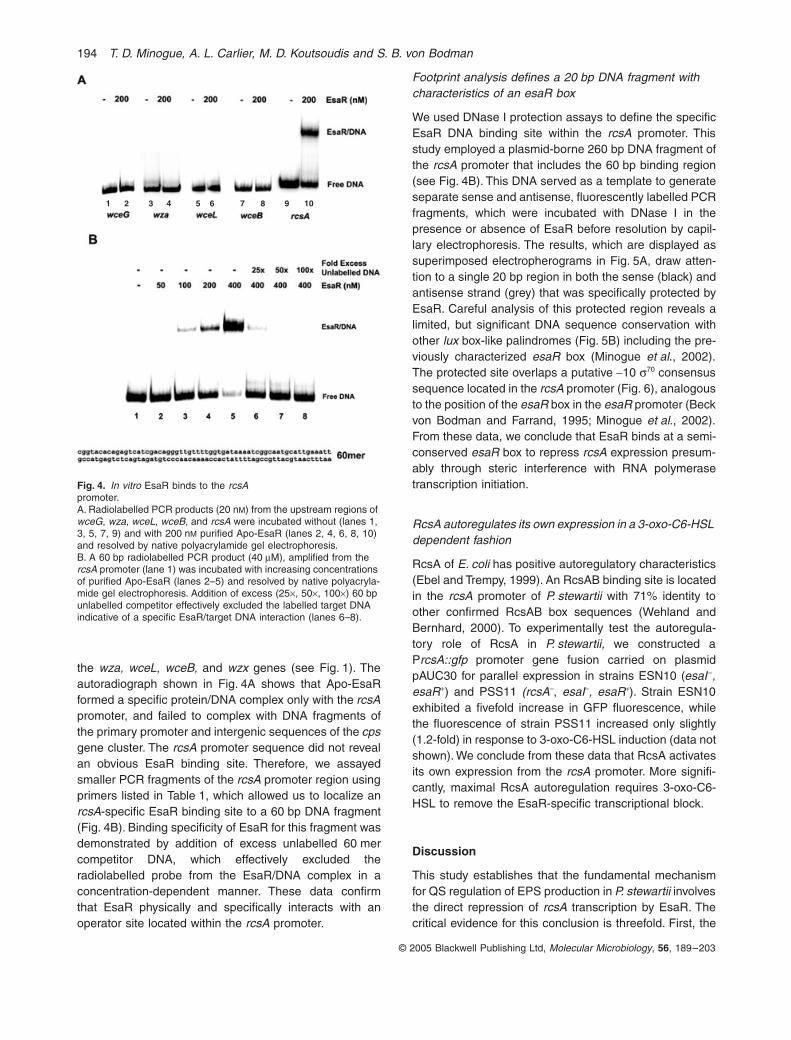
the wza, wceL, wceB, and wzx genes (see Fig. 1). Theautoradiograph shown in Fig. 4A shows that Apo-EsaRformed a specific protein/DNA complex only with the rcsApromoter, and failed to complex with DNA fragments ofthe primary promoter and intergenic sequences of the cpsgene cluster. The rcsA promoter sequence did not revealan obvious EsaR binding site. Therefore, we assayedsmaller PCR fragments of the rcsA promoter region usingprimers listed in Table 1, which allowed us to localize anrcsA-specific EsaR binding site to a 60 bp DNA fragment(Fig. 4B). Binding specificity of EsaR for this fragment wasdemonstrated by addition of excess unlabelled 60 mercompetitor DNA, which effectively excluded theradiolabelled probe from the EsaR/DNA complex in aconcentration-dependent manner. These data confirmthat EsaR physically and specifically interacts with anoperator site located within the rcsA promoter.
Footprint analysis defines a 20 bp DNA fragment with characteristics of an esaR box
We used DNase I protection assays to define the specificEsaR DNA binding site within the rcsA promoter. Thisstudy employed a plasmid-borne 260 bp DNA fragment ofthe rcsA promoter that includes the 60 bp binding region(see Fig. 4B). This DNA served as a template to generateseparate sense and antisense, fluorescently labelled PCRfragments, which were incubated with DNase I in thepresence or absence of EsaR before resolution by capil-lary electrophoresis. The results, which are displayed assuperimposed electropherograms in Fig. 5A, draw atten-tion to a single 20 bp region in both the sense (black) andantisense strand (grey) that was specifically protected byEsaR. Careful analysis of this protected region reveals alimited, but significant DNA sequence conservation withother lux box-like palindromes (Fig. 5B) including the pre-viously characterized esaR box (Minogue et al., 2002).The protected site overlaps a putative -10 s70 consensussequence located in the rcsA promoter (Fig. 6), analogousto the position of the esaR box in the esaR promoter (Beckvon Bodman and Farrand, 1995; Minogue et al., 2002).From these data, we conclude that EsaR binds at a semi-conserved esaR box to repress rcsA expression presum-ably through steric interference with RNA polymerasetranscription initiation.
RcsA autoregulates its own expression in a 3-oxo-C6-HSL dependent fashion
RcsA of E. coli has positive autoregulatory characteristics(Ebel and Trempy, 1999). An RcsAB binding site is locatedin the rcsA promoter of P. stewartii with 71% identity toother confirmed RcsAB box sequences (Wehland andBernhard, 2000). To experimentally test the autoregula-tory role of RcsA in P. stewartii, we constructed aPrcsA::gfp promoter gene fusion carried on plasmidpAUC30 for parallel expression in strains ESN10 (esaI–,esaR+) and PSS11 (rcsA–, esaI–, esaR+). Strain ESN10exhibited a fivefold increase in GFP fluorescence, whilethe fluorescence of strain PSS11 increased only slightly(1.2-fold) in response to 3-oxo-C6-HSL induction (data notshown). We conclude from these data that RcsA activatesits own expression from the rcsA promoter. More signifi-cantly, maximal RcsA autoregulation requires 3-oxo-C6-HSL to remove the EsaR-specific transcriptional block.
Discussion
This study establishes that the fundamental mechanismfor QS regulation of EPS production in P. stewartii involvesthe direct repression of rcsA transcription by EsaR. Thecritical evidence for this conclusion is threefold. First, the
Fig. 4. In vitro EsaR binds to the rcsA promoter.A. Radiolabelled PCR products (20 nM) from the upstream regions of wceG, wza, wceL, wceB, and rcsA were incubated without (lanes 1, 3, 5, 7, 9) and with 200 nM purified Apo-EsaR (lanes 2, 4, 6, 8, 10) and resolved by native polyacrylamide gel electrophoresis.B. A 60 bp radiolabelled PCR product (40 mM), amplified from the rcsA promoter (lane 1) was incubated with increasing concentrations of purified Apo-EsaR (lanes 2–5) and resolved by native polyacryla-mide gel electrophoresis. Addition of excess (25¥, 50¥, 100¥) 60 bp unlabelled competitor effectively excluded the labelled target DNA indicative of a specific EsaR/target DNA interaction (lanes 6–8).
1 2 3 4 5 6 7 8 9 10

Quorum sensing control of stewartan EPS 195
© 2005 Blackwell Publishing Ltd, Molecular Microbiology, 56, 189–203
esaI mutant strain ESN51 (esaR+) can be induced for EPSsynthesis by exogenous addition of 3-oxo-C6-HSL, whilethe corresponding esaI/rcsA double mutant strain, PSS11,is non-inducible and remains blocked for EPS synthesiseven in presence of the signal. Second, Apo-EsaR bindsspecifically to the rcsA promoter, but does not interact withsequences of the primary wceG promoter or intergenicregions within the cps operon. Third, DNase protectionassays identify a region within the rcsA promoter thatcorresponds to a semiconserved esaR box element. This
element spans the predicted -10 promoter consensussequence. We also show that the activation of rcsA issubject to positive feedback regulation by RcsA, similar torcsA in E. coli and Erwinia amylovora (Ebel and Trempy,1999; Wehland et al., 1999). These data permit us toformulate a hierarchical model for QS regulation of EPSsynthesis in P. stewartii, as summarized in Fig. 7. At lowcell density, in absence of threshold concentrations of 3-oxo-C6-HSL ligand, Apo-EsaR is DNA binding competentand acts as a direct repressor of rcsA transcription. Even
Fig. 5. DNase I footprinting analysis to detect the EsaR binding motif in the rcsA promoter.A. DNase I digestion reactions of a 260 bp rcsA promoter fragment spanning the full-length rcsA promoter (35 nM) were resolved by capillary electrophoresis on a Beckman Coulter CEQ 2000XL. The electropherograms shown are of the sense (�) and antisense ( ) strands in the absence (upper panel) or presence of purified EsaR (0.9 mM) (lower panel). Fluorescence Intensity, y-axis, is proportional to the relative fragment abundance and elution time, x-axis, correlates to fragment length. Numeric values (bottom scale) refer to nucleotide position relative to the putative transcription start of the rcsA promoter. The protected sequence is as indicated and falls between nucleotides -24 and -5.B. The EsaR binding site from the rcsA promoter was compared to other lux box-like DNA sequences found in P. stewartii (esaR) (Minogue et al., 2002), Vibrio fischeri (luxI) (Egland and Greenberg, 1999), Serratia marcescens (spnR) (Horng et al., 2002) and Pseudomonas aeruginosa (lasB) (Rust et al., 1996). Highly conserved nucleotides are highlighted in black, while other conserved nucleotides are displayed in grey; R = A or G; Y = T or C; M = A or C; K = G or T; H = A, C or T; V = A, C or G; D = A, G or T.
A
B
ATCGGCAATGCATTGAAATT

196 T. D. Minogue, A. L. Carlier, M. D. Koutsoudis and S. B. von Bodman
© 2005 Blackwell Publishing Ltd, Molecular Microbiology, 56, 189–203
so, the rcsA gene expresses at measurable basal levelsunder EsaR repressive conditions (data not shown). Thisis likely a function of the less stringent conservation of theesaR box in the rcsA promoter compared to the corre-sponding palindrome in the esaR promoter (Minogueet al., 2002). RcsA is also subject to rapid proteolysis byLon protease presumably to keep cellular RcsA proteinbelow a functionally relevant concentration. Consistentwith this assessment is the observed enhanced mucoidyof lon mutant strains of P. stewartii analogous to lonmutants of E. coli and E. amylovora (Stout et al., 1991;Eastgate et al., 1995; M. D. Koutsoudis, unpubl.). At highcell density, or after exogenous addition of 3-oxo-C6-HSL,EsaR repressor activity relaxes, thus permitting the rapidexpression of the rcsA gene. In this context, it is importantto note that EsaR does not govern the expression of thercsB gene, and rcsB transcript levels remain constantunder inducing conditions. This model also indicates thatEsaR-mediated QS is functionally dominant to the RcsC-YojN-RcsB environmental sensing phosphorelay system.
The genetic evidence for this assertion is twofold. First,the esaI mutant strains ESN10 and ESN51 grown on highglucose medium remain repressed for EPS synthesisunless exposed to inducing levels of 3-oxo-C6-HSL. Sec-ond, an esaR or esaI/esaR double mutant strain ofP. stewartii exhibits a mucoid phenotype even when grownon low glucose medium (von Bodman et al., 1998).
The role of RcsA/B activation of several group 1 capsulegene clusters including cps of P. stewartii is well docu-mented (Poetter and Coplin, 1991; Stout et al., 1991;Kelm et al., 1997; Wehland et al., 1999). Our data confirmthat the RcsA protein is essential for the activation of thecps gene system and stewartan EPS synthesis inP. stewartii. However, it is interesting to note that the pro-moter proximal genes (wceG, wza) of the cps gene clusterare induced to a significantly higher degree by 3-oxo-C6-HSL than genes located toward the middle (wceL) and 3¢end (galE) of the ~18 kb operon. The primary promoterupstream of wceG features a conserved JUMPstartsequence (just upstream of many polysaccharide starts)
Fig. 6. Schematic depiction of the rcsA promoter. The promoter features a well-conserved RcsA/B binding site (grey), an EsaR binding motif (black) positioned between a putative -35 (underlined) and overlapping a putative -10 (underlined) s70 recognition sequence. An arrow indicates a possible transcriptional start. Comparison of the published rcsA DNA sequence (GenBank Accession X58707) with the sequences obtained from the flanking regions of the rcsA transposon insertions revealed minor, but significant discrepancies (black circles above individual nucleotides). Sequence analyses of PCR fragments amplified from the genome of several different P. stewartii strains confirmed the discrepancies. We deposited a corrected DNA sequence for the rcsA promoter in GenBank as Accession AY819768.
Fig. 7. A model depicting the hierarchical EsaR QS regulatory pathway. At low cell density, ligand-free EsaR represses the transcription of rcsA, yielding basal levels of RcsA protein that is subject to degradation by Lon protease (Gottesman and Stout, 1991) preventing significant RcsA/RcsB activation complex formation. At high cell density or 3-oxo-C6-HSL inducing conditions, EsaR repression of rcsA is relieved resulting in RcsA levels exceeding the degradation capacity of Lon. RcsA recruits RcsB to form an activation complex for the positive feedback regulation of rcsA and activation of the cps gene cluster.

Quorum sensing control of stewartan EPS 197
© 2005 Blackwell Publishing Ltd, Molecular Microbiology, 56, 189–203
(Hobbs and Reeves, 1994; Wehland et al., 1999). Thissequence contains an eight bp element termed ops (oper-ons polarity suppressor), which recruits the RfaH antiter-mination protein into the transcription complex to promotethe synthesis of full-length operonic transcripts (Baileyet al., 1997; Stevens et al., 1997; Marolda and Valvano,1998; Rahn et al., 1999; Artsimovitch and Landick, 2002).The cps operon of P. stewartii also features a putativestem-loop structure at the 3¢ end of the wzc gene, analo-gous to the Rho-independent termination stem-loop struc-ture found in the K30 cps cluster of E. coli (Rahn et al.,1999; Rahn and Whitfield, 2003). By analogy, this termi-nator region separates genes involved in higher-orderpolymerization and surface expression of EPS from thegenes that encode specific glycosyltransferase enzymesfor the biosynthesis of oligosaccharide repeat units. Weassume that this putative stem-loop structure plays animportant role in the differential expression of these twoblocks of cps genes in P. stewartii. It is conceivable thatunder optimal RcsA/B-mediated activation of the cpsoperon, a subpopulation of transcripts escape antitermi-nation leading to the accumulation of truncated tran-scripts. This scenario would explain the differentialtranscript levels detected of genes located upstream anddownstream of the termination loop in response to 3-oxo-C6-HSL induction (Fig. 2). The dual galF/galE genes posi-tioned at the 3¢ end of the cps gene system could beexpressed from an independent promoter (Torres-Cabassa et al., 1987; Dolph et al., 1988), although addi-tional experiments are needed for unequivocal proof. Inany case, we show that EsaR and 3-oxo-C6-HSL do notsignificantly control the expression of the galE gene. TheGalF and GalE enzymes serve important functions inUDP-glucose and UDP-galactose synthesis, which areimportant precursors of stewartan EPS (Dolph et al.,1988; Nimtz et al., 1996).
Group 1 polysaccharides can be produced in severaldistinct forms. A short, or low molecular weight formassembles on a lipid A-core and consists of one or a fewoligosaccharide repeat units (MacLachlan et al., 1993;Drummelsmith and Whitfield, 1999; Rahn and Whitfield,2003). This form is referred to as KLPS. Multiple oligosac-charide repeat units attached to the lipid A-core leads tothe synthesis of O-antigen, sometimes referred to assmooth LPS (S-LPS). A high molecular weight capsularor EPS form is assembled on the cell surface in a trans-location pathway that requires functions including Wza, anouter membrane lipoprotein, Wzb, an acid phosphatase,and Wzc, an inner membrane tyrosine kinase (Stevensonet al., 1996; Drummelsmith and Whitfield, 1999; Geider,2000; Beis et al., 2004). These proteins are encoded bygenes located between wceG and the putative stem-loopstructure upstream of wceL in P. stewartii. We thereforeenvision a model in which basal level expression of the
cps gene system directs oligosaccharide repeat units intoO-antigen and or KLPS synthesis, while RcsA/B-mediatedactivation of the cps gene system may be a mechanismto shunt these same oligosaccharide repeat units into highmolecular weight stewartan EPS biosynthesis. Thus,EsaR-mediated QS regulation may serve as a key switchbetween LPS and EPS synthesis in P. stewartii. It shouldbe noted that the cps gene cluster of P. stewartii lacks awzi gene, which encodes an outer membrane proteinthought to anchor the high molecular weight polymer tothe cell surface typical of capsular polysaccharides (CPS)(Rahn et al., 2003). It is therefore likely that most of stew-artan is in the cell-free EPS form.
We recognize that EPS synthesis is controlled by otherglobal regulatory mechanisms in addition to QS. Forexample, the role of Lon protease in EPS synthesis is wellestablished (Gottesman et al., 1985). Chatterjee and col-leagues reported a role of the CsrA/csrB (Romeo et al.,1993) homologue pair, RsmA/rsmB, in the control of EPSsynthesis in several Erwinia strains and P. stewartii (Cuiet al., 1995). Additionally, this group and others showedthat RsmA functions by destabilizing transcripts of LuxIhomologue QS signal synthases in different Erwinia spe-cies (Cui et al., 1995; Whitehead et al., 2002). It is there-fore possible that the effect of RsmA on EPS synthesis isa consequence of controlled intrinsic levels of 3-oxo-C6-HSL.
Finally, one must ask why EsaR, a reasonably con-served LuxR orthologue, should have evolved to functionas a repressor with affinity for its DNA binding target in aligand-free state while LuxR requires the signal cofactorfor DNA binding and transcriptional activation? Werecently reported that EsaR retains the ability to functionalso as a transcriptional activator in the ligand free stateif provided a properly positioned cis binding site (vonBodman et al., 2003). Preliminary data indicate thatEsaR may positively control one or more genes inP. stewartii under signal-limiting conditions. In the overallcontext of QS regulation, such dual functionality would bepossible only if EsaR is DNA binding proficient inabsence of the signal ligand to correspondingly activategenes required at low cell density, while repressinggenes needed for cellular function at a higher cell density.This regulatory scenario would be an attractive mecha-nism for bacteria to transition between different stages ofgrowth or development particularly when colonizing aspecific niche or host.
Experimental procedures
Bacterial strains, growth conditions and DNA techniques
The E. coli strains used as cloning hosts include DH5a (LifeTechnologies), Top10 (Invitrogen), DH10B (Invitrogen), and
198 T. D. Minogue, A. L. Carlier, M. D. Koutsoudis and S. B. von Bodman
© 2005 Blackwell Publishing Ltd, Molecular Microbiology, 56, 189–203
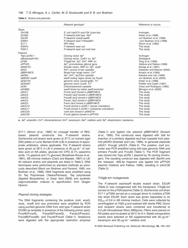
S17-1 (Simon et al., 1982) for conjugal transfer of RK2-based plasmid constructs into P. stewartii strains.Escherichia coli strains were grown at 37∞C on nutrient agar(NA) plates or Luria–Bertani broth (LB) in presence of appro-priate antibiotics, where applicable. The P. stewartii strainswere grown at 28∞C in LB in presence of 30 mg ml-1 of nali-dixic acid on NA plates, glucose-rich CPG (0.1% casaminoacids, 1% peptone and 1% glucose) (Bradshaw-Rouse et al.,1981), AB minimal medium (Clark and Maaløe, 1967) or LB.All relevant strains and plasmids are listed in Table 2. DNAtechniques were performed by standard methods as previ-ously described (Beck von Bodman and Farrand, 1995; vonBodman et al., 1998). DNA fragments were amplified usingEx Taq Polymerase (Takera/Panvera), Taq polymerase(Applied Biosystems), or Deep Vent (NEB) and syntheticoligonucleotides ordered to specification from QiagenOperon.
Plasmid cloning strategies
The DNA fragments containing the putative rcsA, wceG,wceL, wceB and wzx promoters were amplified by PCRusing purified genomic DNA from wild type P. stewartii strainDC283 as template in presence of the following primer pairs:PrcsA5/PrcsA3, PwceG5/PwceG3, PwceL5/PwceL3,PwceB5/PwceB3, and Pwzx5/Pwzx3 (Table 1). Ampliconswere digested with the appropriate restriction enzymes
(Table 1) and ligated into plasmid pBBR1MCS (Kovachet al., 1995). The constructs were digested with SalI forinsertion of a similarly digested lacZ-kan cassette from plas-mid pKOK5 (Kokotek and Lotz, 1989), to generate plasmidspAUC1 through pAUC5 (Table 2). The putative rcsA pro-moter was PCR amplified using wild type genomic DNA andprimers PrcsA5 and PrcsA3 (Table 1). The PCR fragmentwas cloned into Topo pCR2.1 plasmid by TA cloning (Invitro-gen). The resulting construct was digested with BamHI andthe released ~800 bp fragment was ligated into pFPV25plasmid (Valdivia and Falkow, 1996), resulting in pAUC30(Table 2).
Tn5gfp-km mutagenesis
The P. stewartii esaI/esaR double mutant strain, ESDIR(Table 2) was mutagenized with the transposon Tn5gfp-kmcarried on the pTGN plasmid (Table 2). Escherichia coli strainS17-1 (pTGN) served as a conjugal donor to mobilize pTGNinto strain ESDIR. Each strain was grown separately to anOD600 of 0.6 in AB minimal medium. Cells were collected bycentrifugation at 7000 g and washed with sterile PBS. Donorand recipient strains were combined and transferred to0.2 mM nitrocellulose filters (Millipore). Filters were placed onNA plates and incubated at 28∞C for 6 h. Stable transpositionevents were selected on NA supplemented with 30 mg ml-1
kanamycin and 30 mg ml-1 nalidixic acid.
Table 2. Strains and plasmids.
Relevant genotypea Reference or source
StrainDH10B E. coli DlacX74 araD139 D(ara-leu) InvitrogenDC283 P. stewartii wild type, NalR Dolph et al. (1988)ESDIR P. stewartii D(esaI-esaR) von Bodman et al. (1998)ESN51 P. stewartii esaI::Tn5seqN51 (von Bodman et al. (1998)S17-1 RP4 Mob+ Simon et al. (1982)ESN10 P. stewartii esaI::cat This studyPSS11 P. stewartii esaI::cat rcsA::kan This study
PlasmidTopo pCR2.1 Cloning vector ApR InvitrogenpBluescriptII KS+ Cloning vector, ColE1 ori, ApR StratagenepTGN Tn5gfp-km, ApR, KmR, R6K ori Tang et al. (1999)pFPV25 ApR, promoterless gfpmut gene Valdivia and Falkow (1996)pKNG101 Suicide vector, R6K ori, SpR, sacB Kaniga et al. (1991)pKD4 KmR, source of kan cassette Datsenko and Wanner (2000)pBBR1MCS Broad host range vector, CmR Kovach et al. (1995)pKOK5 ApR, KmR, lacZ-Km cassette Kokotek and Lotz (1989)pBAD22::esaR esaR coding region driven by ParaC von Bodman et al. (2003)pES2144 genomic clone (wceG-galE), TcR Dolph et al. (1988)pES4507 genomic clone (rcsA), TcR Poetter and Coplin (1991)pCM1 Chloramphenicol cassette Close and Rodriguez (1982)pSVB60 esaR driven by native esaR promoter Minogue et al. (2002)pAUC1 PrcsA::lacZ cloned in pBBR1MCS This studypAUC2 PwceG::lacZ cloned in pBBR1MCS This studypAUC3 PwceB::lacZ cloned in pBBR1MCS This studypAUC4 PwceL::lacZ cloned in pBBR1MCS This studypAUC5 Pwzx::lacZ cloned in pBBR1MCS This studypAUC10 PrcsA cloned in pCR2.1 (sense orientation) This studypAUC11 PrcsA cloned in pCR2.1 (antisense orientation) This studypAUC20 rcsA::kan cloned in pKNG101 This studypAUC30 PrcsA::gfpmut cloned in pFPV25 This study
a. ApR, ampicillin; CmR, chloramphenicol; KmR, kanamycin; NalR, nalidixic acid; SpR, streptomycin; resistance.

Quorum sensing control of stewartan EPS 199
© 2005 Blackwell Publishing Ltd, Molecular Microbiology, 56, 189–203
Screening Tn5gfp-km mutants
Colonies exhibiting stable expression of Tn5gfp-km wereviewed under a stereomicroscope for identification of EPSdeficient mutants. Such mutants were patched onto AB min-imal medium to ensure prototrophy. Secondary screeningwas based on the comparative expression of the Tn5gfp-kmencoded GFP in the presence or absence of EsaR. PlasmidpSVB60 (Table 2) was introduced into independent mutantsusing E. coli S17-1 (Table 2) as a conjugal donor. The ESDIRTn5gfp-km mutants and corresponding strains carryingpSVB60 were cultured in AB minimal medium and grown toan OD600 of 0.6. Aliquots of 5 ml cell suspensions, standard-ized to OD600 of 1.0, were spotted onto fresh AB minimalplates in replicates of six, and evaluated over the course of3 days using a Molecular Imager FX (FITC) (Bio-Rad). GFPlevels of each sample were quantified using QUANTITY ONE
software (Bio-Rad). Insertional mutants showing less than50% GFP-specific fluorescence in the presence of EsaRwere selected for further characterization.
Cloning and sequencing of the genomic DNA flanking Tn5gfp-km insertions
Genomic DNA was extracted using the MasterPureTM DNAPurification Kit (Epicentre) and digested to completion withKpnI (Invitrogen), HinDIII (Invitrogen) or XmaI (NEB).Digested DNA was cloned into pBluescriptII KS+ (Stratagene)using T4 ligase (Invitrogen). Ligation reactions were trans-formed into E. coli strain DH10B and transformants wereanalysed for GFP production. Plasmid DNA from GFPexpressing, KmR/ApR transformants was isolated usingQIAPrep Spin Mini-prep Kit (Qiagen). The purified DNA wassequenced at the W.M. Keck Foundation BiotechnologyResource Center (Yale University) using a primer specific tothe 5¢ region of Tn5gfp-km (Tn5 seq, Table 1).
Sequence analysis of the rcsA promoter
The rcsA promoter region was PCR amplified from the fol-lowing sources: plasmid pES4507, DC283, ESN51 andESDIR. PCR amplicons were cloned using the pCR®2.1-TOPO® TA cloning kit (Invitrogen) as per manufacturer’s rec-ommendations. Resulting plasmids were isolated usingQIAPrep Spin Mini-prep Kit (Qiagen) and sequenced at theW.M. Keck Foundation Biotechnology Resource Center (YaleUniversity). The rcsA promoter sequence was deposited toGenBank under Accession (AY819768).
Real time RT-PCR analysis
Pantoea stewartii strains were grown in AB minimal mediumto an OD600 of 0.6. Total RNA was extracted using theRibopureTM-Bacteria (Ambion) RNA extraction kit followingthe manufacturer’s instructions. RNA concentrations werequantified by absorbance at 260 nm. Total cDNAs were syn-thesized using 500 ng of total RNA and the iScript™ cDNASynthesis Kit (Bio-Rad). Reactions were incubated for 5 minat 25∞C, 30 min at 42∞C, 5 min at 85∞C. Real time RT-PCRwas performed using iQ™SYBR®Green Supermix and aniCycler (Bio-Rad) using the appropriate primers (Table 1).
Primers were designed using the Primer3 algorithm (Rozenand Skaletsky, 1998). The 25 ml standard reaction volumeconsisted of 12.5 ml of iQ™SYBR® Green Supermix, 1 ml ofcDNA, 1.25 ml of each 5¢- and 3¢ primer (10 mM), and 9 ml ofwater. Amplifications were performed using the following con-ditions: an initial 4 min incubation at 95∞C followed by 40cycles of 30 s at 95∞C, 30 s at 55∞C, and 30 s at 72∞C.Fluorescence was monitored at the end of each cycle usinga SYBR-490 filter setting. Melt curve analysis, 80 incrementsof 0.5∞C every 30 s starting at 55∞C, was performed afterexperiment completion to check for primer-dimer formation.All experiments were performed in triplicate. The 16S rRNAor rseC mRNAs were used as internal references. Data anal-ysis to determine the cycle threshold (CT) values was per-formed using the MyiQ software (Bio-Rad) and DCT valueswere calculated as the average CT of target DNA – averageCT of reference DNA. The calculation of comparative expres-sion levels, or relative fold induction (RFI) (Applied Biosys-tems), used the formula 2(DCT target gene – DCT internal reference gene)or (2–DDCT) to reflect the difference between each samples DCT
and the baseline or reference DCT. Statistical analysis wasperformed using Microsoft Excel (Microsoft).
Deletion mutagenesis and allelic replacement
The ESN10 mutant (Table 2) was created by cloning theesaI/esaR locus into pUC18 as a SmaI/PstI fragment result-ing in plasmid pMDK2. The chloramphenicol acetyl-tran-ferase cassette (cat) was released from pCM1 (Close andRodriguez, 1982) as a SalI fragment. This fragment wasinserted into the SalI restriction site located in the esaI gene.The resulting construct was digested with SmaI/HpaI andthe released 2.8 kb fragment was cloned into the SmaIdigested pKNG101 (Kaniga et al., 1991) to create pMDK10.This plasmid was mobilized into P. stewartii, wild type strain,DC283 by conjugal transfer using E. coli S17-1 (pMDK10)as donor strain. Allelic replacement events were select onthe basis of chloramphenicol resistance and sucrosesensitivity.
The PSS11 (esaI–, rcsA–) double mutant strain was createdby the amplification of partial 5¢- and 3¢ fragments of the rcsAgene using the primer pairs PrcsA5¢/RcsADup and RcsAD-low/RcsA3¢ (Table 1). The PCR products were digested withthe appropriate endonucleases and ligated into pBluescriptSK+ cloning vector. The resulting construct lacking a 355 bpinternal fragment was digested with EcoRI to allow the inser-tion of a kanamycin resistance cassette (kan) released fromplasmid pKD4 (Datsenko and Wanner, 2000). The construct,which contained the 5¢- and 3¢ regions of rcsA and an internalKmR cassette, was excised from the pBluescript and sub-cloned into the suicide vector pKNG101 to yield pAUC20.This plasmid was introduced into E. coli strain S17-1 andtransferred by conjugation into the P. stewartii, ESN10. Allelicreplacement events were select on the basis of chloram-phenicol resistance and sucrose sensitivity. Southern Blothybridization (DIG Detection Kit, Roche) and PCR analysiswere used to verify all allelic replacement events.
Purification of EsaR
Native EsaR was purified from E. coli strain DH10B carrying
200 T. D. Minogue, A. L. Carlier, M. D. Koutsoudis and S. B. von Bodman
© 2005 Blackwell Publishing Ltd, Molecular Microbiology, 56, 189–203
the pBAD22::esaR essentially as previously described(Minogue et al., 2002).
Gel retardation assays
DNA/protein complexes were resolved essentially as previ-ously described (Minogue et al., 2002). DNA fragmentswere amplified from genomic DNA using the primers listedin Table 1 to obtain the desired rcsA and cps promoter frag-ments. PCR products were digested with the appropriateenzymes (Table 1), and labelled by a fill-in reaction usingDeep Vent polymerase in presence of [a-32P]-dATP, specificactivity 3000 Ci mmol-1 (Perkin Elmer). DNA binding reac-tions, using varying concentrations of EsaR and labelledDNA product, were incubated at 28∞C for 30 min The reac-tion buffer consisted of 20 mM Hepes (pH 7.6), 1 mMEDTA, 10 mM (NH4)2SO4, 1 mM DTT, 0.2% Tween-20,30 mM KCl, 50 mg ml-1 l-DNA, and 150 mg ml-1 BSA. Eachreaction was resolved by electrophoresis on a native 6%polyacrylamide gel in 0.25 ¥ TBE buffer (pH 8.3) (FisherScientific). Gels were dried using a vacuum gel drier. Radio-activity was detected using a Molecular Imager FX phos-phorimager system and analysed using QUANTITY ONE
software (Bio-Rad).
DNase I nucleotide protection assay
A 260 bp DNA fragment was PCR amplified from the rcsApromoter using the primers PrcsA5 and PrcsA3-1 (Table 1).Products were cloned into the vector pCR2.1® Topo® (Invitro-gen) (Table 2). Inserts in both orientations yielded, respec-tively, plasmids pAUC10 (sense) and pAUC11 (antisense).Insert DNAs were confirmed by automated DNA sequencing.The LightSabre Green Primer, FPprimer (Synthegen)(Table 1), was used to generate fluorescently labelled doublestranded DNA by PCR using pAUC10 and pAUC11 as tem-plates. The PCR product was purified using the Qiagen PCRPurification kit. Binding reactions of 20 ml consisted of bindingbuffer (20 mM Hepes (pH 7.6), 1 mM EDTA, 10 mM(NH4)2SO4, 1 mM DTT, 0.2% Tween-20, 30 mM KCl), 100 ngof labelled DNA (0.4 pmol), 500 ng of l DNA (NEB), and 9 mgof total protein (0.9 mM purified EsaR + BSA or BSA alone).Binding reactions were incubated for 30 min at 25∞C. Foot-print assays were performed using a protocol adapted fromYindeeyoungyeon and Schell (2000). DNase I digestion wasperformed by adding 10 ml of DNase I (Amersham) diluted to10-2 units ml-1 in dilution buffer (10 mM Tris-HCl (pH 7.5),10 mM MgCl2, 5 mM CaCl2 and 0.1 mg ml-1 (BSA) and incu-bating at 26∞C for 4 min The DNase I digestions werestopped by the addition 30 ml of 0.5 M EDTA (pH 8.0).Digested DNA was extracted using the Qiagen NucleotideRemoval Kit and resuspended in 40 ml of Sample LoadingSolution (Beckman Coulter). Before loading, 0.5 ml of sizestandard 400 (Beckman Coulter) was added to each sample.Samples were resolved using a Beckman Coulter CEQ2000XL capillary electrophoresis unit under the followingconditions: denaturation for 2 min at 90∞C; injection at 2.0 kVfor 30 s; separation at 7.5 kV for 45 min The resulting elec-tropherograms were analysed using Beckman Coulter CEQ2000 software (Beckman Coulter).
Acknowledgements
The authors gratefully acknowledge Dr David Coplin (TheOhio State University) for providing cps clones used forgenetic complementation in this study and Tonia Vassilovitchfor assisting in the transposon mutagenesis phase of thisstudy. This work was supported by Grant MCB-0211687 fromthe National Science Foundation.
References
Artsimovitch, I., and Landick, R. (2002) The transcriptionalregulator RfaH stimulates RNA chain synthesis afterrecruitment to elongation complexes by the exposed non-template DNA strand. Cell 109: 193–203.
Bailey, M.J., Hughes, C., and Koronakis, V. (1997) RfaH andthe ops element, components of a novel system controllingbacterial transcription elongation. Mol Microbiol 26: 845–851.
Beck von Bodman, S., and Farrand, S.K. (1995) Capsularpolysaccharide biosynthesis and pathogenicity in Erwiniastewartii require induction by an N-acylhomoserine lactoneautoinducer. J Bacteriol 177: 5000–5008.
Beis, K., Collins, R.F., Ford, R.C., Kamis, A.B., Whitfield, C.,and Naismith, J.H. (2004) Three-dimensional structure ofWza, the protein required for translocation of group 1 cap-sular polysaccharide across the outer membrane ofEscherichia coli. J Biol Chem 279: 28227–28232.
Bernhard, F., Poetter, K., Geider, K., and Coplin, D.L. (1990)The rcsA gene from Erwinia amylovora: identification,nucleotide sequence, and regulation of exopolysaccharidebiosynthesis. Mol Plant Microbe Interact 3: 429–437.
Bernhard, F., Coplin, D.L., and Geider, K. (1993) A genecluster for amylovoran synthesis in Erwinia amylovora:characterization and relationship to cps genes in Erwiniastewartii. Mol Gen Genet 239: 158–168.
von Bodman, S.B., Majerczak, D.R., and Coplin, D.L. (1998)A negative regulator mediates quorum-sensing control ofexopolysaccharide production in Pantoea stewartii subsp.stewartii. Proc Natl Acad Sci USA 95: 7687–7692.
von Bodman, S.B., Ball, J.K., Faini, M.A., Herrera, C.M.,Minogue, T.D., Urbanowski, M.L., and Stevens, A.M.(2003) The quorum sensing negative regulators EsaR andExpR (Ecc), homologues within the LuxR family, retain theability to function as activators of transcription. J Bacteriol185: 7001–7007.
Bradshaw-Rouse, J., Whatley, M., Coplin, D., Woods, A.,Sequeria, L., and Kelman, A. (1981) Agglutination ofErwinia stewartii strains with a corn agglutinin: correlationwith extracellular polysaccharide production and pathoge-nicity. Appl Environ Microbiol 42: 344–350.
Chen, X., Schauder, S., Potier, N., Van Dorsselaer, A., Pel-czer, I., Bassler, B.L., and Hughson, F.M. (2002) Structuralidentification of a bacterial quorum-sensing signal contain-ing boron. Nature 415: 545–549.
Clark, J.D., and Maaløe, O. (1967) DNA replication and thedivision cycle in Escherichia coli. J Mol Biol 23: 99–112.
Close, T.J., and Rodriguez, R.L. (1982) Construction andcharacterization of the chloramphenicol-resistance genecartridge: a new approach to the transcriptional mappingof extrachromosomal elements. Gene 20: 305–316.

Quorum sensing control of stewartan EPS 201
© 2005 Blackwell Publishing Ltd, Molecular Microbiology, 56, 189–203
Coplin, D.L., Frederick, R.D., Majerzak, D.R., and Tuttle, L.D.(1992) Characterization of a gene cluster that specifiespathogenicity in Erwinia stewartii. Mol Plant Microbe Inter-act 3: 271–279.
Cui, Y., Chatterjee, A., Liu, Y., Dumenyo, C.K., and Chatter-jee, A.K. (1995) Identification of a global repressor gene,rsmA, of Erwinia carotovora subsp. carotovora that controlsextracellular enzymes, N-(3-oxohexanoyl)-L-homoserinelactone, and pathogenicity in soft-rotting Erwinia spp. JBacteriol 177: 5108–5115.
Datsenko, K.A., and Wanner, B.L. (2000) One-step inactiva-tion of chromosomal genes in Escherichia coli K-12 usingPCR products. Proc Natl Acad Sci USA 97: 6640–6645.
Davies, D.G., Parsek, M.R., Pearson, J.P., Iglewski, B.H.,Costerton, J.W., and Greenberg, E.P. (1998) The involve-ment of cell-to-cell signals in the development of a bacterialbiofilm. Science 280: 295–298.
Dolph, P.J., Majerczak, D.R., and Coplin, D.L. (1988) Char-acterization of a gene cluster for exopolysaccharide bio-synthesis and virulence in Erwinia stewartii. J Bacteriol170: 865–871.
Drummelsmith, J., and Whitfield, C. (1999) Gene productsrequired for surface expression of the capsular form of thegroup 1 K antigen in Escherichia coli (O9a: K30). MolMicrobiol 31: 1321–1332.
Dunny, G.M., and Leonard, B.A. (1997) Cell-cell communi-cation in gram-positive bacteria. Annu Rev Microbiol 51:527–564.
Eastgate, J.A., Taylor, N., Coleman, M.J., Healy, B., Thomp-son, L., and Roberts, I.S. (1995) Cloning, expression, andcharacterization of the lon gene of Erwinia amylovora: evi-dence for a heat shock response. J Bacteriol 177: 932–937.
Ebel, W., and Trempy, J.E. (1999) Escherichia coli RcsA, apositive activator of colanic acid capsular polysaccharidesynthesis, functions to activate its own expression. J Bac-teriol 181: 577–584.
Egland, K.A., and Greenberg, E.P. (1999) Quorum sensingin Vibrio fischeri: elements of the luxl promoter. Mol Micro-biol 31: 1197–1204.
Engebrecht, J., and Silverman, M. (1987) Nucleotidesequence of the regulatory locus controlling expression ofbacterial genes for bioluminescence. Nucleic Acids Res15: 10455–10467.
Federle, M.J., and Bassler, B.L. (2003) Interspecies commu-nication. J Clin Invest 112: 1291–1299.
Fuqua, C., Winans, S.C., and Greenberg, E.P. (1996) Cen-sus and consensus in bacterial ecosystems: the LuxR-LuxIfamily of quorum-sensing transcriptional regulators. AnnuRev Microbiol 50: 727–751.
Fuqua, C., Parsek, M.R., and Greenberg, E.P. (2001) Regu-lation of gene expression by cell-to-cell communication:acyl-homoserine lactone quorum sensing. Annu RevGenet 35: 439–468.
Fuqua, W.C., Winans, S.C., and Greenberg, E.P. (1994)Quorum sensing in bacteria: the LuxR-LuxI family of celldensity-responsive transcriptional regulators. J Bacteriol176: 269–275.
Geider, K. (2000) Exopolysaccharides of Erwinia amylovora:structure, biosynthesis, regulation, role in pathogenicity ofamylovoran and levan. In Fire Blight: the Disease and
Causative Agent, Erwinia Amylovora. Vanneste, J.L. (ed.).Wallingford, UK: CAB International, pp. 117–140.
Gottesman, S., and Stout, V. (1991) Regulation of capsularpolysaccharide synthesis in Escherichia coli K12. MolMicrobiol 5: 1599–1606.
Gottesman, S., Trisler, P., and Torres-Cabassa, A. (1985)Regulation of capsular polysaccharide synthesis inEscherichia coli K-12: characterization of three regulatorygenes. J Bacteriol 162: 1111–1119.
Henke, J.M., and Bassler, B.L. (2004) Bacterial socialengagements. Trends Cell Biol 14: 648–656.
Hobbs, M., and Reeves, P.R. (1994) The JUMPstartsequence: a 39 bp element common to several polysac-charide gene clusters. Mol Microbiol 12: 855–856.
Horng, Y.T., Deng, S.C., Daykin, M., Soo, P.C., Wei, J.R.,Luh, K.T., et al. (2002) The LuxR family protein SpnRfunctions as a negative regulator of N-acylhomoserinelactone-dependent quorum sensing in Serratia marce-scens. Mol Microbiol 6: 1655–1671.
Kaniga, K., Delor, I., and Cornelis, G.R. (1991) A wide-host-range suicide vector for improving reverse genetics ingram-negative bacteria: inactivation of the blaA gene ofYersinia enterocolitica. Gene 109: 137–141.
Kaplan, H.B., and Greenberg, E. (1985) Diffusion of autoin-ducer is involved in regulation of the Vibrio fischeri lumi-nescence system. J Bacteriol 163: 1210–1214.
Kelm, O., Kiecker, C., Geider, K., and Bernhard, F. (1997)Interaction of the regulator proteins RcsA and RcsB withthe promoter of the operon for amylovoran biosynthesis inErwinia amylovora. Mol Gen Genet 256: 72–83.
Kleerebezem, M., and Quadri, L.E. (2001) Peptidepheromone-dependent regulation of antimicrobial peptideproduction in Gram-positive bacteria: a case of multicellu-lar behavior. Peptides 22: 1579–1596.
Kokotek, W., and Lotz, W. (1989) Construction of a lacZ-kanamycin-resistance cassette, useful for site-directedmutagenesis and as a promoter probe. Gene 84: 467–471.
Kovach, M.E., Elzer, P.H., Hill, D.S., Robertson, G.T., Farris,M.A., Roop, R.M., 2nd, and Peterson, K.M. (1995) Fournew derivatives of the broad-host-range cloning vectorpBBR1MCS, carrying different antibiotic-resistance cas-settes. Gene 166: 175–176.
Lazazzera, B.A., Solomon, J.M., and Grossman, A.D. (1997)An exported peptide functions intracellularly to contributeto cell density signaling in B. subtilis. Cell 89: 917–925.
Leigh, J.A., and Coplin, D.L. (1992) Exopolysaccharides inplant–bacterial interactions. Annu Rev Microbiol 46: 307–346.
MacLachlan, P.R., Keenleyside, W.J., Dodgson, C., andWhitfield, C. (1993) Formation of the K30 (group I) capsulein Escherichia coli O9: K30 does not require attachment tolipopolysaccharide lipid A-core. J Bacteriol 175: 7515–7522.
Marolda, C.L., and Valvano, M.A. (1998) Promoter region ofthe Escherichia coli O7-specific lipopolysaccharide genecluster: structural and functional characterization of anupstream untranslated mRNA sequence. J Bacteriol 180:3070–3079.
Miller, M.B., and Bassler, B.L. (2001) Quorum sensing inbacteria. Annu Rev Microbiol 55: 165–199.
Minogue, T.D., Wehland-von Trebra, M., Bernhard, F., and
202 T. D. Minogue, A. L. Carlier, M. D. Koutsoudis and S. B. von Bodman
© 2005 Blackwell Publishing Ltd, Molecular Microbiology, 56, 189–203
von Bodman, S.B. (2002) The autoregulatory role of EsaR,a quorum-sensing regulator in Pantoea stewartii ssp. stew-artii: evidence for a repressor function. Mol Microbiol 44:1625–1635.
Missiakas, D., Mayer, M.P., Lemaire, M., Georgopoulos, C.,and Raina, S. (1997) Modulation of the Escherichia colisigmaE (RpoE) heat-shock transcription-factor activity bythe RseA, RseB and RseC proteins. Mol Microbiol 24:355–371.
Nesper, J., Hill, C.M., Paiment, A., Harauz, G., Beis, K.,Naismith, J.H., and Whitfield, C. (2003) Translocation ofgroup 1 capsular polysaccharide in Escherichia coli sero-type K30. Structural and functional analysis of the outermembrane lipoprotein Wza. J Biol Chem 278: 49763–49772.
Nimtz, M., Mort, A., Wray, V., Domke, T., Zhang, Y., Coplin,D.L., and Geider, K. (1996) Structure of stewartan, thecapsular exopolysaccharide from the corn pathogenErwinia stewartii. Carbohydr Res 288: 189–201.
Ophir, T., and Gutnick, D.L. (1994) A role for exopolysaccha-rides in the protection of microorganisms from desiccation.Appl Environ Microbiol 60: 740–745.
Parker, C.T., Kloser, A.W., Schnaitman, C.A., Stein, M.A.,Gottesman, S., and Gibson, B.W. (1992) Role of the rfaGand rfaP genes in determining the lipopolysaccharide corestructure and cell surface properties of Escherichia coli K-12. J Bacteriol 174: 2525–2538.
Pearson, J.P., Van Delden, C., and Iglewski, B.H. (1999)Active efflux and diffusion are involved in transport ofPseudomonas aeruginosa cell-to-cell signals. J Bacteriol181: 1203–1210.
Poetter, K., and Coplin, D.L. (1991) Structural and functionalanalysis of the rcsA gene from Erwinia stewartii. Mol GenGenet 229: 155–160.
Qin, Y., Luo, Z.Q., Smyth, A.J., Gao, P., Beck von Bodman,S., and Farrand, S.K. (2000) Quorum-sensing signal bind-ing results in dimerization of TraR and its release frommembranes into the cytoplasm. EMBO J 19: 5212–5221.
Rahn, A., and Whitfield, C. (2003) Transcriptional organiza-tion and regulation of the Escherichia coli K30 group 1capsule biosynthesis (cps) gene cluster. Mol Microbiol 47:1045–1060.
Rahn, A., Drummelsmith, J., and Whitfield, C. (1999) Con-served organization in the cps gene clusters for expressionof Escherichia coli group 1 K antigens: relationship to thecolanic acid biosynthesis locus and the cps genes fromKlebsiella pneumoniae. J Bacteriol 181: 2307–2313.
Rahn, A., Beis, K., Naismith, J.H., and Whitfield, C. (2003) Anovel outer membrane protein,. Wzi, is involved in surfaceassembly of the Escherichia coli K30 group 1 capsule. JBacteriol 185: 5882–5890.
Reeves, P.R., Hobbs, M., Valvano, M.A., Skurnik, M., Whit-field, C., Coplin, D., et al. (1996) Bacterial polysaccharidesynthesis and gene nomenclature. Trends Microbiol 4:495–503.
Rogov, V.V., Bernhard, F., Lohr, F., and Dotsch, V. (2004)Solution structure of the Escherichia coli YojN histidine-phosphotransferase domain and its interaction with cognatephosphoryl receiver domains. J Mol Biol 343: 1035–1048.
Romeo, T., Gong, M., Liu, M.Y., and Brun-Zinkernagel, A.M.(1993) Identification and molecular characterization of
csrA, a pleiotropic gene from Escherichia coli that affectsglycogen biosynthesis, gluconeogenesis, cell size, andsurface properties. J Bacteriol 175: 4744–4755.
Rozen, S., and Skaletsky, H.J. (1998) Primer3 SoftwareDistribution. [URL] http://www-genome.wi.mit.edu/genome_software/other/primer3.html
Rust, L., Pesci, E.C., and Iglewski, B.H. (1996) Analysis ofthe Pseudomonas aeruginosa elastase (lasB) regulatoryregion. J Bacteriol 178: 1134–1140.
Simon, R., Priefer, U., and Pühler, A. (1982) A broad hostrange mobilization system for in vivo genetic engineering:transposon mutagenesis in gram-negative bacteria. Bio-technology 1: 784–769.
Sledjeski, D., and Gottesman, S. (1995) A small RNA actsas an antisilencer of the H-NS-silenced rcsA gene ofEscherichia coli. Proc Natl Acad Sci USA 92: 2003–2007.
Sledjeski, D.D., and Gottesman, S. (1996) Osmotic shockinduction of capsule synthesis in Escherichia coli K-12. JBacteriol 178: 1204–1206.
Stevens, A.M., and Greenberg, E.P. (1997) Quorum sensingin Vibrio fischeri: essential elements for activation of theluminescence genes. J Bacteriol 179: 557–562.
Stevens, M.P., Clarke, B.R., and Roberts, I.S. (1997) Regu-lation of the Escherichia coli K5 capsule gene cluster bytranscription antitermination. Mol Microbiol 24: 1001–1012.
Stevenson, G., Andrianopoulos, K., Hobbs, M., and Reeves,P.R. (1996) Organization of the Escherichia coli K-12 genecluster responsible for production of the extracellularpolysaccharide colanic acid. J Bacteriol 178: 4885–4893.
Stout, V., Torres-Cabassa, A., Maurizi, M.R., Gutnick, D., andGottesman, S. (1991) RcsA, an unstable positive regulatorof capsular polysaccharide synthesis. J Bacteriol 173:1738–1747.
Sturme, M.H., Kleerebezem, M., Nakayama, J., Akkermans,A.D., Vaugha, E.E., and de Vos, W.M. (2002) Cell to cellcommunication by autoinducing peptides in gram-positivebacteria. Antonie Van Leeuwenhoek 81: 233–243.
Takeda, S., Fujisawa, Y., Matsubara, M., Aiba, H., andMizuno, T. (2001) A novel feature of the multistep phos-phorelay in Escherichia coli: a revised model of the RcsCÆ YojN Æ RcsB signalling pathway implicated in capsularsynthesis and swarming behaviour. Mol Microbiol 40: 440–450.
Tang, X., Lu, B.F., and Pan, S.Q. (1999) A bifunctional trans-poson mini-Tn5gfp-km which can be used to select forpromoter fusions and report gene expression levels inAgrobacterium tumefaciens. FEMS Microbiol Lett 179: 37–42.
Torres-Cabassa, A., Gottesman, S., Frederick, R.D., Dolph,P.J., and Coplin, D.L. (1987) Control of extracellularpolysaccharide synthesis in Erwinia stewartii and Escher-ichia coli K-12: a common regulatory function. J Bacteriol169: 4525–4531.
Valdivia, R.H., and Falkow, S. (1996) Bacterial genetics byflow cytometry: rapid isolation of Salmonella typhimuriumacid-inducible promoters by differential fluorescence induc-tion. Mol Microbiol 22: 367–378.
Wehland, M., and Bernhard, F. (2000) The RcsAB box. Char-acterization of a new operator essential for the regulationof exopolysaccharide biosynthesis in enteric bacteria. JBiol Chem 275: 7013–7020.

Quorum sensing control of stewartan EPS 203
© 2005 Blackwell Publishing Ltd, Molecular Microbiology, 56, 189–203
Wehland, M., Kiecker, C., Coplin, D.L., Kelm, O., Saenger,W., and Bernhard, F. (1999) Identification of an RcsA/RcsBrecognition motif in the promoters of exopolysaccharidebiosynthetic operons from Erwinia amylovora and Pantoeastewartii subspecies stewartii. J Biol Chem 274: 3300–3307.
Whitehead, N.A., Byers, J.T., Commander, P., Corbett, M.J.,Coulthurst, S.J., Everson, L., et al. (2002) The regulationof virulence in phytopathogenic Erwinia species: quorumsensing, antibiotics and ecological considerations. AntonieVan Leeuwenhoek 81: 223–231.
Whitfield, C., and Roberts, I.S. (1999) Structure, assembly
and regulation of expression of capsules in Escherichiacoli. Mol Microbiol 31: 1307–1319.
Williams, P., Camara, M., Hardman, A., Swift, S., Milton, D.,Hope, V.J., et al. (2000) Quorum sensing and the popula-tion-dependent control of virulence. Philos Trans R SocLond B Biol Sci 355: 667–680.
Withers, H., Swift, S., and Williams, P. (2001) Quorum sensingas an integral component of gene regulatory networks inGram-negative bacteria. Curr Opin Microbiol 4: 186–193.
Yindeeyoungyeon, W., and Schell, M.A. (2000) Footprintingwith an automated capillary DNA sequencer. Biotech-niques 29: 1034–1036, 1038, 1040–1031.