paradoxial changes in the expression of estrogen receptor alpha in breast cancer multicellular...
TRANSCRIPT
S
Pi
LJa
b
a
ARRA
KMTEB
1
stlCtdeaaa2hr
pd
g
v
0d
Tissue and Cell 42 (2010) 334–337
Contents lists available at ScienceDirect
Tissue and Cell
journa l homepage: www.e lsev ier .com/ locate / t i ce
hort communication
aradoxial changes in the expression of estrogen receptor alphan breast cancer multicellular spheroids
aura Munoza, Magali Espinosab, Valeria Quintanar-Juradob, Alfredo Hidalgob,orge Melendez-Zajglab,∗, Vilma Maldonadoa,∗∗
Molecular Biology Laboratory, Sub-Direction of Basic Research, National Institute of Cancerology, Av. San Fernando 22, Tlalpan 14080, Mexico City, MexicoNational Institute of Genomic Medicine, Periferico Sur 4124, Alvaro Obregon 01900, Mexico City, Mexico
r t i c l e i n f o
rticle history:eceived 18 January 2010eceived in revised form 1 July 2010ccepted 8 July 2010
a b s t r a c t
Multicellular spheroids are excellent models for the analysis of cancer behavior. Just like small avas-cular tumors, they present a marked zonal heterogeneity which influences gene expression and thus,growth and response to chemotherapy. In the present paper, we sought to analyze the effects of three-dimensional culture in the expression and distribution of estrogen receptor alpha. Using MCF-7 breastcancer cells, we found that multicellular spheroids in estrogen-containing medium presented a paradox-
eywords:ulticellular spheroids
hree-dimensional culturestrogen receptorreast cancer
ical regulation of estrogen receptor alpha, with a decrease in protein expression and a marked increase inmRNA steady-state levels. Immunohistochemistry showed that only sparse cells in the periphery of thespheroid expressed estrogen receptor, in sharp contrast with progresterone receptor, which was moreextensively expressed and HIF-alpha, which was expressed in the central core of the spheroid. This couldmean that both hypoxia and ERA activation by estrogen participate in the expression heterogeneity of
breahisto
this hormone receptor ininterpretation of immuno
. Introduction
Cancer cells cultivated as three-dimensional spheroids repre-ent a useful model of small avascular tumors. The behavior ofhese cells changes considerably from the commonly used mono-ayer cultures, due to a more physiological micro-ambient (Lin andhang, 2008). This micro-ambient is conformed by more realis-ic cell–cell and matrix–cell interactions, and, most important, byifferential oxygen and nutrient supply among the spheroid lay-rs (Walenta et al., 2000). The central zone is thus subjected tocidic and hypoxic conditions, which lead to a core of necrotic andpoptotic cells. In contrast, peripheral cells have adequate oxygennd nutrient concentrations, so are able to proliferate (LaRue et al.,004; Rofstad et al., 1996). As a result, three-dimensional culturesave a marked zonal heterogeneity, very useful to provide more
elevant information of cancer biology (Lin and Chang, 2008).Estrogen receptor alpha (ERA) is key in the diagnosis andrognosis of breast cancer. This receptor is the most important pre-ictive factor for anti-estrogen therapy, being also a key factor for
Abbreviations: ERA, estrogen receptor alpha; PR, progesterone receptor; GAPDH,lyceraldehyde 3-phosphate dehydrogenase.∗ Corresponding author. Tel.: +52 55 5350 1920; fax: +52 55 5350 1968.
∗∗ Corresponding author. Tel.: +52 55 5628 0439; fax: +52 55 5628 0432.E-mail addresses: [email protected] (J. Melendez-Zajgla),
[email protected] (V. Maldonado).
040-8166/$ – see front matter © 2010 Elsevier Ltd. All rights reserved.oi:10.1016/j.tice.2010.07.006
st cancer These results are important to considerate in the analysis andchemistry of ERA and downstream targets in samples of solid tumors.
© 2010 Elsevier Ltd. All rights reserved.
defining the specific breast cancer subtype (Hurvitz and Pietras,2008). Nevertheless, there are practically no studies exploring thedistribution of ERA in mammary epithelium and its relation withtissue hierarchy. Similarly, there is a need to understand and char-acterize the factors that regulate the expressional heterogeneityin tumors, in order to provide clearer diagnostic and prognos-tic factors, and moreover, to provide tools for avoid intrinsic andacquired anti-estrogen resistance (Nadji, 2008). One of the ele-ments that could be leading to a heterogeneous ERA expression ishypoxia. It has been shown that hypoxia induce degradation of ERAby a proteosomal-dependent pathway in ZR-75 breast cancer cells(Stoner et al., 2002). Similarly, Cho et al. demonstrated that expo-sure of MCF-7 cells to cobalt chloride, a chemical agent that mimichypoxia, induces a down-regulation of ERA which was dependentin the hipoxia inducible factor-1 (HIF-1) (Cho et al., 2005). Nev-ertheless, cancer cells are not only subjected to hypoxia, but to acomplex microenvironment in which several local stress factorsinteract. In the present paper, we sought to analyze the effectsof three-dimensional culture in the expression and distribution ofestrogen receptor alpha.
2. Materials and methods
2.1. Cell culture
MCF-7 cells were obtained from the American type culture col-lection (ATCC) and grown in Dulbeccoıs minimal essential medium

and C
(3o(ccs
2
lsrwuC
2
wvGw
2
mIeIe
Fm
L. Munoz et al. / Tissue
DMEM) with 8% fetal bovine serum in a 5% CO2 atmosphere at7 ◦C. Estrogen was not removed from the serum. Spheroids werebtained as previously described using the liquid overlay techniqueYuhas et al., 1978). Briefly, cells were seeded into Petri dishesoated with a layer of 2% agarose for 1 week and then aggregatesultured in a rotatory incubator at 37 ◦C to form the multicellularpheroids.
.2. Reverse transcription-polymerase chain reaction
Total RNA was extracted from monolayer cultures and multicel-ular spheroids using TriZol reagent (Invitrogen, CA, USA) and thenubjected to reverse transcription using the superscript III kit (Invit-ogen, CA, USA) following the manufacturer’s instructions. cDNAas amplified using Amplitaq Gold (Applied Biosystems, CA, USA),sing the following primers: GAPDH: forward 5′ CCCCTTCATTGAC-TCAACT 3′ and reverse 5′ TTGTCATGGATGACCTTGGC 3′.
.3. Estrogen receptor alpha
The products were the electrophoresed in agarose gels stainedith ethidium bromide. A curve was generated for each sample to
erify that the amplification reactions proceeded logarithmically.APDH was used a loading control. Gene expression measurementsere repeated at least three times.
.4. Western blot
Total proteins were extracted from monolayer cultures and
ulticellular spheroids using RIPA buffer (150 mM NaCl, 1%gepal, 0.5% deoxycholic acid, 0.1% SDS and 50 mM Tris, pH 8.0),lectrophoresed in 10% SDS-polyacrylamide gels, transferred tommobilon P membranes (Millipore, MA, USA) and blotted againststrogen receptor alpha (Santa Cruz Biotechnology, catalog sc-
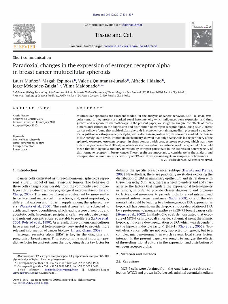
ig. 1. Expression of ERA and HIF-alpha in MCF-7 breast cancer cells grown as multicellulicroscopy. (B) Expression of ER alpha in MCF-7 grows in monolayer or spheroids by We
ell 42 (2010) 334–337 335
542). A GAPDH antibody (Santa Cruz Biotechnology 6C5, catalogsc-32233) was used as a loading control. Specific antibody bind-ing was visualized using Enhanced Chemiluminiscence (ECLII kit,Millipore, MA) with a peroxidase-labeled secondary antibody, asdescribed by the manufacturer.
2.5. Immunohistochemistry
Monolayer and multicellular spheroid cultures were fixed withbuffered 10% (V/V) formaldehyde in PBS. Spheroids were dehy-drated and included in paraffin. Subsequently, sections wereobtained, dewaxed and rehydrated. To uncover epitopes, the slideswere immersed in a preheated citrate buffer (10 mM sodium citratein PBS 1×, pH 6) then placed for 10 min in a water bath at 95 ◦C.The samples were allowed to cool to room temperature. The slideswere incubated with a primary antibody against ER alpha (SantaCruz Biotechnology) in a 1:25 dilution in 1× phosphate-bufferedsaline, and the antibody detected using a secondary biotinylatedanti-mouse antibody followed by streptavidin–biotin–peroxidase,using the streptavidin–biotin–peroxidase kit CSA Catalyzed Sig-nal Amplification System (Dako). The signal was visualized withdiaminobenzidine/hydrogen peroxide, followed by a hematoxylincounterstain. Finally, the samples were dehydrated in alcohol andxylene and were then mounted.
3. Results and discussion
MCF-7 breast cancer cells grew readily into well-formedspheroids 2 weeks after initial seeding (Fig. 1A). Analysis of ERA
expression by Western blot assays showed a decrease of more than50% in cells grown as multicellular spheroids, relative to mono-layer cultures (Fig. 1B). It is interesting to note that Cooper et al.found that intermittent hypoxia in breast tumors was associatedwith reduced levels of ERA expression (Cooper et al., 2004). Sincear spheroids. (A) Monolayer cultures and multicellular spheroids visualized by lightstern blot analysis. GAPDH was used as a loading control.

336 L. Munoz et al. / Tissue and Cell 42 (2010) 334–337
F own aE
iatSsgs(seCeEferstch(ceiiotdtisfisttabdapAaAeotptac
presented a paradoxical regulation of estrogen receptor alpha,with a decrease in protein expression and a marked increase inmRNA steady-state levels. Immunohistochemistry showed thatonly sparse cells in the periphery of the spheroid expressedestrogen receptor, in sharp contrast with progresterone receptor,
Fig. 3. Immunohistochemistry of ER alpha, progesterone receptor (PR) and HIF in
ig. 2. mRNA expression of ER alpha and HIF-alpha in MCF-7 breast cancer cells grxpression of HIF-alpha by RT-PCR. GAPDH was used as a loading control.
t has been shown that the central layers of multicellular spheroidsre hypoxic (le Roux et al., 2008; Rofstad et al., 1996), the reduc-ion of ERA protein expression could be due to lack of oxygenupporting this fact, we found that HIF-alpha protein, a key tran-cription factor responsive to hypoxia, was over-expressed in cellrown as spheroids (Fig. 3). In addition, the expression of HIF inmall spheroids was low and limited to a few cells at the centerFig. 3F). Interestingly, the expression of ER alpha was higher inmall spheroids and found in most cells, particularly at the periph-ry (Fig. 3G and H). In accordance to previous reports (Salceda andaro, 1997), this increase was not due to modulation of mRNAxpression, as shown by RT-PCR assays (Fig. 2B). To further supportRA regulation, we analyzed its expression by RT-PCR assays. Weound a marked increase (5.1 times) in the steady-state mRNA lev-ls in cell grown as multicellular spheroids (Fig. 2A). This divergentesult contrasts with the absence of mRNA regulation after hypoxiahown by Cooper et al. (2004). The differences may be due to addi-ional micro-ambient factors provided by the three-dimensionalulture. This is supported by a previous report showing that, in vivo,ypoxia downregulates ERA mRNA expression in cerebral cortexWestberry et al., 2008) and that a reduced response to estrogen inells is found in breast cancer cells exposed to hypoxia (Kurebayashit al., 2001). Alternatively, ERA protein itself could be participatingn a negative feedback. This is supported by a recent finding show-ng that ERA promoter activity is negatively regulated by a complexf ERA and Sin3A (Ellison-Zelski et al., 2009). In this scenario,he negative feedback would be disrupted by hypoxia-mediatedown-regulation of ERA protein, which has been attributed to pro-eosomal degradation (Cooper et al., 2004). Finally, we performedmmunohistochemistry on monolayer cultures and multicellularpheroids. As expected, a clear down-regulation of ERA protein wasound in three-dimensional cultures (Fig. 3). In these cultures, onlysolated cells expressed this protein, mainly at the periphery of thepheroid, with a marked nuclear staining. It is interesting to notehat progesterone receptor (PR), a key ERA target, was expressedhorough the spheroid, unrelated to ERA expression. This could bereflection of the activation of ERA in our conditions, since it haseen shown that estrogen induces proliferation and simultaneouslyecreases ERA in a model of multicellular spheroids (Truchet etl., 2008). Alternatively, it has been shown that the expression ofrogesterone receptors can be independent of ERA. For example,rpino et al. has shown that 25% and 3% of breast cancer patientsre ERA+/PR− and ERA−/PR+, respectively (Arpino et al., 2005).dditional experiments using a panel of cell lines should help tolucidate the relationship between both receptors. Alternatively,ur results could be explained by the mentioned report showing
hat the activation of ERA by estrogen leads to a decrease in itsrotein levels (Truchet et al., 2008). Nevertheless, we found thathe ERA positive cells in three-dimensional cultures were locatedt the periphery. Since these cells are in contact with the estrogen-ontaining medium, and the staining is nuclear, it is unlikely thats multicellular spheroids or monolayer. (A) Expression of ER alpha by RT-PCR. (B)
an activation-dependent mechanism could the only reason for theprotein decrease. Our results support the notion that both hypoxiaand estrogen activity could be leading to activation and decrease inthe ERA protein levels.
In brief, multicellular spheroids of MCF-7 breast cancer cells
MCF-7 breast cancer cells. (A) Expression of ER alpha in cells grown as a monolayer.(B) Expression of PR in cells grown as a monolayer. (C) Expression of ER alpha incells grown as multicellular spheroid. (D) Expression of PR in cells grown as mul-ticellular spheroid. (E) Expression of HIF in cells grown as multicellular spheroid.(F) Expression of HIF-alpha in small spheroids. (G and H) Expression of ER alpha insmall spheroids. Bar scale = 100 �m.

and C
webecit
R
A
C
C
E
H
K
L
Westberry, J.M., Prewitt, A.K., Wilson, M.E., 2008. Epigenetic regulation of the estro-
L. Munoz et al. / Tissue
hich was more extensively expressed and HIF-alpha, which wasxpressed in the central core of the spheroid. This could mean thatoth hypoxia and ERA activation by estrogen participate in thexpression heterogeneity of this hormone receptor in breast can-er. These results are important to considerate in the analysis andnterpretation of immunohistochemistry of ERA and downstreamargets in samples of solid tumors.
eferences
rpino, G., Weiss, H., Lee, A.V., Schiff, R., De Placido, S., Osborne, C.K., Elledge,R.M., 2005. Estrogen receptor-positive, progesterone receptor-negative breastcancer: association with growth factor receptor expression and tamoxifen resis-tance. J. Natl. Cancer Inst. 97, 1254–1261.
ho, J., Kim, D., Lee, S., Lee, Y., 2005. Cobalt chloride-induced estrogen receptor alphadown-regulation involves hypoxia-inducible factor-1alpha in MCF-7 humanbreast cancer cells. Mol. Endocrinol. 19, 1191–1199.
ooper, C., Liu, G.Y., Niu, Y.L., Santos, S., Murphy, L.C., Watson, P.H., 2004. Intermittenthypoxia induces proteasome-dependent down-regulation of estrogen receptoralpha in human breast carcinoma. Clin. Cancer Res. 10, 8720–8727.
llison-Zelski, S.J., Solodin, N.M., Alarid, E.T., 2009. Repression of ESR1 throughactions of estrogen receptor-alpha and Sin3A at the proximal promoter. Mol.Cell. Biol..
urvitz, S.A., Pietras, R.J., 2008. Rational management of endocrine resistance inbreast cancer: a comprehensive review of estrogen receptor biology, treatment
options, and future directions. Cancer 113, 2385–2397.urebayashi, J., Otsuki, T., Moriya, T., Sonoo, H., 2001. Hypoxia reduces hormoneresponsiveness of human breast cancer cells. Jpn. J. Cancer Res. 92, 1093–1101.
aRue, K.E., Khalil, M., Freyer, J.P., 2004. Microenvironmental regulation of prolifer-ation in multicellular spheroids is mediated through differential expression ofcyclin-dependent kinase inhibitors. Cancer Res. 64, 1621–1631.
ell 42 (2010) 334–337 337
le Roux, L., Volgin, A., Maxwell, D., Ishihara, K., Gelovani, J., Schellingerhout, D.,2008. Optimizing imaging of three-dimensional multicellular tumor spheroidswith fluorescent reporter proteins using confocal microscopy. Mol. Imaging 7,214–221.
Lin, R.Z., Chang, H.Y., 2008. Recent advances in three-dimensional multicellularspheroid culture for biomedical research. Biotechnol. J. 3, 1172–1184.
Nadji, M., 2008. Quantitative immunohistochemistry of estrogen receptor in breastcancer: “much ado about nothing!”. Appl. Immunohistochem. Mol. Morphol. 16,105–107.
Rofstad, E.K., Eide, K., Skoyum, R., Hystad, M.E., Lyng, H., 1996. Apopto-sis, energy metabolism, and fraction of radiobiologically hypoxic cells: astudy of human melanoma multicellular spheroids. Int. J. Radiat. Biol. 70,241–249.
Salceda, S., Caro, J., 1997. Hypoxia-inducible factor 1alpha (HIF-1alpha) protein israpidly degraded by the ubiquitin–proteasome system under normoxic condi-tions. Its stabilization by hypoxia depends on redox-induced changes. J. Biol.Chem. 272, 22642–22647.
Stoner, M., Saville, B., Wormke, M., Dean, D., Burghardt, R., Safe, S., 2002. Hypoxiainduces proteasome-dependent degradation of estrogen receptor alpha in ZR-75breast cancer cells. Mol. Endocrinol. 16, 2231–2242.
Truchet, I., Jozan, S., Baron, S., Frongia, C., Balaguer, P., Richard-Foy, H., Valette, A.,2008. Estrogen and antiestrogen-dependent regulation of breast cancer cell pro-liferation in multicellular spheroids: influence of cell microenvironment. Int. J.Oncol. 32, 1033–1039.
Walenta, S., Doetsch, J., Mueller-Klieser, W., Kunz-Schughart, L.A., 2000. Metabolicimaging in multicellular spheroids of oncogene-transfected fibroblasts. J. His-tochem. Cytochem. 48, 509–522.
gen receptor alpha promoter in the cerebral cortex following ischemia in maleand female rats. Neuroscience 152, 982–989.
Yuhas, J.M., Tarleton, A.E., Molzen, K.B., 1978. Multicellular tumor spheroid for-mation by breast cancer cells isolated from different sites. Cancer Res. 38,2486–2491.