part b: methods for the determination of ......contraindication the in vitro methods are strongly...
TRANSCRIPT

PART B: METHODS FOR THE DETERMINATION OF TOXICITY AND OTHER HEALTHEFFECTSGENERAL INTRODUCTION: PART BA. EXPLANATORY NOTEFor the purposes of the General Introduction, the following numbering applies:B.15. Gene mutation-Saccharomyces cerevisiaeB.16. Mitotic recombination-Saccharomyces cerevisiaeB.17. In vitro mammalian cell gene mutation testB.18. DNA damage and repair-unscheduled DNA synthesis-mammalian cells in vitroB.19. Sister chromatid exchange assay in vitroB.20. Sex-linked recessive lethal test in Drosophila melanogasterB.21. In vitro mammalian cell transformation testsB.22. Rodent dominant lethal testB.23. Mammalian spermatogonial CHROMOSOME ABERRATION TESTB.24. Mouse spot testB.25. Mouse heritable translocationB.26. Sub-chronic oral toxicity test: 90-day repeated oral dose using rodent speciesB.27. Sub-chronic oral toxicity test: 90-day repeated oral dose using non-rodent speciesB.28. Sub-chronic dermal toxicity study: 90-day repeated dermal dose study using rodent speciesB.29. Sub-chronic inhalation toxicity study: 90-day repeated inhalation dose study using rodentspeciesB.30. Chronic toxicity testB.31. Teratogenicity test - rodent and non-rodentB.32. Carcinogenicity testB.33. Combined chronic toxicity/carcinogenicity testB.34. One-generation reproduction toxicity testB.35. Two-generation reproduction toxicity testB.36. ToxicokineticsB. GENERAL DEFINITIONS FOR TERMS USED IN THE TEST METHODS IN THISANNEX(i) Acute toxicity comprises the adverse effects occurring within a given time (usually 14 days),after administration of a single dose of a substance.(ii) Evident toxicity is a general term describing clear signs of toxicity following administrationof test substance. These should be sufficient for hazard assessment and should be such that anincrease in the dose administered can be expected to result in the development of severe toxicsigns and probable mortality.(iii) Dose is the amount of test substance administered. Dose is expressed as weight (grams ormilligrams) or as weight of test substance per unit weight of test animal (e.g. milligrams perkilogram body weight), or as constant dietary concentrations (parts per million or milligrams perkilogram of food).(iv) Discriminating dose is the highest out of the four fixed dose levels which can beadministered without causing compound-related mortality (including humane kills).(v) Dosage is a general term comprising of dose, its frequency and the duration of the dosing.(vi) LD50 (median lethal dose) is a statistically derived single dose of a substance that can beexpected to cause death in 50 % of dosed animals. The LD50 value is expressed in terms ofweight of test substance per unit weight of test animal (milligrams per kilogram).(vii) LC50 (median lethal concentration) is a statistically derived concentration of a substancethat can be expected to cause death during exposure or within a fixed time after exposure in 50 %of animals exposed for a specified time.The LC50 value is expressed as weight of test substance per standard volume of air (milligramsper litre).

(viii) NOAEL is the abbreviation for no observed adverse effect level and is the highest dose orexposure level where no adverse treatment-related findings are observed.(ix) Repeated dose/Sub-chronic toxicity comprises the adverse effects occurring in experimentalanimals as a result of repeated daily dosing with, or exposure to, a chemical for a short part oftheir expected life-span.(x) Maximum Tolerated Dose (MTD) is the highest dose level eliciting signs of toxicity inanimals without having major effects on survival relative to the test in which it is used.(xi) Skin irritation is the production of inflammatory changes in the skin following theapplication of a test substance.(xii) Eye irritation is the production of changes in the eye following the application of a testsubstance to the anterior surface of the eye.(xiii) Skin sensitisation (allergic contact dermatitis) is an immunologically mediated cutaneousreaction to a substance.(xiv) Dermal corrosion is the production of irreversible tissue damage in the skin following theapplication of a test substance for the duration period of 3 minutes up to 4 hours.(xv) Toxicokinetics is the study of the absorption, distribution, metabolism and excretion of testsubstances.(xvi) Absorption is the process(es) by which an administered substance enters the body.(xvii) Excretion is the process(es) by which the administered substance and/or its metabolites areremoved from the body.(xviii) Distribution is the process(es) by which the absorbed substance and/or its metabolitespartition within the body.(xvv) Metabolism is the process(es) by which the administered substances are structurallychanged in the body by either enzymatic or non enzymatic reactions.B.I Acute - repeated dose / subchronic and chronic toxicityThe acute toxic effects and organ or system toxicity of a substance may be evaluated using avariety of toxicity tests (Methods B.1-B.5) from which, following a single dose, a preliminaryindication of toxicity may be obtained.Dependent on the toxicity of the substance, a limit test approach to a full LD50 may beconsidered, although no limit test is specified in inhalation studies, because it has not beenpossible to define a single inhalation exposure limit value.Consideration should be given to methods which use as few animals as possible and minimiseanimal suffering, for example the fixed dose method (Method B.1 bis) and acute toxic class(Method B.1 tris). In level 1 testing, a study in a second species may complement theconclusions drawn from the first study. In this case, a standard test method may be used or themethod may be adapted for a smaller number of animals.The repeated dose toxicity test (Methods B.7, B.8 and B.9) includes assessment of toxic effectsarising from repeated exposure. The need for careful clinical observations of the animals isstressed, so as to obtain as much information as possible. These tests should help to identify thetarget organs of toxicity and the toxic and non-toxic doses. Further in-depth investigation ofthese aspects may be required in long term studies (Methods B.26 - B.30 and B.33).B.II Mutagenicity - GenotoxicityMutagenicity refers to the induction of permanent transmissible changes in the amount orstructure of the genetic material of cells or organisms. These changes, "mutations", may involvea single gene or gene segments, a block of genes, or whole chromosomes. Effects on wholechromosomes may be structural and/or numerical.The mutagenic activity of a substance is assessed by in vitro assays for gene (point) mutations inbacteria (Method B.13/14) and/or for structural chromosome aberrations in mammalian cells,(Method B.10).Acceptable are also in vivo procedures, e.g. the micronucleus test (Method B.12) or themetaphase analysis of bone marrow cells, (Method B.11). However, in the absence of any

contraindication the in vitro methods are strongly preferred.Additional studies to investigate mutagenicity further or to pre-screen for carcinogenicity may berequired for higher production volumes and/or to conduct or follow-up a risk assessment, andthese can be used for a number of purposes: to confirm results obtained in the base set; toinvestigate end-points not studied in the base set; to initiate or extend in vivo studies.For these purposes, methods B.15 to B.25 include both in vivo and in vitro eukaryotic systemsand an extended range of biological end-points. These tests provide information on pointmutations and other end-points in organisms more complex than the bacteria used for the baseset.As a general principle, when a programme of further mutagenicity studies is considered, itshould be designed so as to provide relevant additional information on the mutagenic and/orcarcinogenic potential of that substance.The actual studies which may be appropriate in a specific instance will depend on numerousfactors, including the chemical and physical characteristics of the substance, the results of theinitial bacterial and cytogenetic assays, the metabolic profile of the substance, the results of othertoxicity studies, and the known uses of the substance. A rigid schedule for selection of tests istherefore inappropriate in view of the variety of factors which may require consideration.Some general principles for the testing strategy are laid down by Dir. 93/67/EEC, but cleartesting strategies may be found in the technical guidance document for Risk Assessment, whichnevertheless is flexible and can be adapted as appropriate to specific circumstances.Methods for further investigation are however grouped below, on the basis of their principalgenetic end-point:Studies to investigate gene (point) mutations(a) Forward or reverse mutation studies using eukaryotic micro-organisms (Saccharomycescerevisiae) (Method B.15)(b) In vitro studies to investigate forward mutation in mammalian cells, (Method B.17)(c) The sex-linked recessive lethal assay in Drosophila melanogaster, (Method B.20)(d) In vivo somatic cell mutation assay, the mouse spot test, (Method B.24)Studies to investigate chromosome aberrations(a) In vivo cityogenetic studies in mammals; In vivo metaphase analysis of bone marrow cellsshould be considered if it has not been included in the initial assessment (Method B.11). Inaddition, in vivo germ cell cytogenetics may be investigated, (Method B.23)(b) In vitro citogenetic studies in mammalian cells, if this has not been included in the initialassessment, (Method B.10)(c) Dominant lethal studies in rodents, (Method B.22)(d) Mouse heritable translocation test, (Method B.25)Genotoxic effects - effects on DNAGenotoxicity, identified as potentially harmful effects on genetic material not necessarilyassociated with mutagenicity, may be indicated by induced damage to DNA without directevidence of mutation. The following methods using eukaryotic micro-organisms or mammaliancells may be appropriate for such investigation:(a) Mitotic recombination in saccharomyces cerevisiae, (Methods B.16)(b) DNA damage and repair - unscheduled DNA synthesis - mammalian cells - in vitro, (MethodB.18)(c) Sister chromatid exchange in mammalian cells - in vitro, (Method B.19)Alternative methods for investigating carcinogenic potentialMammalian cell-transformation assays are available which measure the ability of a substance toinduce morphological and behavioural changes in cell cultures, which are thought to beassociated with malignant transformation - in vivo, (Method B.21). A number of different celltypes and criteria for transformation may be used.Risk assessment for heritable effects in mammals

There are methods available to measure heritable effects in whole mammals produced by gene(point) mutations, e. g. the mouse specific locus test, to measure germ-cell mutation in the firstgeneration, (not included in this Annex), or for chromosome aberrations, e. g. the mouseheritable translocation test, (Method B.25). Such methods may be used when estimating thepossible genetic risk of a substance to man. However, in view of the complexities involved inthese tests and the very large number of animals necessary, particularly for the specific locustest, a strong justification is needed before undertaking these studies.B.III CarcinogenicityChemicals may be described as genotoxic or non-genotoxic carcinogens, dependent on thepresumed mechanism of action.Pre-screening information for genotoxic carcinogenic potential of a substance may be obtainedfrom the mutagenicity/genotoxicity studies. Additional information may be obtained from therepeated dose, subchronic or chronic toxicity tests. The repeated dose toxicity test, Method B.7and longer repeated dose studies include assessment on histopathological changes observed inrepeated dose toxicity tests, e. g. hyperplasia in certain tissues which could be of concern. Thesestudies and toxicokinetic information may help to identify chemicals with carcinogenic potential,which may require further in-depth investigation of this aspect, in a carcinogenicity test (MethodB.32) or often in a combined chronic toxicity/carcinogenicity study (Method B.33)B.IVReproductive ToxicityReproductive toxicity may be detected in different ways e. g. impairment of male and femalereproductive functions or capacity, identified as "effects on fertility", or induction of non-inheritable harmful effects on the progreny, identified as "development toxicity" whereteratogenicity and effects during lactation are also included.For teratogenicity studies, as part of the developmental toxicity testing, the test method (MethodB.31), is primarily directed to administration by the oral route. Alternatively, other routes may beused depending on the physical properties of the test substance or likely route of humanexposure. In such cases, the test method should be suitably adapted taking into consideration theappropriate elements of the 28-day test methods.Where a three-generation reproduction (fertility) test is required, the described method for thetwo-generation reproduction test (Method B.35), can be extended to cover a third generation.B.V NeurotoxicityNeurotoxicity may be detected in different ways e. g. functional changes and/or structural andbiochemical changes in the central or peripheral nervous system. A preliminary indication ofneurotoxicity may be obtained from acute toxicity tests. The repeated dose toxicity test, MethodB.7, includes assessment of neurotoxicological effects, and the need for careful clinicalobservations of the animals is stressed, so as to obtain as much information as possible. Themethod should help to identify chemicals with neurotoxic potential, which may require furtherin-depth investigation of this aspect. Additionally, it is important to consider the potential ofsubstances to cause specific neurotoxic effects that may not be detected in other toxicity studies.For example, certain organophosphorous substances have been observed to cause delayedneurotoxicity and can be evaluated in methods B.37 and B.38, following single or repeated-doseexposure.B.VI ImmunotoxicityImmunotoxicity may be detected in different ways e. g. immunosupression and/or enhancementof the responsiveness of the immune system resulting in either hypersensitivity or inducedautoimmunity. The repeated dose toxicity test, Method B.7, includes assessment of immunotoxiceffects. The method should help to identify chemicals with immunotoxic potential, which mayrequire further in-depth investigation of this aspect.B.VII ToxicokineticsToxicokinetic studies help in the interpretation and evaluation of toxicity data. These studies areintended to elucidate particular aspects of the toxicity of the chemical under test and the results

may assist in the design of further toxicity studies. It is not envisaged that in every case allparameters will need to be determined. Only in rare cases will the whole sequence oftoxicokinetic studies (absorption, excretion, distribution and metabolism) be necessary. Forcertain compounds, changes in this sequence may be advisable or a single-dose study may besufficient (Method B.36).Information on chemical structure (SAR) and physico-chemical properties may also provide anindication of the absorption characteristics by the intended route of administration and themetabolic and tissue distribution possibilities. There may also be information on toxicokineticparameters from preceding toxicity and toxicokinetic studies.C. CHARACTERISATION OF THE TEST SUBSTANCEThe composition of the test substance, including major impurities, and its relevant physico-chemical properties including stability, should be known prior to the initiation of any toxicitystudy.The physico-chemical properties of the test substance provide important information for theselection of the route of administration, the design of each particular study and the handling andstorage of the test substance.The development of an analytical method for qualitative and quantitative determination of thetest substance (including major impurities when possible) in the dosing medium and thebiological material should precede the initiation of the study.All information relating to the identification, the physico-chemical properties, the purity, andbehaviour of the test substance should be included in the test report.D. ANIMAL CAREStringent control of environmental conditions and proper animal care techniques are essential intoxicity testing.(i) Housing conditionsThe environmental conditions in the experimental animal rooms or enclosures should beappropriate to the test species. For rats, mice and guinea pigs, suitable conditions are a roomtemperature of 22° C ± 3° C with a relative humidity of 30 to 70 %; for rabbits the temperatureshould be 20 ± 3° C with a relative humidity of 30 to 70 %.Some experimental techniques are particularly sensitive to temperature effects and, in thesecases, details of appropriate conditions are included in the description of the test method. In allinvestigations of toxic effects, the temperature and humidity should be monitored, recorded, andincluded in the final report of the study.Lighting should be artificial, the sequence being 12 hours light, 12 hours dark. Details of thelighting pattern should be recorded and included in the final report of the study.Unless otherwise specified in the method, animals may be housed individually, or be caged insmall groups of the same sex; for group caging, no more than five animals should be housed percage.In reports of animal experiments, it is important to indicate the type of caging used and thenumber of animals housed in each cage both during exposure to the chemical and any subsequentobservation period.(ii) Feeding conditionsDiets should meet all the nutritional requirements of the species under test. Where testsubstances are administered to animals in their diet the nutritional value may be reduced byinteraction between the substance and a dietary constituent. The possibility of such a reactionshould be considered when interpreting the results of tests. Conventional laboratory diets may beused with an unlimited supply of drinking water. The choice of the diet may be influenced by theneed to ensure a suitable admixture of a test substance when administered by this method.Dietary contaminants which are known to influence the toxicity should not be present ininterfering concentrations.E. ANIMAL WELFARE

When elaborating the test methods due consideration was given to animal welfare. Someexamples are briefly given below, but this list is not exhaustive. The exact wording and/orconditions should be read in the text of the methods:- For the determination of acute oral toxicity, two alternative methods, the "Fixed DoseProcedure" and the "Acute Toxic Class method" should be considered. The "Fixed DoseProcedure" does not utilise death as specific endpoint and it uses fewer animals. The "AcuteToxic Class method" uses on average 70 % less animals than Method B.1 for Acute Oraltoxicity. Both these alternative methods result in less pain and distress than the classicalmethodology.- The number of animals used is reduced to the scientifically acceptable minimum: only 5animals of the same sex are tested per dose level for methods B.1 and B.3; only 10 animals (andonly 5 for the negative control group) are used for the determination of the skin sensitisation bythe guinea pig maximisation test (method B.6); the number of animals needed for the positivecontrol when testing mutagenicity in vivo is also lowered (methods B.11 and B.12)- Pain and distress of animals during the tests are minimised: animals showing severe andenduring signs of distress and pain may need to be humanely killed; dosing test substances in away known to cause marked pain and distress due to corrosive or irritating properties need not tobe carried out (methods B.1, B.2 and B.3).- Testing with irrelevantly high doses is avoided by the introduction of limit tests, not only in theacute toxicity tests (methods B.1, B.2 and B.3) but also in the in vivo tests for mutagenicity(methods B.11 and B.12).- A strategy of testing for irritancy now allows the non-performance of a test, or its reduction to asingle animal study, when sufficient scientific evidence can be provided.Such scientific evidence can be based on the physico-chemical properties of the substance, theresults of other tests already performed, or the results of well validated in vitro tests. Forexample, if an acute toxicity study by the dermal route has been conducted at the limit test dosewith the substance (method B.3), and no skin irritation was observed, further testing for skinirritation (method B.4) may be unnecessary; materials which have demonstrated definitecorrosion or severe skin irritancy in a dermal irritation study (method B.4) should not be furthertested for eye irritancy (method B.5).F. ALTERNATIVE TESTINGA scientific objective for the European Union is the development and validation of alternativetechniques which can provide the same level of information as current animal tests, but whichuse fewer animals, cause less suffering or avoid the use of animals completely.Such methods, as they become available, must be considered wherever possible for hazardcharacterisation and consequent classification and labelling for intrinsic hazards.G. EVALUATION AND INTERPRETATIONWhen tests are evaluated and interpreted, limitations in the extent to which the results of animaland in vitro studies can be extrapolated directly to man must be considered and therefore,evidence of adverse effects in humans, where available, may be used for confirmation of testingresults.These results, can be used for the classification and labelling of the new and the existingchemicals for human health effects, on the basis of their intrinsic properties, identified andquantified by these methods. Corresponding Annex VI criteria for classification and labellingrelate also to the end-points of the testing protocols included in these testing methods.These results can also be used for risk assessment studies, for new and existing chemicals, andappropriate testing strategies for these purposes are indicated in the corresponding guidancedocuments.H. LITERATURE REFERENCESMost of these methods are developed within the framework of the OECD programme for TestingGuidelines, and should be performed in conformity with the principles of Good Laboratory

Practice, in order to ensure as wide as possible "mutual acceptance of data".Additional information may be found in the references found in the OECD guidelines and therelevant literature published elsewhere.


B.2. ACUTE TOXICITY (INHALATION)
1. METHOD1.1. INTRODUCTIONIt is useful to have preliminary information on the particle size distribution, the vapour pressure,the melting point, the boiling point, the flash point and explosivity (if applicable) of thesubstance.See also General Introduction Part B (A).
1.2. DEFINITIONSSee General Introduction Part B (B).
1.3. REFERENCE SUBSTANCESNone.
1.4. PRINCIPLE OF THE TEST METHODSeveral groups of experimental animals are exposed for a defined period to the test substance ingraduated concentrations, one concentration being used per group. Subsequently observations ofeffects and deaths are made. Animals which die during the test are necropsied and at theconclusion of the test surviving animals are necropsied.Animals showing severe and enduring signs of distress and pain may need to be humanely killed.Dosing test substances in a way known to cause marked pain and distress due to corrosive orsevere irritating properties need not be carried out.
1.5. QUALITY CRITERIANone.
1.6. DESCRIPTION OF THE TEST METHOD1.6.1. PreparationsThe animals are kept under the experimental housing and feeding conditions for at least five daysprior to the experiment. Before the test healthy young animals are randomized and assigned tothe required number of groups. They need not be subjected to simulated exposure unless this isindicated by the type of exposure apparatus being used.Solid test substances may need to be micronised in order to achieve particles of an appropriatesize.Where necessary a suitable vehicle may be added to the test substance to help generate anappropriate concentration of the test substance in the atmosphere and a vehicle control groupshould then be used. If a vehicle or other additives are used to facilitate dosing, they should beknown not to produce toxic effects. Historical data can be used if appropriate.
1.6.2. Test Conditions1.6.2.1. Experimental AnimalsUnless there are contra-indications the rat is the preferred species. Commonly used laboratorystrains should be employed. For each sex, at the start of the test the range of weight variation inthe animals used should not exceed ± 20 % of the appropriate mean value.
1.6.2.2. Number and SexAt least 10 rodents (five female and five male) are used at each concentration level. The femalesshould be nulliparous and non-pregnant.Note: In acute toxicity tests with animals of a higher order than rodents, the use of smallernumbers should be considered. Doses should be carefully selected, and every effort should be

made not to exceed moderately toxic doses. In such tests administration of lethal doses of the testsubstance should be avoided.
1.6.2.3. Exposure ConcentrationsThese should be sufficient in number, at least three, and spaced appropriately to produce testgroups with a range of toxic effects and mortality rates. The data should be sufficient to producea concentration mortality curve and, where possible, permit an acceptable determination of anLC50.
1.6.2.4. Limit TestIf an exposure of five male and five female test animals to 20 mg per litre of a gas or 5 mg per
litre of an aerosol or a particulate for four hours (or, where this is not possible due to the physicalor chemical, including explosive, properties of the test substance, the maximum attainableconcentration) produces no compound related mortality within 14 days further testing may not beconsidered necessary.
1.6.2.5. Exposure TimeThe period of exposure should be four hours.
1.6.2.6. EquipmentThe animals should be tested with inhalation equipment designed to sustain a dynamic airflow ofat least 12 air changes per hour, to ensure an adequate oxygen content and an evenly distributedexposure atmosphere. Where a chamber is used its design should minimize crowding of the testanimals and maximize their exposure by inhalation to the test substance. As a general rule toensure stability of a chamber atmosphere the total 'volume` of the test animals should not exceed5 % of the volume of the test chamber. Oro-nasal, head only, or whole body individual chamberexposure may be used; the first two will help to minimize the uptake of the test substance byother routes.
1.6.2.7. Observation PeriodThe observation period should be at least 14 days. However, the duration of observations shouldnot be rigidly fixed. It should be determined by the toxic reactions, their rate of onset and thelength of the recovery period; it may thus be extended when considered necessary. The time atwhich signs of toxicity appear and disappear and the time of death are important, especially ifthere is a tendency for deaths to be delayed.
1.6.3. ProcedureShortly before exposure, the animals are weighed, and then exposed to the test concentration inthe designated apparatus for a period of four hours, after equilibration of the chamberconcentration. Time for equilibration should be short. The temperature at which the test isperformed should be maintained at 22 ± 3 C. Ideally the relative humidity should be maintainedbetween 30 % and 70 %, but in certain instances (e.g. tests of some aerosols) this may not bepracticable. Maintenance of a slight negative pressure inside the chamber ( & ge; 5 mm of water)will prevent leakage of the test substance into the surrounding area. Food and water should bewithheld during exposure. Suitable systems for the generation and monitoring of the testatmosphere should be used. The system should ensure that stable exposure conditions areachieved as rapidly as possible. The chamber should be designed and operated in such a way thata homogeneous distribution of the test atmosphere within the chamber is maintained.Measurements or monitoring should be made:(a) of the rate of air flow (continuously).(b) of the actual concentration of the test substance measured in the breathing zone at least three

times during exposure (some atmospheres, e.g. aerosols at high concentrations, may need morefrequent monitoring). During the exposure period the concentration should not vary by more than± 15 % of the mean value. However in the case of some aerosols, this level of control may not beachievable and a wider range would then be acceptable. For aerosols, particle size analysisshould be performed as often as necessary (at least once per test group).(c) of temperature and humidity, continuously if possible.During and following exposure, observations are made and recorded systematically; individualrecords should be maintained for each animal. Observations should be made frequently duringthe first day. A careful clinical examination should be made at least once each working day,other observations should be made daily with appropriate actions taken to minimize loss ofanimals from the study, e.g. necropsy or refrigeration of those animals found dead and isolationor sacrifice of weak or moribund animals.Observations should include changes in the skin and fur, eyes, mucous membranes, respiratory,circulatory, autonomic and central nervous systems, and somatomotor activity and behaviourpattern. Particular attention should be directed to observation of respiratory behaviour, tremors,convulsions, salivation, diarrhoea, lethargy, sleep and coma. The time of death should berecorded as precisely as possible. Individual weights of animals should be determined weeklyafter exposure, and at death.Animals that die during the test and those surviving at the termination of the test are subjected tonecropsy with particular reference to any changes in the upper and lower respiratory tract. Allgross pathological changes should be recorded. Where indicated, tissues should be taken forhistopathological examination.
2. DATAData should be summarized in tabular form showing for each test group the number of animals atthe start of the test, time of death of individual animals, number of animals displaying other signsof toxicity, description of toxic effects and necropsy findings. Changes in weight must becalculated and recorded when survival exceeds one day. Animals which are humanely killed dueto compound-related distress and pain are recorded as compound-related deaths. The LC50should be determined by a recognized method. Data evaluation should include the relationship, ifany, between the animal's exposure to the test substance and the incidence and severity of allabnormalities, including behavioural and clinical abnormalities, gross lesions, body weightchanges, mortality and any other toxic effects.
3. REPORTING
3.1. TEST REPORTThe test report shall, if possible, include the following information:- species, strain, source, environmental conditions, diet etc.;- test conditions: description of exposure apparatus including design, type, dimensions, source ofair, system for generating aerosols, method of conditioning air and the method of housinganimals in a test chamber when this is used. The equipment for measuring temperature,humidity, and aerosol concentrations and particle size distribution should be described.Exposure dataThese should be tabulated and presented with mean values and a measure of variability (e.g.standard deviation) and shall, if possible, include:(a) airflow rates through the inhalation equipment;(b) temperature and humidity of the air;(c) nominal concentrations (total amount of test substance fed into the inhalation equipmentdivided by volume of air);(d) nature of vehicle, if used;

(e) actual concentrations in test breathing zone;(f) The mass median aerodynamic diameter (MMAD) and the geometric standard deviation(GSD);(g) equilibration period;(h) exposure period;- tabulation of response data by sex and exposure level (i.e. number of animals that died or werekilled during the test; number of animals showing signs of toxicity; number of animals exposed);- time of death during or following exposure, reasons and criteria used for humane killing ofanimals;- all observations;- LC50 value for each sex determined at the end of the observation period (with method ofcalculation specified);- 95 % confidence interval for the LC50 (where this can be provided);- dose/mortality curve and slope (where permitted by the method of determination);- necropsy findings;- any histopathological findings;- discussions of the results (particular attention should be given to the effect that humane killingof animals during the test may have on the calculated LC50 value);- interpretation of the results.
3.2. EVALUATION AND INTERPRETATIONSee General Introduction Part B (D).
4. REFERENCESSee General Introduction Part B (E).

B.3. ACUTE TOXICITY (DERMAL)
1. METHOD1.1. INTRODUCTIONSee General Introduction Part B (A).
1.2. DEFINITIONSee General Introduction Part B (B).
1.3. REFERENCE SUBSTANCESNone.
1.4. PRINCIPLE OF THE TEST METHODThe test substance is applied to the skin in graduated doses to several groups of experimentalanimals, one dose being used per group. Subsequently, observations of effects and deaths aremade. Animals which die during the test are necropsied and at the conclusion of the testsurviving animals are necropsied.Animals showing severe and enduring signs of distress and pain may need to be humanely killed.Dosing test substances in a way known to cause marked pain and distress due to corrosive orirritating properties need not be carried out.
1.5. QUALITY CRITERIANone.
1.6. DESCRIPTION OF THE TEST METHOD
1.6.1. PreparationsThe animals are kept in their experimental cages under the experimental housing and feedingconditions for at least five days prior to the experiment. Before the test, healthy young adultanimals are randomized and assigned to the treatment groups. Approximately 24 hours before thetest, fur should be removed by clipping or shaving from the dorsal area of the trunk of theanimals. When clipping or shaving the fur, care must be taken to avoid abrading the skin whichcould alter its permeability. Not less than 10 % of the body surface should be clear for theapplication of the test substance. When testing solids, which may be pulverized if appropriate,the test substance should be moistened sufficiently with water or, where necessary, a suitablevehicle to ensure good contact with the skin. When a vehicle is used, the influence of the vehicleon penetration of skin by the test substance should be taken into account. Liquid test substancesare generally used undiluted.
1.6.2. Test Conditions1.6.2.1. Experimental AnimalsThe adult rat or rabbit may be used. Other species may be used but their use would requirejustification. Commonly used laboratory strains should be employed. For each sex, at the start ofthe test the range of weight variation in the animals used should not exceed ± 20 % of theappropriate mean value.
1.6.2.2. Number and SexAt least 5 animals are used at each dose level. They should all be of the same sex. If females areused, they should be nulliparous and non-pregnant. Where information is availabledemonstrating that a sex is markedly more sensitive, animals of this sex should be dosed.Note: In acute toxicity tests with animals of a higher order than rodents, the use of smaller

numbers should be considered. Doses should be carefully selected, and every effort should bemade not to exceed moderately toxic doses. In such tests, administration of lethal doses of thetest substance should be avoided.
1.6.2.3. Dose LevelsThese should be sufficient in number, at least three, and spaced appropriately to produce testgroups with a range of toxic effects and mortality rates. Any irritant or corrosive effects shouldbe taken into account when deciding on dose levels. The data should be sufficient to produce adose/response curve and, where possible, permit an acceptable determination of the LD50.
1.6.2.4. Limit TestA limit test at one dose level of at least 2 000 mg/kg bodyweight may be carried out in a group of5 male and 5 female animals, using the procedures described above. If compound-relatedmortality is produced, a full study may need to be considered.
1.6.2.5. Observation PeriodThe observation period should be at least 14 days. However, the duration of observation shouldnot be rigidly fixed. It should be determined by the toxic reactions, their rate of onset and thelength of the recovery period; it may thus be extended when considered necessary. The time atwhich signs of toxicity appear and disappear, their duration and the time of death are important,especially if there is a tendency for deaths to be delayed.
1.6.3. ProcedureAnimals should be caged individually. The test substance should be applied uniformly over anarea which is approximately 10 % of the total body surface area. With highly toxic substancesthe surface area covered may be less but as much of the area should be covered with a layer asthin and uniform as possible.Test substances should be held in contact with the skin with a porous gauze dressing and non-irritating tape throughout a 24-hour exposure period. The test site should be further covered in asuitable manner to retain the gauze dressing and test substance and ensure that the animalscannot ingest the test substance. Restrainers may be used to prevent the ingestion of the testsubstance but complete immobilisation is not a recommended method.At the end of the exposure period, residual test substance should be removed, where practicable,using water or some other appropriate method of cleansing the skin.Observations should be recorded systematically as they are made. Individual records should bemaintained for each animal. Observations should be made frequently during the first day. Acareful clinical examination should be made at least once each working day, other observationsshould be made daily with appropriate actions taken to minimize loss of animals to the study,e.g. necropsy or refrigeration of those animals found dead and isolation or sacrifice of weak ormoribund animals.Observations should include changes in fur, treated skin, eyes and mucous membranes, and alsorespiratory, circulatory, autonomic and central nervous systems, and somatomotor activity andbehaviour pattern. Particular attention should be directed to observations of tremors, convulsions,salivation, diarrhoea, lethargy, sleep and coma. The time of death must be recorded as preciselyas possible. Animals that die during the test and those surviving at the termination of the test aresubjected to necropsy. All gross pathological changes should be recorded. Where indicated,tissues should be taken for histopathological examination.Assessment of toxicity in the other sexAfter completion of the study in one sex, at least one group of 5 animals of the other sex is dosedto establish that animals of this sex are not markedly more sensitive to the test substance. The useof fewer animals may be justified in individual circumstances. Where adequate information is
available to demonstrate that animals of the sex tested are markedly more sensitive, testing inanimals of the other sex may be dispensed with.
2. DATAData should be summarized in tabular form, showing for each test group the number of animalsat the start of the test, time of death of individual animals, number of animals displaying othersigns of toxicity, description of toxic effects and necropsy findings. Individual weights ofanimals should be determined and recorded shortly before the test substance is applied, weeklythereafter, and at death; changes in weight should be calculated and recorded when survivalexceeds one day. Animalswhich are humanely killed due to compound-related distress and pain are recorded as compound-related deaths. The LD50 should be determined by a recognized method.Data evaluation should include an evaluation of relationships, if any, between the animal'sexposure to the test substance and the incidence and severity of all abnormalities, includingbehavioural and clinical abnormalities, gross lesions, body weight changes, mortality, and anyother toxicological effects.
3. REPORTING3.1. TEST REPORTThe test report shall, if possible, include the following information:- species, strain, source, environmental conditions, diet, etc.;- test conditions (including method of skin cleansing and type of dressing: occlusive or notocclusive);- dose levels (with vehicle, if used, and concentrations),- sex of animals dosed;- tabulation of response data by sex and dose level (i.e. number of animals that died or werekilled during the test; number of animals showing signs of toxicity; number of animals exposed);- time of death after dosing, reasons and criteria used for humane killing of animals;- all observations;- LD50 value for the sex subjected to a full study, determined at 14 days with the method ofdetermination specified;- 95 % confidence interval for the LD50 (where this can be provided);- dose/mortality curve and slope where permitted by the method of determination;- necropsy findings;- any histopathological findings;- results of any test on the other sex;- discussion of results (particular attention should be given to the effect that humane killing ofanimals during the test may have on the calculated LD50 value);- interpretation of the results.
3.2. EVALUATION AND INTERPRETATIONSee General Introduction Part B (D).
4. REFERENCESSee General Introduction Part B (E).


B.7 REPEATED DOSE (28 DAYS) TOXICITY (ORAL)
1. METHOD1.1. IntroductionSee General Introduction Part B.1.2. DefinitionsSee General Introduction Part B.1.3. Principle of the test methodThe test substance is orally administered daily in graduated doses to several groups ofexperimental animals, one dose level per group for a period of 28 days. During the period ofadministration the animals are observed closely, each day for signs of toxicity. Animals whichdie or are killed during the test are necropsied and at the conclusion of the test surviving animalsare killed and necropsied.This method places more emphasis on neurological effects as a specific endpoint, and the needfor careful clinical observations of the animals, so as to obtain as much information as possible,is stressed. The method should identify chemicals with neurotoxic potential, which may warrantfurther in-depth investigation of this aspect. In addition, the method may give an indication ofimmunological effects and reproductive organ toxicity.1.4. Description of the test method1.4.1. PreparationsHealthy young adult animals are randomly assigned to the control and treatment groups. Cagesshould be arranged in such a way that possible effects due to cage placement are minimized. Theanimals are identified uniquely and kept in their cages for at least five days prior to the start ofthe study to allow for acclimatisation to the laboratory conditions.The test substance is administered by gavage or via the diet or drinking water. The method oforal administration is dependent on the purpose of the study, and the physical/chemicalproperties of the substance.Where necessary, the test substance is dissolved or suspended in a suitable vehicle. It isrecommended that, wherever possible, the use of an aqueous solution/suspension be consideredfirst, followed by consideration of a solution/emulsion in oil (e.g. corn oil) and then by possiblesolution in other vehicles. For vehicles other than water the toxic characteristics of the vehiclemust be known. The stability of the test substance in the vehicle should be determined.1.4.2. Test conditions1.4.2.1. Test animalsThe preferred rodent species is the rat, although other rodent species may be used. Commonlyused laboratory strains of young healthy adult animals should be employed. The females shouldbe nulliparous and non-pregnant. Dosing should begin as soon as possible after weaning and, inany case, before the animals are nine weeks old.At the commencement of the study the weight variation of animals used should be minimal andnot exceed ± 20 % of the mean weight of each sex.Where a repeated dose oral study is conducted as a preliminary to a long term study, preferablyanimals from the same strain and source should be used in both studies.1.4.2.2. Number and sexAt least 10 animals (five female and five male) should be used at each dose level. If interim killsare planned, the number should be increased by the number of animals scheduled to be killedbefore the completion of the study.In addition, a satellite group of 10 animals (five animals per sex) may be treated with the highdose level for 28 days and observed for reversibility, persistence, or delayed occurrence of toxiceffects for 14 days post-treatment. A satellite group of 10 control animals (five animals per sex)is also used.1.4.2.3. Dose levels

Generally, at least three test groups and a control group should be used. Except for treatmentwith the test substance, animals in the control group should be handled in an identical manner tothe test group subjects. If a vehicle is used in administering the test substance, the control groupshould receive the vehicle in the highest volume used.If from assessment of other data, no effects would be expected at a dose of 1 000 mg/kg bw/d, alimit test may be performed. If there are no suitable data available, a range finding study may beperformed to aid the determination of the doses to be used.Dose levels should be selected taking into account any existing toxicity and (toxico-) kinetic dataavailable for the test substance or related materials. The highest dose level should be chosen withthe aim of inducing toxic effects but not death or severe suffering. Thereafter, a descendingsequence of dose levels should be selected with a view to demonstrating any dosage relatedresponse and no-observed-adverse effects at the lowest dose level (NOAEL). Two to four foldintervals are frequently optimal for setting the descending dose levels and addition of a fourthtest group is often preferable to using very large intervals (e.g. more than a factor of 10) betweendosages.For substances administered via the diet or drinking water it is important to ensure that thequantities of the test substance involved do not interfere with normal nutrition or water balance.When the test substance is administered in the diet either a constant dietary concentration (ppm)or a constant dose level in terms of the animals' body weight may be used; the alternative usedmust be specified. For a substance administered by gavage, the dose should be given at similartimes each day, and adjusted as necessary to maintain a constant dose level in terms of animalbody weight.Where a repeated dose study is used as a preliminary to a long term study, a similar diet shouldbe used in both studies.1.4.2.4. Limit testIf a test at one dose level of at least 1 000 mg/kg body weight/day or, for dietary or drinkingwater administration, an equivalent percentage in the diet or drinking water (based upon bodyweight determinations), using the procedures described for this study, produces no observabletoxic effects and if toxicity would not be expected based upon data from structurally relatedsubstances, then a full study using three dose levels may not be considered necessary. The limittest applies except when human exposure indicates the need for a higher dose level to be used.1.4.2.5. Observation periodThe observation period should be 28 days. Animals in a satellite group scheduled for follow-upobservations should be kept for at least a further 14 days without treatment to detect delayedoccurrence, or persistence of, or recovery from toxic effects.1.4.3. ProcedureThe animals are dosed with the test substance daily seven days each week for a period of 28days; use of a five-day per week dosing regime needs to be justified. When the test substance isadministered by gavage, this should be done in a single dose to the animals using a stomach tubeor a suitable intubation cannula. The maximum volume of liquid that can be administered at onetime depends on the size of the test animal. The volume should not exceed 1 ml/100 g bodyweight, except in the case of aqueous solutions where 2 ml/100 g body weight may be used.Except for irritating or corrosive substances which will normally reveal exacerbated effects withhigher concentrations, variability in test volume should be minimised by adjusting theconcentration to ensure a constant volume at all dose levels.1.4.3.1. General observationsGeneral clinical observations should be made at least once a day, preferably at the same time(s)each day and considering the peak period of anticipated effects after dosing. The health conditionof the animals should be recorded. At least twice daily, all animals are observed for morbidityand mortality. Moribund animals and animals in severe distress or pain should be removed whennoticed, humanely killed and necropsied.

Once before the first exposure (to allow for within-subject comparisons), and at least once aweek thereafter, detailed clinical observations should be made in all animals. These observationsshould be made outside the home cage in a standard arena and preferably at the same time, eachtime. They should be carefully recorded, preferably using scoring systems, explicitly defined bythe testing laboratory. Effort should be made to ensure that variations in the test conditions areminimal and that observations are preferably conducted by observers unaware of the treatment.Signs noted should include but not be limited to, changes in skin, fur, eyes, mucous membranes,occurrence of secretions and excretions and autonomic activity (e.g. lacrimation, piloerection,pupil size, unusual respiratory pattern). Changes in gait, posture and response to handling as wellas the presence of clonic or tonic movements, stereotypes (e.g. excessive grooming, repetitivecircling) or bizarre behaviour (e.g. self-mutilation, walking backwards) should also be recorded.In the fourth exposure week sensory reactivity to stimuli of different types (e.g. auditory, visualand proprioceptive stimuli), assessment of grip strength and motor activity assessment should beconducted. Further details of the procedures that could be followed are given in the literature (seeGeneral Introduction Part B).Functional observations conducted in the fourth exposure week may be omitted when the studyis conducted as a preliminary study to a subsequent subchronic (90-day) study. In that case, thefunctional observations should be included in this follow-up study. On the other hand, theavailability of data on functional observations from the repeated dose study may enhance theability to select dose levels for a subsequent subchronic study.Exceptionally, functional observations may also be omitted for groups that otherwise revealsigns of toxicity to an extent that would significantly interfere with the functional testperformance.1.4.3.2. Body weight and food/water consumptionAll animals should be weighed at least once a week. Measurements of food and waterconsumption should be made at least weekly. If the test substance is administered via thedrinking water, water consumption should also be measured at least weekly.1.4.3.3. HaematologyThe following haematological examinations should be made at the end of the test period:haematocrit, haemoglobin concentration, erythrocyte count, total an differential leucocyte count,platelet count and a measure of blood clotting time/potential.Blood samples should be taken from a named site just prior to or as part of the procedure forkilling the animals, and stored under appropriate conditions.1.4.3.4. Clinical biochemistryClinical biochemistry determinations to investigate major toxic effects in tissues and,specifically, effects on kidney and liver, should be performed on blood samples obtained of allanimals just prior to or as part of the procedure for killing the animals (apart from those foundmoribund and/or intercurrently killed). Overnight fasting of the animals prior to blood samplingis recommended (1). Investigations of plasma or serum shall include sodium, potassium, glucose,total cholesterol, urea, creatinine, total protein and albumin, at least two enzymes indicative ofhepatocellular effects (such as alanine aminotransferase, aspartate aminotransferase, alkalinephosphatase, gamma glutamyl transpeptidase, and sorbitol dehydrogenase). Measurements ofadditional enzymes (of hepatic or other origin) and bile acids may provide useful informationunder certain circumstances.(1) For a number of measurements in serum and plasma, most notably for glucose, overnightfasting would be preferable. The major reason for this preference is that the increased variabilitywhich would inevitably result from non-fasting, would tend to mask more subtle effects andmake interpretation difficult. On the other hand, however, overnight fasting may interfere withthe general metabolism of the animals and, particularly in feeding studies, may disturb the dailyexposure to the test substance. If overnight fasting is adopted, clinical biochemicaldeterminations should be performed after the conduct of functional observations in week 4 of the

study.Optionally, the following urine analysis determinations could be performed during the last weekof the study using timed urine volume collection; appearance, volume, osmolality or specificgravity, pH, protein, glucose and blood/blood cells.In addition, studies to investigate serum markers of general tissue damage should be considered.Other determinations that should be carried out if the known properties of the test substance may,or are suspected to, affect related metabolic profiles include calcium, phosphate, fastingtriglycerides, specific hormones, methaemoglobin and cholinesterase. These need to be identifiedfor substances in certain classes or on a case-by-case basis.Overall, there is a need for a flexible approach, depending on the species and the observed and/orexpected effect with a given substance.If historical baseline data are inadequate, consideration should be given to determination ofhaematological and clinical biochemistry variables before dosing commences.1.4.3.5. Gross necropsyAll animals in the study shall be subjected to a full, detailed gross necropsy which includescareful examination of the external surface of the body, all orifices, and the cranial, thoracic andabdominal cavities and their contents. The liver, kidneys, adrenals, testes, epididymides, thymus,spleen, brain and heart of all animals should be trimmed of any adherent tissue, as appropriate,and their wet weight taken as soon as possible after dissection to avoid drying.The following tissues should be preserved in the most appropriate fixation medium for both thetype of tissue and the intended subsequent histopathological examination: all gross lesions, brain(representative regions including cerebrum, cerebellum and pons), spinal cord, stomach, smalland large intestines (including Peyer's patches), liver, kidneys, adrenals, spleen, heart, thymus,thyroid, trachea and lungs (preserved by inflation with fixative and then immersion), gonads,accessory sex organs (e.g. uterus, prostate), urinary bladder, lymph nodes (preferably one lymphnode covering the route of administration and another one distant from the route ofadministration to cover systemic effects), peripheral nerve (sciatic or tibial) preferably in closeproximity to the muscle, and a section of bone marrow (or, alternatively, a fresh mounted bonemarrow aspirate). The clinical and other findings may suggest the need to examine additionaltissues. Also any organs considered likely to be target organs based on the known properties ofthe test substance should be preserved.1.4.3.6. Histopathological examinationFull histopathology should be carried out on the preserved organs and tissues of all animals inthe control and high dose groups. These examinations should be extended to animals of all otherdosage groups, if treatment-related changes are observed in the high dose group.All gross lesions shall be examined.When a satellite group is used, histopathology should be performed on tissues and organsidentified as showing effects in the treated groups.2. DATAIndividual data should be provided. Additionally, all data should be summarised in tabular formshowing for each test group the number of animals at the start of the test, the number of animalsfound dead during the test or killed for humane reasons and the time of any death or humane kill,the number showing signs of toxicity, a description of the signs of toxicity observed, includingtime of onset, duration, and severity of any toxic effects, the number of animals showing lesions,the type of lesions and the percentage of animals displaying each type of lesion.When possible, numerical results should be evaluated by an appropriate and generally acceptablestatistical method. The statistical methods should be selected during the design of the study.3. REPORTINGTest reportThe test report shall, if possible, include the following information:Test animals:

- species/strain used;- number, age and sex of animals;- source, housing conditions, diet, etc.;- individual weights of animals at the start of the test in weekly intervals thereafter and at the endof the testTest conditions:- justification for choice of vehicle, if other than water;- rationale for dose level selection;- details of test substance formulation/diet preparation, achieved concentration, stability andhomogeneity of the preparation;- details of the administration of the test substance;- conversion from diet/drinking water test substance concentration (ppm) to the actual dose(mg/kg body weight/day), if applicable;- details of food and water qualityResults:- body weight/body weight changes;- food consumption, and water consumption, if applicable;- toxic response data by sex and dose level, including signs of toxicity;- nature, severity and duration of clinic observations (whether reversible or not);- sensory activity, grip strength and motor activity assessments;- haematological tests with relevant base-line values;- clinical biochemistry tests with relevant base-line values;- body weight at killing and organ weight data;- necropsy findings;- a detailed description of all histopathological findings- absorption data if available;- statistical treatment of results, where appropriate.Discussion of results.Conclusions.4. REFERENCESThis method is analogous to OECD TG 407.

B.8. REPEATED DOSE (28 DAYS) TOXICITY (INHALATION)
1. METHOD1.1. INTRODUCTIONIt is useful to have preliminary information on the particle size distribution, the vapour pressure,the melting point, the boiling point, the flash point and explosivity (if applicable) of thesubstance.See also General Introduction Part B (A).
1.2. DEFINITIONSee General Introduction Part B (B).
1.3. REFERENCE SUBSTANCESNone.
1.4. PRINCIPLE OF THE TEST METHODSeveral groups of experimental animals are exposed daily for a defined period to the testsubstance in graduated concentrations, one concentration being used per group, for a period of 28days. Where a vehicle is used to help generate an appropriate concentration of the test substancein the atmosphere, a vehicle control group should be used. During the period of administrationthe animals are observed daily to detect signs of toxicity. Animals which die during the test arenecropsied and at the conclusion of the test surviving animals are necropsied.
1.5. QUALITY CRITERIANone.
1.6. DESCRIPTION OF THE TEST METHOD1.6.1. PreparationsThe animals are kept under the experimental housing and feeding conditions for at least five daysprior to the experiment. Before the test, healthy young animals are randomized and assigned tothe required number of groups. Where necessary, a suitable vehicle may be added to the testsubstance to help generate an appropriate concentration of the substance in the atmosphere. If avehicle or other additive is used to facilitate dosing, it should be known not to produce toxiceffects. Historical data can be used if appropriate.
1.6.2. Test Conditions1.6.2.1. Experimental AnimalsUnless there are contra-indications, the rat is the preferred species. Commonly used laboratorystrains of young healthy animals should be employed.At the commencement of the study the range of weight variation in the animals used should notexceed ± 20 % of the appropriate mean value.
1.6.2.2. Number and SexAt least 10 animals (five female and five male) should be used for each test group. The femalesshould be nulliparous and non-pregnant. If interim sacrifices are planned, the numbers should beincreased by the number of animals scheduled to be sacrificed before the completion of thestudy. In addition, a satellite group of 10 animals (five animals per sex) may be treated with thehigh concentration level for 28 days and observed for reversibility, persistence, or delayedoccurrence of toxic effects for 14 days post-treatment. A satellite group of 10 control animals(five animals per sex) is also used.

1.6.2.3. Exposure ConcentrationAt least three concentrations are required, with a control or a vehicle control (corresponding tothe concentration of vehicle at the highest level) if a vehicle is used. Except for treatment withthe test substance, animals in the control group should be handled in an identical manner to thetest-group animals. The highest concentration should result in toxic effects but no, or few,fatalities. The lowest concentration should not produce any evidence of toxicity. Where there is ausable estimation of human exposure, the lowest concentration should exceed this. Ideally, theintermediate concentration should produce minimal observable toxic effects. If more than oneintermediate concentration is used the concentrations should be spaced to produce a gradation oftoxic effects. In the low and intermediate groups and in the controls, the incidence of fatalitiesshould be low to permit a meaningful evaluation of the results.
1.6.2.4. Exposure TimeThe duration of daily exposure should be six hours but other periods may be needed to meetspecific requirements.
1.6.2.5. EquipmentThe animals should be tested in inhalation equipment designed to sustain a dynamic airflow of atleast 12 air changes per hour to ensure an adequate oxygen content and an evenly distributedexposure atmosphere. Where a chamber is used, its design should minimize crowding of the testanimals and maximize their exposure by inhalation of the test substance. As a general rule toensure stability of a chamber atmosphere the total 'volume` of the test animals should not exceed5 % of the volume of the test chamber. Oro-nasal, head only, or individual whole body chamberexposure may be used; the first two will minimize uptake by other routes.
1.6.2.6. Observation PeriodThe experimental animals should be observed daily for signs of toxicity during the entiretreatment and recovery period. The time of death and the time at which signs of toxicity appearand disappear should be recorded.
1.6.3. ProcedureThe animals are exposed to the test substance daily, five to seven days per week, for a period of28 days. Animals in any satellite groups scheduled for follow-up observations should be kept fora further 14 days without treatment to detect recovery from, or persistence of, toxic effects. Thetemperature at which the test is performed should be maintained at 22 ± 3 C.Ideally, the relative humidity should be maintained between 30 and 70 %, but in certain instances(e.g. tests of some aerosols) this may not be practicable. Maintenance of a slight negativepressure inside the chamber (≤ 5 mm of water) will prevent leakage of the test substance into thesurrounding area. Food and water should be withheld during exposure.A dynamic inhalation system with a suitable analytical concentration control system should beused. To establish suitable exposure concentrations a trial test is recommended. The airflowshould be adjusted to ensure that conditions throughout the exposure chamber are homogeneous.The system should ensure that stable exposure conditions are achieved as rapidly as possible.Measurements or monitoring should be made:(a) of the rate of airflow (continuously);(b) of the actual concentration of the test substance measured in the breathing zone. During thedaily exposure period the concentration should not vary by more than ± 15 % of the mean value.However, in the case of some aerosols, this level of control may not be achievable and a widerrange would then be acceptable. During the total duration of the study, the day-to-dayconcentrations should be held as constant as practicable. For aerosols, at least one particle sizeanalysis should be performed per test group weekly;

(c) of temperature and humidity, continuously if possible.During and following exposure observations are made and recorded systematically; individualrecords should be maintained for each animal. All the animals should be observed daily andsigns of toxicity recorded including the time of onset, their degree and duration. Observationsshould include changes in the skin and fur, eyes, mucous membranes, respiratory, circulatory,autonomic and central nervous systems, somatomotor activity and behaviour pattern.Measurements should be made weekly of the animals' weight. It is also recommended that foodconsumption is measured weekly. Regular observation of the animals is necessary to ensure thatanimals are not lost from the study due to causes such as cannibalism, autolysis of tissues ormisplacement. At the end of the study period, all survivors in the non-satellite treatment groupsare necropsied. Moribund animals and animals in severe distress or pain should be removedwhen noticed, humanely killed and necropsied.The following examinations shall be made at the end of the test on all animals including thecontrols:(i) haematology, including at least haematocrit, haemoglobin concentration, erythrocyte count,total and differential leucocyte count and a measure of clotting potential;(ii) clinical blood biochemistry including at least one parameter of liver and kidney function:serum alanine aminotransferase (formerly known as glutamic pyruvic transaminase), serumaspartate aminotransferase (formerly known as glutamic oxaloacetic transaminase), ureanitrogen, albumin, blood creatinine, total bilirubin and total serum protein measurements;Other determinations which may be necessary for an adequate toxicological evaluation includecalcium, phosphorus, chloride, sodium, potassium, fasting glucose analysis of lipids, hormones,acid/base balance, methaemoglobin and cholinesterase activity.Additional clinical biochemistry may be employed, where necessary, to extend the investigationof observed toxic effects.
1.6.3.1. Gross NecropsyAll animals in the study should be subjected to a full gross necropsy. At least the liver, kidneys,adrenals, lungs, and testes should be weighed wet as soon as possible after dissection to avoiddrying. Organs and tissues (the respiratory tract, liver, kidneys, spleen, testes, adrenals, heart,and any organs showing gross lesions or changes in size) should be preserved in a suitablemedium for possible future histopathological examination. The lungs should be removed intact,weighed and treated with a suitable fixative to ensure that lung structure is maintained.
1.6.3.2. Histopathological ExaminationIn the high-concentration group and in the control(s), histological examination should beperformed on preserved organs and tissues. Organs and tissues showing defects attributable tothe test substance at the highest dosage level should be examined in all lower-dosage groups.Animals in any satellite groups should be examined histologically with particular emphasis onthose organs and tissues identified as showing effects in the other treated groups.
2. DATAData should be summarized in tabular form, showing for each test group the number of animalsat the start of the test and the number of animals displaying each type of lesion.All observed results should be evaluated by an appropriate statistical method. Any recognizedstatistical method may be used.
3. REPORTING
3.1. TEST REPORTThe test report shall, if possible, include the following information:

- species, strain, source, environmental conditions, diet, etc.;- test conditions:Description of exposure apparatus including design, type, dimensions, source of air, system forgenerating aerosols, method of conditioning air, treatment of exhaust air and the method ofhousing animals in a test chamber when this is used. The equipment for measuring temperature,humidity and, where appropriate, stability of aerosol concentrations or particle size distribution,should be described.Exposure data:These should be tabulated and presented with mean values and a measure of variability (e.g.standard deviation) and shall, if possible, include:a) airflow rates through the inhalation equipment;b) temperature and humidity of air;c) nominal concentrations (total amount of test substance fed into the inhalation equipmentdivided by the volume of air);d) nature of vehicle, if used;e) actual concentrations in test breathing zone;f) the mass median aerodynamic diameter (MMAD) and the geometric standard deviation(GSD);- toxic response data by sex and concentration;- time of death during the study or whether animals survived to termination;- description of toxic or other effects; no-effect level;- the time of observation of each abnormal sign and its subsequent course;- food and body-weight data;- haematological tests employed and results;- clinical biochemistry tests employed and results;- necropsy findings;- a detailed description of all histopathological findings;- a statistical treatment of results where possible;- discussion of the results;- interpretation of results.
3.2. EVALUATION AND INTERPRETATIONSee General Introduction Part B (D).
4. REFERENCESSee General Introduction Part B (E).

B.9. REPEATED DOSE (28 DAYS) TOXICITY (DERMAL)
1. METHOD1.1. INTRODUCTIONSee General Introduction Part B (A).
1.2. DEFINITIONSSee General Introduction Part B (B).
1.3. REFERENCE SUBSTANCESNone.
1.4. PRINCIPLE OF THE TEST METHODThe test substance is applied daily to the skin in graduated doses to several groups ofexperimental animals, one dose per group, for a period of 28 days. During the period ofapplication, the animals are observed daily to detect signs of toxicity. Animals which die duringthe test are necropsied and at the conclusion of the test surviving animals are necropsied.
1.5. QUALITY CRITERIANone.
1.6. DESCRIPTION OF THE TEST METHOD1.6.1. PreparationsThe animals are kept under the experimental housing and feeding conditions for at least five daysprior to the test. Before the test, healthy young animals are randomized and assigned to thetreatment and control groups. Shortly before testing, fur is clipped from the dorsal area of thetrunk of the test animals. Shaving may be employed but it should be carried out approximately24 hours before the test. Repeat clipping or shaving is usually needed at approximately weeklyintervals. When clipping or shaving the fur, care must be taken to avoid abrading the skin. Notless than 10 % of the body surface area should be clear for the application of the test substance.The weight of the animal should be taken into account when deciding on the area to be clearedand on the dimensions of the covering. When testing solids, which may be pulverized ifappropriate, the test substance should be moistened sufficiently with water or, where necessary, asuitable vehicle to ensure good contact with the skin. Liquid test substances are generally usedundiluted. Daily application on a five to seven-day per week basis is used.
1.6.2. Test Conditions1.6.2.1. Experimental AnimalsThe adult rat, rabbit or guinea-pig may be used. Other species may be used but their use wouldrequire justification.At the commencement of the study, the range of weight variation in the animals used should notexceed ± 20 % of the appropriate mean value.
1.6.2.2. Number and SexAt least 10 animals (five female and five male) with healthy skin should be used at each doselevel. The females should be nulliparous and non-pregnant. If interim sacrifices are planned, thenumbers should be increased by the number of animals scheduled to be sacrificed before thecompletion of the study. In addition, a satellite group of 10 animals (five animals per sex) maybe treated with the high dose level for 28 days and observed for reversibility, persistence, ordelayed occurrence of toxic effects for 14 days post-treatment. A satellite group of 10 controlanimals (five animals per sex) is also used.

1.6.2.3. Dose levelsAt least three dose levels are required with a control or a vehicle control if a vehicle is used. Theexposure period should be at least six hours per day. The application of the test substance shouldbe made at similar times each day, and adjusted at intervals (weekly or bi-weekly) to maintain aconstant dose level in terms of animal body-weight. Except for treatment with the test substance,animals in the control group should be handled in an identical manner to the test group subjects.Where a vehicle is used to facilitate dosing, the vehicle control group should be dosed in thesame way as the treated groups, and receive the same amount as that received by the highest doselevel group. The highest dose level should result in toxic effects but produce no, or few,fatalities. The lowest dose level should not produce any evidence or toxicity. Where there is ausable estimation of human exposure, the lowest level should exceed this. Ideally, theintermediate dose level should produce minimal observable toxic effects. If more than oneintermediate dose is used the dose levels should be spaced to produce a gradation of toxiceffects. In the low and intermediate groups and in the controls, the incidence of fatalities shouldbe low in order to permit a meaningful evaluation of the results.If application of the test substance produces severe skin irritation, the concentrations should bereduced and this may result in a reduction in, or absence of, other toxic effects at the high doselevel. Moreover if the skin has been badly damaged it may be necessary to terminate the studyand undertake a new study at lower concentrations.
1.6.2.4. Limit TestIf a preliminary study at a dose level of 1 000 mg/kg, or a higher dose level related to possiblehuman exposure where this is known, produces no toxic effects, further testing may not beconsidered necessary.
1.6.2.5. Observation PeriodThe experimental animals should be observed daily for signs of toxicity. The time of death andthe time at which signs of toxicity appear and disappear should be recorded.
1.6.3. ProcedureAnimals should be caged individually. The animals are treated with the test substance, ideally onseven days per week, for a period of 28 days. Animals in any satellite groups scheduled forfollow-up observations should be kept for a further 14 days without treatment to detect recoveryfrom or persistence of toxic effects. Exposure time should be at least six hours per day.The test substance should be applied uniformly over an area which is approximately 10 % of thetotal body surface area. With highly toxic substances, the surface area covered may be less but asmuch of the area as possible should be covered with as thin and uniform a layer as possible.During exposure the test substance is held in contact with the skin with porous gauze dressingand non-irritating tape. The test site should be further covered in a suitable manner to retain thegauze dressing and test substance and ensure that the animals cannot ingest the test substance.Restrainers may be used to prevent the ingestion of the test substance but completeimmobilization is not a recommended method. As an alternative a 'collar protective device` maybe used.At the end of the exposure period, residual test substance should be removed, where practicable,using water or some other appropriate method of cleansing the skin.All the animals should be observed daily and signs of toxicity recorded including the time ofonset, their degree and duration. Observations should include changes in skin and fur, eyes andmucous membranes as well as respiratory, circulatory, autonomic and central nervous systems,somatomotor activity and behaviour pattern. Measurements should be made weekly of theanimals' weight. It is also recommended that food consumption is measured weekly. Regular

observation of the animals is necessary to ensure that animals are not lost from the study due tocauses such as cannibalism, autolysis of tissues or misplacement. At the end of the study period,all survivors in the non-satellite treatment groups are necropsied. Moribund animals and animalsin severe distress or pain should be removed when noticed, humanely killed and necropsied.The following examinations shall be made at the end of the test on all animals including thecontrols:1) haematology, including at least haematocrit, haemoglobin concentration, erythrocyte count,total and differential leucocyte count, and a measure of clotting potential;2) clinical blood biochemistry including at least one parameter of liver and kidney function:serum alanine aminotransferase (formerly known as glutamic pyruvic transaminase), serumaspartate aminotransferase (formerly known as glutamic oxaloacetic transaminase), ureanitrogen, albumin, blood creatinine, total bilirubin and total serum protein;Other determinations which may be necessary for an adequate toxicological evaluation includecalcium, phosphorus, chloride, sodium, potassium, fasting glucose, analysis of lipids, hormones,acid/base balance, methaemoglobin and cholinesterase activity.Additional clinical biochemistry may be employed, where necessary, to extend the investigationof observed effects.
1.6.4. Gross NecropsyAll animals in the study should be subjected to a full gross necropsy. At least the liver, kidneys,adrenals, and testes should be weighed wet as soon as possible after dissection, to avoid drying.Organs and tissues, i.e. normal and treated skin, liver, kidney, spleen, testes, adrenals, heart, andtarget organs (that is those organs showing gross lesions or changes in size), should be preservedin a suitable medium for possible future histopathological examination.
1.6.5. Histopathological ExaminationIn the high dose group and in the control group, histological examination should be performed onthe preserved organs and tissues. Organs and tissues showing defects attributable to the testsubstance at the highest dosage level should be examined in all lower-dosage groups. Animals inthe satellite group should be examined histologically with particular emphasis on those organsand tissues identified as showing effects in the other treated groups.
2. DATAData should be summarized in tabular form, showing for each test group the number of animalsat the start of the test and the number of animals displaying each type of lesion.All observed results should be evaluated by an appropriate statistical method. Any recognizedstatistical method may be used.
3. REPORTING
3.1. TEST REPORTThe test report shall, if possible, include the following information:- animal data (species, strain, source, environmental conditions, diet, etc.);- test conditions (including the type of dressing: occlusive or not-occlusive);- dose levels (including vehicle, if used) and concentrations;- no-effect level, where possible;- toxic response data by sex and dose;- time of death during the study or whether animals survived to termination;- toxic or other effects;- the time of observation of each abnormal sign and its subsequent course;- food and body-weight data;

- haematological tests employed and results;- clinical biochemistry tests employed and results;- necropsy findings;- a detailed description of all histopathological findings;- statistical treatment of results where possible;- discussion of the results;- interpretation of the results.
3.2. EVALUATION AND INTERPRETATIONSee General Introduction Part B (D).
4. REFERENCESSee General Introduction Part B (E).

B. 10. MUTAGENICITY – IN VITRO MAMMALIAN CHROMOSOME ABERRATION TEST
1. METHOD
This method is a replicate of the OECD TG 473, In Vitro Mammalian Chromosome Aberration Test (1997).
1.1 INTRODUCTION
The purpose of the in vitro chromosomal aberration test is to identify agents that cause structural chromosomeaberrations in cultured mammalian cells (1)(2)(3). Structural aberrations may be of two types, chromosome orchromatid. With the majority of chemical mutagens, induced aberrations are of the chromatid type, butchromosome-type aberrations also occur. An increase in polyploidy may indicate that a chemical has thepotential to induce numerical aberrations. However, this method is not designed to measure numericalaberrations and is not routinely used for that purpose. Chromosome mutations and related events are the causeof many human genetic diseases and there is substantial evidence that chromosome mutations and related eventscausing alterations in oncogenes and tumour suppressor genes of somatic cells are involved in cancer inductionin humans and experimental animals.
The in vitro chromosome aberration test may employ cultures of established cell lines, cell strains or primarycell cultures. The cells used are selected on the basis of growth ability in culture, stability of the karyotype,chromosome number, chromosome diversity and spontaneous frequency of chromosome aberrations.
Tests conducted in vitro generally require the use of an exogenous source of metabolic activation. Thismetabolic activation system cannot mimic entirely the mammalian in vivo conditions. Care should be taken toavoid conditions which would lead to positive results which do not reflect intrinsic mutagenicity and may arisefrom changes in pH, osmolality or high levels of cytotoxicity (4)(5).
This test is used to screen for possible mammalian mutagens and carcinogens. Many compounds that arepositive in this test are mammalian carcinogens; however, there is not a perfect correlation between this test andcarcinogenicity. Correlation is dependent on chemical class and there is increasing evidence that there arecarcinogens that are not detected by this test because they appear to act through mechanisms other than directDNA damage.
See also General Introduction Part B.
1.2 DEFINITIONS
Chromatid-type aberration: structural chromosome damage expressed as breakage of single chromatids orbreakage and reunion between chromatids.
Chromosome-type aberration: structural chromosome damage expressed as breakage, or breakage andreunion, of both chromatids at an identical site
.

Endoreduplication: a process in which after an S period of DNA replication, the nucleus does not go intomitosis but starts another S period. The result is chromosomes with 4, 8, 16, . . .chromatids.
Gap: an achromatic lesion smaller than the width of one chromatid, and with minimum misalignment of thechromatids).
Mitotic index: the ratio of cells in metaphase divided by the total number of cells observed in a population ofcells; an indication of the degree of proliferation of that population.
Numerical aberration: a change in the number of chromosomes from the normal number characteristic of thecells utilised.
Polyploidy: a multiple of the haploid chromosome number (n) other than the diploid number (i.e. 3n, 4n and soon).
Structural aberration: a change in chromosome structure detectable by microscopic examination of themetaphase stage of cell division, observed as deletions and fragments, intrachanges or interchanges.
1.3 PRINCIPLE OF THE TEST METHOD
Cell cultures are exposed to the test substance both with and without metabolic activation. At predeterminedintervals after exposure of cell cultures to the test substance, they are treated with a metaphase-arrestingsubstance (e.g. Colcemid or colchicine), harvested, stained and metaphase cells are analysed microscopicallyfor the presence of chromosome aberrations.
1.4 DESCRIPTION OF THE TEST METHOD
1.4.1 Preparations
1.4.1.1 Cells
A variety of cell lines, strains or primary cell cultures, including human cells, may be used (e.g. Chinesehamster fibroblasts, human or other mammalian peripheral blood lymphocytes).
1.4.1.2 Media and culture conditions
Appropriate culture media, and incubation conditions (culture vessels, CO2 concentration, temperature andhumidity) should be used in maintaining cultures. Established cell lines and strains should be checked routinelyfor stability in the modal chromosome number and the absence of mycoplasma contamination and should not beused if contaminated. The normal cell cycle time for the cells and culture conditions used should be known.

1.4.1.3 Preparation of cultures
Established cell lines and strains: cells are propagated from stock cultures, seeded in culture medium at adensity such that the cultures will not reach confluency before the time of harvest, and incubated at 37°C.
Lymphocytes: whole blood treated with an anti-coagulant (e.g. heparin) or separated lymphocytes obtainedfrom healthy subjects are added to the culture medium containing a mitogen (e.g. phytohaemagglutinin) andincubated at 37°C.
1.4.1.4 Metabolic activation
Cells should be exposed to the test substance both in the presence and absence of an appropriate metabolicactivation system. The most commonly used system is a cofactor-supplemented post-mitochondrial fraction(S9) prepared from the livers of rodents treated with enzyme-inducing agents such as: Aroclor 1254(6)(7)(8)(9), or a mixture of phenobarbitone and ß–naphthoflavone (10)(11)(12).
The post-mitochondrial fraction is usually used at concentrations in the range from 1-10% v/v in the final testmedium. The condition of a metabolic activation system may depend upon the class of chemical being tested. Insome cases it may be appropriate to utilise more than one concentration of post-mitochondrial fraction.
A number of developments, including the construction of genetically engineered cell lines expressing specificactivating enzymes, may provide the potential for endogenous activation. The choice of the cell lines usedshould be scientifically justified (e.g. by the relevance of the cytochrome P450 isoenzyme for the metabolism ofthe test substance).
1.4.1.5 Test substance/Preparation
Solid test substances should be dissolved or suspended in appropriate solvents or vehicles and diluted ifappropriate prior to treatment of the cells. Liquid test substances may be added directly to the test systemsand/or diluted prior to treatment. Fresh preparations of the test substance should be employed unless stabilitydata demonstrate the acceptability of storage.
1.4.2 Test conditions
1.4.2.1 Solvent/vehicle
The solvent/vehicle should not be suspected of chemical reaction with the test substance and should becompatible with the survival of the cells and the S9 activity. If other than well-known solvent/vehicles are used,their inclusion should be supported by data indicating their compatibility. It is recommended that whereverpossible, the use of an aqueous solvent/vehicle be considered first. When testing water-unstable substances, theorganic solvents used should be free of water. Water can be removed by adding a molecular sieve.

1.4.2.2 Exposure concentrations
Among the criteria to be considered when determining the highest concentration are cytotoxicity, solubility inthe test system and changes in pH or osmolality.
Cytotoxicity should be determined with and without metabolic activation in the main experiment using anappropriate indication of cell integrity and growth, such as degree of confluency, viable cell counts, or mitoticindex. It may be useful to determine cytotoxicity and solubility in a preliminary experiment.
At least three analysable concentrations should be used. Where cytotoxicity occurs, these concentrations shouldcover a range from the maximum to little or no toxicity; this will usually mean that the concentrations should beseparated by no more than a factor between 2 and √10. At the time of harvesting, the highest concentrationshould show a significant reduction in degree of confluency, cell count or mitotic index, (all greater than 50%).The mitotic index is only an indirect measure of cytotoxic/cytostatic effects and depends on the time aftertreatment. However, the mitotic index is acceptable for suspension cultures in which other toxicitymeasurements may be cumbersome and impractical. Information on cell cycle kinetics, such as averagegeneration time (AGT), could be used as supplementary information. AGT, however, is an overall average thatdoes not always reveal the existence of delayed subpopulations, and even slight increases in average generationtime can be associated with very substantial delay in the time of optimal yield of aberrations.
For relatively non-cytotoxic substances, the maximum test concentration should be 5 µl/ml, 5 mg/ml or 0.01 M,whichever is the lowest.
For relatively insoluble substances that are not toxic at concentrations lower than the insoluble concentration,the highest dose used should be a concentration above the limit of solubility in the final culture medium at theend of the treatment period. In some cases (e.g. when toxicity occurs only at higher than the lowest insolubleconcentration) it is advisable to test at more than one concentration with visible precipitation. It may be usefulto assess solubility at the beginning and the end of the treatment, as solubility can change during the course ofexposure in the test system due to presence of cells, S9, serum etc. Insolubility can be detected by using theunaided eye. The precipitate should not interfere with the scoring.
1.4.2.3 Negative and positive controls
Concurrent positive and negative (solvent or vehicle) controls, both with and without metabolic activation,should be included in each experiment. When metabolic activation is used, the positive control chemical shouldbe the one that requires activation to give a mutagenic response.
Positive controls should employ a known clastogen at exposure levels expected to give a reproducible anddetectable increase over background which demonstrates the sensitivity of the test system.

Positive control concentrations should be chosen so that the effects are clear but do not immediately reveal theidentity of the coded slides to the reader. Examples of positive control substances include:
Metabolic Activation condition Substance CAS No. EINECS No.
Absence of Exogenous Metabolic Methyl methanesulphonate 66-27-3 200-625-0
Activation Ethyl methanesulphonate 62-50-0 200-536-7
Ethyl nitrosourea 759-73-9 212-072-2
Mitomycin C 50-07-7 200-008-6
4–Nitroquinoline–N–oxide 56-57-5 200-281-1
Presence of Exogenous Metabolic Benzo[a]pyrene 50-32-8 200-028-5
Activation Cyclophosphamide
Cyclophosphamide monohydrate
50-18-0
6055-19-2
200-015-4
Other appropriate positive control substances may be used. The use of chemical class-related positive controlchemicals should be considered, when available.
Negative controls, consisting of solvent or vehicle alone in the treatment medium, and treated in the same wayas the treatment cultures, should be included for every harvest time. In addition, untreated controls should alsobe used unless there are historical control data demonstrating that no deleterious or mutagenic effects areinduced by the chosen solvent.
1.4.3 Procedure
1.4.3.1 Treatment with the test substance
Proliferating cells are treated with the test substance in the presence and absence of a metabolic activationsystem. Treatment of lymphocytes should commence at about 48 hours after mitogenic stimulation.
1.4.3.2 Duplicate cultures should normally be used at each concentration, and are strongly recommended fornegative/solvent control cultures. Where minimal variation between duplicate cultures can be demonstrated(13)(14), from historical data, it may be acceptable for single cultures to be used at each concentration.
Gaseous or volatile substances should be tested by appropriate methods, such as in sealed culture vessels(15)(16).

1.4.3.3 Culture harvest time
In the first experiment, cells should be exposed to the test substance, both with and without metabolicactivation, for 3-6 hours, and sampled at a time equivalent to about 1.5 normal cell cycle length after thebeginning of treatment (12). If this protocol gives negative results both with and without activation, anadditional experiment without activation should be done, with continuous treatment until sampling at a timeequivalent to about 1.5 normal cell cycle lengths. Certain chemicals may be more readily detected bytreatment/sampling times longer than 1.5 cycle lengths. Negative results with metabolic activation need to beconfirmed on a case-by-case basis. In those cases where confirmation of negative results is not considerednecessary, justification should be provided.
1.4.3.4 Chromosome preparation
Cell cultures are treated with Colcemid or colchicine usually for one to three hours prior to harvesting. Eachcell culture is harvested and processed separately for the preparation of chromosomes. Chromosome preparationinvolves hypotonic treatment of the cells, fixation and staining.
1.4.3.5 Analysis
All slides, including those of positive and negative controls, should be independently coded before microscopicanalysis. Since fixation procedures often result in the breakage of a proportion of metaphase cells with loss ofchromosomes, the cells scored should therefore contain a number of centromeres equal to the modal number ± 2for all cell types. At least 200 well spread metaphases should be scored per concentration and control, equallydivided amongst the duplicates, if applicable. This number can be reduced when high number of aberrations areobserved.
Though the purpose of the test is to detect structural chromosome aberrations, it is important to recordpolyploidy and endoreduplication when these events are seen.
2. DATA
2.1 TREATMENT OF RESULTS
The experimental unit is the cell, and therefore the percentage of cells with structural chromosome aberration(s)should be evaluated. Different types of structural chromosome aberrations should be listed with their numbersand frequencies for experimental and control cultures. Gaps are recorded separately and reported but generallynot included in the total aberration frequency.
Concurrent measures of cytotoxicity for all treated and negative control cultures in the main aberrationexperiments should also be recorded.
Individual culture data should be provided. Additionally, all data should be summarised in tabular form.
There is no requirement for verification of a clear positive response. Equivocal results should be clarified byfurther testing preferably using modification of experimental conditions. The need to confirm negative resultshas been discussed in 1.4.3.3. Modification of study parameters to extend the range of conditions assessedshould be considered in follow-up experiments. Study parameters that might be modified include theconcentration spacing and the metabolic activation conditions.
2.2 EVALUATION AND INTERPRETATION OF RESULTS
There are several criteria for determining a positive result, such as a concentration-related increase or areproducible increase in the number of cells with chromosome aberrations. Biological relevance of the resultsshould be considered first. Statistical methods may be used as an aid in evaluating the test results (3)(13).Statistical significance should not be the only determining factor for a positive response.
An increase in the number of polyploid cells may indicate that the test substance has the potential to inhibitmitotic processes and to induce numerical chromosome aberrations. An increase in the number of cells withendoreduplicated chromosomes may indicate that the test substance has the potential to inhibit cell cycleprogression (17)(18).
A test substance for which the results do not meet the above criteria is considered non-mutagenic in this system.
Although most experiments will give clearly positive or negative results, in rare cases the data set will precludemaking a definite judgement about the activity of the test substance. Results may remain equivocal orquestionable regardless of the number of times the experiment is repeated.
Positive results from the in vitro chromosome aberration test indicate that the test substance induces structuralchromosome aberrations in cultured mammalian somatic cells. Negative results indicate that, under the testconditions, the test substance does not induce chromosome aberrations in cultured mammalian somatic cells.
3. REPORTING
TEST REPORT
The test report must include the following information:
Solvent/Vehicle
— justification for choice of vehicle;
— solubility and stability of the test substance in solvent/vehicle, if known;

Cells:
— type and source of cells;
— karyotype features and suitability of the cell type used;
— absence of mycoplasma, if applicable;
— information on cell cycle length;
— sex of blood donors, whole blood or separated lymphocytes, mitogen used;
— number of passages, if applicable;
— methods for maintenance of cell culture, if applicable;
— modal number of chromosomes.
Test conditions :
— identity of metaphase arresting substance, its concentration and duration of cell exposure;
— rationale for selection of concentrations and number of cultures including, e.g. cytotoxicity data andsolubility limitations, if available;
— composition of media, CO2 concentration if applicable;
— concentration of test substance;
— volume of vehicle and test substance added;
— incubation temperature;
— incubation time;
— duration of treatment;
— cell density at seeding, if appropriate;
— type and composition of metabolic activation system, including acceptability criteria;
— positive and negative controls;
— methods of slide preparation;
— criteria for scoring aberrations;
— number of metaphases analysed;
— methods for the measurements of toxicity;
— criteria for considering studies as positive, negative or equivocal.

Results :
— signs of toxicity, e.g. degree of confluency, cell cycle data, cell counts, mitotic index;
— signs of precipitation;
— data on pH and osmolality of the treatment medium, if determined;
— definition for aberrations, including gaps;
— number of cells with chromosome aberrations and type of chromosome aberrations given separately foreach treated and control culture;
— changes in ploidy if seen;
— dose-response relationship, where possible;
— statistical analyses, if any;
— concurrent negative (solvent/vehicle) and positive control data;
— historical negative (solvent/vehicle) and positive control data, with ranges, means and standard deviations.
Discussion of results.
Conclusions.
4. REFERENCES
1. Evans, H.J. (1976). Cytological Methods for Detecting Chemical Mutagens. In: Chemical mutagens,Principles and Methods for their Detection, Vol. 4, Hollaender, A. (ed) Plenum Press, New York andLondon, pp. 1-29.
2. Ishidate, M.Jr. and Sofuni, T. (1985). The in Vitro Chromosomal Aberration Test Using Chinese HamsterLung (CHL) Fibroblast Cells in Culture. In: Progress in Mutation Research, Vol. 5, Ashby, J. et al., (Eds)Elsevier Science Publishers, Amsterdam-New York-Oxford, 427-432.
3. Galloway, S.M., Armstrong, M.J., Reuben, C., Colman, S., Brown, B., Cannon, C., Bloom, A.D.,Nakamura, F., Ahmed, M., Duk, S., Rimpo, J., Margolin, G.H., Resnick, MA, Anderson, G. and Zeiger, E.(1978). Chromosome aberration and sister chromatic exchanges in Chinese hamster ovary cells: Evaluationof 108 chemicals. Environs. Molec. Mutagen 10 (suppl.10), 1-175.
4. Scott, D., Galloway, S.M., Marshall, R.R., Ishidate, M.Jr., Brusick, D., Ashby, J. and Myhr, B.C. (1991).Genotoxicity under Extreme Culture Conditions. A report from ICPEMC Task Group 9. Mutation Res,.257, 147-204.
5. Morita, T., Nagaki, T., Fukuda, I. and Okumura, K., (1992). Clastogenicity of low pH to Various CulturedMammalian Cells. Mutation Res., 268, 297-305.

6. Ames, B.N., McCann, J. and Yamasaki, E. (1975). Methods for Detecting Carcinogens and Mutagens withthe Salmonella/Mammalian Microsome Mutagenicity Test. Mutation Res., 31, 347-364.
7. Maron, D.M. and Ames, B.N. (1983). Revised Methods for the Salmonella Mutagenicity Test. MutationRes., 113, 173-215.
8. Natarajan, A.T., Tates, A.D., van Buul, P.P.W., Meijers, M. and de Vogel, N. (1976). Cytogenetic Effectsof Mutagen/Carcinogens after Activation in a Microsomal System in Vitro, I. Induction of ChromosomeAberrations and Sister Chromatid Exchange by Diethylnitrosamine (DEN) and Dimethylnitrosamine(DMN) in CHO Cells in the Presence of Rat-Liver Microsomes. Mutation Res., 37, 83-90.
9. Matsuoka, A., Hayashi, M. and Ishidate, M. Jr. (1979). Chromosomal Aberration Tests on 29 ChemicalsCombined with S9 Mix In Vitro . Mutation Res., 66, 277-290.
10. Elliot, B.M., Combes, R.D., Elcombe, C.R., Gatehouse, D.G., Gibson, G.G., Mackay, J.M. and Wolf, R.C.(1992). Report of UK Environmental Mutagen Society Working Party. Alternatives to Aroclor 1254-induced S9 in In Vitro Genotoxicity Assays. Mutagenesis, 7, 175-177.
11. Matsushima, T., Sawamura, M., Hara, K. and Sugimura, T. (1976). A Safe Substitute for PolychlorinatedBiphenyls as an Inducer of Metabolic Activation Systems. In: de Serres, F.J., Fouts, J.R., Bend, J.R. andPhilpot, R.M. (eds) In Vitro Metabolic Activation in Mutagenesis Testing, Elsevier, North-Holland, pp. 85-88.
12. Galloway, S.M., Aardema, M.J., Ishidate, M.Jr., Ivett, J.L., Kirkland, D.J., Morita, T., Mosesso, P., Sofuni,T. (1994). Report from Working Group on in In Vitro Tests for Chromosomal Aberrations. Mutation Res.,312, 241-261.
13. Richardson, C., Williams, D.A., Allen, J.A., Amphlett, G., Chanter, D.O. and Phillips, B. (1989). Analysisof Data from In Vitro Cytogenetic Assays. In: Statistical Evaluation of Mutagenicity Test Data. Kirkland,D.J., (ed) Cambridge University Press, Cambridge, pp. 141-154.
14. Soper, K.A. and Galloway, S.M. (1994). replicate Flasks are not Necessary for In Vitro ChromosomeAberration Assays in CHO Cells. Mutation Res., 312, 139-149.
15. Krahn, D.F., Barsky, F.C. and McCooey, K.T. (1982). CHO/HGPRT Mutation Assay: Evaluation of Gasesand Volatile Liquids. In: Tice, R.R., Costa, D.L., Schaich, K.M. (eds). Genotoxic Effects of AirborneAgents. New York, Plenum, pp. 91-103.
16. Zamora, P.O., Benson, J.M., Li, A.P. and Brooks, A.L. (1983). Evaluation of an Exposure System UsingCells Grown on Collagen Gels for Detecting Highly Volatile Mutagens in the CHO/HGPRT MutationAssay. Environmental Mutagenesis, 5, 795-801.
17. Locke-Huhle, C. (1983). Endoreduplication in Chinese hamster cells during alpha-radiation induced G2arrest. Mutation Res., 119, 403-413.
18. Huang, Y., Change, C. and Trosko, J.E. (1983). Aphidicolin - induced endoreduplication in Chinesehamster cells. Cancer Res., 43, 1362-1364.

B.11. MUTAGENICITY – IN VIVO MAMMALIAN BONE MARROW CHROMOSOME
ABERRATION TEST
1. METHOD
This method is a replicate of the OECD TG 475, Mammalian Bone Marrow Chromosome Aberration Test(1997).
1.1 INTRODUCTION
The mammalianin vivo chromosome aberration test is used for the detection of structural chromosomeaberrations induced by the test substance to the bone marrow cells of animals, usually rodents (1)(2)(3)(4).Structural chromosome aberrations may be of two types, chromosome or chromatid. An increase in polyploidymay indicate that a chemical has the potential to induce numerical aberrations. With the majority of chemicalmutagens, induced aberrations are of the chromatid-type, but chromosome-type aberrations also occur.Chromosome mutations and related events are the cause of many human genetic diseases and there issubstantial evidence that chromosome mutations and related events causing alterations in oncogenes and tumoursuppressor genes are involved in cancer in humans and experimental systems.
Rodents are routinely used in this test. Bone marrow is the target tissue in this test, since it is a highlyvascularised tissue, and it contains a population of rapidly cycling cells that can be readily isolated andprocessed. Other species and target tissues are not the subject of this method.
This chromosome aberration test is especially relevant to assessing mutagenic hazard in that it allowsconsideration of factors ofin vivo metabolism, pharmacokinetics and DNA-repair processes although these mayvary among species and among tissues. Anin vivo test is also useful for further investigation of a mutageniceffect detected byin vitro test.
If there is evidence that the test substance, or a reactive metabolite, will not reach the target tissue, it is notappropriate to use this test.
See also General Introduction Part B.
1.2 DEFINITIONS
Chromatid-type aberration: structural chromosome damage expressed as breakage of single chromatids orbreakage and reunion between chromatids.
Chromosome-type aberration: structural chromosome damage expressed as breakage, or breakage andreunion, of both chromatids at an identical site.
Endoreduplication: a process in which after an S period of DNA replication, the nucleus does not go intomitosis but starts another S period. The result is chromosomes with 4, 8, 16...chromatids.

Gap: an achromatic lesion smaller than the width of one chromatid, and with minimum misalignment of thechromatid(s).
Numerical aberration: a change in the number of chromosomes from the normal number characteristic of thecells utilised.
Polyploidy: a multiple of the haploid chromosome number (n) other than the diploid number (i.e. 3n, 4n and soon).
Structural aberration: a change in chromosome structure detectable by microscopic examination of themetaphase stage of cell division, observed as deletions and fragments, intrachanges or interchanges.
1.3 PRINCIPLE OF THE TEST METHOD
Animals are exposed to the test substance by an appropriate route of exposure and are sacrificed at appropriatetimes after treatment. Prior to sacrifice, animals are treated with a metaphase arresting agent (e.g. colchicine orColcemid). Chromosome preparations are then made from the bone marrow cells and stained, and metaphasecells are analysed for chromosome aberrations.
1.4 DESCRIPTION OF THE TEST METHOD
1.4.1 Preparations
1.4.1.1 Selection of animal species
Rats, mice and Chinese hamsters are commonly used, although any appropriate mammalian species may beused. Commonly used laboratory strains of young healthy adult animals should be employed. At thecommencement of the study, the weight variation of animals should be minimal and not exceed± 20% of themean weight of each sex.
1.4.1.2 Housing and feeding conditions
General conditions referred in the General Introduction to Part B are applied although the aim for humidityshould be 50-60%.
1.4.1.3 Preparation of the animals
Healthy young adult animals are randomly assigned to the control and treatment groups. Cages should bearranged in such a way that possible effects due to cage placement are minimised. The animals are identifieduniquely. The animals are acclimated to the laboratory conditions for at least five days.

1.4.1.4 Preparation of doses
Solid test substances should be dissolved or suspended in appropriate solvents or vehicles and diluted, ifappropriate, prior to dosing of the animals. Liquid test substances may be dosed directly or diluted prior todosing. Fresh preparations of the test substance should be employed unless stability data demonstrate theacceptability of storage.
1.4.2 Test conditions
1.4.2.1 Solvent/Vehicle
The solvent/vehicle should not produce toxic effects at the dose levels used, and should not be suspected ofchemical reaction with the test substance. If other than well-known solvents/vehicles are used, their inclusionshould be supported with data indicating their compatibility. It is recommended that wherever possible, the useof an aqueous solvent/vehicle should be considered first.
1.4.2.2 Controls
Concurrent positive and negative (solvent/vehicle) controls should be included for each sex in each test. Exceptfor treatment with the test substance, animals in the control groups should be handled in an identical manner tothe animals in the treated groups.
Positive controls should produce structural aberrationsin vivo at exposure levels expected to give a detectableincrease over background. Positive control doses should be chosen so that the effects are clear but do notimmediately reveal the identity of the coded slides to the reader. It is acceptable that the positive control beadministered by a route different from the test substance and sampled at only a single time. The use of chemicalclass related positive control chemicals may be considered, when available. Examples of positive controlsubstances include:
Substance CAS No. EINECS No.
Ethyl methanesulphonate 62-50-0 200-536-7
Ethyl nitrosourea 759-73-9 212-072-2
Mitomycin C 50-07-7 200-008-6
Cyclophosphamide
Cyclophosphamide monohydrate
50-18-0
6055-19-2
200-015-4
Triethylenemelamine 51-18-3 200-083-5

Negative controls, treated with solvent or vehicle alone, and otherwise treated in the same way as the treatmentgroups, should be included for every sampling time, unless acceptable inter-animal variability and frequenciesof cells with chromosome aberrations are available from historical control data. If single sampling is applied fornegative controls, the most appropriate time is the first sampling time. In addition, untreated controls shouldalso be used unless there are historical or published control data demonstrating that no deleterious or mutageniceffects are induced by the chosen solvent/vehicle.
1.5 PROCEDURE
1.5.1 Number and sex of animals
Each treated and control group must include at least 5 analysable animals per sex. If at the time of the studythere are data available from studies in the same species and using the same route of exposure that demonstratethat there are no substantial differences in toxicity between sexes, then testing in a single sex will be sufficient.Where human exposure to chemicals may be sex-specific, as for example with some pharmaceutical agents, thetest should be performed with animals of the appropriate sex.
1.5.2 Treatment schedule
Test substances are preferably administered as a single treatment. Test substances may also be administered as asplit dose, i.e. two treatments on the same day separated by no more than a few hours, to facilitate administeringa large volume of material. Other dose regimens should be scientifically justified.
Samples should be taken at two separate times following treatment on one day. For rodents, the first samplinginterval is 1.5 normal cell cycle length (the latter being normally 12-18 hr) following treatment. Since the timerequired for uptake and metabolism of the test substance as well as its effect on cell cycle kinetics can affect theoptimum time for chromosome aberration detection, a later sample collection 24 hr after the first sample time isrecommended. If dose regimens of more than one day are used, one sampling time at 1.5 normal cell cyclelengths after the final treatment should be used.
Prior to sacrifice, animals are injected intraperitoneally with an appropriate dose of a metaphase arresting agent(e.g. Colcemid or colchicine). Animals are sampled at an appropriate interval thereafter. For mice this intervalis approximately 3-5 hours; for Chinese hamsters this interval is approximately 4-5 hours. Cells are harvestedfrom the bone marrow and analysed for chromosome aberrations.

1.5.3 Dose levels
If a range finding study is performed because there are no suitable data available, it should be performed in thesame laboratory, using the same species, strain, sex, and treatment regimen to be used in the main study (5). Ifthere is toxicity, three dose levels are used for the first sampling time. These dose levels should cover a rangefrom the maximum to little or no toxicity. At the later sampling time only the highest dose needs to be used. Thehighest dose is defined as the dose producing signs of toxicity such that higher dose levels, based on the samedosing regimen, would be expected to produce lethality. Substances with specific biological activities at lownon-toxic doses (such as hormones and mitogens) may be exceptions to the dose-setting criteria and should beevaluated on a case-by-case basis. The highest dose may also be defined as a dose that produces some indicationof toxicity in the bone marrow (e.g. greater than 50% reduction in mitotic index).
1.5.4 Limit test
If a test at one dose level of at least 2000 mg/kg body weight using a single treatment, or as two treatments onthe same day, produces no observable toxic effects, and if genotoxicity would not be expected based on datafrom structurally related substances, then a full study using three dose levels may not be considered necessary.For studies of a longer duration, the limit dose is 2000 mg/kg/body weight/day for treatment up to 14 days, and1000 mg/kg/body weight/day for treatment longer than 14 days. Expected human exposure may indicate theneed for a higher dose level to be used in the limit test.
1.5.5 Administration of doses
The test substance is usually administered by gavage using a stomach tube or a suitable intubation cannula, orby intraperitoneal injection. Other routes of exposure may be acceptable where they can be justified. Themaximum volume of liquid that can be administered by gavage or injection at one time depends on the size ofthe test animal. The volume should not exceed 2 ml/100g body weight. The use of volumes higher than thesemust be justified. Except for irritating or corrosive substances which will normally reveal exacerbated effectswith higher concentrations, variability in test volume should be minimised by adjusting the concentration toensure a constant volume at all dose levels.
1.5.6 Chromosome preparation
Immediately after sacrifice, bone marrow is obtained, exposed to hypotonic solution and fixed. The cells arethen spread on slides and stained.

1.5.7 Analysis
The mitotic index should be determined as a measure of cytotoxicity in at least 1000 cells per animal for alltreated animals (including positive controls) and untreated negative control animals.
At least 100 cells should be analysed for each animal. This number could be reduced when high numbers ofaberrations are observed. All slides, including those of positive and negative controls, should be independentlycoded before microscopic analysis. Since slide preparation procedures often result in the breakage of aproportion of metaphases with loss of chromosomes, the cells scored should therefore contain a number ofcentromeres equal to the number 2n± 2.
2. DATA
2.1 TREATMENT OF RESULTS
Individual animal data should be presented in tabular form. The experimental unit is the animal. For eachanimal the number of cells scored, the number of aberrations per cell and the percentage of cells with structuralchromosome aberration(s) should be evaluated. Different types of structural chromosome aberrations should belisted with their numbers and frequencies for treated and control groups. Gaps are recorded separately andreported but generally not included in the total aberration frequency. If there is no evidence for a difference inresponse between the sexes, the data from both sexes may be combined for statistical analysis.
2.2 EVALUATION AND INTERPRETATION OF RESULTS
There are several criteria for determining a positive result, such as a dose-related increase in the relative numberof cells with chromosome aberrations or a clear increase in the number of cells with aberrations in a single dosegroup at a single sampling time. Biological relevance of the results should be considered first. Statisticalmethods may be used as an aid in evaluating the test results (6). Statistical significance should not be the onlydetermining factor for a positive response. Equivocal results should be clarified by further testing preferablyusing a modification of experimental conditions.
An increase in polyploidy may indicate that the test substance has the potential to induce numericalchromosome aberrations. An increase in endoreduplication may indicate that the test substance has the potentialto inhibit cell cycle progression (7)(8).
A test substance for which the results do not meet the above criteria is considered non-mutagenic in this test.
Although most experiments will give clearly positive or negative results, in rare cases the data set will precludemaking a definite judgement about the activity of the test substance. Results may remain equivocal orquestionable regardless of the number of experiments performed.
Positive results from thein vivo chromosome aberration test indicate that a substance induces chromosomeaberrations in the bone marrow of the species tested. Negative results indicate that, under the test conditions, thetest substance does not induce chromosome aberrations in the bone marrow of the species tested.
The likelihood that the test substance or its metabolites reach the general circulation or specifically the targettissue (e.g. systemic toxicity) should be discussed.

3. REPORTING
TEST REPORT
The test report must include the following information :
Solvent/Vehicle
— justification for choice of vehicle;
— solubility and stability of the test substance in solvent/vehicle, if known;
Test animals:
— species/strain used;
— number, age and sex of animals;
— source, housing conditions, diet, etc.;
— individual weight of the animals at the start of the test, including body weight range, mean and standarddeviation for each group;
Test conditions :
— positive and negative (vehicle/solvent) controls;
— data from range-finding study, if conducted;
— rationale for dose level selection;
— details of test substance preparation;
— details of the administration of the test substance;
— rationale for route of administration;
— methods for verifying that the test substance reached the general circulation or target tissue, if applicable;
— conversion from diet/drinking water test substance concentration (ppm) to the actual dose (mg/kg bodyweight/day), if applicable;
— details of food and water quality;
— detailed description of treatment and sampling schedules;
— methods for measurements of toxicity;
— identity of metaphase arresting substance, its concentration and duration of treatment;
— methods of slide preparation;
— criteria for scoring aberrations;
— number of cells analysed per animal;
— criteria for considering studies as positive, negative or equivocal.

Results :
— signs of toxicity;
— mitotic index;
— type and number of aberrations, given separately for each animal;
— total number of aberrations per group with means and standard deviations;
— number of cells with aberrations per group with means and standard deviations;
— changes in ploidy, if seen;
— dose-response relationship, where possible;
— statistical analyses, if any;
— concurrent negative control data;
— historical negative control data with ranges, means and standard deviations;
— concurrent positive control data.
Discussion of the results.
Conclusions.
4. REFERENCES
1. Adler, I.D. (1984). Cytogenetic Tests in Mammals. In: Mutagenicity Testing: a Practical Approach. S.Venitt and J.M. Parry (Eds). IRL Press, Oxford, Washington D.C., pp. 275-306.
2. Preston, R.J., Dean, B.J., Galloway, S., Holden, H., McFee, A.F. and Shelby, M. (1987). MammalianInVivo Cytogenetic Assays: Analysis of Chromosome Aberrations in Bone Marrow Cells. Mutation Res., 189,157-165.
3. Richold, M., Chandley, A., Ashby, J., Gatehouse, D.G., Bootman, J. and Henderson, L. (1990).In VivoCytogenetic Assays. In: D.J. Kirkland (Ed.) Basic Mutagenicity Tests, UKEMS Recommended Procedures.UKEMS Sub-Committee on Guidelines for Mutagenicity Testing. Report. Part I revised. CambridgeUniversity Press, Cambridge, New York, Port Chester, Melbourne, Sydney, pp. 115-141.
4. Tice, R.R., Hayashi, M., MacGregor, J.T., Anderson, D., Blakey, D.H., Holden, H.E., Kirsch-Volders, M.,Oleson Jr., F.B., Pacchierotti, F., Preston, R.J., Romagna, F., Shimada, H., Sutou, S. and Vannier, B.(1994). Report from the Working Group on thein Vivo Mammalian Bone Marrow ChromosomalAberration Test. Mutation Res., 312, 305-312.
5. Fielder, R.J., Allen, J.A., Boobis, A.R., Botham, P.A., Doe, J., Esdaile, D.J., Gatehouse, D.G., Hodson-Walker, G., Morton, D.B., Kirkland, D.J. and Richold, M. (1992). Report of British Toxicology Society/UKEnvironmental Mutagen Society Working Group: Dose setting inIn Vivo Mutagenicity Assays.Mutagenesis, 7, 313-319.
6. Lovell, D.P., Anderson, D., Albanese, R., Amphlett, G.E., Clare, G., Ferguson, R., Richold, M., Papworth,D.G. and Savage, J.R.K. (1989). Statistical Analysis ofIn Vivo Cytogenetic Assays. In: UKEMS Sub-Committee on Guidelines for Mutagenicity Testing. Report PartIII. Statistical Evaluation of MutagenicityTest Data. D.J. Kirkland, (Ed.) Cambridge University Press, Cambridge. pp. 184-232.
7. Locke-Huhle, C. (1983). Endoreduplication in Chinese hamster cells during alpha-radiation induced G2arrest. Mutation Res. 119, 403-413.
8. Huang, Y., Change, C. and Trosko, J.E. (1983). Aphidicolin - induced endoreduplication in Chinesehamster cells. Cancer Res., 43, 1362-1364.

B. 12. MUTAGENICITY – IN VIVO MAMMALIAN ERYTHROCYTE MICRONUCLEUS TEST
1. METHOD
This method is a replicate of the OECD TG 474, Mammalian Erythrocyte Micronucleus Test (1997).
1.1 INTRODUCTION
The mammalianin vivo micronucleus test is used for the detection of damage induced by the test substance tothe chromosomes or the mitotic apparatus of erythroblasts by analysis of erythrocytes as sampled in bonemarrow and/or peripheral blood cells of animals, usually rodents.
The purpose of the micronucleus test is to identify substances that cause cytogenetic damage which results inthe formation of micronuclei containing lagging chromosome fragments or whole chromosomes.
When a bone marrow erythroblast develops into a polychromatic erythrocyte, the main nucleus is extruded; anymicronucleus that has been formed may remain behind in the otherwise anucleated cytoplasm. Visualisation ofmicronuclei is facilitated in these cells because they lack a main nucleus. An increase in the frequency ofmicronucleated polychromatic erythrocytes in treated animals is an indication of induced chromosome damage.
The bone marrow of rodents is routinely used in this test since polychromatic erythrocytes are produced in thattissue. The measurement of micronucleated immature (polychromatic) erythrocytes in peripheral blood isequally acceptable in any species in which the inability of the spleen to remove micronucleated erythrocytes hasbeen demonstrated, or which has shown an adequate sensitivity to detect agents that cause structural ornumerical chromosome aberrations. Micronuclei can be distinguished by a number of criteria. These includeidentification of the presence or absence of a kinetochore or centromeric DNA in the micronuclei. Thefrequency of micronucleated immature (polychromatic) erythrocytes is the principal endpoint. The number ofmature (normochromatic) erythrocytes in the peripheral blood that contain micronuclei among a given numberof mature erythrocytes can also be used as the endpoint of the assay when animals are treated continuously for 4weeks or more.
This mammalianin vivo micronucleus test is especially relevant to assessing mutagenic hazard in that it allowsconsideration of factors ofin vivo metabolism, pharmacokinetics and DNA-repair processes although these mayvary among species, among tissues and among genetic endpoints. Anin vivo assay is also useful for furtherinvestigation of a mutagenic effect detected by anin vitro system.
If there is evidence that the test substance, or a reactive metabolite, will not reach the target tissue, it is notappropriate to use this test.
See also General Introduction Part B.
1.2 DEFINITIONS
Centromere (Kinetochore): Region(s) of a chromosome with which spindle fibers are associated during celldivision, allowing orderly movement of daughter chromosomes to the poles of the daughter cells.
Micronuclei: Small nuclei, separate from and additional to the main nuclei of cells, produced during telophaseof mitosis (meiosis) by lagging chromosome fragments or whole chromosomes.
Normochromatic erythrocyte: Mature erythrocyte that lacks ribosomes and can be distinguished fromimmature, polychromatic erythrocytes by stains selective for ribosomes.
Polychromatic erythrocyte: Immature erythrocyte, in an intermediate stage of development, that still containsribosomes and therefore can be distinguished from mature, normochromatic erythrocytes by stains selective forribosomes.

1.3 PRINCIPLE OF THE TEST METHOD
Animals are exposed to the test substance by an appropriate route. If bone marrow is used, the animals aresacrificed at appropriate times after treatment, the bone marrow extracted, and preparations made and stained(1)(2)(3)(4)(5)(6)(7). When peripheral blood is used, the blood is collected at appropriate times after treatmentand smear preparations are made and stained (4)(8)(9)(10). For studies with peripheral blood, as little time aspossible should elapse between the last exposure and cell harvest. Preparations are analysed for the presence ofmicronuclei.
1.4 DESCRIPTION OF THE TEST METHOD
1.4.1 Preparations
1.4.1.1 Selection of animal species
Mice or rats are recommended if bone marrow is used, although any appropriate mammalian species may beused. When peripheral blood is used, mice are recommended. However, any appropriate mammalian speciesmay be used provided it is a species in which the spleen does not remove micronucleated erythrocytes or aspecies which has shown an adequate sensitivity to detect agents that cause structural or numerical chromosomeaberrations. Commonly used laboratory strains of young healthy animals should be employed. At thecommencement of the study, the weight variation of animals should be minimal and not exceed±20% of themean weight of each sex.
1.4.1.2 Housing and feeding conditions
General conditions referred in the General Introduction to Part B are applied although the aim for humidityshould be 50-60%.
1.4.1.3 Preparation of the animals
Healthy young adult animals are randomly assigned to the control and treatment groups. The animals areidentified uniquely. The animals are acclimated to the laboratory conditions for at least five days. Cages shouldbe arranged in such a way that possible effects due to cage placement are minimised.
1.4.1.4 Preparation of doses
Solid test substances should be dissolved or suspended in appropriate solvents or vehicles and diluted, ifappropriate, prior to dosing of the animals. Liquid test substances may be dosed directly or diluted prior todosing. Fresh preparations of the test substance should be employed unless stability data demonstrate theacceptability of storage.
1.4.2 Test conditions
1.4.2.1 Solvent/Vehicle
The solvent/vehicle should not produce toxic effects at the dose levels used, and should not be suspected ofchemical reaction with the test substance. If other than well-known solvents/vehicles are used, their inclusionshould be supported with reference data indicating their compatibility. It is recommended that whereverpossible, the use of an aqueous solvent/vehicle should be considered first.
1.4.2.2 Controls
Concurrent positive and negative (solvent/vehicle) controls should be included for each sex in each test. Exceptfor treatment with the test substance, animals in the control groups should be handled in an identical manner toanimals of the treatment groups.
Positive controls should produce micronucleiin vivo at exposure levels expected to give a detectable increaseover background. Positive control doses should be chosen so that the effects are clear but do not immediatelyreveal the identity of the coded slides to the reader. It is acceptable that the positive control be administered by aroute different from the test substance and sampled at only a single time. In addition, the use of chemical class-related positive control chemicals may be considered, when available. Examples of positive control substancesinclude:

Substance CAS No. EINECS No.
Ethyl methanesulphonate 62-50-0 200-536-7
N-ethyl-N-nitrosourea 759-73-9 212-072-2
Mitomycin C 50-07-7 200-008-6
Cyclophosphamide
Cyclophosphamidemonohydrate
50-18-0
6055-19-2
200-015-4
Triethylenemelamine 51-18-3 200-083-5
Negative controls, treated with solvent or vehicle alone, and otherwise treated in the same way as the treatmentgroups should be included for every sampling time, unlessacceptable inter-animal variability and frequencies ofcells with micronuclei are demonstrated by historical control data. If single sampling is applied for negativecontrols, the most appropriate time is the first sampling time. In addition, untreated controls should also be usedunless there are historical or published control data demonstrating that no deleterious or mutagenic effects areinduced by the chosen solvent/vehicle.
If peripheral blood is used, a pre-treatment sample may also be acceptable as a concurrent negative control, butonly in the short peripheral blood studies (e.g., 1-3 treatment(s)) when the resulting data are in the expectedrange for the historical control.
1.5 PROCEDURE
1.5.1 Number and sex of animals
Each treated and control group must include at least 5 analysable animals per sex (11). If at the time of the studythere are data available from studies in the same species and using the same route of exposure that demonstratethat there are no substantial differences between sexes in toxicity, then testing in a single sex will be sufficient.Where human exposure to chemicals may be sex-specific, as for example with some pharmaceutical agents, thetest should be performed with animals of the appropriate sex.
1.5.2 Treatment schedule
No standard treatment schedule (i.e. 1, 2 or more treatments at 24 h intervals) can be recommended. Thesamples from extended dose regimens are acceptable as long as a positive effect has been demonstrated for thisstudy or, for a negative study, as long as toxicity has been demonstrated or the limit dose has been used, anddosing continued until the time of sampling. Test substances may also be administered as a split dose, i.e., twotreatments on the same day separated by no more than a few hours, to facilitate administering a large volume ofmaterial.
The test may be performed in two ways:
(a) Animals are treated with the test substance once. Samples of bone marrow are taken at least twice,starting not earlier than 24 hours after treatment, but not extending beyond 48 hours after treatment withappropriate intervals between samples. The use of sampling times earlier than 24 hours after treatmentshould be justified. Samples of peripheral blood are taken at least twice, starting not earlier than 36 hoursafter treatment, with appropriate intervals following the first sample, but not extending beyond 72 hours.When a positive response is recognised at one sampling time, additional sampling is not required.
(b) If two or more daily treatments are used (e.g. two or more treatments at 24-hour intervals), samplesshould be collected once between 18 and 24 hours following the final treatment for the bone marrow andonce between 36 and 48 hours following the final treatment for the peripheral blood (12).
Other sampling times may be used in addition, when relevant.

1.5.3 Dose levels
If a range finding study is performed because there are no suitable data available, it should be performed in thesame laboratory, using the same species, strain, sex, and treatment regimen to be used in the main study (13). Ifthere is toxicity, three dose levels are used for the first sampling time. These dose levels should cover a rangefrom the maximum to little or no toxicity. At the later sampling time only the highest dose needs to be used. Thehighest dose is defined as the dose producing signs of toxicity such that higher dose levels, based on the samedosing regimen, would be expected to produce lethality. Substances with specific biological activities at lownon-toxic doses (such as hormones and mitogens) may be exceptions to the dose-setting criteria and should beevaluated on a case-by-case basis. The highest dose may also be defined as a dose that produces some indicationof toxicity in the bone marrow (e.g. a reduction in the proportion of immature erythrocytes among totalerythrocytes in the bone marrow or peripheral blood).
1.5.4 Limit test
If a test at one dose level of at least 2000 mg/kg body weight using a single treatment, or as two treatments onthe same day, produces no observable toxic effects, and if genotoxicity would not be expected based upon datafrom structurally related substances, then a full study using three dose levels may not be considered necessary.For studies of a longer duration, the limit dose is 2000 mg/kg/body weight/day for treatment up to 14 days, and1000 mg/kg/body weight/day for treatment longer than 14 days. Expected human exposure may indicate theneed for a higher dose level to be used in the limit test.
1.5.5 Administration of doses
The test substance is usually administered by gavage using a stomach tube or a suitable intubation cannula, orby intraperitoneal injection. Other routes of exposure may be acceptable where they can be justified. Themaximum volume of liquid that can be administered by gavage or injection at one time depends on the size ofthe test animal. The volume should not exceed 2 ml/100g body weight. The use of volumes higher than thesemust be justified. Except for irritating or corrosive substances which will normally reveal exacerbated effectswith higher concentrations, variability in test volume should be minimised by adjusting the concentration toensure a constant volume at all dose levels.
1.5.6 Bone marrow / blood preparation
Bone marrow cells are usually obtained from the femurs or tibias immediately following sacrifice. Commonly,cells are removed from femurs or tibias, prepared and stained using established methods. Peripheral blood isobtained from the tail vein or other appropriate blood vessel. Blood cells are immediately stained supravitally(8)(9)(10) or smear preparations are made and then stained. The use of a DNA specific stain [e.g. acridineorange (14) or Hoechst 33258 plus pyronin-Y (15)] can eliminate some of the artifacts associated with using anon DNA specific stain. This advantage does not preclude the use of conventional stains (e.g., Giemsa).Additional systems [e.g. cellulose columns to remove nucleated cells (16)] can also be used provided that thesesystems have been shown to adequately work for micronucleus preparation in the laboratory.
1.5.7 Analysis
The proportion of immature among total (immature + mature) erythrocytes is determined for each animal bycounting a total of at least 200 erythrocytes for bone marrow and 1000 erythrocytes for peripheral blood (17).All slides, including those of positive and negative controls, should be independently coded before microscopicanalysis. At least 2000 immature erythrocytes per animal are scored for the incidence of micronucleatedimmature erythrocytes. Additional information may be obtained by scoring mature erythrocytes for micronuclei.When analysing slides, the proportion of immature erythrocytes among total erythrocytes should not be lessthan 20% of the control value. When animals are treated continuously for 4 weeks or more, at least 2000 matureerythrocytes per animal can also be scored for the incidence of micronuclei. Systems for automated analysis(image analysis and cell suspensions flow cytometry) are acceptable alternatives to manual evaluation ifappropriately justified and validated.
2. DATA
2.1 TREATMENT OF RESULTS
Individual animal data should be presented in tabular form. The experimental unit is the animal. The number ofimmature erythrocytes scored, the number of micronucleated immature erythrocytes, and the number ofimmature among total erythrocytes should be listed separately for each animal analysed. When animals aretreated continuously for 4 weeks or more, the data on mature erythrocytes should also be given if it is collected.

The proportion of immature among total erythrocytes and, if considered applicable, the percentage ofmicronucleated erythrocytes is given for each animal. If there is no evidence for a difference in responsebetween the sexes, the data from both sexes may be combined for statistical analysis.
2.2 EVALUATION AND INTERPRETATION OF RESULTS
There are several criteria for determining a positive result, such as a dose-related increase in the number ofmicronucleated cells or a clear increase in the number of micronucleated cells in a single dose group at a singlesampling time. Biological relevance of the results should be considered first. Statistical methods may be used asan aid in evaluating the test results (18)(19). Statistical significance should not be the only determining factorfor a positive response. Equivocal results should be clarified by further testing preferably using a modificationof experimental conditions.
A test substance for which the results do not meet the above criteria is considered non-mutagenic in this test.
Although most experiments will give clearly positive or negative results, in rare cases the data set will precludemaking a definite judgement about the activity of the test substance. Results may remain equivocal orquestionable regardless of the number of times the experiment is repeated.
Positive results in the micronucleus test indicate that the substance induces micronuclei which are the result ofchromosomal damage or damage to the mitotic apparatus in the erythroblasts of the test species. Negativeresults indicate that, under the test conditions, the test substance does not produce micronuclei in the immatureerythrocytes of the test species.
The likelihood that the test substance or its metabolites reach the general circulation or specifically the targettissue (e.g. systemic toxicity) should be discussed.
3. REPORTING
TEST REPORT
The test report should include the following information :
Solvent/Vehicle
— justification for choice of vehicle;
— solubility and stability of the test substance in solvent/vehicle, if known;
Test animals:
— species/strain used;
— number, age and sex of animals;
— source, housing conditions, diet, etc.;
— individual weight of the animals at the start of the test, including body weight range, mean and standarddeviation for each group;
Test conditions :
— positive and negative (vehicle/solvent) control data;
— data from range-finding study, if conducted;
— rationale for dose level selection;
— details of test substance preparation;
— details of the administration of the test substance;
— rationale for route of administration;
— methods for verifying that the test substance reached the general circulation or target tissue, if applicable;
— conversion from diet/drinking water test substance concentration (ppm) to the actual dose (mg/kg bodyweight/day), if applicable;

— details of food and water quality;
— detailed description of treatment and sampling schedules;
— methods of slide preparation;
— methods for measurements of toxicity;
— criteria for scoring micronucleated immature erythrocytes;
— number of cells analysed per animal;
— criteria for considering studies as positive, negative or equivocal.
Results :
— signs of toxicity;
— proportion of immature erythrocytes among total erythrocytes;
— number of micronucleated immature erythrocytes, given separately for each animal;
— mean± standard deviation of micronucleated immature erythrocytes per group;
— dose-response relationship, where possible;
— statistical analyses and methods applied;
— concurrent and historical negative control data;
— concurrent positive control data.
Discussion of the results.
Conclusions.
4. REFERENCES
1. Heddle, J.A. (1973). A RapidIn Vivo Test for Chromosomal Damage, Mutation Res., 18, 187-190.
2. Schmid, W. (1975). The Micronucleus Test, Mutation Res., 31, 9-15.
3. Heddle, J.A., Salamone, M.F., Hite, M., Kirkhart, B., Mavournin, K., MacGregor, J.G. and Newell, G.W.(1983). The Induction of Micronuclei as a Measure of Genotoxicity. Mutation Res. 123: 61-118.
4. Mavournin, K.H., Blakey, D.H., Cimino, M.C., Salamone, M.F. and Heddle, J.A. (1990). TheIn VivoMicronucleus Assay in Mammalian Bone Marrow and Peripheral Blood. A report of the U.S.Environmental Protection Agency Gene-Tox Program, Mutation Res., 239, 29-80.
5. MacGregor, J.T., Schlegel, R. Choy, W.N., and Wehr, C.M. (1983). Micronuclei in CirculatingErythrocytes: A Rapid Screen for Chromosomal Damage During Routine Toxicity Testing in Mice. in:“Developments in Science and Practice of Toxicology“, Ed. A.W. Hayes, R.C. Schnell and T.S. Miya,Elsevier, Amsterdam, 555-558.
6. MacGregor, J.T., Heddle, J.A. Hite, M., Margolin, G.H., Ramel, C., Salamone, M.F., Tice, R.R., and Wild,D. (1987) Guidelines for the Conduct of Micronucleus Assays in Mammalian Bone Marrow Erytrocytes.Mutation Res. 189: 103-112.
7. MacGregor, J.T., Wehr, C.M., Henika, P.R., and Shelby, M.E. (1990). Thein vivo ErythrocyteMicronucleus Test: Measurement at Steady State Increases Assay Efficiency and Permits Integration withToxicity Studies. Fund. Appl. Toxicol. 14: 513-522.
8. Hayashi, M., Morita, T., Kodama, Y., Sofuni, T. and Ishidate, M. Jr. (1990). The Micronucleus Assay withMouse Peripheral Blood Reticulocytes Using Acridine Orange-Coated Slides. Mutation Res., 245, 245-249.

9. The Collaborative Study Group for the Micronucleus Test (1992). Micronucleus Test with MousePeripheral Blood Erythrocytes by Acridine Orange Supravital Staining: The Summary Report of the 5thCollaborative Study by CSGMT/JEMMS. MMS. Mutation Res., 278, 83-98.
10. The Collaborative Study Group for the Micronucleus Test (CSGMT/JEMMS. MMS: The MammalianMutagenesis Study Group of the Environmental Mutagen Society of Japan)(1995). Protocol recommendedfor the short-term mouse peripheral blood micronucleus test. Mutagenesis, 10, 153-159.
11. Hayashi, M., Tice, R.R., MacGregor, J.T., Anderson, D., Blackey, D.H., Kirsch-Volders, M., Oleson,Jr.F.B., Pacchierotti, F., Romagna, F., Shimada, H., Sutou, S. and Vannier, B. (1994).In Vivo RodentErythrocyte Micronucleus Assay. Mutation Res., 312, 293-304.
12. Higashikuni, N. and Sutou, S. (1995). An optimal, generalized sampling time of 30± 6 h after doubledosing in the mouse peripheral blood micronucleus test. Mutagenesis, 10, 313-319.
13. Fielder, R.J., Allen, J.A., Boobis, A.R., Botham, P.A., Doe, J., Esdaile, D.J., Gatehouse, D.G., Hodson-Walker, G., Morton, D.B., Kirkland, D.J. and Rochold, M. (1992). Report of British ToxicologySociety/UK Environmental Mutagen Society Working Group: Dose Setting inIn Vivo Mutagenicity Assays.Mutagenesis, 7, 313-319.
14. Hayashi, M., Sofuni, T. and Ishidate, M. Jr. (1983). An Application of Acridine Orange FluorescentStaining to the Micronucleus Test. Mutation Res., 120, 241-247.
15. MacGregor, J.T., Wehr, C.M. and Langlois, R.G. (1983). A Simple Fluorescent Staining Procedure forMicronuclei and RNA in Erythrocytes Using Hoechst 33258 and Pyronin Y. Mutation Res., 120, 269-275.
16. Romagna, F. and Staniforth, C.D. (1989). The automated bone marrow micronucleus test. Mutation Res.,213, 91-104.
17. Gollapudi, B. and McFadden, L.G. (1995). Sample size for the estimation of polychromatic tonormochromatic erythrocyte ratio in the bone marrow micronucleus test. Mutation Res., 347, 97-99.
18. Richold, M., Ashby, J., Bootman, J., Chandley, A., Gatehouse, D.G. and Henderson, L. (1990).In VivoCytogenetics Assay. In: D.J. Kirkland (Ed.) Basic Mutagenicity tests. UKEMS Recommended Procedures.UKEMS Sub-Committee on Guidelines for Mutagenicity Testing. Report, Part I, revised. CambridgeUniversity Press, Cambridge, New York, Port Chester, Melbourne, Sydney, pp. 115-141.
19. Lovell, D.P., Anderson, D., Albanese, R., Amphlett, G.E., Clare, G., Ferguson, R., Richold, M., Papworth,D.G. and Savage, J.R.K. (1989). Statistical Analysis ofIn Vivo Cytogenetic Assays. In: D.J. Kirkland (Ed.)Statistical Evaluation of Mutagenicity Test Data. UKEMS Sub-Committee on Guidelines for MutagenicityTesting. Report, Part III. Cambridge University Press, Cambridge, New York, Port Chester, Melbourne,Sydney, pp. 184-232.

B.13/14. Mutagenicity – reverse mutation test using bacteria.
1. METHOD
This method is a replicate of the OECD TG 471, Bacterial Reverse Mutation Test (1997).
1.1. INTRODUCTION
The bacterial reverse mutation test uses amino-acid requiring strains ofSalmonella typhimuriumandEscherichia colito detect point mutations, which involve substitution, addition or deletion of one or a few DNAbase pairs (1)(2)(3). The principle of this bacterial reverse mutation test is that it detects mutations which revertmutations present in the test strains and restore the functional capability of the bacteria to synthesise anessential amino acid. The revertant bacteria are detected by their ability to grow in the absence of the amino-acid required by the parent test strain.
Point mutations are the cause of many human genetic diseases and there is substantial evidence that pointmutations in oncogenes and tumour suppressor genes of somatic cells are involved in tumour formation inhumans and experimental animals. The bacterial reverse mutation test is rapid, inexpensive and relatively easyto perform. Many of the test strains have several features that make them more sensitive for the detection ofmutations including responsive DNA sequences at the reversion sites, increased cell permeability to largemolecules and elimination of DNA repair systems or enhancement of error-prone DNA repair processes. Thespecificity of the test strains can provide some useful information on the types of mutations that are induced bygenotoxic agents. A very large data base of results for a wide variety of structures is available for bacterialreverse mutation tests and well-established methodologies have been developed for testing chemicals withdifferent physico-chemical properties, including volatile compounds.
See also General Introduction Part B.
1.2. DEFINITIONS
A reverse mutation test in eitherSalmonella typhimuriumor Escherichia colidetects mutation in an amino-acid requiring strain (histidine or tryptophan, respectively) to produce a strain independent of an outside supplyof amino-acid.
Base pair substitution mutagensare agents that cause a base change in DNA. In a reversion test this changemay occur at the site of the original mutation, or at a second site in the bacterial genome.
Frameshift mutagensare agents that cause the addition or deletion of one or more base pairs in the DNA, thuschanging the reading frame in the RNA.
1.3. INITIAL CONSIDERATIONS
The bacterial reverse mutation test utilises prokaryotic cells, which differ from mammalian cells in such factorsas uptake, metabolism, chromosome structure and DNA repair processes. Tests conductedin vitro generallyrequire the use of an exogenous source of metabolic activation.In vitro metabolic activation systems cannotmimic entirely the mammalianin vivo conditions. The test therefore does not provide direct information on themutagenic and carcinogenic potency of a substance in mammals.
The bacterial reverse mutation test is commonly employed as an initial screen for genotoxic activity and, inparticular, for point mutation-inducing activity. An extensive data base has demonstrated that many chemicalsthat are positive in this test also exhibit mutagenic activity in other tests. There are examples of mutagenicagents which are not detected by this test; reasons for these shortcomings can be ascribed to the specific natureof the endpoint detected, differences in metabolic activation, or differences in bioavailability. On the otherhand, factors which enhance the sensitivity of the bacterial reverse mutation test can lead to an overestimationof mutagenic activity.
The bacterial reverse mutation test may not be appropriate for the evaluation of certain classes of chemicals, forexample highly bactericidal compounds (e.g. certain antibiotics) and those which are thought (or known) tointerfere specifically with the mammalian cell replication system (e.g. some topoisomerase inhibitors and somenucleoside analogues). In such cases, mammalian mutation tests may be more appropriate.

Although many compounds that are positive in this test are mammalian carcinogens, the correlation is notabsolute. It is dependent on chemical class and there are carcinogens that are not detected by this test becausethey act through other, non-genotoxic, mechanisms or mechanisms absent in bacterial cells.
1.4. PRINCIPLE OF THE TEST METHOD
Suspensions of bacterial cells are exposed to the test substance in the presence and in the absence of anexogenous metabolic activation system. In the plate incorporation method, these suspensions are mixed with anoverlay agar and plated immediately onto minimal medium. In the preincubation method, the treatment mixtureis incubated and then mixed with an overlay agar before plating onto minimal medium. For both techniques,after two or three days of incubation, revertant colonies are counted and compared to the number ofspontaneous revertant colonies on solvent control plates.
Several procedures for performing the bacterial reverse mutation test have been described. Among thosecommonly used are the plate incorporation method (1)(2)(3)(4), the preincubation method (2)(3)(5)(6)(7)(8),the fluctuation method (9)(10), and the suspension method (11). Modifications for the testing of gases orvapours have been described (12).
The procedures described in the method pertain primarily to the plate incorporation and preincubation methods.Either of them is acceptable for conducting experiments both with and without metabolic activation. Somesubstances may be detected more efficiently using the preincubation method. These substances belong tochemical classes that include short chain aliphatic nitrosamines, divalent metals, aldehydes, azo-dyes and diazocompounds, pyrollizidine alkaloids, allyl compounds and nitro compounds (3). It is also recognised that certainclasses of mutagens are not always detected using standard procedures such as the plate incorporation methodor preincubation method. These should be regarded as “special cases” and it is strongly recommended thatalternative procedures should be used for their detection. The following “special cases” could be identified(together with examples of procedures that could be used for their detection): azo-dyes and diazo compounds(3)(5)(6)(13), gases and volatile chemicals (12)(14)(15)(16) and glycosides (17)(18). A deviation from thestandard procedure needs to be scientifically justified.
1.5. DESCRIPTION OF THE TEST METHOD
1.5.1. Preparations
1.5.1.1. Bacteria
Fresh cultures of bacteria should be grown up to the late exponential or early stationary phase of growth(approximately 109 cells per ml). Cultures in late stationary phase should not be used. It is essential that thecultures used in the experiment contain a high titre of viable bacteria. The titre may be demonstrated either fromhistorical control data on growth curves, or in each assay through the determination of viable cell numbers by aplating experiment.
The recommended incubation temperature is 37°C.
At least five strains of bacteria should be used. These should include four strains ofS. typhimurium(TA 1535;TA 1537 or TA97a or TA97; TA98; and TA100) that have been shown to be reliable and reproduciblyresponsive between laboratories. These fourS. typhimuriumstrains have GC base pairs at the primary reversionsite and it is known that may not detect certain oxidising mutagens, cross-linking agents and hydrazines. Suchsubstances may be detected byE. coli WP2 strains orS. typhimuriumTA102 (19) which have an AT base pairat the primary reversion site. Therefore the recommended combination of strains is:
• S. typhimuriumTA1535, and
• S. typhimuriumTA1537 or TA97 or TA97a, and
• S. typhimuriumTA98, and
• S. typhimuriumTA100, and
• E. coli WP2 uvrA, orE. coli WP2 uvrA (pKM101), orS. typhimuriumTA102.
In order to detect cross-linking mutagens it may be preferable to include TA102 or to add a DNA repair-proficient strain ofE. coli [e.g.E. coli WP2 orE. coli WP2 (pKM101)]
Established procedures for stock culture preparation, marker verification and storage should be used. Theamino-acid requirement for growth should be demonstrated for each frozen stock culture preparation (histidine

for S. typhimuriumstrains, and tryptophan forE. coli strains). Other phenotypic characteristics should besimilarly checked, namely: the presence or absence of R-factor plasmids where appropriate [i.e. ampicillinresistance in strains TA98, TA100 and TA97a or TA97, WP2 uvrA and WP2 uvrA (pKM101), and ampicillin +tetracycline resistance in strain TA102]; the presence of characteristic mutations (i.e. rfa mutation inS.typhimurium through sensitivity to crystal violet, and uvrA mutation inE. coli or uvrB mutation in S.typhimurium,through sensitivity to ultra-violet light) (2)(3). The strains should also yield spontaneous revertantcolony plate counts within the frequency ranges expected from the laboratory's historical control data andpreferably within the range reported in the literature.
1.5.1.2. Medium
An appropriate minimal agar (e.g. containing Vogel-Bonner minimal medium E and glucose), and an overlayagar containing histidine and biotin or tryptophan to allow for a few cell divisions, is used (1)(2)(9).
1.5.1.3. Metabolic activation
Bacteria should be exposed to the test substance both in the presence and absence of an appropriate metabolicactivation system. The most commonly used system is a cofactor-supplemented post-mitochondrial fraction(S9) prepared from the livers of rodents treated with enzyme-inducing agents such as Aroclor1254 (1)(2) or acombination of Phenobarbitone and ß–naphthoflavone (18)(20)(21). The post-mitochondrial fraction is usuallyused at concentrations in the range from 5 to 30% v/v in the S9-mix. The choice and condition of a metabolicactivation system may depend upon the class of chemical being tested. In some cases, it may be appropriate toutilize more than one concentration of post-mitochondrial fraction. For azo-dyes and diazo-compounds, using areductive metabolic activation system may be more appropriate (6)(13).
1.5.1.4. Test substance/Preparation
Solid test substances should be dissolved or suspended in appropriate solvents or vehicles and diluted ifappropriate prior to treatment of the bacteria. Liquid test substances may be added directly to the test systemsand/or diluted prior to treatment. Fresh preparations should be employed unless stability data demonstrate theacceptability of storage.
The solvent/vehicle should not be suspected of chemical reaction with the test substance and should becompatible with the survival of the bacteria and the S9 activity (22). If other than well-known solvent/vehiclesare used, their inclusion should be supported by data indicating their compatibility. It is recommended thatwherever possible, the use of an aqueous solvent/vehicle be considered first. When testing water-unstablesubstances, the organic solvents used should be free of water.
1.5.2. Test conditions
1.5.2.1. Test strains (see 1.5.1.1)
1.5.2.2. Exposure concentration
Amongst the criteria to be taken into consideration when determining the highest amount of the test substance tobe used are the cytotoxicity and the solubility in the final treatment mixture.
It may be useful to determine toxicity and insolubility in a preliminary experiment. Cytotoxicity may bedetected by a reduction in the number of revertant colonies, a clearing or diminution of the background lawn, orthe degree of survival of treated cultures. The cytotoxicity of a substance may be altered in the presence ofmetabolic activation systems. Insolubility should be assessed as precipitation in the final mixture under theactual test conditions and evident to the unaided eye.
The recommended maximum test concentration for soluble non-cytotoxic substances is 5 mg/plate or 5 µl/plate. Fornon-cytotoxic substances that are not soluble at 5 mg/plate or 5 µl/plate, one or more concentrations testedshould be insoluble in the final treatment mixture. Test substances that are cytotoxic already below 5 mg/plateor 5 µl/plate should be tested up to a cytotoxic concentration. The precipitate should not interfere with thescoring.
At least five different analysable concentrations of the test substance should be used with approximately halflog (i.e.√10) intervals between test points for an initial experiment. Smaller intervals may be appropriate whena concentration-response is being investigated. Testing above the concentration of 5 mg/plate or 5 µl/plate maybe considered when evaluating substances containing substantial amounts of potentially mutagenic impurities.

1.5.2.3. Negative and positive controls
Concurrent strain-specific positive and negative (solvent or vehicle) controls, both with and without metabolicactivation, should be included in each assay. Positive control concentrations that demonstrate the effectiveperformance of each assay should be selected.
For assays employing a metabolic activation system, the positive control reference substance(s) should beselected on the basis of the type of bacteria strains used.
The following substances are examples of suitable positive controls for assays with metabolic activation:
CA numbers EINECS numbers Names
781-43-1 212-308-4 9,10-dimethylanthracene
57-97-6 200-359-5 7,12-dimethylbenz[a]anthracene
50-32-8 200-028-5 benzo[a]pyrene
613-13-8 210-330-9 2-aminoanthracene
50-18-0
6055-19-2
200-015-4 cyclophosphamide
cyclophosphamide monohydrate
The following substance is a suitable positive control for the reductive metabolic activation method:
573-58-0 209-358-4 Congo Red
2–Aminoanthracene should not be used as the sole indicator of the efficacy of the S9-mix. If 2-aminoanthraceneis used, each batch of S9 should also be characterised with a mutagen that requires metabolic activation bymicrosomal enzymes, e.g., benzo[a]pyrene, dimethylbenzanthracene.
The following substances are examples of strain-specific positive controls for assays performed withoutexogenous metabolic activation system:
CAS numbers EINECS numbers Names Strain
26628-22-8 247-852-1 sodium azide TA 1535 and TA 100
607-57-8 210-138-5 2-nitrofluorene TA 98
90-45-9 201-995-6 9-aminoacridine TA 1537, TA 97 andTA 97a
17070-45-0 241-129-4 ICR 191 TA 1537, TA 97 andTA 97a
80-15-9 201-254-7 cumene hydroperoxide TA 102
50-07-7 200-008-6 mitomycin C WP2 uvrA andTA102
70-25-7 200-730-1 N-ethyl-N-nitro-N-nitrosoguanidine
WP2, WP2uvrA andWP2uvrA(pKM101)
56-57-5 200-281-1 4-nitroquinoline-1-oxide WP2, WP2uvrA andWP2uvrA(pKM101)
3688-53-7 furylfuramide
(AF2)
plasmid-containingstrains

Other appropriate positive control reference substances may be used. The use of chemical class-related positivecontrol chemicals should be considered, when available.
Negative controls, consisting of solvent or vehicle alone, without test substance, and otherwise treated in thesame way as the treatment groups, should be included. In addition, untreated controls should also be used unlessthere are historical control data demonstrating that no deleterious or mutagenic effects are induced by thechosen solvent.
1.5.3. Procedure
For the plate incorporation method (1)(2)(3)(4), without metabolic activation, usually 0.05 ml or 0.1 ml of thetest solutions, 0.1 ml of fresh bacterial culture (containing approximately 108 viable cells) and 0.5 ml of sterilebuffer are mixed with 2.0 ml of overlay agar. For the assay with metabolic activation, usually 0.5 ml ofmetabolic activation mixture containing an adequate amount of post-mitochondrial fraction (in the range from 5to 30% v/v in the metabolic activation mixture) are mixed with the overlay agar (2.0 ml), together with thebacteria and test substance/test solution. The contents of each tube are mixed and poured over the surface of aminimal agar plate. The overlay agar is allowed to solidify before incubation.
For the preincubation method (2)(3)(5)(6), the test substance/test solution is preincubated with the test strain(containing approximately 108 viable cells) and sterile buffer or the metabolic activation system (0.5 ml)usually for 20 min. or more at 30-37°C prior to mixing with the overlay agar and pouring onto the surface of aminimal agar plate. Usually, 0.05 or 0.1 ml of test substance/test solution, 0.1 ml of bacteria, and 0.5 ml ofS9-mix or sterile buffer are mixed with 2.0 ml of overlay agar. Tubes should be aerated during pre-incubationby using a shaker.
For an adequate estimate of variation, triplicate plating should be used at each dose level. The use of duplicateplating is acceptable when scientifically justified. The occasional loss of a plate does not necessarily invalidatethe assay.
Gaseous or volatile substances should be tested by appropriate methods, such as in sealed vessels(12)(14)(15)(16).
1.5.4. Incubation
All plates in a given assay should be incubated at 37°C for 48-72 hours. After the incubation period, the numberof revertant colonies per plate is counted.
2. DATA
2.1. TREATMENT OF RESULTS
Data should be presented as the number of revertant colonies per plate. The number of revertant colonies onboth negative (solvent control, and untreated control if used) and positive control plates should also be given.Individual plate counts, the mean number of revertant colonies per plate and the standard deviation should bepresented for the test substance and positive and negative (untreated and/or solvent) controls.
There is no requirement for verification of a clear positive response. Equivocal results should be clarified byfurther testing preferably using a modification of experimental conditions. Negative results need to beconfirmed on a case-by-case basis. In those cases where confirmation of negative results is not considerednecessary, justification should be provided. Modification of study parameters to extend the range of conditionsassessed should be considered in follow-up experiments. Study parameters that might be modified include theconcentration spacing, the method of treatment (plate-incorporation or liquid pre-incubation), and metabolicactivation conditions.
2.2. EVALUATION AND INTERPRETATION OF RESULTS
There are several criteria for determining a positive result, such as a concentration-related increase over therange tested and/or a reproducible increase at one or more concentrations in the number of revertantcolonies per plate in at least one strain with or without metabolic activation system (23). Biological relevance ofthe results should be considered first. Statistical methods may be used as an aid in evaluating the test results(24). However, statistical significance should not be the only determining factor for a positive response.

A test substance for which the results do not meet the above criteria is considered non-mutagenic in this test.
Although most experiments will give clearly positive or negative results, in rare cases the data set will precludemaking a definite judgement about the activity of the test substance. Results may remain equivocal orquestionable regardless of the number of times the experiment is repeated.
Positive results from the bacterial reverse mutation test indicate that the substance induces point mutations bybase substitutions or frameshifts in the genome of eitherSalmonella typhimuriumand/or Escherichia coli.Negative results indicate that under the test conditions, the test substance is not mutagenic in the tested species.
3. REPORTING
TEST REPORT
The test report must include the following information :
Solvent/Vehicle
- justification for choice of solvent/vehicle;
- solubility and stability of the test substance in solvent/vehicle, if known.
Strains :
- strains used;
- number of cells per culture;
- strain characteristics.
Test conditions :
- amount of test substance per plate (mg/plate or µl/plate) with rationale for selection of dose and number ofplates per concentration;
- media used;
- type and composition of metabolic activation system, including acceptability criteria;
- treatment procedures.
Results :
- signs of toxicity;
- signs of precipitation;
- individual plate counts;
- the mean number of revertant colonies per plate and standard deviation;
- dose-response relationship, where possible;
- statistical analyses, if any;
- concurrent negative (solvent/vehicle) and positive control data, with ranges, means and standard deviations;
- historical negative (solvent/vehicle) and positive control data with ranges, means and standard deviations.
Discussion of results.
Conclusions.

4. REFERENCES
1. Ames, B.N., McCann, J. and Yamasaki E. (1975). Methods for Detecting Carcinogens and Mutagens withthe Salmonella/Mammalian-Microsome Mutagenicity Test. Mutation Res., 31, 347-364.
2. Maron, D.M. and Ames, B.N. (1983). Revised Methods for the Salmonella Mutagenicity Test. MutationRes., 113, 173-215.
3. Gatehouse, D., Haworth, S., Cebula, T., Gocke, E., Kier, L., Matsushima, T., Melcion, C., Nohmi, T.,Venitt, S. and Zeiger, E. (1994). Recommendations for the Performance of Bacterial Mutation Assays.Mutation Res., 312, 217-233.
4. Kier, L.D., Brusick D.J., Auletta, A.E., Von Halle, E.S., Brown, M.M., Simmon, V.F., Dunkel, V.,McCann, J., Mortelmans, K., Prival, M., Rao, T.K. and Ray V. (1986). TheSalmonellatyphimurium/Mammalian Microsomal Assay: A Report of the U.S. Environmental Protection AgencyGene-Tox Program. Mutation Res., 168, 69-240.
5. Yahagi, T., Degawa, M., Seino, Y.Y., Matsushima, T., Nagao, M., Sugimura, T. and Hashimoto, Y.(1975). Mutagenicity of Carcinogen Azo Dyes and their Derivatives, Cancer Letters, 1, 91-96.
6. Matsushima, M., Sugimura, T., Nagao, M., Yahagi, T., Shirai, A. and Sawamura, M. (1980). FactorsModulating Mutagenicity Microbial Tests. In: Short-term Test Systems for Detecting Carcinogens. Ed.Norpoth K.H. and Garner, R.C., Springer, Berlin-Heidelberg-New York. pp. 273-285.
7. Gatehouse, D.G., Rowland, I.R., Wilcox, P., Callender, R.D. and Foster, R. (1980). Bacterial MutationAssays. In: Basic Mutagenicity Tests: UKEMS Part 1 Revised. Ed. D.J. Kirkland, Cambridge UniversityPress, pp. 13-61.
8. Aeschacher, H.U., Wolleb, U. and Porchet, L. (1987). Liquid Preincubation Mutagenicity Test for Foods.J. Food Safety, 8, 167-177.
9. Green, M.H.L., Muriel, W.J. and Bridges, B.A. (1976). Use of a simplified fluctuation test to detect lowlevels of mutagens. Mutation Res., 38, 33-42.
10. Hubbard, S.A., Green, M.H.L., Gatehouse, D. and J.W. Bridges (1984). The Fluctuation Test in Bacteria.In: Handbook of Mutagenicity Test Procedures. 2nd Edition. Ed. Kilbey, B.J., Legator, M., Nichols, W.and Ramel, C., Elsevier, Amsterdam-New York-Oxford, pp. 141-161.
11. Thompson, E.D. and Melampy, P.J. (1981). An Examination of the Quantitative Suspension Assay forMutagenesis with Strains ofSalmonella typhimurium. Environmental Mutagenesis, 3, 453-465.
12. Araki, A., Noguchi, T., Kato, F. and T. Matsushima (1994). Improved Method for Mutagenicity Testing ofGaseous Compounds by Using a Gas Sampling Bag. Mutation Res.,307, 335-344.
13. Prival, M.J., Bell, S.J., Mitchell, V.D., Reipert, M.D. and Vaughn, V.L. (1984). Mutagenicity ofBenzidine and Benzidine-Congener Dyes and Selected Monoazo Dyes in a Modified Salmonella Assay.Mutation Res., 136, 33-47.
14. Zeiger, E., Anderson, B.E., Haworth, S., Lawlor, T. and Mortelmans, K. (1992). Salmonella MutagenicityTests. V. Results from the Testing of 311 Chemicals. Environ. Mol. Mutagen., 19, 2-141.
15. Simmon, V., Kauhanen, K. and Tardiff, R.G. (1977). Mutagenic Activity of Chemicals Identified inDrinking Water. In Progress in Genetic Toxicology, D. Scott, B. Bridges and F. Sobels (Eds.) Elsevier,Amsterdam, pp. 249-258.
16. Hughes, T.J., Simmons, D.M., Monteith, I.G. and Claxton, L.D. (1987). Vaporization Technique toMeasure Mutagenic Activity of Volatile Organic Chemicals in the Ames/Salmonella Assay.Environmental Mutagenesis, 9, 421-441.

17. Matsushima, T., Matsumoto, A., Shirai, M., Sawamura, M., and Sugimura, T. (1979). Mutagenicity of theNaturally Occurring Carcinogen Cycasin and Synthetic Methylazoxy Methane Conjugates inSalmonellatyphimurium. Cancer Res., 39, 3780-3782.
18. Tamura, G., Gold, C., Ferro-Luzzi, A. and Ames, B.N. (1980). Fecalase: A Model for Activation ofDietary Glycosides to Mutagens by Intestinal Flora. Proc. Natl. Acad. Sci. USA, 77, 4961-4965.
19. Wilcox, P., Naidoo, A., Wedd, D.J. and Gatehouse, D.G. (1990). Comparison ofSalmonella typhimuriumTA 102 with Escherichia coliWP2 Tester strains. Mutagenesis, 5, 285-291.
20. Matsushima, T., Sawamura, M., Hara, K. and Sugimura, T. (1976). A Safe Substitute for PolychlorinatedBiphenyls as an Inducer or Metabolic Activation Systems. In: “In Vitro metabolic Activation inMutagenesis Testing” Eds. F.J. de Serres et al. Elsevier, North Holland, pp. 85-88.
21. Elliot, B.M., Combes, R.D., Elcombe, C.R., Gatehouse, D.G., Gibson, G.G., Mackay, J.M. and Wolf, R.C.(1992). Alternatives to Aroclor 1254-induced S9 inin vitro Genotoxicity Assays. Mutagenesis, 7, 175-177.
22. Maron, D., Katzenellenbogen, J. and Ames, B.N. (1981). Compatibility of Organic Solvents with theSalmonella/Microsome Test. Mutation Res., 88 343-350.
23. Claxton, L.D., Allen, J., Auletta, A., Mortelmans, K., Nestmann, E. and Zeiger, E. (1987). Guide for theSalmonella typhimurium/Mammalian Microsome Tests for Bacterial Mutagenicity. Mutation Res. 189,83-91.
24. Mahon, G.A.T., Green, M.H.L., Middleton, B., Mitchell, I., Robinson, W.D. and Tweats, D.J. (1989).Analysis of Data from Microbial Colony Assays. In: UKEMS Sub-Committee on Guidelines forMutagenicity Testing. Part II. Statistical Evaluation of Mutagenicity Test Data. Ed. Kirkland, D.J.,Cambridge University Press, pp. 28-65.


B. 17. MUTAGENICITY – IN VITRO MAMMALIAN CELL GENE MUTATION TEST
1. METHOD
This method is a replicate of the OECD TG 476,In Vitro Mammalian Cell Gene Mutation Test (1997).
1.1 INTRODUCTION
The in vitro mammalian cell gene mutation test can be used to detect gene mutations induced by chemicalsubstances. Suitable cell lines include L5178Y mouse lymphoma cells, the CHO, CHO-AS52 and V79 lines ofChinese hamster cells, and TK6 human lymphoblastoid cells (1). In these cell lines the most commonly-usedgenetic endpoints measure mutation at thymidine kinase (TK) and hypoxanthine-guanine phosphoribosyltransferase (HPRT), and a transgene of xanthine-guanine phosphoribosyl transferase (XPRT). The TK, HPRTand XPRT mutation tests detect different spectra of genetic events. The autosomal location of TK and XPRTmay allow the detection of genetic events (e.g. large deletions) not detected at the HPRT locus on X-chromosomes (2)(3)(4)(5)(6).
In the in vitro mammalian cell gene mutation test, cultures of established cell lines or cell strains can be used.The cells used are selected on the basis of growth ability in culture and stability of the spontaneous mutationfrequency.
Tests conductedin vitro generally require the use of an exogenous source of metabolic activation. Thismetabolic activation system cannot mimic entirely the mammalianin vivo conditions. Care should be taken toavoid conditions which would lead to results not reflecting intrinsic mutagenicity. Positive results which do notreflect intrinsic mutagenicity may arise from changes in pH, osmolality or high levels of cytotoxicity (7).
This test is used to screen for possible mammalian mutagens and carcinogens. Many compounds that arepositive in this test are mammalian carcinogens; however, there is not a perfect correlation between this test andcarcinogenicity. Correlation is dependent on chemical class and there is increasing evidence that there arecarcinogens that are not detected by this test because they appear to act through other, non genotoxicmechanisms or mechanisms absent in bacterial cells (6).
See also General Introduction Part B.
1.2 DEFINITIONS
Forward mutation: a gene mutation from the parental type to the mutant form which gives rise to an alterationor a loss of the enzymatic activity of the function of the encoded protein.
Base pair substitution mutagens:substances which cause substitution of one or several base pairs in the DNA.
Frameshift mutagens:Substances which cause the addition or deletion of single or multiple base pairs in theDNA molecule.

Phenotypic expression time:a period during which unaltered gene products are depleted from newly mutatedcells.
Mutant frequency: the number of mutant cells observed divided by the number of viable cells.
Relative total growth: increase in cell number over time compared to a control population of cells; calculatedas the product of suspension growth relative to the negative control times cloning efficiency relative to negativecontrol.
Relative suspension growth:increase in cell number over the expression period relative to the negativecontrol.
Viability: the cloning efficiency of the treated cells at the time of plating in selective conditions after theexpression period.
Survival: the cloning efficiency of the treated cells when plated at the end of the treatment period; survival isusually expressed in relation to the survival of the control cell population.
1.3 PRINCIPLE OF THE TEST METHOD
Cells deficient in thymidine kinase (TK) due to the mutation TK+/- -> TK-/- are resistant to the cytotoxic effectsof the pyrimidine analogue trifluorothymidine (TFT). Thymidine kinase proficient cells are sensitive to TFT,which causes the inhibition of cellular metabolism and halts further cell division. Thus mutant cells are able toproliferate in the presence of TFT, whereas normal cells, which contain thymidine kinase, are not. Similarly,cells deficient in HPRT or XPRT are selected by resistance to 6-thioguanine (TG) or 8-azaguanine (AG). Theproperties of the test substance should be considered carefully if a base analogue or a compound related to theselective agent is tested in any of the mammalian cell gene mutation tests. For example, any suspected selectivetoxicity by the test substance for mutant and non-mutant cells should be investigated. Thus, performance of theselection system/agent must be confirmed when testing chemicals structurally related to the selective agent (8).
Cells in suspension or monolayer culture are exposed to the test substance, both with and without metabolicactivation, for a suitable period of time and subcultured to determine cytotoxicity and to allow phenotypicexpression prior to mutant selection (9)(10)(11)(12)(13). Cytotoxicity is usually determined by measuring therelative cloning efficiency (survival) or relative total growth of the cultures after the treatment period. Thetreated cultures are maintained in growth medium for a sufficient period of time, characteristic of each selectedlocus and cell type, to allow near-optimal phenotypic expression of induced mutations. Mutant frequency isdetermined by seeding known numbers of cells in medium containing the selective agent to detect mutant cellsand in medium without selective agent to determine the cloning efficiency (viability). After a suitableincubation time, colonies are counted. The mutant frequency is derived from the number of mutant colonies inselective medium and the number of colonies in non-selective medium.

1.4 DESCRIPTION OF THE TEST METHOD
1.4.1 Preparations
1.4.1.1 Cells
A variety of cell types are available for use in this test including subclones of L5178Y, CHO, CHO-AS52, V79or TK6 cells. Cell types used in this test should have a demonstrated sensitivity to chemical mutagens, a highcloning efficiency and a stable spontaneous mutant frequency. Cells should be checked for mycoplasmacontamination and should not be used if contaminated.
The test should be designed to have a predetermined sensitivity and power. The number of cells, cultures andconcentrations of test substance used should reflect these defined parameters (14). The minimal number ofviable cells surviving treatment and used at each stage in the test should be based on the spontaneous mutationfrequency. A general guide is to use a cell number which is at least ten times the inverse of the spontaneousmutation frequency. However, it is recommended to utilise at least 106 cells. Adequate historical data on the cellsystem used should be available to indicate consistent performance of the test.
1.4.1.2 Media and culture conditions
Appropriate culture media, and incubation conditions (culture vessels, temperature, CO2 concentration, andhumidity) should be used. Media should be chosenaccording to the selective systems and cell type used in thetest. It is particularly important that culture conditions should be chosen that ensure optimal growth of cellsduring the expression period and colony forming ability of both mutant and non-mutant cells.
1.4.1.3 Preparation of cultures
Cell are propagated from stock cultures, seeded in culture medium and incubated at 37°C. Prior to use in thistest, cultures may need to be cleansed of pre-existing mutant cells.
1.4.1.4 Metabolic activation
Cells should be exposed to the test substance both in the presence and absence of an appropriate metabolicactivation system. The most commonly used system is a cofactor-supplemented post-mitochondrial fraction(S9) prepared from the livers of rodents treated with enzyme-inducing agents such as Aroclor 1254(15)(16)(17)(18) or a combination of phenobarbitone and ß–naphthoflavone (19)(20).
The post-mitochondrial fraction is usually used at concentrations in the range from 1-10% v/v in the final testmedium. The choice and condition of a metabolic activation system may depend upon the class of chemicalbeing tested. In some cases it may be appropriate to utilise more than one concentration of post-mitochondrialfraction.
A number of developments, including the construction of genetically engineered cell lines expressing specificactivating enzymes, may provide the potential for endogenous activation. The choice of the cell lines usedshould be scientifically justified (e.g. by the relevance of the cytochrome P450 isoenzyme for the metabolism ofthe test substance).

1.4.1.5 Test substance/Preparation
Solid test substances should be dissolved or suspended in appropriate solvents or vehicles and diluted ifappropriate prior to treatment of the cells. Liquid test substances may be added directly to the test systemsand/or diluted prior to treatment. Fresh preparations of the test substance should be employed unless stabilitydata demonstrate the acceptability of storage.
1.4.2 Test conditions
1.4.2.1 Solvent/vehicle
The solvent/vehicle should not be suspected of chemical reaction with the test substance and should becompatible with the survival of the cells and the S9 activity. If other than well-known solvent/vehicles are used,their inclusion should be supported by data indicating their compatibility. It is recommended that whereverpossible, the use of an aqueous solvent/vehicle be considered first. When testing water-unstable substances, theorganic solvents used should be free of water. Water can be removed by adding a molecular sieve.
1.4.2.2 Exposure concentrations
Among the criteria to be considered when determining the highest concentration are cytotoxicity, solubility inthe test system and changes in pH or osmolality.
Cytotoxicity should be determined with and without metabolic activation in the main experiment using anappropriate indication of cell integrity and growth, such as relative cloning efficiency (survival) or relative totalgrowth. It may be useful to determine cytotoxicity and solubility in a preliminary experiment.
At least four analysable concentrations should be used. Where there is cytotoxicity, these concentrations shouldcover a range from the maximum to little or no toxicity; this will usually mean that the concentration levelsshould be separated by no more than a factor between 2 and√10. If the maximum concentration is based oncytotoxicity then it should result in approximately 10-20% (but not less than 10%) relative survival (relativecloning efficiency) or relative total growth. For relatively non-cytotoxic substances, the maximum testconcentration should be 5 mg/ml 5 µl/ml, or 0.01 M, whichever is the lowest.
Relatively insoluble substances should be tested up to or beyond their limit of solubility under cultureconditions. Evidence of insolubility should be determined in the final treatment medium to which cells areexposed. It may be useful to assess solubility at the beginning and the end of the treatment, as solubility canchange during the course of exposure in the test system due to presence of cells, S9, serum etc. Insolubility canbe detected by using the unaided eye. The precipitate should not interfere with the scoring.
1.4.2.3 Controls
Concurrent positive and negative (solvent or vehicle) controls, both with and without metabolic activationshould be included in each experiment. When metabolic activation is used, the positive control chemical shouldbe the one that requires activation to give a mutagenic response.

Examples of positive control substances include:
Metabolic Activationcondition
Locus Substance CAS No. EINECS No.
Absence of ExogenousMetabolic Activation
HPRT Ethyl methanesulphonate
Ethyl nitrosourea
62-50-0
759-73-9
200-536-7
212-072-2
TK (small andlarge colonies)
Methyl methanesulphonate 66-27-3 200-625-0
XPRT Ethyl methanesulphonate
Ethyl nitrosourea
62-50-0
759-73-9
200-536-7
212-072-2
Presence of ExogenousMetabolic Activation
HPRT 3-Methylcholanthrene
N-Nitrosodimethylamine
56-49-5
62-75-9
200-276-4
200-549-8
7,12-Dimethylbenzanthracene
57-97-6 200-359-5
TK (small andlarge colonies)
Cyclophosphamide
Cyclophosphamidemonohydrate
Benzo[a]pyrene
3-Methylcholanthrene
50-18-0
6055-19-2
50-32-8
56-49-5
200-015-4
200-028-5
200-276-5
XPRT N-Nitrosodimethylamine (forhigh levels of S-9)
Benzo[a]pyrene
62-75-9
50-32-8
200-549-8
200-028-5
Other appropriate positive control reference substances may be used, e.g., if a laboratory has a historical database on 5-Bromo 2’-deoxyuridine[CAS n. 59-14-3, EINECS n. 200-415-9], this reference substance could beused as well. The use of chemical class-related positive control chemicals should be considered, when available.
Negative controls, consisting of solvent or vehicle alone in the treatment medium, and treated in the same wayas the treatment groups, should be included. In addition, untreated controls should also be used unless there arehistorical control data demonstrating that no deleterious or mutagenic effects are induced by the chosen solvent.

1.4.3 Procedure
1.4.3.1 Treatment with the test substance
Proliferating cells should be exposed to the test substance both with and without metabolic activation. Exposureshould be for a suitable period of time (usually three to six hours is effective). Exposure time may be extendedover one or more cell cycles.
Either duplicate or single treated cultures may be used at each concentration tested. When single cultures areused, the number of concentrations should be increased to ensure an adequate number of cultures for analysis(e.g. at least 8 analysable concentrations). Duplicate negative (solvent) control cultures should be used.
Gaseous or volatile substances should be tested by appropriate methods, such as in sealed culture vessels(21)(22).
1.4.3.2 Measurement of survival, viability and mutant frequency
At the end of the exposure period, cells are washed and cultured to determine survival and to allow forexpression of the mutant phenotype. Measurement of cytotoxicity by determining the relative cloning efficiency(survival) or relative total growth of the cultures is usually initiated after the treatment period.
Each locus has a defined minimum time requirement to allow near optimal phenotypic expression of newlyinduced mutants (HPRT and XPRT require at least 6-8 days, and TK at least 2 days). Cells are grown inmedium with and without selective agent(s) for determination of numbers of mutants and cloning efficiency,respectively. The measurement of viability (used to calculate mutant frequency) is initiated at the end of theexpression time by plating in non-selective medium.
If the test substance is positive in the L5178Y TK+/- test, colony sizing should be performed on at least one ofthe test cultures (the highest positive concentration) and on the negative and positive controls. If the testsubstance is negative in the L5178Y TK+/- test, colony sizing should be performed on the negative and positivecontrols. In studies using TK6TK+/-, colony sizing may also be performed.
2. DATA
2.1 TREATMENT OF RESULTS
Data should include cytotoxicity and viability determination, colony counts and mutant frequencies for thetreated and control cultures. In the case of a positive response in the L5178Y TK+/- test, colonies are scoredusing the criteria of small and large colonies on at least one concentration of the test substance (highest positiveconcentration) and on the negative and positive control. The molecular and cytogenetic nature of both large andsmall colony mutants has been explored in detail (23)(24). In the TK+/- test, colonies are scored using the criteriaof normal growth (large) and slow growth (small) colonies (25). Mutant cells that have suffered the mostextensive genetic damage have prolonged doubling times and thus form small colonies. This damage typicallyranges in scale from the losses of the entire gene to karyotypically visible chromosome aberrations. Theinduction of small colony mutants has been associated with chemicals that induce gross chromosomeaberrations (26). Less seriously affected mutant cells grow at rates similar to the parental cells and form largecolonies.
Survival (relative cloning efficiencies) or relative total growth should be given. Mutant frequency should beexpressed as number of mutant cells per number of surviving cells.
Individual culture data should be provided. Additionally, all data should be summarised in tabular form.
There is no requirement for verification of a clear positive response. Equivocal results should be clarified byfurther testing preferably using modification of experimental conditions. Negative results need to be confirmedon a case-by-case basis. In those cases where confirmation of negative results is not considered necessary,justification should be provided. Modification of study parameters to extend the range of conditions assessedshould be considered in follow-up experiments for either equivocal or negative results. Study parameters thatmight be modified include the concentration spacing and the metabolic activation conditions.
2.2 EVALUATION AND INTERPRETATION OF RESULTS
There are several criteria for determining a positive result, such as a concentration-related increase or areproducible increase in mutant frequency. Biological relevance of the results should be considered first.Statistical methods may be used as an aid in evaluating the test results. Statistical significance should not be theonly determining factor for a positive response.
A test substance for which the results do not meet the above criteria is considered non-mutagenic in this system.
Although most studies will give clearly positive or negative results, in rare cases the data set will precludemaking a definite judgement about the activity of the test substance. Results may remain equivocal orquestionable regardless of the number of times the experiment is repeated.
Positive results from thein vitro mammalian cell gene mutation test indicate that the test substance induces genemutations in the cultured mammalian cells used. A positive concentration-response that is reproducible is mostmeaningful. Negative results indicate that, under the test conditions, the test substance does not induce genemutations in the cultured mammalian cells used.

3. REPORTING
TEST REPORT
The test report must include the following information :
Solvent/Vehicle
— justification for choice of vehicle/solvent;
— solubility and stability of the test substance in solvent/vehicle, if known;
Cells:
— type and source of cells;
— number of cell cultures;
— number of cell passages, if applicable;
— methods for maintenance of cell culture, if applicable;
— absence of mycoplasma.
Test conditions :
— rationale for selection of concentrations and number of cultures including, e.g. cytotoxicity data andsolubility limitations, if available;
— composition of media, CO2 concentration;
— concentration of test substance;
— volume of vehicle and test substance added;
— incubation temperature;
— incubation time;
— duration of treatment;
— cell density during treatment;
— type and composition of metabolic activation system, including acceptability criteria;
— positive and negative controls;
— length of expression period (including number of cells seeded, and subcultures and feeding schedules, ifappropriate);
— selective agents;
— criteria for considering tests as positive, negative or equivocal;
— methods used to enumerate numbers of viable and mutant cells.
— definition of colonies of which size and type are considered (including criteria for “small” and “large”colonies, as appropriate).

Results :
— signs of toxicity;
— signs of precipitation;
— data on pH and osmolality during the exposure to the test substance, if determined;
— colony size if scored for at least negative and positive controls;
— laboratory’s adequacy to detect small colony mutants with the L5178Y TK+/- system, where appropriate;
— dose-response relationship, where possible;
— statistical analyses, if any;
— concurrent negative (solvent/vehicle) and positive control data;
— historical negative (solvent/vehicle) and positive control data with ranges, means and standard deviations;
— mutant frequency.
Discussion of results.
Conclusions.

4. REFERENCES
1. Moore, M.M., DeMarini, D.M., DeSerres, F.J. and Tindall, K.R. (Eds.) (1987). Banbury Report 28:Mammalian Cell Mutagenesis, Cold Spring Harbor Laboratory, New York.
2. Chu, E.H.Y. and Malling, H.V. (1968). Mammalian Cell Genetics. II. Chemical Induction of Specific LocusMutations in Chinese Hamster CellsIn Vitro, Proc. Natl. Acad. Sci., USA, 61, 1306-1312.
3. Liber, H.L. and Thilly, W.G. (1982). Mutation Assay at the Thymidine Kinase Locus in Diploid HumanLymphoblasts. Mutation Res., 94, 467-485.
4. Moore, M.M., Harington-Brock, K., Doerr, C.L. and Dearfield, K.L. (1989). Differential MutantQuantitation at the Mouse Lymphoma TK and CHO HGPRT Loci. Mutagenesis, 4, 394-403.
5. Aaron, C.S. and Stankowski, Jr.L.F. (1989). Comparison of the AS52/XPRT and the CHO/HPRT Assays:Evaluation of Six Drug Candidates. Mutation Res., 223, 121-128.
6. Aaron, C.S., Bolcsfoldi, G., Glatt, H.R., Moore, M., Nishi, Y., Stankowski, Jr.L.F., Theiss, J. andThompson, E. (1994). Mammalian Cell Gene Mutation Assays Working Group Report. Report of theInternational Workshop on Standardisation of Genotoxicity Test Procedures. Mutation Res., 312, 235-239.
7. Scott, D., Galloway, S.M., Marshall, R.R., Ishidate, M., Brusick, D., Ashby, J. and Myhr, B.C. (1991).Genotoxicity Under Extreme Culture Conditions. A report from ICPEMC Task Group 9. Mutation Res.,257, 147-204.
8. Clive, D., McCuen, R., Spector, J.F.S., Piper, C. and Mavournin, K.H. (1983). Specific Gene Mutations inL5178Y Cells in Culture. A Report of the U.S. Environmental Protection Agency Gene-Tox Program.Mutation Res., 115, 225-251.
9. Li, A.P., Gupta, R.S., Heflich, R.H. and Wasson, J.S. (1988). A Review and Analysis of the ChineseHamster Ovary/Hypoxanthine Guanine Phosphoribosyl Transferase System to Determine the Mutagenicityof Chemical Agents: A Report of Phase III of the U.S. Environmental Protection Agency Gene-ToxProgram. Mutation Res., 196, 17-36.
10. Li, A.P., Carver, J.H., Choy, W.N., Hsie, A.W., Gupta, R.S., Loveday, K.S., O’Neill, J.P., Riddle, J.C.,Stankowski, L.F. Jr. and Yang, L.L. (1987). A Guide for the Performance of the Chinese Hamster OvaryCell/Hypoxanthine-Guanine Phosphoribosyl Transferase Gene Mutation Assay. Mutation Res., 189, 135-141.
11. Liber, H.L., Yandell, D.W and Little, J.B. (1989). A Comparison of Mutation Induction at the TK andHPRT Loci in Human Lymphoblastoid Cells: Quantitative Differences are Due to an Additional Class ofMutations at the Autosomal TK Locus. Mutation Res., 216, 9-17.
12. Stankowski, L.F. Jr., Tindall, K.R. and Hsie, A.W. (1986). Quantitative and Molecular Analyses of EthylMethanosulphonate -and ICR191- Induced Molecular Analyses of Ethyl Methanosulphonate -and ICR 191-Induced Mutation in AS52 Cells. Mutation Res., 160, 133-147.
13. Turner, N.T., Batson, A.G. and Clive, D. (1984). Procedures for the L5178Y/TK+/- - TK+/- MouseLymphoma Cell Mutagenicity Assay. In: Kilbey, B.J. et al (eds.) Handbook of Mutagenicity TestProcedures, Elsevier Science Publishers, New York, pp. 239-268.
14. Arlett, C.F., Smith, D.M., Clarke, G.M., Green, M.H.L., Cole, J., McGregor, D.B. and Asquith, J.C. (1989).Mammalian Cell Gene Mutation Assays Based upon Colony Formation. In: Statistical Evaluation ofMutagenicity Test Data, Kirkland, D.J., Ed., Cambridge University Press, pp. 66-101.
15. Abbondandolo, A., Bonatti, S., Corti, G., Fiorio, R., Loprieno, N. and Mazzaccaro, A. (1977). Induction of6-Thioguanine-Resistant Mutants in V79 Chinese Hamster Cells by Mouse-Liver Microsome-ActivatedDimethylnitrosamine. Mutation Res., 46, 365-373.

16. Ames, B.N., McCann, J. and Yamasaki, E. (1975). Methods for Detecting Carcinogens and Mutagens withthe Salmonella/Mammalian-Microsome Mutagenicity Test. Mutation Res., 31, 347-364.
17. Clive, D., Johnson, K.O., Spector, J.F.S., Batson, A.G. and Brown M.M.M. (1979). Validation andCharacterization of the L5178Y/TK+/- Mouse Lymphoma Mutagen Assay System. Mutat. Res., 59, 61-108.
18. Maron, D.M. and Ames, B.N. (1983). Revised Methods for the Salmonella Mutagenicity Test. MutationRes., 113, 173-215.
19. Elliott, B.M., Combes, R.D., Elcombe, C.R., Gatehouse, D.G., Gibson, G.G., Mackay, J.M. and Wolf, R.C.(1992). Alternatives to Aroclor 1254-Induced S9 inIn Vitro Genotoxicity Assays. Mutagenesis, 7, 175-177.
20. Matsushima, T., Sawamura, M., Hara, K. and Sugimura, T. (1976). A Safe Substitute for PolychlorinatedBiphenyls as an Inducer of Metabolic Activation Systems. In:In Vitro Metabolic Activation in MutagenesisTesting, de Serres, F.J., Fouts, J.R., Bend, J.R. and Philpot, R.M. (eds), Elsevier, North-Holland, pp. 85-88.
21. Krahn, D.F., Barsky, F.C. and McCooey, K.T. (1982). CHO/HGPRT Mutation Assay: Evaluation of Gasesand Volatile Liquids. In: Tice, R.R., Costa, D.L., Schaich, K.M. (eds). Genotoxic Effects of AirborneAgents. New York, Plenum, pp. 91-103.
22. Zamora, P.O., Benson, J.M., Li, A.P. and Brooks, A.L. (1983). Evaluation of an Exposure System UsingCells Grown on Collagen Gels for Detecting Highly Volatile Mutagens in the CHO/HGPRT MutationAssay. Environmental Mutagenesis, 5, 795-801.
23. Applegate, M.L., Moore, M.M., Broder, C.B., Burrell, A. and Hozier, J.C. (1990). Molecular Dissection ofMutations at the Heterozygous Thymidine Kinase Locus in Mouse Lymphoma Cells. Proc. Natl. Acad. Sci.USA, 87, 51-55.
24. Moore, M.M., Clive, D., Hozier, J.C., Howard, B.E., Batson, A.G., Turner, N.T. and Sawyer, J. (1985).Analysis of Trifluorothymidine-Resistant (TFTr) Mutants of L5178Y/TK+/- Mouse Lymphoma Cells.Mutation Res., 151, 161-174.
25. Yandell, D.W., Dryja, T.P. and Little, J.B. (1990). Molecular Genetic Analysis of Recessive Mutations at aHeterozygous Autosomal Locus in Human Cells. Mutation Res., 229, 89-102.
26. Moore, M.M. and Doerr, C.L. (1990). Comparison of Chromosome Aberration Frequency and Small-Colony TK-Deficient Mutant Frequency in L5178Y/TK+/- -3.7.2C Mouse Lymphoma Cells. Mutagenesis,5, 609-614.


B.23. MAMMALIAN SPERMATOGONIAL CHROMOSOME ABERRATION TEST
1. METHOD
This method is a replicate of the OECD TG 483, Mammalian Spermatogonial Chromosome Aberration Test(1997).
1.1 INTRODUCTION
The purpose of thein vivo mammalian spermatogonial chromosome aberration test is to identify thosesubstances that cause structural chromosome aberrations in mammalian spermatogonial cells (1)(2)(3)(4)(5).Structural aberrations may be of two types, chromosome or chromatid. With the majority of chemical mutagens,induced aberrations are of the chromatid type, but chromosome-type aberrations also occur. This method is notdesigned to measure numerical aberrations and is not routinely used for that purpose. Chromosome mutationsand related events are the cause of many human genetic diseases.
This test measures chromosome events in spermatogonial germ cells and is, therefore, expected to be predictiveof induction of inheritable mutations in germ cells.
Rodents are routinely used in this test. Thisin vivo cytogenetic test detects chromosome aberrations inspermatogonial mitoses. Other target cells are not the subject of this method.
To detect chromatid-type aberrations in spermatogonial cells, the first mitotic cell division following treatmentshould be examined before these lesions are lost in subsequent cell divisions. Additional information fromtreated spermatogonial stem cells can be obtained by meiotic chromosome analysis for chromosome-typeaberrations at diakinesis-metaphase I when the treated cells become spermatocytes.
This in vivo test is designed to investigate whether somatic cell mutagens are also active in germ cells. Inaddition, the spermatogonial test is relevant to assessing mutagenicity hazard in that it allows consideration offactors ofin vivo metabolism, pharmacokinetics and DNA-repair processes.
A number of generations of spermatogonia are present in the testis with a spectrum of sensitivity to chemicaltreatment. Thus, the aberrations detected represent an aggregate response of treated spermatogonial cellpopulations, with the more numerous differentiated spermatogonial cells predominating. Depending on theirposition within the testis, different generations of spermatogonia may or may not be exposed to the generalcirculation, because of the physical and physiological Sertoli cell barrier and the blood-testis barrier.
If there is evidence that the test substance, or a reactive metabolite, will not reach the target tissue, it is notappropriate to use this test.
See also General Introduction Part B.

1.2 DEFINITIONS
Chromatid-type aberration: structural chromosome damage expressed as breakage of single chromatids orbreakage and reunion between chromatids.
Chromosome-type aberration: structural chromosome damage expressed as breakage, or breakage andreunion, of both chromatids at an identical site.
Gap: an achromatic lesion smaller than the width of one chromatid, and with minimum misalignment of thechromatids.
Numerical aberration: a change in the number of chromosomes from the normal number characteristic of theanimals utilised.
Polyploidy: a multiple of the haploid chromosome number (n) other than the diploid number (i.e. 3n, 4n and soon).
Structural aberration: a change in chromosome structure detectable by microscopic examination of themetaphase stage of cell division, observed as deletions, intrachanges or interchanges.
1.3 PRINCIPLE OF THE TEST METHOD
Animals are exposed to the test substance by an appropriate route of exposure and are sacrificed at appropriatetimes after treatment. Prior to sacrifice, animals are treated with a metaphase-arresting substance (e.g.colchicine or Colcemid). Chromosome preparations are then made from germ cells and stained, andmetaphase cells are analysed for chromosome aberrations.
1.4 DESCRIPTION OF THE TEST METHOD
1.4.1 Preparations
1.4.1.1 Selection of animal species
Male Chinese hamsters and mice are commonly used. However, males of other appropriate mammalian speciesmay be used. Commonly used laboratory strains of young healthy adult animals should be employed. At thecommencement of the study the weight variation of animals should be minimal and not exceed± 20% of themean weight.
1.4.1.2 Housing and feeding conditions
General conditions referred in the General Introduction to Part B are applied although the aim for humidityshould be 50-60%.
1.4.1.3 Preparation of the animals
Healthy young adult males are randomly assigned to the control and treatment groups. Cages should bearranged in such a way that possible effects due to cage placement are minimized. The animals are identifieduniquely. The animals are acclimated to the laboratory conditions for at least five days prior to the start of thestudy.

1.4.1.4 Preparation of doses
Solid test substances should be dissolved or suspended in appropriate solvents or vehicles and diluted, ifappropriate, prior to dosing of the animals. Liquid test substances may be dosed directly or diluted prior todosing. Fresh preparations of the test substance should be employed unless stability data demonstrate theacceptability of storage.
1.4.2 Test conditions
1.4.2.1 Solvent/vehicle
The solvent/vehicle should not produce toxic effects at the dose levels used and should not be suspected ofchemical reaction with the test substance. If other than well-known solvents/vehicles are used, their inclusionshould be supported by data indicating their compatibility. It is recommended that wherever possible, the use ofan aqueous solvent/vehicle should be considered first.
1.4.2.2 Controls
Concurrent positive and negative (solvent/vehicle) controls should be included in each test. Except for treatmentwith the test substance, animals in the control groups should be handled in an identical manner to animals in thetreated groups.
Positive controls should produce structural chromosome aberrationsin vivo in spermatogonial cells whenadministered at exposure levels expected to give a detectable increase over background.
Positive control doses should be chosen so that the effects are clear but do not immediately reveal the identity ofthe coded slides to the reader. It is acceptable that the positive control be administered by a route different fromthe test substance and sampled at only a single time. In addition, the use of chemical class-related positive controlchemicals may be considered, when available. Examples of positive control substances include:
Substance CAS No. EINECS No.
Cyclophosphamide
Cyclophosphamide monohydrate
50-18-0
6055-19-2
200-015-4
Cyclohexylamine 108-91-8 203-629-0
Mitomycin C 50-07-7 200-008-6
Monomeric acrylamide 79-06-1 201-173-7
Triethylenemelamine 51-18-3 200-083-5
Negative controls, treated with solvent or vehicle alone, and otherwise treated in the same way as the treatmentgroups, should be included for every sampling time, unless acceptable inter-animal variability and frequency ofcells with chromosome aberrations are demonstrated by historical control data. In addition, untreated controlsshould also be used unless there are historical or published control data demonstrating that no deleterious ormutagenic effects are induced by the chosen solvent/vehicle.

1.5 PROCEDURE
1.5.1 Number of animals
Each treated and control group must include at least 5 analysable males.
1.5.2 Treatment schedule
Test substances are preferably administered once or twice (i.e. as a single treatment or as two treatments). Testsubstances may also be administered as a split dose, i.e. two treatments on the same day separated by no morethan a few hours, to facilitate administering a large volume of material. Other dose regimens should bescientifically justified.
In the highest dose group two sampling times after treatment are used. Since cell cycle kinetics can beinfluenced by the test substance, one early and one late sampling time are used around 24 and 48 hours aftertreatment. For doses other than the highest dose, a sampling time of 24 hours or 1.5 cell cycle length aftertreatment should be taken, unless another sampling time is known to be more appropriate for detection ofeffects (6).
In addition, other sampling times may be used. For example in the case of chemicals which may inducechromosome lagging, or may exert S-independent effects, earlier sampling times may be appropriate (1).
The appropriateness of a repeated treatment schedule needs to be identified on a case-by-case basis. Following arepeated treatment schedule the animals should then be sacrificed 24 hours (1.5 cell cycle length) after the lasttreatment. Additional sampling times may be used where appropriate.
Prior to sacrifice, animals are injected intraperitoneally with an appropriate dose of a metaphase arrestingsubstance (e.g. Colcemid or colchicine). Animals are sampled at an appropriate interval thereafter. For micethis interval is approximately 3 - 5 hours, for Chinese hamsters this interval is approximately 4 - 5 hours.
1.5.3 Dose levels
If a range finding study is performed because there are no suitable data available, it should be performed in thesame laboratory, using the same species, strain and treatment regimen to be used in the main study (7). If thereis toxicity, three dose levels are used for the first sampling time. These dose levels should cover a range fromthe maximum to little or no toxicity. At the later sampling time only the highest dose needs to be used. Thehighest dose is defined as the dose producing signs of toxicity such that higher dose levels, based on the samedosing regimen, would be expected to produce lethality.
Substances with specific biological activities at lownon-toxic doses (such as hormones and mitogens) may beexceptions to the dose-setting criteria and should be evaluated on a case-by-case basis. The highest dose mayalso be defined as a dose that produces some indication of toxicity in the spermatogonial cells (e.g. a reductionin the ratio of spermatogonial mitoses to first and second meiotic metaphases; this reduction should not exceed50%).

1.5.4 Limit test
If a test at one dose level of at least 2000 mg/kg body weight/day using a single treatment, or as two treatmentson the same day, produces no observable toxic effects, and if genotoxicity would not be expected based upondata from structurally related substances, then a full study using three dose levels may not be considerednecessary. Expected human exposure may indicate the need for a higher dose level to be used in the limit test.
1.5.5 Administration of doses
The test substance is usually administered by gavage using a stomach tube or a suitable intubation cannula, orby intraperitoneal injection. Other routes of exposure may be acceptable where they can be justified. Themaximum volume of liquid that can be administered by gavage or injection at one time depends on the size ofthe test animal. The volume should not exceed 2 ml/100g body weight. The use of volumes higher than thesemust be justified. Except for irritating or corrosive substances, which will normally reveal exacerbated effectswith higher concentrations, variability in test volume should be minimised by adjusting the concentration toensure a constant volume at all dose levels.
1.5.6 Chromosome preparation
Immediately after sacrifice, cell suspensions are obtained from one or both testes, exposed to hypotonic solutionand fixed. The cells are then spread on slides and stained.
1.5.7 Analysis
For each animal at least 100 well-spread metaphase should be analysed (i.e. a minimum of 500 metaphases pergroup). This number could be reduced when high numbers of aberrations are observed. All slides, includingthose of positive and negative controls, should be independently coded before microscopic analysis. Sincefixation procedures often result in the breakage of a proportion of metaphases with loss of chromosomes, thecells scored should contain a number of centromeres equal to the number 2n ± 2.
2. DATA
2.1 TREATMENT OF RESULTS
Individual animal data should be presented in a tabular form. The experimental unit is the animal. For eachindividual animal the number of cells with structural chromosome aberrations and the number of chromosomeaberrations per cell should be evaluated. Different types of structural chromosome aberrations should be listedwith their numbers and frequencies for treated and control groups. Gaps are recorded separately and reportedbut generally not included in the total aberration frequency.
If mitosis as well as meiosis is observed, the ratio of spermatogonial mitoses to first and second meioticmetaphases should be determined as a measure of cytotoxicity for all treated and negative control animals in atotal sample of 100 dividing cells per animal to establish a possible cytotoxic effect. If only mitosis is observed,the mitosis index should be determined in at least 1000 cells for each animal.

2.2 EVALUATION AND INTERPRETATION OF RESULTS
There are several criteria for determining a positive result, such as a dose-related increase in the relative numberof cells with chromosome aberrations or a clear increase in the number of cells with aberrations in a single doseat a single sampling time. Biological relevance of the results should be considered first. Statistical methods maybe used as an aid in evaluating the test results (8). Statistical significance should not be the only determiningfactor for a positive response. Equivocal results should be clarified by further testing preferably using amodification of experimental conditions.
A test substance for which the results do not meet the above criteria is considered non-mutagenic in this test.
Although most experiments will give clearly positive or negative results, in rare cases the data set will precludemaking a definite judgement about the activity of the test substance. Results may remain equivocal orquestionable regardless of the number of times the experiment is repeated.
Positive results from thein vivo spermatogonial chromosome aberration test indicate that the test substanceinduces structural chromosome aberrations in the germ cells of the species tested. Negative results indicate that,under the test conditions, the test substance does not induce chromosome aberrations in the germ cells of thespecies tested.
The likelihood that the test substance or its metabolites reach the target tissue should be discussed.
3. REPORTING
TEST REPORT
The test report must include the following information:
Solvent/Vehicle
— justification for choice of vehicle;
— solubility and stability of the test substance in solvent/vehicle, if known;
Test animals:
— species/strain used;
— number and age of animals;
— source, housing conditions, diet, etc.;
— individual weight of the animals at the start of the test, including body weight range, mean and standarddeviation for each group;

Test conditions :
— data from range finding study, if conducted;
— rationale for dose level selection;
— rationale for route of administration;
— details of test substance preparation;
— details of the administration of the test substance;
— rationale for sacrifice times;
— conversion from diet/drinking water test substance concentration (ppm) to the actual dose (mg/kg bodyweight/day), if applicable;
— details of food and water quality;
— detailed description of treatment and sampling schedules;
— methods for measurement of toxicity;
— identity of metaphase arresting substance, its concentration and duration of treatment;
— methods of slide preparation;
— criteria for scoring aberrations;
— number of cells analysed per animal;
— criteria for considering studies as positive, negative or equivocal;
Results:
— signs of toxicity;
— mitotic index;
— ratio of spermatogonial mitoses cells to first and second meiotic metaphases;
— type and number of aberrations, given separately for each animal;
— total number of aberrations per group;
— number of cells with aberrations per group;
— dose-response relationship, if possible;
— statistical analyses, if any;
— concurrent negative control data;
— historical negative control data with ranges, means and standard deviations;
— concurrent positive control data;
— changes in ploidy, if seen.
Discussion of results.
Conclusions.

4. REFERENCES
1. Adler, I.D. (1986). Clastogenic Potential in Mouse Spermatogonia of Chemical Mutagens Related to theirCell-Cycle Specifications. In: Genetic Toxicology of Environmental Chemicals, Part B: Genetic Effects andApplied Mutagenesis, Ramel, C., Lambert, B. and Magnusson, J. (Eds.) Liss, New York, pp. 477-484.
2. Adler, I.D., (1984). Cytogenetic tests in Mammals. In: Mutagenicity Testing: a Practical Approach. Ed. S.Venitt and J.M. Parry. IRL Press, Oxford, Washington DC, pp. 275-306.
3. Evans, E.P., Breckon, G. and Ford, C.E., (1964). An Air-Drying Method for Meiotic Preparations fromMammalian Testes. Cytogenetics and Cell Genetics, 3, 289-294.
4. Richold, M., Ashby, J., Chandley, A., Gatehouse, D.G. and Henderson, L. (1990).In Vivo CytogeneticAssays, In: D.J. Kirkland (Ed.) Basic Mutagenicity Tests, UKEMS Recommended Procedures. UKEMSSubcommittee on Guidelines for Mutagenicity Testing. Report. Part I revised. Cambridge University Press,Cambridge, New York, Port Chester, Melbourne, Sydney, pp. 115-141.
5. Yamamoto, K. and Kikuchi, Y. (1978). A New Method for Preparation of Mammalian SpermatogonialChromosomes. Mutation Res., 52, 207-209.
6. Adler, I.D., Shelby M.D., Bootman, J., Favor, J., Generoso, W., Pacchierotti, F., Shibuya, T. and Tanaka N.(1994). International Workshop on Standardisation of Genotoxicity Test Procedures. Summary Report ofthe Working Group on Mammalian Germ Cell Tests. Mutation Res., 312, 313-318.
7. Fielder, R.J., Allen, J.A., Boobis, A.R., Botham, P.A., Doe, J., Esdaile, D.J., Gatehouse, D.G., Hodson-Walker, G., Morton, D.B., Kirkland, D.J. and Richold, M. (1992). Report of British Toxicology Society/UKEnvironmental Mutagen Society Working group: Dose setting inIn Vivo Mutagenicity Assays.Mutagenesis, 7, 313-319.
8. Lovell, D.P., Anderson, D., Albanese, R., Amphlett, G.E., Clare, G., Ferguson, R., Richold, M., Papworth,D.G. and Savage, J.R.K. (1989). Statistical Analysis ofIn Vivo Cytogenetic Assays In: D.J. Kirkland (Ed.)Statistical Evaluation of Mutagenicity Test Data. UKEMS Sub-Committee on Guidelines for MutagenicityTesting, report, Part III. Cambridge University Press, Cambridge, New York, Port Chester, Melbourne,Sydney, pp. 184-232.


B.26. SUB-CHRONIC ORAL TOXICITY TEST
REPEATED DOSE 90 - DAY ORAL TOXICITY STUDY IN RODENTS
1. METHOD
This sub-chronic oral toxicity test method is a replicate of the OECD TG 408 (1998).
1.1 INTRODUCTION
In the assessment and evaluation of the toxic characteristics of a chemical, the determination of sub-chronic oraltoxicity using repeated doses may be carried out after initial information on toxicity has been obtained fromacute or repeated dose 28-day toxicity tests. The 90-day study provides information on the possible healthhazards likely to arise from repeated exposure over a prolonged period of time covering post-weaningmaturation and growth well into adulthood. The study will provide information on the major toxic effects,indicate target organs and the possibility of accumulation, and can provide an estimate of a no-observed-adverse-effect level of exposure which can be used in selecting dose levels for chronic studies and forestablishing safety criteria for human exposure.
The method places additional emphasis on neurological endpoints and gives an indication of immunological andreproductive effects. The need for careful clinical observations of the animals, so as to obtain as muchinformation as possible, is also stressed. This study should allow for the identification of chemicals with thepotential to cause neurotoxic, immunological or reproductive organ effects, which may warrant further in-depthinvestigation.
See also General Introduction Part B.
1.2 DEFINITIONS
Dose: is the amount of test substance administered. Dose is expressed as weight (g, mg) or as weight of testsubstance per unit weight of test animal (e.g., mg/kg), or as constant dietary concentrations (ppm).
Dosage:is a general term comprising of dose, its frequency and the duration of dosing.
NOAEL: is the abbreviation for no-observed-adverse-effect level and is the highest dose level where no adversetreatment-related findings are observed.
1.3 PRINCIPLE OF THE TEST METHOD
The test substance is orally administered daily in graduated doses to several groups of experimental animals,one dose level per group for a period of 90 days. During the period of administration the animals are observedclosely for signs of toxicity. Animals which die or are killed during the test are necropsied and, at theconclusion of the test, surviving animals are also killed and necropsied.
1.4 DESCRIPTION OF THE TEST METHOD
1.4.1 Preparations of animals
Healthy animals, which have been acclimated to laboratory conditions for at least 5 days and have not beensubjected to previous experimental procedures, should be used. The test animals should be characterised as tospecies, strain, source, sex, weight and/or age. Animals should be randomly assigned to the control andtreatment groups. Cages should be arranged in such a way that possible effects due to cage placement areminimised. Each animal should be assigned a unique identification number.

1.4.2 Preparations of doses
The test substance is administered by gavage or via the diet or drinking water. The method of oraladministration is dependent on the purpose of the study, and the physical/chemical properties of the testmaterial.
Where necessary, the test substance is dissolved or suspended in a suitable vehicle. It is recommended that,wherever possible, the use of an aqueous solution/suspension be considered first, followed by consideration of asolution/emulsion in oil (e.g., corn oil) and then by possible solution in other vehicles. For vehicles other thanwater the toxic characteristics of the vehicle must be known. The stability of the test substanceunder theconditions of administration should be determined.
1.4.3 Test conditions
1.4.3.1 Experimental animals
The preferred species is the rat, although other rodent species, e.g., the mouse, may be used. Commonly usedlaboratory strains of young healthy adult animals should be employed. The females should be nulliparous andnon-pregnant. Dosing should begin as soon as possible after weaning and, in any case, before the animals arenine weeks old. At the commencement of the study the weight variation of animals used should be minimal andnot exceed ± 20 % of the mean weight of each sex. Where the study is conducted as a preliminary to a long termchronic toxicity study, animals from the same strain and source should be used in both studies.
1.4.3.2 Number and sex
At least 20 animals (ten female and ten male) should be used at each dose level. If interim kills are planned, thenumber should be increased by the number of animals scheduled to be killed before the completion of the study.Based on previous knowledge of the chemical or a close analogue, consideration should be given to includingan additional satellite group of ten animals (five per sex) in the control and in the top dose group forobservation, after the treatment period, of reversibility or persistence of any toxic effects. The duration of thispost-treatment period should be fixed appropriately with regard to the effects observed.
1.4.3.3 Dose levels
At least three dose levels and a concurrent control shall be used, except where a limit test is conducted(see 1.4.3.4). Dose levels may be based on the results of repeated dose or range finding studies and should takeinto account any existing toxicological and toxicokinetic data available for the test substance or relatedmaterials. Unless limited by the physical-chemical nature or biological effects of the test substance, the highestdose level should be chosen with the aim to induce toxicity but not death or severe suffering. A descendingsequence of dose levels should be selected with a view to demonstrating any dosage related response and a no-observed-adverse-effect level (NOAEL) at the lowest dose level. Two to four fold intervals are frequentlyoptimal for setting the descending dose levels and addition of a fourth test group is often preferable to usingvery large intervals (e.g., more than a factor of about 6-10) between dosages.
The control group shall be an untreated group or a vehicle-control group if a vehicle is used in administering thetest substance. Except for treatment with the test substance, animals in the control group should be handled inan identical manner to those in the test groups. If a vehicle is used, the control group shall receive the vehicle inthe highest volume used. If a test substance is administered in the diet, and causes reduced dietary intake, then apair-fed control group may be useful in distinguishing between reductions due to palatability or toxicologicalalterations in the test model.
Consideration should be given to the following characteristics of the vehicle and other additives, as appropriate:effects on the absorption, distribution, metabolism, or retention of the test substance; effects on the chemicalproperties of the test substance which may alter its toxic characteristics; and effects on the food or waterconsumption or the nutritional status of the animals.
1.4.3.4 Limit test
If a test at one dose level, equivalent to at least 1000 mg/kg body weight/day, using the procedures described forthis study, produces no-observed-adverse-effects and if toxicity would not be expected based upon data fromstructurally related substances, then a full study using three dose levels may not be considered necessary. Thelimit test applies except when human exposure indicates the need for a higher dose level to be used.

1.5 PROCEDURE
1.5.1 Administration of doses
The animals are dosed with the test substance daily seven days each week for a period of 90 days. Any otherdosing regime, e.g., five days per week, needs to be justified. When the test substance is administered bygavage, this should be done in a single dose to the animals using a stomach tube or a suitable intubationcannula. The maximum volume of liquid that can be administered at one time depends on the size of the testanimal. The volume should not exceed 1 ml/100g body weight, except in the case of aqueous solutions where2 ml/100g body weight may be used. Except for irritating or corrosive substances which will normally revealexacerbated effects with higher concentrations, variability in test volume should be minimised by adjusting theconcentration to ensure a constant volume at all dose levels.
For substances administered via the diet or drinking water it is important to ensure that the quantities of the testsubstance involved do not interfere with normal nutrition or water balance. When the test substance isadministered in the diet either a constant dietary concentration (ppm) or a constant dose level in terms of theanimal’s body weight may be used; the alternative used must be specified. For a substance administered bygavage, the dose should be given at similar times each day, and adjusted as necessary to maintain a constantdose level in terms of animal body weight. Where a 90-day study is used as a preliminary to a long term chronictoxicity study, a similar diet should be used in both studies.
1.5.2 Observations
The observation period should be at least 90 days. Animals in a satellite group scheduled for follow-upobservations should be kept for an appropriate period without treatment to detect persistence of, or recoveryfrom toxic effects.
General clinical observations should be made at least once a day, preferably at the same time(s) each day,taking into consideration the peak period of anticipated effects after dosing. The clinical condition of theanimals should be recorded. At least twice daily, usually at the beginning and end of each day, all animals areinspected for signs of morbidity and mortality.
At least once prior to the first exposure (to allow for within-subject comparisons), and once a week thereafter,detailed clinical observations should be made in all animals. These observations should be made outside thehome cage, preferably in a standard arena and at similar times on each occasion. They should be carefullyrecorded, preferably using scoring systems, explicitly defined by the testing laboratory. Effort should be madeto ensure that variations in the observation conditions are minimal. Signs noted should include, but not belimited to, changes in skin, fur, eyes, mucous membranes, occurrence of secretions and excretions andautonomic activity (e.g., lacrimation, pilo-erection, pupil size, unusual respiratory pattern). Changes in gait,posture and response to handling as well as the presence of clonic or tonic movements, stereotypes (e.g.,excessive grooming, repetitive circling) or bizarre behaviour (e.g., self-mutilation, walking backwards) shouldalso be recorded (1).
Ophthalmological examination, using an ophthalmoscope or equivalent suitable equipment, should be madeprior to the administration of the test substance and at the termination of the study, preferably in all animals butat least in the high dose and control groups. If changes in the eyes are detected all animals should be examined.
Towards the end of the exposure period and in any case not earlier than in week 11, sensory reactivity to stimuliof different types (1) (e.g., auditory, visual and proprioceptive stimuli) (2), (3), (4), assessment of grip strength(5) and motor activity assessment (6) should be conducted. Further details of the procedures that could befollowed are given in the respective references. However, alternative procedures than those referenced couldalso be used.
Functional observations conducted towards the end of the study may be omitted when data on functionalobservations are available from other studies and the daily clinical observations did not reveal any functionaldeficits.
Exceptionally, functional observations may also be omitted for groups that otherwise reveal signs of toxicity toan extent that would significantly interfere with the functional test performance.
1.5.2.1 Body weight and food/water consumption
All animals should be weighed at least once a week. Measurements of food consumption should be made atleast weekly. If the test substance is administered via the drinking water, water consumption should also be

measured at least weekly. Water consumption may also be considered for dietary or gavage studies duringwhich drinking activity may be altered.
1.5.2.2 Haematology and Clinical Biochemistry
Blood samples should be taken from a named site and stored, if applicable, under appropriate conditions. At theend of the test period, samples are collected just prior to or as part of the procedure for killing the animals.
The following haematological examinations should be made at the end of the test period and when any interimblood samples may have been collected: haematocrit, haemoglobin concentration, erythrocyte count, total anddifferential leukocyte count, platelet count and a measure of blood clotting time/potential.
Clinical biochemistry determinations to investigate major toxic effects in tissues and, specifically, effects onkidney and liver, should be performed on blood samples obtained from each animal just prior to or as part of theprocedure for killing the animals (apart from those found moribund and/or intercurrently killed). In a similarmanner to haematological investigations, interim sampling for clinical biochemical tests may be performed.Overnight fasting of the animals prior to blood sampling is recommended(1). Determinations in plasma or serumshould include sodium, potassium, glucose, total cholesterol, urea, blood urea nitrogen, creatinine, total proteinand albumin, and more than two enzymes indicative of hepatocellular effects (such as alanine aminotransferase,aspartate aminotransferase, alkaline phosphatase, gamma glutamyl transpeptidase, and sorbitol dehydrogenase).Measurements of additional enzymes (of hepatic or other origin) and bile acids, which may provide usefulinformation under certain circumstances, may also be included.
Optionally, the following urinalysis determinations could be performed during the last week of the study usingtimed urine volume collection: appearance, volume, osmolality or specific gravity, pH, protein, glucose andblood/blood cells.
In addition, studies to investigate serum markers of general tissue damage should be considered. Otherdeterminations that should be carried out if the known properties of the test substance may, or are suspected to,affect related metabolic profiles include calcium, phosphorus, fasting triglycerides, specific hormones,methaemoglobin and cholinesterase. These need to be identified for chemicals in certain classes or on a case-by-case basis.
Overall, there is a need for a flexible approach, depending on the species and the observed and/or expectedeffect from a given substance.
If historical baseline data are inadequate, consideration should be given as to whether haematological andclinical biochemistry variables need to be determined before dosing commences; it is generally notrecommended that this data be generated before treatment (7).
1.5.2.3 Gross necropsy
All animals in the study shall be subjected to a full, detailed gross necropsy which includes careful examinationof the external surface of the body, all orifices, and the cranial, thoracic and abdominal cavities and theircontents. The liver, kidneys, adrenals, testes, epididymides, uterus, ovaries, thymus, spleen, brain and heart ofall animals (apart from those found moribund and/or intercurrently killed) should be trimmed of any adherenttissue, as appropriate, and their wet weight taken as soon as possible after dissection to avoid drying.
The following tissues should be preserved in the most appropriate fixation medium for both the type of tissueand the intended subsequent histopathological examination: all gross lesions, brain (representative regionsincluding cerebrum, cerebellum and medulla/pons), spinal cord (at three levels: cervical, mid-thoracic andlumbar), pituitary, thyroid, parathyroid, thymus, oesophagus, salivary glands, stomach, small and largeintestines (including Peyer's patches), liver, pancreas, kidneys, adrenals, spleen, heart, trachea and lungs(preserved by inflation with fixative and then immersion), aorta, gonads, uterus,accessory sex organs, female
(1) For a number of measurements in serum and plasma, most notably for glucose, overnight fasting would bepreferable. The major reason for this preference is that the increased variability which would inevitably result fromnon-fasting, would tend to mask more subtle effects and make interpretation difficult. On the other hand, however,overnight fasting may interfere with the general metabolism of the animals and, particularly in feeding studies, maydisturb the daily exposure to the test substance. If overnight fasting is adopted, clinical biochemical determinationsshould be performed after the conduct of functional observations of the study.

mammary gland, prostate, urinary bladder, gall bladder (mouse), lymph nodes (preferably one lymph nodecovering the route of administration and another one distant from the route of administration to cover systemiceffects), peripheral nerve (sciatic or tibial) preferably in close proximity to the muscle, a section of bonemarrow (and/or a fresh bone marrow aspirate ), skin and eyes (if changes were observed duringophthalmological examinations). The clinical and other findings may suggest the need to examine additionaltissues. Also any organs considered likely to be target organs based on the known properties of the testsubstance should be preserved.
1.5.2.4 Histopathology
Full histopathology should be carried out on the preserved organs and tissues of all animals in the control andhigh dose groups. These examinations should be extended to animals of all other dosage groups, if treatment-related changes are observed in the high dose group.
All gross lesions should be examined.
When a satellite group is used, histopathology should be performed on tissues and organs identified as showingeffects in the treated groups.
2. DATA AND REPORTING
2.1 DATA
Individual data should be provided. Additionally, all data should be summarised in tabular form showing foreach test group the number of animals at the start of the test, the number of animals found dead during the testor killed for humane reasons and the time of any death or humane kill, the number showing signs of toxicity, adescription of the signs of toxicity observed, including time of onset, duration, and severity of any toxic effects,the number of animals showing lesions, the type of lesions and the percentage of animals displaying each typeof lesion.
When applicable, numerical results should be evaluated by an appropriate and generallyacceptable statisticalmethod. The statistical methods and the data to be analysed should be selected during the design of the study.
2.2 TEST REPORT
The test report must include the following information:
2.2.1 Test substance:
— physical nature, purity and physico-chemical properties;
— identification data;
— vehicle (if appropriate): justification for choice of vehicle, if other than water.
2.2.2 Test species:
— species and strain used;
— number, age and sex of animals;
— source, housing conditions, diet etc.;
— individual weights of animals at the start of the test.
2.2.3 Test conditions:
— rationale for dose level selection;
— details of test substance formulation/diet preparation, achieved concentration, stability and homogeneityof the preparation;
— details of the administration of the test substance;

— actual doses (mg/kg body weight/day), and conversion factor from diet/drinking water test substanceconcentration (ppm) to the actual dose, if applicable;
— details of food and water quality.
2.2.4 Results:
— body weight and body weight changes;
— food consumption, and water consumption, if applicable;
— toxic response data by sex and dose level, including signs of toxicity;
— nature, severity and duration of clinical observations (whether reversible or not);
— results of ophthalmological examination;
— sensory activity, grip strength and motor activity assessments (when available);
— haematological tests with relevant base-line values;
— clinical biochemistry tests with relevant base-line values;
— terminal body weight, organ weights and organ/body weight ratios;
— necropsy findings;
— a detailed description of all histopathological findings;
— absorption data if available;
— statistical treatment of results, where appropriate;
Discussion of results.
Conclusions.
3. REFERENCES
(1) IPCS (1986). Principles and Methods for the Assessment of Neurotoxicity Associated with Exposure toChemicals. Environmental Health Criteria Document No. 60.
(2) Tupper, D.E., Wallace, R.B. (1980). Utility of the Neurologic Examination in Rats. Acta Neurobiol. Exp., 40,999-1003.
(3) Gad, S.C. (1982). A Neuromuscular Screen for Use in Industrial Toxicology. J.Toxicol. Environ. Health, 9,691-704.
(4) Moser, V.C., Mc Daniel, K.M., Phillips, P.M. (1991). Rat Strain and Stock Comparisons Using a FunctionalObservational Battery: Baseline Values and Effects of Amitraz. Toxicol. Appl. Pharmacol., 108, 267-283.
(5) Meyer O.A., Tilson H.A., Byrd W.C., Riley M.T. (1979). A Method for the Routine Assesment of Fore- andHind- limb grip Strength of Rats and Mice. Neurobehav. Toxivol., 1, 233-236.
(6) Crofton K.M., Howard J.L., Moser V.C., Gill M.W., Reiter L.W., Tilson H.A., MacPhail R.C. (1991).Interlaboratory Comparison of Motor Activity Experiments: Implication for Neurotoxicological Assessments.Neurotoxicol. Teratol., 13, 599-609.
(7) Weingand K., Brown G., Hall R.et al. (1996). "Harmonisation of Animal Clinic Pathology Testing in Toxicityand Safety Studies", Fundam. & Appl. Toxicol., 29,198-201.

B.27. SUB-CHRONIC ORAL TOXICITY TEST
REPEATED DOSE 90 - DAY ORAL TOXICITY STUDY IN NON-RODENTS
1. METHOD
This sub-chronic oral toxicity test method is a replicate of the OECD TG 409 (1998).
1.1 INTRODUCTION
In the assessment and evaluation of the toxic characteristics of a chemical, the determination of sub-chronic oraltoxicity using repeated doses may be carried out after initial information on toxicity has been obtained fromacute or repeated dose 28-day toxicity tests. The 90-day study provides information on the possible healthhazards likely to arise from repeated exposure over a period of rapid growth and into young adulthood. Thestudy will provide information on the major toxic effects, indicate target organs and the possibility ofaccumulation, and can provide an estimate of a no-observed-adverse-effect level of exposure which can be usedin selecting dose levels for chronic studies and for establishing safety criteria for human exposure.
The test method allows for the identification in non-rodent species of adverse effects of chemical exposure andshould only be used:
– where effects observed in other studies indicate a need for clarification/characterisation in a second, non-rodent species, or
– where toxicokinetic studies indicate that the use of a specific non-rodent species is the most relevant choiceof laboratory animal, or
– where other specific reasons justify the use of a non-rodent species.
See also General Introduction Part B.
1.2 DEFINITIONS
Dose: is the amount of test substance administered. Dose is expressed as weight (g, mg) or as weight of testsubstance per unit weight of test animal (e.g., mg/kg), or as constant dietary concentrations (ppm).
Dosage:is a general term comprising of dose, its frequency and the duration of dosing.
NOAEL: is the abbreviation for no-observed-adverse-effect level and is the highest dose level where no adversetreatment-related findings are observed.
1.3 PRINCIPLE OF THE TEST METHOD
The test substance is orally administered daily in graduated doses to several groups of experimental animals,one dose level per group for a period of 90 days. During the period of administration the animals are observedclosely for signs of toxicity. Animals which die or are killed during the test are necropsied and at the conclusionof the test surviving animals are also killed and necropsied.

1.4 DESCRIPTION OF THE TEST METHOD
1.4.1 Selection of animal species
The commonly used non-rodent species is the dog, which should be of a defined breed; the beagle is frequentlyused. Other species, e.g., swine, mini-pigs, may also be used. Primates are not recommended and their useshould be justified. Young, healthy animals should be employed, and in the case of the dog, dosing should beginpreferably at 4-6 months and not later than 9 months of age. Where the study is conducted as a preliminary to along-term chronic toxicity study, the same species/breed should be used in both studies.
1.4.2 Preparation of animals
Healthy young animals, which have beenacclimated to laboratory conditions and have not been subjected toprevious experimental procedures, should be used. The duration ofacclimatisation will depend upon theselected test species and their source. At least 5 days for dogs or purpose bred swine from a resident colony andat least 2 weeks for these animals if from external sources are recommended. The test animals should becharacterised as to species, strain, source, sex, weight and/or age. Animals should be randomly assigned to thecontrol and treatment groups. Cages should be arranged in such a way that possible effects due to cageplacement are minimised. Each animal should be assigned a unique identification number.
1.4.3 Preparations of doses
The test substance may be administered in the diet or in the drinking water, by gavage or in capsules. Themethod of oral administration is dependent on the purpose of the study, and the physical-chemical properties ofthe test material.
Where necessary, the test substance is dissolved or suspended in a suitable vehicle. It is recommended that,wherever possible, the use of an aqueous solution/suspension be considered first, followed by consideration of asolution/emulsion in oil (e.g., corn oil) and then by possible solution in other vehicles. For vehicles other thanwater the toxic characteristics of the vehicle must be known. The stability of the test substanceunder theconditions of administration should be determined.
1.5 PROCEDURE
1.5.1 Number and sex of animals
At least 8 animals (four female and four male) should be used at each dose level. If interim kills are planned,the number should be increased by the number of animals scheduled to be killed before the completion of thestudy. The number of animals at the termination of the study must be adequate for a meaningful evaluation oftoxic effects. Based on previous knowledge of the substance or a close analogue, consideration should be givento including an additional satellite group of 8 animals (four per sex) in control and in top dose group forobservation after the treatment period of reversibility or persistence of any toxic effects. The duration of thispost-treatment period should be fixed appropriately with regard to the effects observed.
1.5.2 Dosage
At least three dose levels and a concurrent control shall be used, except where a limit test is conducted(see 1.5.3). Dose levels may be based on the results of repeated dose or range finding studies and should takeinto account any existing toxicological and toxicokinetic data available for the test compound or relatedmaterials. Unless limited by the physical-chemical nature or biological effects of the test substance, the highestdose level should be chosen with the aim to induce toxicity but not death or severe suffering. A descendingsequence of dose levels should be selected with a view to demonstrating any dosage related response and a no-observed-adverse-effect level (NOAEL) at the lowest dose level. Two to four fold intervals are frequentlyoptimal for setting the descending dose levels and addition of a fourth test group is often preferable to usingvery large intervals (e.g., more than a factor of about 6 - 10) between dosages.
The control group shall be an untreated group or a vehicle-control group if a vehicle is used in administering thetest substance. Except for treatment with the test substance, animals in the control group should be handled inan identical manner to those in the test groups. If a vehicle is used, the control group shall receive the vehicle inthe highest volume used. If a test substance is administered in the diet, and causes reduced dietary intake, then apair-fed control group may be useful in distinguishing between reductions due to palatability or toxicologicalalterations in the test model.

Consideration should be given to the following characteristics of the vehicle and other additives, as appropriate:effects on the absorption, distribution, metabolism, or retention of the test substance; effects on the chemicalproperties of the test substance which may alter its toxic characteristics; and effects on the food or waterconsumption or the nutritional status of the animals.
1.5.3 Limit test
If a test at one dose level, equivalent to at least 1000 mg/kg body weight/day, using the procedures described forthis study, produces no-observed-adverse-effects and if toxicity would not be expected based upon data fromstructurally related substances, then a full study using three dose levels may not be considered necessary. Thelimit test applies except when human exposure indicates the need for a higher dose level to be used.
1.5.4 Administration of doses
The animals are dosed with the test substance daily seven days each week for a period of 90 days. Any otherdosing regime, e.g., five days per week, needs to be justified. When the test substance is administered bygavage, this should be done in a single dose to the animals using a stomach tube or a suitable intubationcannula. The maximum volume of liquid that can be administered at one time depends on the size of the testanimal. Normally the volume should be kept as low as possible. Except for irritating or corrosive substanceswhich will normally reveal exacerbated effects with higher concentrations, variability in test volume should beminimised by adjusting the concentration to ensure a constant volume at all dose levels.
For substances administered via the diet or drinking water it is important to ensure that the quantities of the testsubstance involved do not interfere with normal nutrition or water balance. When the test substance isadministered in the diet either a constant dietary concentration (ppm) or a constant dose level in terms of theanimal’s body weight may be used; any alternative used must be specified. For a substance administered bygavage or by capsule, the dose should be given at similar times each day, and adjusted as necessary to maintaina constant dose level in terms of animal body weight. Where the 90 day study is used as a preliminary to a longterm chronic toxicity study, a similar diet should be used in both studies.
1.5.5 Observations
The observation period should be at least 90 days. Animals in a satellite group scheduled for follow-upobservations should be kept for an appropriate period without treatment to detect persistence of, or recoveryfrom toxic effects.
General clinical observations should be made at least once a day, preferably at the same time(s) each day,taking into consideration the peak period of anticipated effects after dosing. The clinical condition of theanimals should be recorded. At least twice daily, usually at the beginning and end of each day, all animalsshould be inspected for signs of morbidity and mortality.
At least once prior to the first exposure (to allow for within-subject comparisons), and once a week thereafter,detailed clinical observations should be made in all animals. These observations should be made, wherepractical outside the home cage in a standard arena and preferably at similar times on each occasion. Effortshould be made to ensure that variations in the observation conditions are minimal. Signs of toxicity should becarefully recorded, including time of onset, degree and duration. Observations should include, but not be limitedto, changes in skin, fur, eyes, mucous membranes, occurrence of secretions and excretions and autonomicactivity (e.g., lacrimation, pilo-erection, pupil size, unusual respiratory pattern). Changes in gait, posture andresponse to handling as well as the presence of clonic or tonic movements, stereotypes (e.g., excessivegrooming, repetitive circling) or any bizarre behaviour should also be recorded.
Ophthalmological examination, using an ophthalmoscope or equivalent suitable equipment, should be madeprior to the administration of the test substance and at the termination of the study, preferably in all animals butat least in the high dose and control groups. If treatment related changes in the eyes are detected all animalsshould be examined.
1.5.5.1 Body weight and food/water consumption
All animals should be weighed at least once a week. Measurements of food consumption should be made atleast weekly. If the test substance is administered via the drinking water, water consumption should also bemeasured at least weekly. Water consumption may also be considered for dietary or gavage studies duringwhich drinking activity may be altered.

1.5.5.2 Haematology and Clinical Biochemistry
Blood samples should be taken from a named site and stored, if applicable, under appropriate conditions. At theend of the test period, samples are collected just prior to or as part of the procedure for killing the animals.
Haematology, including haematocrit, haemoglobin concentration, erythrocyte count, total and differentialleukocyte count, platelet count and a measure of clotting potential such as clotting time, prothrombin time, orthromboplastin time should be investigated at the start of the study, then either at monthly intervals or midwaythrough the test period and finally at the end of the test period.
Clinical biochemistry determinations to investigate major toxic effects in tissues and, specifically, effects onkidney and liver, should be performed on blood samples obtained from all animals at the start, then either atmonthly intervals or midway through the test and finally at the end of the test period. Test areas which shouldbe considered are electrolyte balance, carbohydrate metabolism, and liver and kidney function. The selection ofspecific tests will be influenced by observations on the mode of action of the test substance. Animals should befasted for a period appropriate to the species prior to blood sampling. Suggested determinations includecalcium, phosphorus, chloride, sodium, potassium, fasting glucose, alanine aminotransferase, aspartateaminotransferase, ornithine decarboxylase, gamma glutamyl transpeptidase, urea nitrogen, albumin, bloodcreatinine, total bilirubin and total serum protein measurements.
Urinalysis determinations should be performed at least at the start, then midway and finally at the end of thestudy using timed urine volume collection. Urinalysis determinations include appearance, volume, osmolality orspecific gravity, pH, protein, glucose and blood/blood cells. Additional parameters may be employed wherenecessary to extend the investigation of observed effect(s).
In addition, studies to investigate markers of general tissue damage should be considered. Other determinationswhich may be necessary for an adequate toxicological evaluation include analyses of lipids, hormones,acid/base balance, methaemoglobin, and cholinesterase inhibition. Additional clinical biochemistry may beemployed where necessary to extend the investigation of observed effects. These need to be identified forchemicals in certain classes or on a case-by-case basis.
Overall, there is a need for a flexible approach, depending on the species and the observed and/or expectedeffect from a given substance.
1.5.5.3 Gross necropsy
All animals in the study shall be subjected to a full, detailed gross necropsy which includes careful examinationof the external surface of the body, all orifices, and the cranial, thoracic and abdominal cavities and theircontents. The liver with gall bladder, kidneys, adrenals, testes, epididymides, ovaries, uterus, thyroid (withparathyroids), thymus, spleen, brain and heart of all animals (apart from those found moribund and/or inter-currently killed) should be trimmed of any adherent tissue, as appropriate, and their wet weight taken as soon aspossible after dissection to avoid drying.
The following tissues should be preserved in the most appropriate fixation medium for both the type of tissueand the intended subsequent histopathological examination: all gross lesions, brain (representative regionsincluding cerebrum, cerebellum and medulla/pons), spinal cord (at three levels: cervical, mid-thoracic andlumbar), pituitary, eyes, thyroid, parathyroid, thymus, oesophagus, salivary glands, stomach, small and largeintestines (including Peyer's patches), liver, gall bladder, pancreas, kidneys, adrenals, spleen, heart, trachea andlungs, aorta, gonads, uterus,accessory sex organs, female mammary gland, prostate, urinary bladder, lymphnodes (preferably one lymph node covering the route of administration and another one distant from the route ofadministration to cover systemic effects), peripheral nerve (sciatic or tibial) preferably in close proximity to themuscle, a section of bone marrow (and/or a fresh bone marrow aspirate) and skin. The clinical and otherfindings may suggest the need to examine additional tissues. Also any organs considered likely to be targetorgans based on the known properties of the test substance should be preserved.
1.5.5.4 Histopathology
Full histopathology should be carried out on the preserved organs and tissues in at least all animals in controland high dose group. The examination should be extended to animals of all other dosage groups, if treatment-related changes are observed in the high dose group.
All gross lesions should be examined.

When a satellite group is used, histopathology should be performed on tissues and organs identified as showingeffects in the treated groups.
2. DATA AND REPORTING
2.1 DATA
Individual data should be provided. Additionally, all data should be summarised in tabular form showing foreach test group the number of animals at the start of the test, the number of animals found dead during the testor killed for humane reasons and the time of any death or humane kill, the number showing signs of toxicity, adescription of the signs of toxicity observed, including time of onset, duration, and severity of any toxic effects,the number of animals showing lesions, the type of lesions and the percentage of animals displaying each typeof lesion.
When applicable, numerical results should be evaluated by an appropriate and generallyacceptable statisticalmethod. The statistical methods and the data to be analysed should be selected during the design of the study.
2.2 TEST REPORT
The test report must include the following information:
2.2.1 Test substance:
— physical nature, purity and physico-chemical properties;
— identification data;
— vehicle (if appropriate): justification for choice of vehicle, if other than water.
2.2.2 Test species:
— species and strain used;
— number, age and sex of animals;
— source, housing conditions, diet etc.;
— individual weights of animals at the start of the test.
2.2.3 Test conditions:
— rationale for dose level selection;
— details of test substance formulation/diet preparation, achieved concentration, stability and homogeneityof the preparation;
— details of the administration of the test substance;
— actual doses (mg/kg body weight/day), and conversion factor from diet/drinking water test substanceconcentration (ppm) to the actual dose, if applicable;
— details of food and water quality.
2.2.4 Results:
— body weight/body weight changes;
— food consumption, and water consumption, if applicable;
— toxic response data by sex and dose level, including signs of toxicity;
— nature, severity and duration of clinical observations (whether reversible or not);
— ophthalmological examination;
— haematological tests with relevant base-line values;

— clinical biochemistry tests with relevant base-line values;
— terminal body weight, organ weights and organ/body weight ratios;
— necropsy findings;
— a detailed description of all histopathological findings;
— absorption data if available;
— statistical treatment of results, where appropriate.
Discussion of results.
Conclusions.


B.37 DELAYED NEUROTOXICITY OF ORGANOPHOSPHORUS SUBSTANCESFOLLOWING ACUTE EXPOSURE1. METHOD1.1. IntroductionIn the assessment and evaluation of the toxic effects of substances, it is important toconsider the potential of certain classes of substances to cause specific types ofneurotoxicity that might not be detected in other toxicity studies. Certainorganophosphorus substances have been observed to cause delayed neurotoxicity andshould be considered as candidates for evaluation.In vitro screening tests could be employed to identify those substances which maycause delayed polyneuropathy; however, negative findings from in vitro studies donot provide evidence that the test substance is not a neurotoxicant.See General Introduction Part B.1.2. DefinitionsOrganophosphorus substances include uncharged organophosphorus esters, thioestersor anhydrides of organophosphoric, organophosphonic or organophosphoramidicacids or of related phosphorothioic, phosphonothioic or phosphorothioamidic acids, orother substances that may cause the delayed neurotoxicity sometimes seen in thisclass of substances.Delayed neurotoxicity is a syndrome associated with prolonged delayed onset ofataxia, distal axonopathies in spinal cord and peripheral nerve, and inhibition andaging of neuropathy target esterase (NTE) in neural tissue.1.3. Reference substancesA reference substance may be tested with a positive control group as a means ofdemonstrating that under the laboratory test conditions, the response of the testedspecies has not changed significantly.An example of a widely used neurotoxicant is tri-o-tolyl phosphate (CAS 78-30-8,EINECS 201-103-5, CAS nomenclature: phosphoric acid, tris(2-methylphenyl)ester),also known as tris-o-cresylphosphate.1.4. Principle of the test methodThe test substance is administered orally in a single dose to domestic hens which havebeen protected from acute cholinergic effects, when appropriate. The animals areobserved for 21 days for behavioural abnormalities, ataxia, and paralysis.Biochemical measurements, in particular neuropathy target esterase inhibition (NTE),are undertaken on hens randomly selected from each group, normally 24 and 48 hoursafter dosing. Twenty-one days after exposure, the remainder of the hens are killed andhistopathological examination of selected neural tissues is undertaken.1.5. Description of the test method1.5.1. PreparationsHealthy young adult hens free from interfering viral diseases and medication andwithout abnormalities of gait should be randomized and assigned to treatment andcontrol groups and acclimatized to the laboratory conditions for at least 5 days prior tothe start of the study.Cages or enclosures which are large enough to permit free mobility of the hens, andeasy observation of gait should be used.Dosing with the test substance should normally be by the oral route using gavage,gelatine capsules, or a comparable method. Liquids may be given undiluted ordissolved in an appropriate vehicle such as corn oil; solids should be dissolved ifpossible since large doses of solids in gelatine capsules may not be absorbedefficiently. For non-aqueous vehicles the toxic characteristics of the vehicle should be

known, and if not known should be determined before the test.1.5.2. Test conditions1.5.2.1. Test animalsThe young adult domestic laying hen (Gallus gallus domesticus), aged 8 to 12months, is recommended. Standard size breeds and strains should be employed andthe hens normally should have been reared under conditions which permitted freemobility.1.5.2.2. Number and sexIn addition to the treatment group, both a vehicle control group and a positive controlgroup should be used. The vehicle control group should be treated in a manneridentical to the treatment group, except that administration of the test substance isomitted.Sufficient number of hens should be utilized in each group of birds so that at least sixbirds can be killed for biochemical determination (three at each of two time points)and six can survive the 21 day observation period for pathology.The positive control group may be run concurrently or be a recent historical controlgroup. It should contain at least six hens, treated with a known delayed neurotoxicant,three hens for biochemistry and three hens for pathology. Periodic updating ofhistorical data is recommended. New positive control data should be developed whensome essential element (e.g. strain, feed, housing conditions) of the conduct of the testhas been changed by the performing laboratory.1.5.2.3. Dose levelsA preliminary study using an appropriate number of hens and dose levels groupsshould be performed to establish the level to be used in the main study. Some lethalityis typically necessary in this preliminary study to define an adequate main study dose.However, to prevent death due to acute cholinergic effects, atropine or anotherprotective agent, known to not interfere with delayed neurotoxic responses, may beused. A variety of test methods may be used to estimate the maximum non-lethal doseof test substances (See method B.1bis). Historical data in the hen or othertoxicological information may also be helpful in dose selection.The dose level of the test substance in the main study should be as high as possibletaking into account the results of the preliminary dose selection study and the upperlimit dose of 2,000 mg/kg body weight. Any mortality which might occur should notinterfere with the survival of sufficient animals for biochemistry (six) and histology(six) at 21 days. Atropine or another protective agent, known to not interfere withdelayed neurotoxic responses, should be used to prevent death due to acutecholinergic effects.1.5.2.4. Limit testIf a test at a dose level of at least 2,000 mg/kg body weight/day, using the proceduresdescribed for this study, produces no observable toxic effects and if toxicity would notbe expected based upon data from structurally related substances, then a study using ahigher dose may not be considered necessary. The limit test applies except whenhuman exposure indicates the need for a higher dose level to be used.1.5.2.5. Observation periodObservation period should be 21 days.1.5.3. ProcedureAfter administration of a protective agent to prevent death due to acute cholinergiceffect, the test substance is administered in a single dose.1.5.3.1. General observationObservations should start immediately after exposure. All hens should be carefully

observed several times during the first 2 days and thereafter at least once daily for aperiod of 21 days or until scheduled kill. All signs of toxicity should be recorded,including the time of onset, type, severity and duration of behavioural abnormalities.Ataxia should be measured on an ordinal grading scale consisting of at least fourlevels, and paralysis should be noted. At least twice a week the hens selected forpathology should be taken outside the cages and subjected to a period of forced motoractivity, such as ladder climbing, in order to facilitate the observation of minimaltoxic effects. Moribund animals and animals in severe distress or pain should beremoved when noticed, humanely killed and necropsied.1.5.3.2. Body weightAll hens should be weighed just prior to administration of the test substance and atleast once a week thereafter.1.5.3.3. BiochemistrySix hens randomly selected from each of the treatment and vehicle control groups,and three hens from the positive control group (when this group is run concurrently),should be killed within a few days after dosing, and the brain and lumbar spinal cordprepared and assayed for neuropathy target esterase inhibition activity. In addition, itmay also be useful to prepare and assay sciatic nerve tissue for neuropathy targetesterase inhibition activity. Normally, three birds of the control and each treatmentgroup are killed after 24 hours and three at 48 hours, whereas the three hens of thepositive controls should be killed at 24 hours. If observation of clinical signs ofintoxication (this can often be assessed by observation of the time of onset ofcholinergic signs) indicates that the toxic agent may be disposed of very slowly then itmay be preferable to sample tissue from three birds at each of two times between 24and as late as 72 hours after dosing.Analyses of acetylcholinesterase (AChE) may also be performed on these samples, ifdeemed appropriate. However, spontaneous reactivation of AChE may occur in vivo,and so lead to underestimation of the potency of the substance as an AChE inhibitor.1.5.3.4. Gross necropsyGross necropsy of all animals (scheduled killed and killed when moribund) shouldinclude observation of the appearance of the brain and spinal cord.1.5.3.5. Histopathological examinationNeural tissue from animals surviving the observation period and not used forbiochemical studies should be subjected to microscopic examination. Tissues shouldbe fixed in situ, using perfusion techniques. Sections should include cerebellum (mid-longitudinal level), medulla oblongata, spinal cord, and peripheral nerves. The spinalcord sections should be taken from the upper cervical segment, the mid-thoracic andthe lumbo-sacral regions. Sections of the distal region of the tibial nerve and itsbranches to the gastrocnemial muscle and of the sciatic nerve should be taken.Sections should be stained with appropriate myelin and axon-specific stains.2. DATANegative results on the endpoints selected in this method (biochemistry,histopathology and behavioural observation) would not normally require furthertesting for delayed neurotoxicity. Equivocal or inconclusive results for theseendpoints may require further evaluation.Individual data should be provided. Additionally, all data should be summarized intabular form, showing for each test group the number of animals at the start of thetest, the number of animals showing lesions, behavioural or biochemical effects, thetypes and severity of these lesions or effects, and the percentage of animals displayingeach type and severity of lesion or effect.

The findings of this study should be evaluated in terms of the incidence, severity, andcorrelation of behavioural, biochemical and histopathological effects and any otherobserved effects in the treated and control groups.Numerical results should be evaluated by appropriate and generally acceptablestatistical methods. The statistical methods used should be selected during the designof the study.3. REPORTINGTest reportThe test report shall, if possible, include the following information:Test animals:- strain used;- number and age of animals;- source, housing conditions, etc.;- individual weights of animals at the start of the test.Test conditions:- details of test substance preparation, stability and homogeneity; where appropriate;- justification for choice of vehicle;- details of the administration of the test substance;- details of food and water quality;- rationale for dose selection;- specification of doses administered, including details of the vehicle, volume andphysical form of the material administered;- identity and details of the administration of any protective agent.Results:- body weight data;- toxic response data by group, including mortality;- nature, severity and duration of clinic observations (whether reversible or not);- a detailed description of biochemical methods and findings;- necropsy findings;- a detailed description of all histopathological findings;- statistical treatment of results, where appropriate.Discussion of results.Conclusions.4. REFERENCESThis method is analogous to OECD TG 418.
B.38 DELAYED NEUROTOXICITY OF ORGANOPHOSPHORUS SUBSTANCES 28 DAYREPEATED DOSE STUDY
1. METHOD1.1. IntroductionIn the assessment and evaluation of the toxic effects of substances, it is important to consider thepotential of certain classes of substances to cause specific types of neurotoxicity that might notbe detected in other toxicity studies. Certain organophosphorus substances have been observed tocause delayed neurotoxicity and should be considered as candidates for evaluation.In vitro screening tests could be employed to identify those substances which may cause delayedpolyneuropathy; however, negative findings from in vitro studies do not provide evidence thatthe test substance is not a neurotoxicant.This 28-day delayed neurotoxicity test provides information on possible health hazards likely toarise from repeated exposures over a limited period of time. It will provide information on doseresponse and can provide an estimate of a no-observed-adverse effect level which can be of usefor establishing safety criteria for exposure.See also General Introduction Part B.1.2. DefinitionsOrganophosphorus substances include uncharged organophosphorus esters, thioesters oranhydrides of organophosphoric, organophosphonic or organophosphoramidic acids or of relatedphosphorothioic, phosphonothioic or phosphorothioamidic acids or other substances that maycause the delayed neurotoxicity sometimes seen in this class of substances.Delayed neurotoxicity is a syndrome associated with prolonged delayed onset of ataxia, distalaxonopathies in spinal cord and peripheral nerve, and inhibition and ageing of neuropathy targetesterase (NTE) in neural tissue.1.3. Principle of the test methodDaily doses of the test substance are administered orally to domestic hens for 28 days. Theanimals are observed at least daily for behavioural abnormalities, ataxia and paralysis until 14days after the last dose: Biochemical measurements, in particular neuropathy target esteraseinhibition (NTE), are undertaken, on hens randomly selected from each group, normally 24 and48 hours after the last dose. Two weeks after the last dose, the remainder of the hens are killedand histopathological examination of selected neural tissues is undertaken.1.4. Description of the test method1.4.1. PreparationsHealthy young adult hens free from interfering viral diseases and medication, and withoutabnormalities of gait should be randomized and assigned to treatment and control groups andacclimatized to the laboratory conditions for at least 5 days prior to the start of the study.Cages or enclosures which are large enough to permit free mobility of the hens and easyobservation of gait should be used.Oral dosing each day, 7 days per week, should be carried out, preferably by gavage oradministration of gelatine capsules. Liquids may be given undiluted or dissolved in anappropriate vehicle such as corn oil; solids should be dissolved if possible since large doses ofsolids in gelatine capsules may not be absorbed efficiently. For non-aqueous vehicles the toxiccharacteristics of the vehicle should be known, and if not known should be determined before thetest.1.4.2. Test conditions1.4.2.1. Test animalsThe young adult domestic laying hen (Gallus gallus domesticus), aged 8 to 12 months, isrecommended. Standard size, breeds and strains should be employed and the hens normallyshould have been reared under conditions with permitted free mobility.

1.4.2.2. Number and sexGenerally at least three treatment groups and a vehicle control group should be used. The vehiclecontrol group should be treated in a manner identical to the treatment group, except thatadministration of the test substance is omitted.Sufficient number of hens should be utilized in each group of birds so that at least six birds canbe killed for biochemical determinations (three at each of two timepoints) and six birds cansurvive the 14-day post-treatment observation period for pathology.1.4.2.3. Dose levelsDose levels should be selected taking into account three results from an acute test on delayedneurotoxicity and any other existing toxicity or kinetic data available for the test compound. Thehighest dose level should be chosen with the aim of inducing toxic effects, preferably delayedneurotoxicity, but not death nor obvious suffering. Thereafter, a descending sequence of doselevels should be selected with a view to demonstrate any dose-related response and no-observed-adverse effects at the lowest dose level.1.4.2.4. Limit testIf a test at a dose level of at least 1000 mg/kg body weight/day, using the procedures describedfor this study, produces no observable toxic effects and if toxicity would not be expected basedupon data from structurally related substances, then a study using a higher dose may not beconsidered necessary. The limit test applies except when expected human exposure indicates theneed for a higher dose level to be used.1.4.2.5. Observation periodAll the animals should be observed at least daily during the exposure period and 14 days after,unless scheduled necropsy.1.4.3. ProcedureAnimals are dosed with the test substance on seven days per week for a period of 28 days1.4.3.1. General observationsObservations should start immediately after treatment begins. All hens should be carefullyobserved at least once daily on each of the 28 days of treatment, and for 14 days after dosing oruntil scheduled kill. All signs of toxicity should be recorded including their time of onset, type,severity and duration. Observations should include, but not be limited to, behaviouralabnormalities. Ataxia should be measured on an ordinal grading scale consisting of at least fourlevels, and paralysis should be noted. At least twice a week the hens should be taken outside thecages and subjected to a period of forced motor activity, such as ladder climbing, in order tofacilitate the observation of minimal toxic effects. Moribund animals in severe distress or painshould be removed when noticed, humanely killed and necropsied.1.4.3.2. Body weightAll hens should be weighed just prior to the first administration of the test substance and at leastonce a week thereafter.1.4.3.3. BiochemistrySix hens randomly selected from each of the treatment and vehicle control groups should bekilled within a few days after the last dose, and the brain and lumbar spinal cord prepared andassayed for neuropathy target esterase (NTE) inhibition activity. In addition, it may also beuseful to prepare and assay sciatic nerve tissue for neuropathy target esterase (NTE) inhibitionactivity. Normally, three birds of the control and each treatment group are killed after 24 hoursand three at 48 hours after the last dose. If data from the acute study or other studies (e.g.toxicokinetics) indicate that other times of killing after final dosing are preferable then thesetimes should be used and the rationale documented.Analyses of acetylcholinesterase (AChE) may also be performed on these samples, if deemedappropriate. However, spontaneous reactivation of AChE may occur in vivo, and so lead tounderestimation of the potency of the substance as an AChE inhibitor.

1.4.3.4. Gross necropsyGross necropsy of all animals (scheduled killed and killed when moribund) should includeobservation of the appearance of the brain and spinal cord.1.4.3.5. Histopathological examinationNeural tissue from animals surviving the observation period and not used for biochemical studiesshould be subjected to microscopic examination. Tissues should be fixed in situ, using perfusiontechniques. Sections should include cerebellum (mid longitudinal level), medulla oblongata,spinal cord and peripheral nerves. The spinal cord sections should be taken from the uppercervical segment, the mid-thoracic and the lumbo-sacral regions. Sections of the distal region ofthe tibial nerve and its branches to the gastrocnemial muscle and of the sciatic nerve should betaken. Sections should be stained with appropriate myelin and axon-specific stains. Initially,microscopic examination should be carried out on the preserved tissues of all animals in thecontrol and high dose group. When there is evidence of effects in the high dose group,microscopic examination should also be carried out in hens from the intermediate and low dosegroups.2. DATANegative results on the endpoints selected in this method (biochemistry, histopathology andbehavioural observation) would not normally require further testing for delayed neurotoxicity.Equivocal or inconclusive results for these endpoints may require further evaluation.Individual data should be provided. Additionally, all data should be summarized in tabular form,showing for each test group the number of animals at the start of the test, the number of animalsshowing lesions, behavioural or biochemical effects, the types and severity of these lesions oreffects, and the percentage of animals displaying each type and severity of lesion or effect.The findings of this study should be evaluated in terms of the incidence, severity, and correlationof behavioural, biochemical and histopathological effects and any other observed effects in eachof the treated and control groups.Numerical results should be evaluated by appropriate and generally acceptable statisticalmethods. The statistical methods should be selected during the design of the study.3. REPORTINGTest reportThe test report shall, if possible, include the following information:Test animals:- strain used;- number and age of animals;- source, housing conditions, etc.;- individual weights of animals at the start of the test.Test conditions:- details of test substance preparation, stability and homogeneity, where appropriate;- justification for choice of vehicle;- details of the administration of the test substance;- details of food and water quality;- rationale for dose selection;- specification of doses administered, including details of the vehicle, volume and physical formof the material administered;- rationale for choosing other times for biochemical determination, if other than 24 and 48 h.Results:- body weight data;- toxic response data by dose level, including mortality;- no-observed adverse effect level;- nature, severity and duration of clinic observations (whether reversible or not);- a detailed description of biochemical methods and findings;

- necropsy findings;- a detailed description of all histopathological findings;- statistical treatment of results, where appropriate.Discussion of results.Conclusions.4. REFERENCESThis method is analogous to OECD TG 419

B.39. UNSCHEDULED DNA SYNTHESIS (UDS) TEST
WITH MAMMALIAN LIVER CELLS IN VIVO
1. METHOD
This method is a replicate of the OECD TG 486, Unscheduled DNA Synthesis (UDS) Test with MammalianLiver Cells In Vivo (1997).
1.1 INTRODUCTION
The purpose of the unscheduled DNA Synthesis (UDS) test with mammalian liver cells in vivo is to identify testsubstances that induce DNA repair in liver cells of treated animals (see 1,2,3,4).
This in vivo test provides a method for investigating genotoxic effects of chemicals in the liver. The end-pointmeasured is indicative of DNA damage and subsequent repair in liver cells. The liver is usually the major site ofmetabolism of absorbed compounds. It is thus an appropriate site to measure DNA damagein vivo.
If there is evidence that the test substance will not reach the target tissue, it is not appropriate to use this test.
The end-point of unscheduled DNA synthesis (UDS) is measured by determining the uptake of labellednucleosides in cells that are not undergoing scheduled (S-phase) DNA synthesis. The most widely usedtechnique is the determination of the uptake of tritium-labelled thymidine (3H-TdR) by autoradiography. Ratlivers are preferably used forin vivo UDS tests. Tissues other than the livers may be used, but are not thesubject of this method.
The detection of a UDS response is dependent on the number of DNA bases excised and replaced at the site ofthe damage. Therefore, the UDS test is particularly valuable to detect substance-induced “longpatch repair” (20-30 bases). In contrast, “shortpatch repair” (1-3 bases) is detected with much lower sensitivity. Furthermore,mutagenic events may result because of non-repair, misrepair or misreplication of DNA lesions. The extent ofthe UDS response gives no indication of the fidelity of the repair process. In addition, it is possible that amutagen reacts with DNA but the DNA damage is not repaired via an excision repair process. The lack ofspecific information on mutagenic activity provided by the UDS test is compensated for by the potentialsensitivity of this endpoint because it is measured in the whole genome.
See also General Introduction Part B.
1.2 DEFINITIONS
Cells in repair: a net nuclear grain (NNG) higher than a preset value, to be justified at the laboratoryconducting the test.
Net nuclear grains (NNG): quantitative measure for UDS activity of cells in autoradiographic UDS tests,calculated by subtracting the average number of cytoplasmic grains in nucleus-equivalent cytoplasmic areas(CG) from the number of nuclear grains (NG): NNG = NG - CG. NNG counts are calculated for individual cellsand then pooled for cells in a culture, in parallel cultures, etc.
Unscheduled DNA Synthesis (UDS):DNA repair synthesis after excision and removal of a stretch of DNAcontaining a region of damage induced by chemical substances or physical agents.

1.3 PRINCIPLE OF THE TEST METHOD
The UDS test with mammalian liver cellsin vivo indicates DNA repair synthesis after excision and removal of astretch of DNA containing a region of damage induced by chemical substances or physical agents. The test isusually based on the incorporation of3H-TdR into the DNA of liver cells which have a low frequency of cells inthe S-phase of the cell cycle. The uptake of3H-TdR is usually determined by autoradiography, since thistechnique is not as susceptible to interference from S-phrase cells as, for example, liquid scintillation counting.
1.4 DESCRIPTION OF THE METHOD
1.4.1 Preparations
1.4.1.1 Selection of animal species
Rats are commonly used, although any appropriate mammalian species may be used. Commonly usedlaboratory strains of young healthy adult animals should be employed. At the commencement of the study theweight variation of animals should be minimal and not exceed± 20% of the mean weight for each sex.
1.4.1.2 Housing and feeding conditions
General conditions referred in the General Introduction to Part B are applied although the aim for humidityshould be 50-60%.
1.4.1.3 Preparation of the animals
Healthy young adult animals are randomly assigned to the control and treatment groups. Cages should bearranged in such a way that possible effects due to cage placement are minimized. The animals are identifieduniquely and kept in their cages for at least five days prior to the start of the study to allow for acclimatisation tothe laboratory conditions.
1.4.1.4 Test substance/Preparation
Solid test substances should be dissolved or suspended in appropriate solvents or vehicles and diluted, ifappropriate, prior to dosing of the animals. Liquid test substances may be dosed directly or diluted prior todosing. Fresh preparations of the test substance should be employed unless stability data demonstrate theacceptability of storage.
1.4.2 Test conditions
1.4.2.1 Solvent/vehicle
The solvent/vehicle should not produce toxic effects at the dose levels used, and should not be suspected ofchemical reaction with the test substance. If other than well-known solvents/vehicles are used, their inclusionshould be supported with data indicating their compatibility. It is recommended that wherever possible, the useof an aqueous solvent/vehicle should be considered first.

1.4.2.2 Controls
Concurrent positive and negative controls (solvent/vehicle) should be included in each independently performedpart of the experiment. Except for treatment with the test substance, animals in the control group should behandled in an identical manner to the animals in the treated groups.
Positive controls should be substances known to produce UDS when administered at exposure levels expected togive a detectable increase over background. Positive controls needing metabolic activation should be used atdoses eliciting a moderate response (4). The doses may be chosen so that the effects are clear but do notimmediately reveal the identity of the coded slides to the reader. Examples of positive control substances include:
Sampling Times Substance CAS No. EINECS No.
Early sampling times (2-4 hours) N-Nitrosodimethylamine 62-75-9 200-249-8
Late sampling times (12-16 N-2-Fluorenylacetamide (2-AAF) 53-96-3 200-188-6
Other appropriate positive control substances may be used. It is acceptable that the positive control should beadministered by a route different from the test substance.
1.5 PROCEDURE
1.5.1 Number and sex of animals
An adequate number of animals should be used, to take account of natural biological variation in test response.The number of animals should be at least 3 analysable animals per group. Where a significant historicaldatabase has been accumulated, only 1 or 2 animals are required for the concurrent negative and positive controlgroups.
If at the time of the study there are data available from studies in the same species and using the same route ofexposure that demonstrate that there are no substantial differences in toxicity between sexes, then testing in asingle sex, preferably males, will be sufficient. Where human exposure to chemicals may be sex-specific, as forexample with some pharmaceutical agents, the test should be performed with animals of the appropriate sex.
1.5.2 Treatment schedule
Test substances are generally administered as a single treatment.
1.5.3 Dose levels
Normally, at least two dose levels are used. The highest dose is defined as the dose producing signs of toxicitysuch that higher dose levels, based on the same dosing regimen, would be expected to produce lethality. Ingeneral, the lower dose should be 50% to 25% of the high dose.

Substances with specific biological activities at lownon-toxic doses (such as hormones and mitogens) may beexceptions to the dose-setting criteria and should be evaluated on a case-by-case basis. If a range finding studyis performed because there are no suitable data available, it should be performed in the same laboratory, usingthe same species, strain, sex, and treatment regimen to be used in the main study.
The highest dose may also be defined as a dose that produces some indication of toxicity in the liver (e.g.pyknotic nuclei).
1.5.4 Limit test
If a test at one dose level of at least 2000 mg/kg body weight, applied in a single treatment, or in two treatmentson the same day, produces no observable toxic effects, and if genotoxicity would not be expected, based upondata from structurally related substances, then a full study may not be necessary. Expected human exposure mayindicate the need for a higher dose level to be used in the limit test.
1.5.5 Administration of doses
The test substance is usually administered by gavage using a stomach tube or a suitable intubation cannula.Other routes of exposure may be acceptable where they can be justified. However, the intraperitoneal route isnot recommended as it could expose the liver directly to the test substance rather than via the circulatorysystem. The maximum volume of liquid that can be administered by gavage or injection at one time depends onthe size of the test animal. The volume should not exceed 2 ml/100g body weight. The use of volumes higherthan these must be justified. Except for irritating or corrosive substances, which will normally revealexacerbated effects with higher concentrations, variability in test volume should be minimised by adjusting theconcentration to ensure a constant volume at all dose levels.
1.5.6 Preparation of liver cells
Liver cell are prepared from treated animals normally 12-16 hours after dosing. An additional earlier samplingtime (normally 2-4 hours post-treatment) is generally necessary unless there is a clear positive response at 12-16hours. However, alternative sampling times may be used when justified on the basis of toxicokinetic data.
Short-term cultures of mammalian liver cells are usually established by perfusing the liverin situ withcollagenase and allowing freshly dissociated liver cells to attach themselves to a suitable surface. Liver cellsfrom negative control animals should have a viability (5) of at least 50 percent.
1.5.7 Determination of UDS
Freshly isolated mammalian liver cells are incubated usually with medium containing3H-TdR for anappropriate length of time, e.g. 3-8 hours. At the end of the incubation period, medium should be removed fromthe cells, which may then be incubated with medium containing excess unlabelled thymidine to diminishunincorporated radioactivity (“cold chase”). The cells are then rinsed, fixed and dried. For more prolongedincubation times, cold chase may not be necessary. Slides are dipped in autoradiographic emulsion, exposed inthe dark (e.g. refrigerated for 7-14 days), developed, stained, and exposed silver grains are counted. Two tothree slides are prepared from each animal.

1.5.8 Analysis
The slide preparations should contain sufficient cells of normal morphology to permit a meaningful assessmentof UDS. Preparations are examined microscopically for signs of overt cytotoxicity (e.g. pyknosis, reducedlevels of radiolabelling).
Slides should be coded before grain counting. Normally 100 cells are scored from each animal from at least twoslides; the scoring of less than 100 cells/animal should be justified. Grain counts are not scored for S-phasenuclei, but the proportion of S-phase cells may be recorded.
The amount of3H-TdR incorporation in the nuclei and the cytoplasm of morphologically normal cells, asevidenced by the deposition of silver grains, should be determined by suitable methods.
Grain counts are determined over the nuclei (nuclear grains, NG) and nucleus equivalent areas over thecytoplasm (cytoplasmic grains, CG). CG counts are measured by either taking the most heavily labelled area ofcytoplasm, or by taking an average of two to three random cytoplasmic grain counts adjacent to the nucleus.Other counting methods (e.g. whole cell counting) may be used if they can be justified (6).
2. DATA
2.1 TREATMENT OF RESULTS
Individual slide and animal data should be provided. Additionally, all data should be summarised in tabularform. Net nuclear grain (NNG) counts should be calculated for each cell, for each animal and for each dose andtime by subtracting CG counts from NG counts. If “cells in repair” are counted, the criteria for defining “cells inrepair” should be justified and based on historical or concurrent negative control data. Numerical results may beevaluated by statistical methods. If used, statistical tests should be selected and justified prior to conducting thestudy.
2.2 EVALUATION AND INTERPRETATION OF RESULTS
Examples of criteria for positive/negative responses include:
positive (i) NNG values above a pre-set threshold which is justified on the basis of laboratoryhistorical data;
or (ii) NNG values significantly greater than concurrent control.
negative (i) NNG values within/below historical control threshold;
or (ii) NNG values not significantly greater than concurrent control.
The biological relevance of data should be considered: i.e. parameters such as inter-animal variation, dose-response relationship and cytotoxicity should be taken into account. Statistical methods may be used as an aid inevaluating the test results. However, statistical significance should not be the only determining factor for apositive response.

Although most experiments will give clearly positive or negative results, in rare cases the data set will precludemaking a definite judgement about the activity of the test substance. Results may remain equivocal orquestionable regardless of the number of times the experiment is repeated.
A positive result from the UDS test with mammalian liver cellsin vivo indicate that a test substance inducesDNA damage in mammalian liver cellsin vivo that can be repaired by unscheduled DNA synthesisin vitro. Anegative result indicates that, under the test conditions, the test substance does not induce DNA damage that isdetectable by this test.
The likelihood that the test substance reaches the general circulation or specifically the target tissue (e.g.systemic toxicity) should be discussed.
3. REPORTING
TEST REPORT
The test report must include the following information :
Solvent/Vehicle
— justification for choice of vehicle;
— solubility and stability of the test substance in solvent/vehicle, if known.
Test animals:
— species/strain used;
— number, age and sex of animals;
— source, housing conditions, diet, etc.;
— individual weight of the animals at the start of the test, including body weight range, mean and standarddeviation for each group;
Test conditions :
— positive and negative vehicle/solvent controls;
— data from range-finding study, if conducted;
— rationale for dose level selection;
— details of test substance preparation;
— details of the administration of the test substance;
— rationale for route of administration;
— methods for verifying that test agent reached the general circulation or target tissue, if applicable;
— conversion from diet/drinking water test substance concentration (ppm) to the actual dose (mg/kg bodyweight/day), if applicable;
— details of food and water quality;
— detailed description of treatment and sampling schedules;

— methods for measurement of toxicity;
— method of liver cell preparation and culture;
— autoradiographic technique used;
— number of slides prepared and numbers of cells scored;
— evaluation criteria;
— criteria for considering studies as positive, negative or equivocal;
Results:
— individual slide, animal and group mean values for nuclear grains, cytoplasmic grains, and net nucleargrains;
— dose-response relationship, if available;
— statistical evaluation if any;
— signs of toxicity;
— concurrent negative (solvent/vehicle) and positive control data;
— historical negative (solvent/vehicle) and positive control data with range, means and standard deviations;
— number of “cells in repair” if determined;
— number of S-phase cells if determined;
— viability of the cells.
Discussion of results.
Conclusions.
4. REFERENCES
1. Ashby, J., Lefevre, P.A., Burlinson, B. and Penman, M.G. (1985). An Assessment of theIn Vivo RatHepatocyte DNA Repair Assay. Mutation Res., 156, 1-18.
2. Butterworth, B.E., Ashby, J., Bermudez, E., Casciano, D., Mirsalis, J., Probst, G. and Williams, G. (1987).A Protocol and Guide for theIn Vivo Rat Hepatocyte DNA-Repair Assay. Mutation Res. 189, 123-133.
3. Kennelly, J.C., Waters, R., Ashby, J., Lefevre, P.A., Burlinson, B., Benford, D.J., Dean, S.W. and Mitchell,I. de G. (1993). In Vivo Rat Liver UDS Assay. In: Kirkland D.J. and Fox M., (Eds) SupplementaryMutagenicity Tests: UKEM Recommended Procedures. UKEMS Subcommittee on Guidelines forMutagenicity Testing. Report. Part II revised. Cambridge University Press, Cambridge, New York, PortChester, Melbourne, Sydney, pp. 52-77.
4. Madle, S., Dean, S.W., Andrae, U., Brambilla, G., Burlinson, B., Doolittle, D.J., Furihata, C., Hertner, T.,McQueen, C.A. and Mori, H. (1993). Recommendations for the Performance of UDS TestsIn Vitro andInVivo. Mutation Res., 312, 263-285.
5. Fautz, R., Hussain, B., Efstathiou, E. and Hechenberger-Freudl, C. (1993). Assessment of the RelationBetween the Initial Viability and the Attachment of Freshly Isolated Rat Hepatocytes Used for theInVivo/In Vitro DNA Repair Assay (UDS). Mutation Res., 291, 21-27.
6. Mirsalis, J.C., Tyson, C.K. and Butterworth, B.E. (1982). Detection of Genotoxic Carcinogens in theInVivo/In Vitro Hepatocyte DNA Repair Assay. Environ. Mutagen, 4, 553-562.

B.40. SKIN CORROSION
1. METHOD
1.1 INTRODUCTION
Two in vitro tests for skin corrosivity, the rat skin transcutaneous electrical resistance (TER) assay and a testemploying a human skin model, have been endorsed as scientifically valid by the European Centre for theValidation of Alternative Methods (ECVAM, Joint Research Centre, European Commission) (1)(2)(3). TheECVAM validation study demonstrated that both tests were able to reliably discriminate between known skincorrosives and non-corrosives. Furthermore, the test protocol based on a human skin model enabled correctdistinction between degrees of corrosive effects (known severe skin corrosives, R35, and other skin corrosives,R34) (2). Descriptions and procedures for both tests are given; the choice of which test to use depends on thespecific requirements and preferences of the user.
See also General Introduction Part B.
1.2 DEFINITIONS
Skin corrosion: the production of irreversible tissue damage in the skin following the application of a testmaterial.
1.3 REFERENCE SUBSTANCES
None specified, but see points 1.5.3.4 and 1.7.2.3.
1.4 PRINCIPLE OF THE TEST METHOD - RAT SKIN TER ASSAY
The test material is applied for up to 24 hours to the epidermal surfaces of skin discs taken from the pelts ofhumanely killed young rats. Corrosive materials are identified by their ability to produce a loss of normalstratum corneum integrity and barrier function, which is measured as a reduction in the inherent TER below athreshold level (5kΩ) (4)(5). Irritant and non-irritant materials do not reduce the TER below the thresholdlevel. A dye-binding step can be incorporated into the test procedure for surfactants and neutral organics (fordefinition see reference (6)) to reduce the number of false positive results obtained specifically with these chemicaltypes (2) (7).
1.5 DESCRIPTION OF THE TEST METHOD - RAT SKIN TER ASSAY
1.5.1 Animals
Young (20-23 days) rats (Wistar or a comparable strain) are required for the preparation of skin discs. Thedorsal and flank hair is carefully removed with small animal clippers. The animals are then washed by carefulwiping, whilst submerging the area in antibiotic solution (containing, for example, streptomycin, penicillin,chloramphenicol and amphotericin at concentrations effective in inhibiting bacterial growth). Animals are washedwith antibiotics again on the third or fourth day after the first wash, and are used within 3 days (animals must notbe older than 31 days for pelt preparation).
1.5.2 Preparation of the skin discs
Animals are humanely killed. The dorsal skin of each animal is then removed and stripped of excess fat bycarefully peeling it away from the skin. The pelt is placed over the end of a PTFE (polytetrafluoroethylene) tube,ensuring that the epidermal surface is in contact with the tube. A rubber ‘O’ ring is press-fitted over the end of thetube to hold the skin in place and excess tissue is trimmed away. Tube and ‘O’ ring dimensions are shown inFigure 1. The rubber ‘O’ ring is then carefully sealed to the end of the PTFE tube with petroleum jelly. The tube issupported by a spring clip inside a receptor chamber containing magnesium sulphate solution (154mM) (Figure 2).

1.5.3 Test procedure
1.5.3.1 Application of the test material
Liquid test substances (150µl) are applied to the epidermal surface inside the tube (Figure 2). When testing solidmaterials, a sufficient amount of the solid is applied to the disc to ensure that the whole surface of the epidermis iscovered. Deionised water (150µl) is then added on top of the solid and the tubes are gently agitated. Testsubstances should have maximum contact with the skin. For some solids this may be achieved by warming up to30°C to melt the test substance, or by grinding to produce a granular material or powder.
Three skin discs are used for each test substance. Test substances are applied for 24 hours (see also 1.5.3.4). Thetest substance is removed by washing with a jet of tap water at up to 30°C until no further material can beremoved. The removal of test substances which have solidified in the tube can be facilitated by jet washing withwarm water at approximately 30°C.
1.5.3.2 TER measurements
The TER is measured by using a low-voltage, alternating current databridge (e.g. AIM 401 or 6401, or equivalent).Prior to measuring the electrical resistance, the surface tension of the skin is reduced by adding a sufficient volumeof 70% ethanol to cover the epidermis. After a few seconds the ethanol is removed by inverting the tube, and thetissue is then hydrated by the addition of 3ml magnesium sulphate solution (154mM). The databridge electrodesare placed on either side of the skin disc to take the resistance measurement in kΩ/skin disc (Figure 2). Electrodedimensions and the length of the electrode exposed below the crocodile clips are shown in Figure 1. The inner(thick) electrode clip is rested on the top of the PTFE tube during resistance measurement, to ensure that aconsistent length of electrode is submerged in the magnesium sulphate solution. The outer (thin) electrode ispositioned inside the receptor chamber so that it rests on the bottom of the chamber. The distance between thebottom of the spring clip and the bottom of the PTFE tube is maintained as a constant (Figure 1), since thisdistance affects the resistance value obtained.
Note that if the measured resistance value is greater than 20kΩ, this may be due to the test substance coating theepidermal surface of the skin disc. Removal of this coating can be attempted, for example, by sealing the PTFEtube with a gloved thumb and shaking it for approximately 10 seconds; the magnesium sulphate solution isdiscarded and the resistance measurement is repeated with fresh magnesium sulphate.
The mean TER results are accepted on condition that concurrent positive and negative control values fall within theacceptable ranges for the method. The suggested control substances and their associatedacceptable resistanceranges for the methodology and apparatus described are:
Control Substance Resistance range (kΩ)Positive 10M Hydrochloric acid (36%) 0.5 - 1.0Negative Distilled water 10 - 25
1.5.3.3 Modified procedure for surfactants and neutral organics
If the TER values of test substances which are either surfactants or neutral organics are less than or equal to 5kΩ,an assessment of dye penetration can be carried out on the tissues. This procedure will determine whether theresults are false positives (2).

1.5.3.3.1 Sulforhodamine B dye application and removal
Following initial treatment with the test substance, 150µl of a 10% (w/v) dilution of sulforhodamine B dye indistilled water is applied to the epidermal surface of each skin disc for 2hours. The skin discs are then jetwashed with tap water at up to room temperature for approximately 10 seconds to remove any excess/unbounddye. Each skin disc is carefully removed from the PTFE tube and placed in a vial (e.g. a 20ml glassscintillation vial) containing deionised water (8ml). The vials are agitated gently for 5 minutes to remove anyfurther excess/unbound dye. This rinsing procedure is then repeated, after which the skin discs are removedand placed into vials containing 5ml of 30% (w/v) sodium dodecyl sulphate (SDS) in distilled water and areincubated overnight at 60°C. After incubation, each skin disc is removed and discarded and the remainingsolution is centrifuged for 8 minutes at 21°C (relative centrifugal force ~175). A 1ml sample of the supernatant isthen diluted 1 in 5 (v/v) [i.e. 1ml + 4ml] with 30% (w/v) SDS in distilled water. The optical density (OD) of thesolution is measured at approximately 565nm.
1.5.3.3.2 Calculation of dye content
The sulforhodamine B dye content per disc is calculated from the OD values (sulforhodamine B dye molarextinction coefficient at 565nm = 8.7 x 10⁴; molecular weight = 580). The sulforhodamine B dye content isdetermined for each skin disc and a mean dye content is then calculated for the replicates. The mean dye bindingresults are accepted on condition that concurrent control values fall within the acceptable ranges for the method.Suggestedacceptable dye content ranges for the control substances for the methodology and apparatus describedare:
Control Substance Dye content range (µg/disc)Positive 10M Hydrochloric acid (36%) 40 - 100Negative Distilled water 15 - 35
1.5.3.4 Additional information
Test substances can also be applied to the skin discs for shorter periods (e.g. 2 hours) to identify those materialswhich are severely corrosive. However, in the validation study, the TER assay was found to overestimate thecorrosive potential of several test chemicals following their application to the skin discs for 2 hours (2), although itenabled the correct identification of corrosives and non-corrosives after a 24-hour application.
The properties and dimensions of the test apparatus and the experimental procedure used may influence theTER values obtained. The 5kΩ corrosive threshold was developed from data obtained with the specificapparatus and procedure described in this method. Different threshold and control values may apply if the testconditions are altered significantly. Therefore, it is recommended that the methodology and resistancethreshold value are calibrated by testing a series of reference standards chosen from the chemicals used in thevalidation study (3).
1.6 PRINCIPLE OF THE TEST METHOD - HUMAN SKIN MODEL ASSAY
The test material is applied topically for up to 4 hours to a three-dimensional human skin model, comprising areconstructed epidermis with a functional stratum corneum. Corrosive materials are identified by their abilityto produce a decrease in cell viability (as determined, for example, by using the MTT reduction assay) belowdefined threshold levels at specified exposure periods. The principle of the assay is in accordance with thehypothesis that chemicals which are corrosive are those which are able to penetrate the stratum corneum (bydiffusion or erosion) and are sufficiently cytotoxic to cause cell death in the underlying cell layers.
1.7 DESCRIPTION OF THE TEST METHOD - HUMAN SKIN MODEL ASSAY
1.7.1 Human skin models
Human skin models can come from various sources, but they must meet certain criteria. The model must have afunctional stratum corneum with an underlying layer of living cells. The barrier function of the stratumcorneum must be adequate. This can be shown by demonstrating the model's resistance to cytotoxicityfollowing the application of substances which are known to be cytotoxic to cells, but which do not normallypass through the stratum corneum. The model must be shown to give reproducible results under definedexperimental conditions.
The viability of the living cells in the model must be sufficiently high to discriminate well between the positiveand negative control substances. Cell viability (for example, as measured by the amount of MTT reduction, i.e.an OD value) following exposure to the negative control substance must fall within acceptable limits for theparticular model. Similarly, cell viability values with the positive control substance (relative to those for thenegative control) must fall within specified limits. Most importantly, the prediction model used must have beenshown to meet international validation standards.
1.7.2 Test procedure
1.7.2.1 Application of the test material
For liquid materials, sufficient test substance must be applied to cover the skin surface (a minimum of25µl/cm²). For solid materials, sufficient test substance must be applied to cover the skin, and it should then bemoistened to ensure good contact with the skin; where appropriate, solids should be ground to a powder beforeapplication. The application method must be shown to be adequate for a wide range of chemical types (e.g. seereference 2). At the end of the exposure period, the test material must be carefully washed from the skinsurface with saline solution.
1.7.2.2 Cell viability measurements
Any quantitative, validated, method can be used to measure cell viability. The most frequently used assay isMTT reduction, which has been shown to give accurate and reproducible results in various laboratories (2).The skin disc is placed in an MTT solution of 0.3mg/ml at 20-28°C for 3 hours. The precipitated blueformazan product is then extracted (solvent extraction) and the concentration of the formazan is measured bydetermining the OD at a wavelength between 545 and 595 nm.
1.7.2.3 Additional information
The skin model used, and the exact protocol of exposure time and washing procedures, etc. will have a majorimpact on the cell viability results. It is recommended that the methodology and prediction model arecalibrated by testing a series of reference standards chosen from the chemicals used in the ECVAM validationstudy (3). It is critical that the method used has been shown to be reproducible within and between laboratoriesfor a wide range of chemicals, in accordance with international standards. As a minimum, the method shouldmeet the criteria for scientific validity defined previously (2), and the results of such a validation study must bepublished in a peer-reviewed scientific journal.

2. DATA
2.1 TREATMENT OF RESULTS
2.1.1 Rat skin TER assay
Resistance values (kΩ) for the test material, positive and negative controls, and any standard referencechemicals should be reported in tabular form, including data for replicates/repeat experiments, mean valuesand the classification derived.
2.1.2 Human skin model assay
OD values and calculated percentage cell viability data for the test material, positive and negative controls, andany standard reference chemicals should be reported in tabular form, including data for replicates/repeatexperiments, mean values and the classification derived.
2.2 EVALUATION AND INTERPRETATION OF RESULTS
2.2.1 Rat skin TER assay
If the mean TER value obtained for the test substance is greater than 5kΩ, then it is non-corrosive. If the TERvalue is less than or equal to 5kΩ, and the test substance is not a surfactant or neutral organic, then it is corrosive.
For surfactants or neutral organics which give TER values less than or equal to 5kΩ, dye penetration can be carriedout. If the mean disc dye content is greater than or equal to the mean disc dye content of the 36% HCl positivecontrol obtained concurrently, then the test substance is a true positive and is therefore corrosive. If the mean discdye content is less than the mean disc dye content of the 36% HCl positive control obtained concurrently, then thetest substance is a false positive and is thereforenon-corrosive.
2.2.2 Human skin model assay
The negative control OD value represents 100% cell viability; hence, the OD values obtained for each testsample can be used to calculate a percentage viability relative to the negative control. The cut-off percentagecell viability value distinguishing corrosive from non-corrosive test materials (or discriminating betweendifferent corrosive classes) must be clearly defined in the prediction model before the method is validated, andthe subsequent validation study must show that the cut-off value is appropriate (e.g. see reference 2).
3. REPORTING
TEST REPORT
The test report must include at least the following information:
Test substance:
- identification data, physical nature and, where relevant, physicochemical properties. Similar information shouldbe provided for reference substances, if used.
Test conditions:
- details of test procedure used;- description and justification of any modifications.

Results:
- tabulation of resistance values (TER assay) or percentage cell viability values (human skin model assay) for thetest material, positive and negative controls and any standard reference chemicals, including data forreplicates/repeat experiments and mean values;- description of any other effects observed.
Discussion of the results.
Conclusions
4. REFERENCES
1. ECVAM (1998). ECVAM News & Views.ATLA26, 275-280.2. Fentem, J.H., Archer, G.E.B., Balls, M., Botham, P.A., Curren, R.D., Earl, L.K., Esdaile, D.J.,
Holzhutter, H-G. & Liebsch, M. (1998). The ECVAM international validation study onin vitro tests forskin corrosivity. 2. Results and evaluation by the Management Team.Toxicology in Vitro12, 483-
524.3. Barratt, M.D., Brantom, P.G., Fentem, J.H., Gerner, I., Walker, A.P. & Worth, A.P. (1998). The
ECVAM international validation study onin vitro tests for skin corrosivity. 1. Selection and distributionof the test chemicals.Toxicology in Vitro12, 471-482.
4. Oliver, G.J.A., Pemberton, M.A. & Rhodes, C. (1986). Anin vitro skin corrosivity test - modificationsand validation.Food & Chemical Toxicology24, 507-512.
5. Botham, P.A., Hall, T.J., Dennett, R., McCall, J.C., Basketter, D.A., Whittle, E., Cheeseman, M.,Esdaile, D.J. & Gardner, J. (1992). The skin corrosivity testin vitro: results of an interlaboratory trial.Toxicology in Vitro6, 191-194.
6. Worth, A.P., Fentem, J.H., Balls, M., Botham, P.A., Curren, R.D., Earl, L.K., Esdaile, D.J. & Liebsch,M. (1998). An evaluation of the proposed OECD testing strategy for skin corrosion.ATLA26, 709-720.
7. Botham, P.A., Chamberlain, M., Barratt, M.D., Curren, R.D., Esdaile, D.J., Gardner, J.R., Gordon,V.C., Hildebrand, B., Lewis, R.W., Liebsch, M., Logemann, P., Osborne, R., Ponec, M., Regnier, J.F.,Steiling, W., Walker, A.P. & Balls, M. (1995). A prevalidation study onin vitro skin corrosivity testing.The report and recommendations of ECVAM workshop 6.ATLA23, 219-255.

Figure 1
3mm
1mm1mm
0.5mm
0.5mm
95mm
10mm
16mm
13mm
PTFE tube dimensions
Electrode dimensions
81mm
1mm 3mm
Crocodile Clip
75mm 78mm

Figure 2
epidermis of
skin disc
receptor chamber
(disposable tube)
magnesium
sulphate
(154mM)
rubber'O' ring
spring clip
PTFEtube
dermis of skin disc
inner (thick)electrode
outer (thin)
electrode
crocodile clip
crocodile clip
Apparatus for the rat skin TER assay
magnesium
sulphate
(154mM)
B. 41. PHOTOTOXICITY - IN VITRO 3T3 NRU PHOTOTOXICITY TEST
1. METHOD
1.1 INTRODUCTION
Phototoxicity is defined as a toxic response that is elicited after the first exposure of skin to certain chemicalsand subsequent exposure to light, or that is induced similarly by skin irradiation after systemic administration ofa chemical.
Information derived from thein vitro 3T3 NRU phototoxicity test serves to identify the phototoxic potential of atest substance, i.e. the existence or absence of possible hazards likely to arise from a test substance inassociation with exposure to UV and visible light.
Since the toxicological endpoint of thein vitro test is determination ofphotocytotoxicity, induced by thecombined action of a chemical and light, compounds that are phototoxicin vivo after systemic application anddistribution to the skin, as well as compounds that act as photoirritants after topical application to the skin, canbe identified by the test.
The in vitro 3T3 NRU phototoxicity test was developed and validated in a joint EU/COLIPA project from1992-1997 (1)(2)(3), to establish a validin vitro alternative to the variousin vivo tests in use. In 1996 anOECD workshop recommended anin vitro tier testing approach for phototoxicity assessment (4).
Results from thein vitro 3T3 NRU phototoxicity test were compared with acute phototoxicity / photoirritationeffects in vivo in animals and humans, and the test has been shown to give excellent predictivity for theseeffects. The test is not designed to predict other adverse effects that may arise from the combined action of achemical and light, e.g.photogenotoxicity, photoallergy,andphotocarcinogenicity,although many chemicalswhich show these specific properties will react positive in thein vitro 3T3 NRU phototoxicity test. In addition,the test is not designed to permit an assessment ofphototoxic potency.
A sequential approach to phototoxicity testing of chemicals is set out in Annex 1.
1.2 DEFINITIONS
Irradiance: the intensity of ultraviolet (UV) or visible light incident on a surface, measured in W/m² ormW/cm².
Dose of light: the quantity (= intensity× time) of ultraviolet (UV) or visible radiation incident on a surface,expressed in Joules (= W× s) per surface area, e.g. J/m² or J/cm².
UV light wavebands: The designations recommended by the CIE (Commission Internationale de L'Eclairage)are: UVA (315-400nm), UVB (280-315nm) and UVC (100-280nm). Other designations are also used: thedivision between UVB and UVA is often placed at 320nm, and the UVA may be divided into UV-A1 and UV-A2 with a division made at about 340nm.
Cell viability: parameter measuring total activity of a cell population (e.g. uptake of the vital dye Neutral Redinto cellular lysosomes) which, depending on the endpoint measured and the test design used, correlates withthe total number and / or vitality of the cells.
Relative cell viability: cell viability expressed in relation to negative (solvent) controls which have been takenthrough the whole test procedure (either +UV or -UV), but not treated with a test chemical.

Prediction model: an algorithm used to transform the results of a toxicity test into a prediction of toxicpotential. In the present test guideline, PIF and MPE can be used for transformation of the results of the in vitro3T3 NRU phototoxicity test into a prediction of phototoxic potential.
PIF (Photo Irritation Factor): a factor generated by comparing two equally effective cytotoxic concentrations(EC50) of the test chemical obtained in the absence (-UV) and in the presence (+UV) of a non-cytotoxicirradiation with UVA/vis light.
MPE (Mean Photo Effect): a novel measure derived from mathematical analysis of the complete shape of twoconcentration response curves obtained in the absence (-UV) and in the presence (+UV) of a non-cytotoxicirradiation with UVA/vis light.
Phototoxicity: an acute toxic response that is elicited after the first exposure of skin to certain chemicals andsubsequent exposure to light, or that is induced similarly by skin irradiation after the systemic administration ofa chemical.
Photoirritation: a sub-species of the term 'phototoxicity', which is used to describe only those phototoxicreactions which are produced at the skin after exposure to chemicals (topically or orally). These photoxicreactions lead always to non-specific cell damage (sunburn like reactions).
Photoallergy: an acquired immunological reactivity, which does not occur on first treatment with chemical andlight, and needs an induction period of one or two weeks before skin reactivity can be demonstrated.
Photogenotoxicity:a genotoxic response observed with a genetic endpoint, which is elicited after the exposureof cells to a non-genotoxic dose of UV/visible light and a non-genotoxic chemical.
Photocarcinogenicity: carcinogenicity induced by repeated application of light and a chemical. The term'photo co-carcinogenesis', is used if UV induced tumorigenesis is enhanced by a chemical.
1.3 REFERENCE SUBSTANCES
Besides the positive control chemicalChlorpromazine,which should be concurrently tested in each assay, fornewly establishing the 3T3 NRU phototoxicity test it is recommended to use as reference chemicals a subsetfrom the chemicals used in interlaboratory trials with the present test (1)(3)(13).
1.4 INITIAL CONSIDERATIONS
Many types of chemicals have been reported to induce phototoxic effects (5)(6)(7)(8). The only commonfeature is their ability to absorb light energy within the sunlight region. According to the first law ofphotochemistry (Grotthaus-Draper’s Law) photoreaction requires sufficient absorption of light quanta. Thus,before biological testing according to the present test guideline is considered, a UV/vis absorption spectrum ofthe test chemical should be determined (e.g.according to OECD Test Guideline 101). If the molar extinction /absorption coefficient is less than 10 litre× mol-1 × cm-1, the chemical has no photoreactive potential and doesnot need to be tested in thein vitro 3T3 NRU phototoxicity test or any other biological test for adversephotochemical effects (Annex 1).
1.5 PRINCIPLE OF THE TEST METHOD
Four mechanisms have been identified by which absorption of light by a (chemical) chromophore can result in aphototoxic response (7). All of them result in cell damage. Therefore, thein vitro 3T3 NRU phototoxicity test isbased on a comparison of the cytotoxicity of a chemical when tested in the presence and in the absence ofexposure to a non-cytotoxic dose of UVA/vis light. Cytotoxicity in this test is expressed as a concentrationdependent reduction of the uptake of the vital dye, Neutral Red (NR, ( 9)) 24 hours after treatment with the testchemical and irradiation.

Balb/c 3T3 cells are maintained in culture for 24 h for the formation of monolayers. Two 96-well plates per testchemical are then preincubated with eight different concentrations of the chemical for 1 h. Thereafter one of thetwo plates is exposed to a non-cytotoxic UVA/vis light dose of 5 J/cm² UVA (+UV experiment), whereas theother plate is kept in the dark (-UV experiment). In both plates, the treatment medium is then replaced byculture medium and after another 24 h of incubation, cell viability is determined by Neutral Red Uptake (NRU)for 3 h. Relative cell viability, expressed as percentage of untreated negative controls, is calculated for each ofthe eight test concentrations. To predict the phototoxic potential, the concentration responses obtained in thepresence (+UV) and in the absence (-UV) of irradiation are compared, usually at the EC50 level, i.e. at theconcentration inhibiting cell viability by 50 % cf. untreated controls.
1.6 QUALITY CRITERIA
UVA sensitivity of the cells, historical data:Cells should be regularly checked for sensitivity to UVA. Cells areseeded at the density used in thein vitro 3T3 NRU phototoxicity test, irradiated the next day with UVA dosesfrom 1-9 J/cm², and cell viability is determined one day later using the NRU assay. Cells meet the qualitycriteria, if their viability after irradiation with 5 J/cm2 UVA is not less than 80% of the viability of darkcontrols. At the highest UVA dose of 9 J/cm2, viability should not be less than 50% of that of dark controls.This check should be repeated about every 10th passage of the cells.
UVA sensitivity of the negative control cells, current test:The test meets the quality criteria if negative controls(cells in Earl's Balanced Salt Solution (EBSS) with or without 1% dimethylsulfoxide (DMSO) or 1% ethanol(EtOH)) in the +UVA experiment show a viability of not less than 80% of that ofnon-irradiated cells in thesame solvent of the concurrent dark experiment (-UVA).
Viability of negative controls:The absolute optical density (OD540 NRU) measured in the NR extract of thenegative controls indicates whether the 1×10⁴ cells seeded per well have grown with normal doubling timeduring the two days of the assay. A test meets the acceptance criteria if the mean OD540 NRU of untreatedcontrols is≥ 0.2
Positive control:A known phototoxic chemical shall be tested concurrently with eachin vitro 3T3 NRUphototoxicity test. Chlorpromazine (CPZ) was used as positive control in the EU/COLIPA validation study andis therefore recommended. For CPZ tested with the standard protocol in thein vitro 3T3 NRU phototoxicitytest, the following test acceptance criteria were defined:CPZ irradiated (+UVA): EC50= 0.1 to 2.0 µg/ml,CPZnon-irradiated (-UVA): EC50 = 7.0 to 90.0 µg/ml. The Photo Irritation Factor (PIF), i.e. the shift of EC50 shouldbe at least 6.
Other known phototoxic chemicals, suitable for the chemical class or solubility characteristics of the testchemical being evaluated, may be used as the concurrent positive controls, in place of CPZ. In this case, basedon historical data, the ranges of EC50 values and PIF or MPE (Mean Photo Effect) should be adequately definedas acceptance criteria for the test.
1.7 DESCRIPTION OF THE TEST METHOD
1.7.1 Preparations
1.7.1.1 Cells
A permanent mouse fibroblast cell line - Balb/c 3T3, clone 31 - either from ATCC or from ECACC was used inthe validation study, and is therefore recommended. Other cells or cell lines may be successfully used with thesame test protocol, if the culture conditions are adapted to the specific needs of the cells, but equivalency mustbe demonstrated.
Cells should be checked regularly for the absence of mycoplasma contamination and should only be used if theresults of such checking was satisfactory.

Since the UVA sensitivity of cells may increase with the number of passages, Balb/c 3T3 cells of the lowestobtainable passage number should be used, preferably less than100. It is important that UVA sensitivity of theBalb/c 3T3 cells is regularly checked according to the quality control procedure described in this Guideline.
1.7.1.2 Media and culture conditions
Appropriate culture media and incubation conditions should be used for routine cell passage and during the testprocedure. For Balb/c 3T3 cells, these are DMEM supplemented with 10% new-born calf serum, 4 mMGlutamine, Penicillin and Streptomycin, and humidified incubation at 37°C / 7.5% CO2. It is particularlyimportant that cell culture conditions ensure a cell cycle time within the normal historical range of the cells orcell line used.
1.7.1.3 Preparation of cultures
Cells from frozen stock cultures are seeded in culture medium at an appropriate density and subcultured at leastonce before they are used in thein vitro 3T3 NRU phototoxicity test.
For the phototoxicity test cells are seeded in culture medium at a density such that cultures will not reachconfluence by the end of the test, i.e. when cell viability is determined 48 h after the seeding of the cells. ForBalb/c 3T3 cells grown in 96-well plates, 1×10⁴ cells per well is the recommended cell density.
For each test chemical, cells are seeded identically in two separate 96-well plates, which are then takenconcurrently through the whole test procedure under identical culture conditions, except for the time periodwhere one of the plates is irradiated (+UVA/vis) and the other one is kept in the dark (-UVA/vis).
1.7.1.4 Metabolic activation
Whereas the use of metabolising systems is a general requirement for all in vitro tests for the prediction ofgenotoxic and carcinogenic potential, up to now, in the case of phototoxicology, no chemical is known forwhich metabolic transformation is needed for the chemical to act as a phototoxin in vivo or in vitro. Thus, it isneither considered necessary nor scientifically justified for the present test to be performed with a metabolicactivation system.
1.7.1.5 Test chemical / Preparation
Test chemicals must be freshly prepared immediately prior to use, unless stability data demonstrate theacceptability of storage. Preparationunder red light may be required when rapid photodegradation is likely tooccur.
Test chemicals should be dissolved in buffered salt solutions, e.g. Earl's Balanced Salt Solution, (EBSS) orPhosphate Buffered Saline (PBS), which, to avoid interference during irradiation, must be free from proteincomponents and light absorbing pH indicator colours.
Test chemicals of limited solubility in water should be dissolved in appropriate solvents at100-fold the desiredfinal concentration and then diluted 1:100 with the buffered salt solution. If a solvent is used it must be presentat a constant volume of 1% (v/v) in all cultures, i.e. in the negative controls as well as in all concentrations ofthe test chemical.
Dimethylsulphoxide (DMSO) and ethanol (EtOH) are the recommended solvents. Other solvents of lowcytotoxicity (e.g. acetone) may be appropriate, but they should carefully be assessed for specific properties, e.g.reaction with the test chemical, quenching of the phototoxic effect, radical catching properties.
Vortex mixing and / or sonication and / or warming to 37°C may be used, if necessary, to aid solubilization.
1.7.1.6 UV irraditaion / Preparation
Light source: the choice of an appropriate light source and appropriate filtering is the most crucial factor inphototoxicity testing. UVA and visible regions are usually associated with photosensitization (7)(10), whereasUVB is of less relevance and is directly highly cytotoxic, increasing its cytotoxicity through1000 fold from 313to 280 nm (11). Criteria for the choice of an appropriate light source should include the essential requirementthat the light source emits wavelengths absorbed by the test chemical and that the dose of light (achievable in areasonable time) should be sufficient for the detection of known photosensitizers. Furthermore, the wavelengthsand doses employed should not be unduly deleterious to the test system, which includes the emission of heat(infra red region).
The simulation of sunlight with solar simulators is considered the optimal light source. Both, Xenon arcs and(doped) mercury-metal halide arcs are used in solar simulators. The latter have the advantage of emitting lessheat and of being cheaper, but the match to sunlight is not perfect. Since all solar simulators emit significantquantities of UVB, they should be suitably filtered to attenuate the highly cytotoxic UVB wavelengths.
For thein vitro 3T3 NRU phototoxicity test an irradiance spectrum practically devoid of UVB should be used(UVA:UVB ~ 1:20). An example of the spectral irradiance distribution of the filtered solar simulator used inthe validation study of thein vitro 3T3 NRU phototoxicity test has been published (3).
Dosimetry: The intensity of light (irradiance) should be regularly checked before each phototoxicity test, byusing a suitable broadband UV-meter. The UV-meter must have been calibrated to the source. The performanceof the UV-meter should be checked, and for this purpose, the use of a second, reference UV-meter of the sametype and identical calibration is recommended. Ideally, at greater intervals, a spectroradiometer should be usedto measure the spectral irradiance of the filtered light source and to check the calibration of the broadband UV-meter, but such instruments require skilled operation by appropriately trained persons.
A dose of 5 J/cm² (UVA) was determined in the validation study to be non-cytotoxic to Balb/c 3T3 cells andsufficiently potent to excite even weak phototoxic chemicals. To achieve 5 J/cm² within a time period of 50min, irradiance has to be adjusted to 1.666 mW/cm². If another cell line or a different light source are used, theUVA dose may have to be slightly adapted, by using the criteria of being non-deleterious to the cells andsufficient to detect standard phototoxins. The time of light exposure is calculated in the following way:
Irradiation dose (J/cm2 x 1000)
t(min) = ------------------------------------------------- (1 J = 1 W sec)
Irradiance (mW/cm2 x 60)
1.7.2 Test Conditions
The maximum concentration of a test chemical should not exceed 100 µg/ml, since all phototoxic chemicalswere detected at lower concentrations, whereas at higher concentrations the incidence of false positives(overpredictions) increases (13). The pH of the highest concentration of the test chemical should be satisfactory(pH range: 6.5 - 7.8).
The ranges of concentrations of a chemical tested in the presence (+UVA) and in the absence (-UVA) of lightshould be adequately determined in preceding range-finder experiments. Range and intercept of a concentrationseries shall be adjusted in such a way that concentration-response curves are sufficiently supported byexperimental data. Geometric concentration series (with a constant dilution factor) should be used.

1.7.3 Test Procedure1
1.7.3.1 First day
Prepare a cell suspension of 1x10⁵ cells/ml in culture medium and dispense 100 µL culture medium only intothe peripheral wells of a 96-well tissue culture microtiter plate (= blanks). In the remaining wells, dispense 100µL of a cell suspension of 1×10⁵ cells/ml (= 1×10⁴ cells/well). For each test chemical, prepare two plates: onefor determination of cytotoxicity (-UVA), and the other for determination of photocytotoxicity (+UVA).
Incubate the cells for 24 h (7.5% CO2, 37ºC) until they form a half-confluent monolayer. This incubation periodallows for cell recovery and adherence, and for exponential growth.
1.7.3.2 Second day
After incubation, decant the culture medium from the cells and wash twice with 150 µL EBSS/PBS per well.Add 100 µL of EBSS/PBS containing the appropriate concentration of test chemical or just solvent (negativecontrol). Apply 8 different concentrations of the test chemical. Incubate cells with the test chemical in the darkfor 60 minutes (7.5% CO2, 37ºC).
To perform the (+UVA) part of the assay, irradiate the cells at room temperature for 50 minutes through the lidof the 96-well plate with 1.7 mW/cm² UVA (= 5 J/cm²). Ventilate with a fan to prevent H2O condensationunder the lid. Keep duplicate plates (-UVA) at room temperature in a dark box for 50 min (= UVA exposuretime).
Decant test solution and wash twice with 150 µL EBSS/PBS. Replace EBSS/PBS with culture medium andincubate (7.5% CO2, 37 °C) overnight (18-22 h).
1.7.3.3 Third day
Microscopic evaluation
Examine the cells under a phase-contrast microscope. Record changes in morphology of the cells due tocytotoxic effects of the test chemical. This check is recommended, to exclude experimental errors, but theserecords are not used for evaluation of cytotoxicity or phototoxicity
Neutral Red Uptake test
Wash the cells with 150 µL prewarmed EBSS/PBS. Remove the washing solution by gentle tapping. Add 100µl NR medium and incubate at 37 °C, in a humidified atmosphere of 7.5% CO2, for 3 h.
After incubation, remove the NR medium, and wash the cells with 150 µL EBSS/PBS. Decant and blotEBSS/PBS totally. (Optionally: centrifuge reversed plate.)
Add exactly 150 µL NR desorb solution (freshly prepared ethanol/acetic acid)
Shake microtiter plate rapidly on a microtiter plate shaker for 10 min, until the NR has been extracted from thecells and has formed a homogeneous solution.
Measure the optical density of NR extract at 540 nm in a spectrophotometer, using blanks as a reference. Savethe data in appropriate file format (e.g. ASCII) for subsequent analysis.
1 Additional details can be found in reference (12)

2 DATA
2.1 QUALITY AND QUANTITY OF DATA
The data should permit a meaningful analysis of the concentration-response obtained in the presence and in theabsence of UVA/vis irradiation. If cytotoxicity is found, both the concentration range and the intercept ofindividual concentrations should be set in such a way as to allow the fit of a curve to experimental data. Due tothe fact that a test chemical might not be cytotoxic up to the defined limit concentration of 100 µg/ml in thedark experiment (-UVA), but highly cytotoxic when irradiated (+UVA), the concentration ranges to be tested inboth parts of the experiment may need to differ by orders of magnitude to fulfil the requirement of adequatedata quality. If no cytotoxicity is found in both parts of the experiment (-UVA and +UVA), testing with a greatintercept between single doses up to the highest concentration is sufficient.
There is no requirement for verification of a clear positive result by performing a repeat experiment. In addition,clear negative results need not to be verified, provided the test chemical was tested at sufficiently highconcentrations. In such cases, one main experiment, supported by one or more range-finding preliminaryexperiments, is sufficient.
Tests with borderline results near to the cut-off line of the prediction model should be repeated for verification.
If repeat testing is considered necessary, then variation of the experimental conditions may be important toachieve a clear result. A key variable in this test is preparation of solutions of the test chemical. Hence,variation of these conditions (co-solvent, trituration, sonication) may be most relevant in the repetition of a test.Alternatively, variation of the pre-irradiation incubation time may be considered. A shorter time can be relevantfor water-unstable chemicals.
2.2 TREATMENT OF RESULTS
Where possible, the concentration of a test chemical reflecting a 50% inhibition of the cellular NRU (EC50) isdetermined. This can be done by applying any appropriate non-linear regression procedure (preferably a Hillfunction or logistic regression) to the concentration-response data, or by using other fitting procedures (14).Before using an EC50 for further calculations, the quality of the fit should be appropriately checked.Alternatively, graphical fitting methods can be used to calculate the EC50. In this case, the use of probabilitypaper is recommended (x-scale: log, y-scale: probit), as in many cases the concentration response function willbecome almost linear after this transformation.
2.3 EVALUATION OF RESULTS (PREDICTION MODELS)
2.3.1 Prediction model version 1: Photo-Irritation-Factor (PIF)
If both, in the presence (+UVA) and in the absence (-UVA) of light, complete concentration response curvesare obtained, a Photo-Irritation-Factor (PIF) is calculated by means of the following formula:
(a)PIF
EC UV
EC UV=
−
+50
50
( )
( )
A PIF < 5, predicts no phototoxic potential, whereas a PIF≥ 5 predicts phototoxic potential.
If a chemical is only cytotoxic +UVA and is not cytotoxic when tested -UVA, the PIF cannot be calculated,although this is a result that indicates phototoxic potential. In such cases, a "> PIF" can be calculated if the (-UV) cytotoxicity test is performed up to the highest test concentration (Cmax) and this value is used forcalculation of the "> PIF":
(b) > =−
+PIF
Cmax UV
EC50 UV
( )
( )
If only a "> PIF" can be obtained, then any value >1 predicts phototoxic potential.

If both EC50 (-UV) and EC50 (+UV) cannot be calculated due to the fact that a chemical does not show anycytotoxicity up to the highest test concentration, this indicates no phototoxic potential. In such cases, a formal"PIF = *1" is used to characterise the result
(c)PIF = *1
Cmax
UV
Cmax
UV=
−
+
( )
( )
If only a "PIF = *1" can be obtained, this predicts no phototoxic potential.
In cases (b) and (c), concentrations achieved in thein vitro 3T3 NRU phototoxicity test should be carefullytaken into consideration when predicting phototoxic potential
2.3.2 Prediction model version 2: Mean-Photo-Effect (MPE)
Alternatively, a novel version of the model for predicting phototoxic potential can be applied, which has beendeveloped by using data of the EU/COLIPA validation study (15) and tested under blind conditions in asubsequent study on thein vitro phototoxicity of UV filter chemicals (13). This model overcomes the limitationof the PIF model in cases where an EC50 cannot be obtained. The model uses the "Mean Photo Effect" (MPE), ameasure which is based on comparison of the complete concentration response curves. For application of theMPE model, a special computer software was developed at the Humboldt University (Berlin, D), which can beobtained free of charge.
2.4 INTERPRETATION OF RESULTS
A positive result in thein vitro 3T3 NRU phototoxicity test (PIF≥ 5 or MPE ≥ 0.1) indicates that the testsubstance has phototoxic potential. If this result is obtained at concentrations below 10 µg/ml, the test chemicalis also likely to act as phototoxin also under various exposure conditions in vivo. If a positive result is obtainedonly at the highest test concentration of 100 µg/mL, further considerations may be necessary for the assessmentof hazard or phototoxic potency. These may include data on penetration, absorption and possible accumulationof the chemical in the skin, or testing of the chemical in a confirmatory alternative test, e.g. using a humaninvitro skin model.
A negative result from thein vitro 3T3 NRU phototoxicity test (PIF < 5 or MPE < 0.1) indicates that the testsubstance was not phototoxic to the cultured mammalian cells under the conditions used. In cases where thechemical could be tested up to the highest concentration of 100 µg/ml, a negative result indicates that thechemical has no phototoxic potential, and phototoxicityin vivo may be considered unlikely. In cases whereidentical concentration-toxicity responses (EC50+UV and EC50-UV) were obtained at lower concentrations, theinterpretation of data would be the same. In contrast, if no toxicity was demonstrated (+UV and -UV) and ifaqueous solubility limited concentrations to values less than 100 µg/ml, then compatibility of the test substancewith the assay may be questioned and confirmatory testing should be considered (e.g. using anin vitro skinmodel, or anex vivoskin model or anin vivo test).
3 REPORTING
TEST REPORT
The test report must include the following information:
Test chemical:− identification data and CAS no., if known− physical nature and purity− physicochemical properties relevant to conduct of the study− stability and photostability, if known
Solvent:− justification for choice of solvent− solubility of the test chemical in this solvent− percentage of solvent present in treatment medium (EBSS or PBS)

Cells:− type and source of cells− absence of mycoplasma− number of cell passages, if known− UVA sensitivity of cells, determined with the irradiation equipment used in thein vitro 3T3 NRU
phototoxicity test
Test conditions (a);incubation before and after treatment:− type and composition of culture medium− incubation conditions (CO2 concentration, temperature, humidity)− duration of incubation (pre-treatment, post-treatment)
Test conditions (b);treatment with the chemical:− rationale for selection of concentrations of the test chemical used both in the presence and in the absence of
UV/vis irradiation− in case of limited solubility of the test chemical and absence of cytotoxicity, rationale for the highest
concentration tested− type and composition of treatment medium (buffered salt solution)− duration of the chemical treatment
Test conditions (c);irradiation:− rationale for selection of the light source used− spectral irradiance characteristics of the light source− transmission / absorption characteristics of the filter(s) used− characteristics of the radiometer and details on its calibration− distance of the light source from the test system− UVA irradiance at this distance, expressed in mW/cm²− duration of the UV/vis light exposure− UVA dose (irradiance× time), expressed in J/cm²− temperature employed to cell cultures during irradiation and for cell cultures concurrently kept in the dark
Test conditions (d);NRU test− composition of NR medium− duration of NR incubation− incubation conditions (CO2 concentration, temperature, humidity)− NR extraction conditions (extractant, duration)− wavelength used for spectrophotometric reading of NR optical density− second wavelength (reference), if used− content of spectrophotometer blank, if used
Results− cell viability obtained at each concentration of the test chemical, expressed in percent mean viability of
controls− concentration - response curves (test chemical concentration vs. relative cell viability), obtained in
concurrent +UVA and -UVA experiments− data analysis of the concentration response curves: if possible, computation / calculation of EC50 (+UVA)
and EC50 (-UVA)− comparison of the two concentration-response curves obtained in the presence and in the absence of
UVA/vis irradiation, either by calculation of the Photo Irritation Factor (PIF), or by calculation of the MeanPhoto Effect (MPE)
− classification of phototoxic potential− test acceptance criteria (a),concurrent negative control:
- absolute viability (optical density of NR extract) of irradiated and non irradiated cells- historical data of negative control, mean and standard deviation
− test acceptance criteria (b),concurrent positive control:- EC50(+UVA) and EC50(-UVA) and PIF of positive control chemical- historical data of positive control chemical: EC50(+UVA) and EC50(-UVA) and PIF, mean and standarddeviation
Discussion of the results
Conclusions

4 REFERENCES
1. Spielmann, H., Balls, M., Döring, B., Holzhütter, H.G., Kalweit, S., Klecak, G., L’Eplattenier, H., Liebsch,M., Lovell, W.W., Maurer, T., Moldenhauer, F., Moore, L., Pape, W., Pfannbecker, U., Potthast, J., DeSilva, O., Steiling, W. and Willshaw, A. (1994). EEC/COLIPA project on in vitro phototoxicity testing:First results obtained with a Balb/c 3T3 cell phototoxicity assay. Toxicology in Vitro 8, 793-796.
2. Anon (1998). Statement on the scientific validity of the 3T3 NRU PT test (anin vitro test forphototoxicity), European Commission, Joint Research Centre: ECVAM and DGXI/E/2, 3 November1997,ATLA 26, 7-8.
3. Spielmann, H., Balls, M., Dupuis, J., Pape, W. J. W., Pechovitch, G., De Silva, O., Holzhütter, H. G.,Clothier, R., Desolle, P., Gerberick, F., Liebsch, M., Lovell, W. W., Maurer, T., Pfannenbecker, U.,Potthast, J. M., Csato, M., Sladowski, D., Steiling, W and Brantom, P. (1998) EU/COLIPA "In vitrophototoxicity" validation study, results of phase II (blind trial), part 1: the 3T3 NRU phototoxicity test.Toxicology in Vitro 12, 305-327.
4. OECD Test Guidelines Programme, ENV/MC/CHEM/TG(96)9: Final Report of the OECD Workshop onHarmonisation of Validation and Acceptance Criteria of Alternative Toxicological Test Methods -OECDPublications Office, Paris, 1996.
5. Lovell W. W. (1993). A scheme forin vitro screening of substances for photoallergenic potential. Toxicologyin Vitro 7, 95-102.
6. Santamaria, L. and Prino, G. (1972). List of the photodynamic substances. In "Research progress in organic,biological and medicinal chemistry" Vol. 3 part 1. North Holland Publishing Co, Amsterdam, pp. XI-XXXV.
7. Spielmann, H., Lovell, W.W., Hölzle, E., Johnson, B.E., Maurer, T., Miranda, M.A., Pape, W.J.W., Sapora,O. and Sladowski, D. (1994). In vitro phototoxicity testing: The report and recommendations of ECVAMworkshop 2. ATLA 22, 314-348.
8. Spikes, J.D. (1989). Photosensitization. In "The science of photobiology"; edited by KC Smith. Plenum Press,New York; 2nd edition, pp. 79-110.
9. Borenfreund, E. and Puerner, J.A. (1985). Toxicity determination in vitro by morphological alterations andneutral red absorption. Toxicology Letters 24, 119-124.
10. Lambert L. A, Warner W.G. and Kornhauser A. (1996). Animal models for phototoxicity testing. In“Dermatotoxicology”; edited by FN Marzulli and HI Maibach. Published by Taylor & Francis, WashingtonDC; 5th Edition, pp. 515-530.
11. Tyrrell R. M. and Pidoux M (1987). Action spectra for human skin cells: estimates of the relativecytotoxicity of the middle ultraviolet, near ultraviolet and violet regions of sunlight on epidermalkeratinocytes. Cancer Research 47, 1825-1829.
12. ZEBET / ECVAM / COLIPA - Standard Operating Procedure: Balb/c 3T3 NRU Phototoxicity Test.Drafted 23 December 1997 by M. Liebsch and approved 6 March 1998 by the Management Team of theEU/COLIPA project "In Vitro Photoirritation".
13. Spielmann, H., Balls, M., Dupuis, J., Pape, W. J. W., De Silva, O., Holzhütter, H. G., Gerberick, F.,Liebsch, M., Lovell, W. W. and Pfannenbecker (1998). A Study on the Phototoxic Potential of UV FilterChemicals from Annex VII of the EU Directive 76/768/EEC in the 3T3 NRUIn Vitro Phototoxicity Test.ATLA 26, 679-708.
14. Holzhütter, H.G. and Quedenau, J. (1995) Mathematical modelling of cellular responses to external signals.Journal of Biological Systems 3, 127-138.
15. Holzhütter, H.G. (1997). A general measure ofin vitro phototoxicity derived from pairs of dose-responsecurves and its use for predicting the in vivo phototoxicity of chemicals. ATLA 25, 445-462.
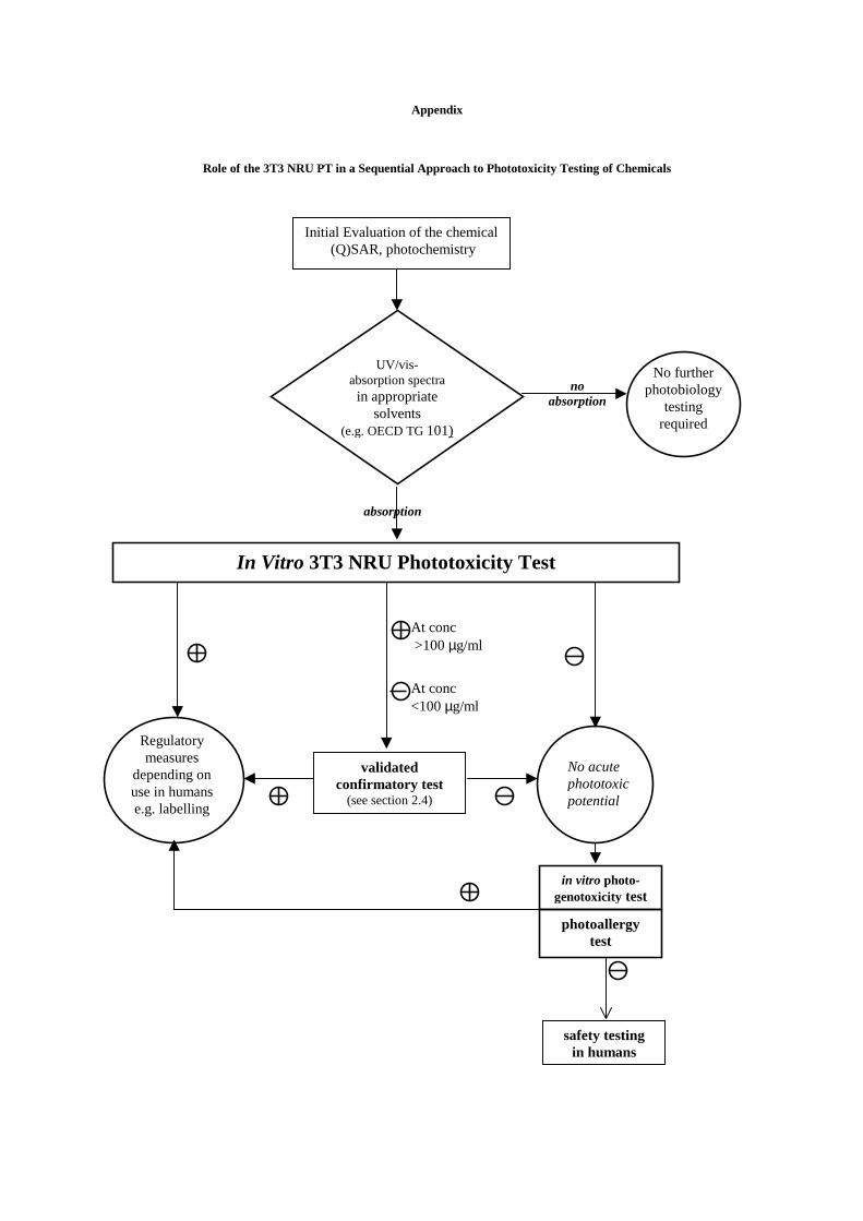
Appendix
Role of the 3T3 NRU PT in a Sequential Approach to Phototoxicity Testing of Chemicals
Initial Evaluation of the chemical(Q)SAR, photochemistry
UV/vis-absorption spectra
in appropriatesolvents
(e.g. OECD TG101)
noabsorption
absorption
In Vitro 3T3 NRU Phototoxicity Test
No acutephototoxicpotential
validatedconfirmatory test
(see section 2.4)
Regulatorymeasures
depending onuse in humanse.g. labelling
At conc>100µg/ml
At conc<100µg/ml
in vitro photo-genotoxicity test
photoallergytest
safety testingin humans
Nophoto
tereq
furtherbiology
stinguired