path lect notes comb 09 b - indiana university...
TRANSCRIPT

330
Ocular Pathology
Robbins Pathologic Basis of Disease, 6th Ed.
I. Congenital and/or developmental
A. Trisomy 21
Hypertelorism (widely spaced eyes)
Keratoconus (cone shaped cornea)
Focal hypoplasia of iris
Cataracts frequently later in life
331
B. Intrauterine infections
A. Rubella
Cataracts (virus can survive for years in the lens)
Congenital glaucoma
Abnormalities of the iris (won’t dilate)
Pigmentary abnormalities of retina
B. Syphilis
Interstitial keratitis (inflammation of the cornea)
II. Nutritional
A. Vitamin A
Night blindness
Keratomalacia
Corneal necrosis and perforation
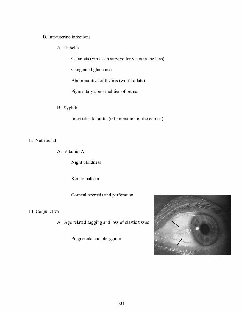
III. Conjunctiva
A. Age related sagging and loss of elastic tissue
Pinguecula and pterygium

Ocular Pathology
332
B. Infectious
Chlamydia trachomatis
Conjunctival and corneal epithelium Multiple stages leading to marked inflammation and scarring
Corneal opacification
C. Tumors and tumor-like conditions
Nevus
Melanoma
Squamous cell carcinoma
Metastatic breast
IV. Cornea
Inflammation and ulceration are the biggies
A. Stromal keratitis
May lead to ulceration
Contact lenses
AIDS
Ocular Pathology
333
Herpes simplex
B. Hereditary abnormalities of cornea
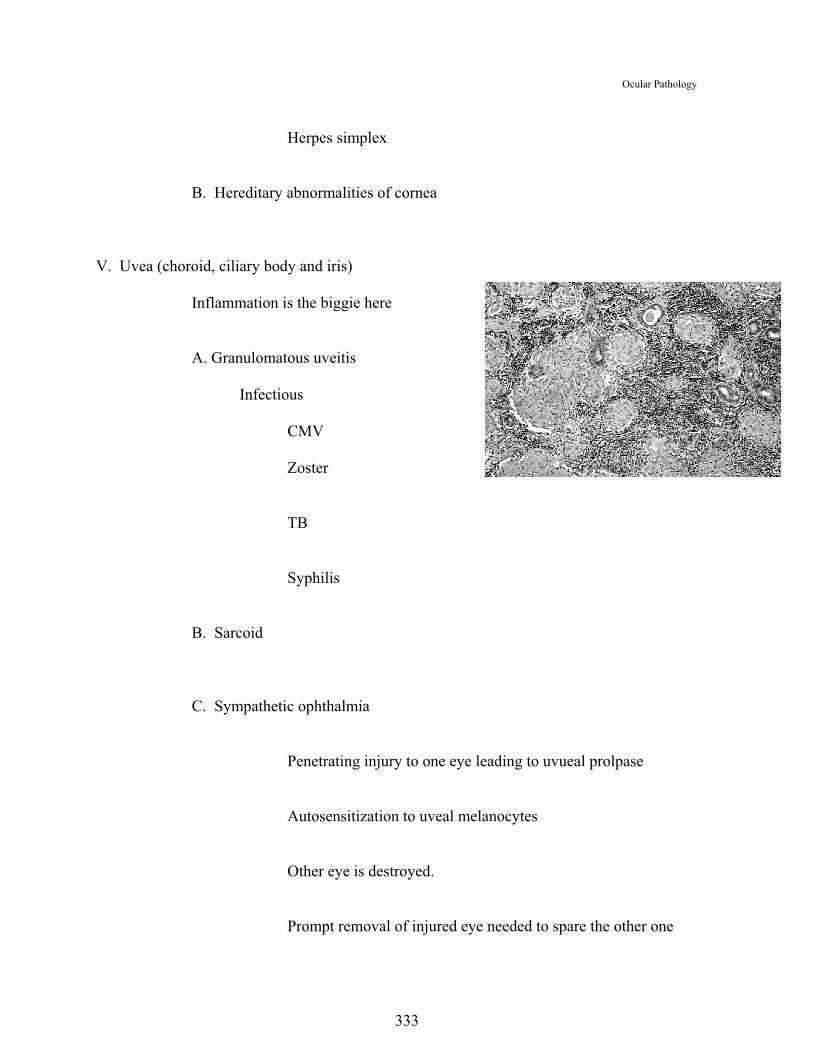
V. Uvea (choroid, ciliary body and iris)
Inflammation is the biggie here
A. Granulomatous uveitis
Infectious
CMV
Zoster
TB
Syphilis
B. Sarcoid
C. Sympathetic ophthalmia
Penetrating injury to one eye leading to uvueal prolpase
Autosensitization to uveal melanocytes
Other eye is destroyed.
Prompt removal of injured eye needed to spare the other one

Ocular Pathology
334
Granulomatous inflammation
D. Tumors of choroid
Nevi
Melanomas are most common
Several histological patterns
Spindle
Epithilioid
Clinically worst
Combined
Liver mets!!! “Ocular hepatic shunt”

Ocular Pathology
335
VI. Lens
A. Cataracts
Overall most common cause of blindness
Opacification of lens (many causes)
Congenital
DM
UV light
Glaucoma
Inflammatory conditions
Atopic disease
Steroids
Age related, so called senile cataract
Trauma related
May calcify

Ocular Pathology
336
VII. Retina
Robbins Pathologic Basis of Disease, 6th Ed.
Color blindness
- 8% of males, .4% of females
- X-linked
- Red/green
-Receptor most times, although can be anywhere

Ocular Pathology
337
A. Developmental
Retrolental fibroplasis, retinopathy of prematurity
Premies that get high levels of oxygen after birth
Seeing it more with kids of very low birth weight.
Developmental vasculature is damaged,
Granulation tissue
Fibrosis
B. DM
1. 60% have it within 15 years of onset of DM
2. Non-proliferative retinopathy
Capillary membrane thickening
Pericyte degeneration
Microaneurysms
Microvascular obstruction
Cotton wool spots
Arteriolar hylinization
3. Proliferative retinopathy
Neovascularization and fibroplasia
Endothelial growth factor (VEGF)
Retinitis prliferans
Retinal detachment (traction detachment)

Ocular Pathology
338
C. Non-diabetic vascular related
Hypertension
Atherosclerosis
Vasculitis
D. Retinitis pigmentosa (a number of conditions fit the picture)
Loss of visual receptor cells, coupled with
Proliferation of retinal pigment epithlium
Hereditary (several patterns, auto dom, sex linked rec, even mitochondrial)
Progressive (sx may begin as early as teens or as late as 60's)
Severity varies
Bilateral
E. Macular degeneration
Macula is a region of the posterior retina rich in photo receptors.
Age related (senile) degeneration
Most common among elderly
UV, genetics, medications
Two grossly identifiable forms or stages
1. Atrophic (dry)
Receptor atrophy and vascular proliferation
2. Exudative (wet)
Vascular leakage and hemorrhage

Ocular Pathology
339
Organization of leakage
Growth of pigment cells
F. Retinal detachement
Separation of retina from choroid with leakage of fluid behind.
Contraction of vitrious collagen
Space occupying lesions
Traction separation (anchor points)
G. Tumors
1. Retinoblastoma
Children 1 in 17,000 live births
Retinoblastoma gene (tumor suppressor gene)
Hereditary (bilateral)
Somatic (unilateral)

Ocular Pathology
340
Flexner-Wintersteiner rosettes
Spread through optic nerve and subarachnoid space
VIII. Optic nerve
A. Papilledema
B. Optic atrophy

Ocular Pathology
341
C. Tumors
Gliomas
Meningiomas
IX. Glaucoma
Several disease grouped under this classification
Robbins Pathologic Basis of Disease, 6th Ed.
A. Angle-closure glaucoma 1/3s of cases
Anatomic predisposition
Small eyes
Hyperopic
Aqueous humor trapped, leading to increased anterior chamber pr.
B. Open angle glaucoma 2/3s of cases
Angel appears adequate, but doesn’t conduct fluid
C. Congenital or juvenile glaucoma (both dom and rec) rubella

Ocular Pathology
342
D. Increased pressure causes optic nerve damage (glaumatous cup)
X. Phthisis bulbi