pathogenesis of preterm birth: bidirectional inflammation
TRANSCRIPT
REVIEW
Pathogenesis of preterm birth: bidirectional inflammationin mother and fetus
Ella Shana Green1& Petra Clara Arck1
Received: 31 March 2020 /Accepted: 14 July 2020# The Author(s) 2020
AbstractPreterm birth (PTB) complicates 5–18% of pregnancies globally and is a leading cause of maternal and fetal morbidity andmortality. Most PTB is spontaneous and idiopathic, with largely undefined causes. To increase understanding of PTB, muchresearch in recent years has focused on using animal models to recapitulate the pathophysiology of PTB. Dysfunctions ofmaternal immune adaptations have been implicated in a range of pregnancy pathologies, including PTB. A wealth of evidencearising from mouse models as well as human studies is now available to support that PTB results from a breakdown in fetal-maternal tolerance, along with excessive, premature inflammation. In this review, we examine the current knowledge of thebidirectional communication between fetal and maternal systems and its role in the immunopathogenesis of PTB. These recentinsights significantly advance our understanding of the pathogenesis of PTB, which is essential to ultimately designing moreeffective strategies for early prediction and subsequent prevention of PTB.
Keywords Preterm birth . Labor . Mouse models . Regulatory T cells . Inflammatory signaling pathways . Microbiome . Fetalsignals
Introduction
Preterm birth (PTB), defined as birth before the 37thweek of gestation, affects up to 15 million pregnancieseach year globally [1]. Being born too soon can lead toneonatal death, but also a high risk for early-life infec-tions and neurodevelopmental, cardiometabolic, and in-flammatory disorders later in the life of surviving in-fants [2–6]. A current understanding of the complexpathogenesis of PTB is still poor, which also explainsthe limited availability of targeted and effective strate-gies for PTB prevention.
Several risk factors for PTB are well recognized, such astwin pregnancies, chorioamnionitis, pre-existing maternal
diseases, genetic factors, previous PTB, and uterine abnormal-ities [7–10]. However, a large proportion of PTBs have noidentifiable cause. These spontaneous PTBs present as patho-logical inflammation, premature rupture of membranes(PROM), and onset of preterm labor.
Interestingly, inflammation is also a key trigger of thephysiological onset of labor at term [11]. Thus, the onset ofinflammation seems to be a common denominator initiatingterm as well as preterm labor. Here, we comprehensively re-view why inflammation may be prematurely initiated in somepregnancies, which then leads to PTB.
A brief overview of maternal immuneadaptation during normally progressingpregnancies
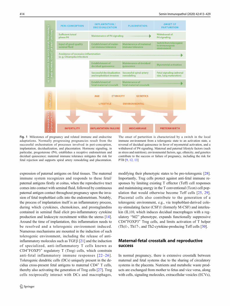
In order to understand the onset of inflammation during preg-nancy, it is pivotal to summarize milestones of pregnancy(Fig. 1), including key features of feto-maternal immune tol-erance, as the breakdown of such immune tolerance is associ-ated with a number of pregnancy complications [14–17].
As covered by a large number of reviews [14, 15, 17–20],the need for feto-maternal immune tolerance arises from the
This article is a contribution to the special issue on Preterm birth:Pathogenesis and clinical consequences revisited – Guest Editors: AnkeDiemert and Petra Arck
* Petra Clara [email protected]
1 Department of Obstetrics and Fetal Medicine, Laboratory forExperimental Feto-Maternal Medicine, University Medical CenterHamburg-Eppendorf, Martinistraße 52, 20251 Hamburg, Germany
https://doi.org/10.1007/s00281-020-00807-y
/ Published online: 7 September 2020
Seminars in Immunopathology (2020) 42:413–429
expression of paternal antigens on fetal tissues. The maternalimmune system recognizes and responds to these fetal/paternal antigens firstly at coitus, when the reproductive tractcomes into contact with seminal fluid, followed by continuouspaternal antigen contact throughout pregnancy upon the inva-sion of fetal trophoblast cells into the endometrium. Notably,the process of implantation itself is an inflammatory process,during which cytokines, chemokines, and prostaglandinscontained in seminal fluid elicit pro-inflammatory cytokineproduction and leukocyte recruitment within the uterus [14].Around the time of implantation, this inflammation needs tobe resolved and a tolerogenic environment induced.Numerous mechanisms are mounted in the induction of suchtolerogenic environment, including the release of anti-inflammatory molecules such as TGFβ [21] and the inductionof specialized, anti-inflammatory T cells known asCD4+FOXP3+ regulatory T (Treg) cells, which constrainanti-fetal inflammatory immune responses [22–26].Tolerogenic dendritic cells (DCs) uniquely present in the de-cidua cross-present fetal antigens to maternal CD4+ T cells,thereby also activating the generation of Treg cells [27]. Tregcells reciprocally interact with DCs and macrophages,
modifying their phenotypic states to be pro-tolerogenic [28].Importantly, Treg cells protect against anti-fetal immune re-sponses by limiting existing T effector (Teff) cell responsesand maintaining anergy in the T conventional (Tcon) cell pop-ulation that would otherwise become Teff cells [25, 29].Placental cells also contribute to the generation of atolerogenic environment, e.g., via trophoblast-derived colo-ny-stimulating factor (CSF)1 (formerly M-CSF) and interleu-kin (IL)10, which induces decidual macrophages with a reg-ulatory “M2” phenotype, expands functionally suppressiveCD4+FOXP3+ Treg cells, and limits activation of T helper(Th)1-, Th17-, and Th2-cytokine-producing Teff cells [30].
Maternal-fetal crosstalk and reproductivesuccess
In normal pregnancy, there is extensive crosstalk betweenmaternal and fetal systems due to the sharing of circulatorysystems in the placenta. Nutrients and metabolic waste prod-ucts are exchanged from mother to fetus and vice versa, alongwith cells, signaling molecules, extracellular vesicles (ECVs),
Fig. 1 Milestones of pregnancy and related immune and endocrineadaptations. Normally progressing pregnancies result from thesuccessful orchestration of processes involved in peri-conception,implantation, decidualization, and placentation. Hormone signaling, inparticular, progesterone (P4), establishes a receptive endometrium anddecidual quiescence; maternal immune tolerance mitigates the risk forfetal rejection and supports spiral artery remodeling and placentation.
The onset of parturition is characterized by a switch in the localimmune environment from a tolerogenic state to an activation state, areversal of decidual quiescence in favor of myometrial activation, and awithdrawal of P4 signaling. Maternal and paternal lifestyle factors (suchas stress and nutrition), environmental factors, age, ethnicity, and geneticscontribute to the success or failure of pregnancy, including the risk forPTB [9, 12, 13]
414 Semin Immunopathol (2020) 42:413–429
and nucleic acids. The exchange of cells results inmicrochimerism in mother or fetus, whereby maternal cellstransferred to the fetus are referred to as maternalmicrochimerism (MMC). Reciprocally, fetal cells transferredto the mother are termed fetal microchimerism (FMC). Both,MMC and FMC can persist for decades after pregnancy in abroad range of tissues in the body, including the brain, skin,heart, lung, bone marrow, spleen, and lymph nodes [31–34].
MMC cells promote tolerance to maternal antigens in fetalimmune cells during pregnancy through the induction of non-inherited maternal antigen (NIMA)–specific CD4+FOXP3+
Treg cells [35]. Furthermore, they may enhance reproductivefitness in the next generation through sustained tolerance toNIMA. However, these insights are solely based on a mousemodel to date and limited to settings where the paternal anti-gens and NIMA were shared [36].
In addition, microvesicles, exosomes, and cell-free (cf) pro-teins and nucleic acids such and cfRNA and cfDNA are ex-changed via the placenta. In general, the concentration ofcfRNA in maternal circulation increases over the course of ges-tation [37, 38], and fetal cfDNA and RNA transcripts also in-crease in maternal circulation late in gestation [39–42]. Somefetal cfDNA and RNA are contained within apoptotic fetal cells[39, 41], but immunosuppressive CD71+ fetal erythroid cells arealso an abundant source of fetal DNA in maternal circulation[37]. The cfDNA and RNA transcripts could be transferred pas-sively, or via ECVs. Exosomes are a type of ECV released byexocytosis into the extracellular environment. Notably, the pla-centa releases exosomes into maternal circulation during preg-nancy [43], which increase with increasing gestational agepeaking at term [44, 45]. The functional importance of variousECVs in pregnancy is largely unknown, but they are suspectedto mediate fetal-maternal communication at key stages of preg-nancy including implantation and parturition [43, 46]. Humanvillous trophoblasts secrete exosomes containing placenta-specific miRNAs into maternal circulation, which play key rolesin regulating immune signaling [47, 48]. Maternal-derivedexosomes are also enriched during pregnancy, and the transferof exosomes between the fetus and mother is bidirectional [49].
Immune tolerance versus inflammationin the development of preterm birth
As mentioned above, inflammation has been linked to thepathogenesis of idiopathic PTB, and also other pregnancycomplications in mice and humans [7, 50–55]. Furthermore,PTB is considered to have early pregnancy origins, prior toplacental development. Thus, it has been proposed that PTB isa disorder that results from defective deep placentation [56,57], similar to preeclampsia.
As the maternal immune adaptation is involved in the pro-cesses sustaining pregnancy success throughout the entire
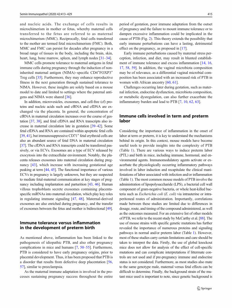
period of gestation, poor immune adaptation from the outsetof pregnancy and the failure to mount immune tolerance or todampen excessive inflammation could be implicated in thecause of PTB (Fig. 2). This theory extends the possibility thatearly immune perturbations can have a lasting, detrimentaleffect on the pregnancy, as proposed in [17].
Early immune perturbations caused by maternal stress per-ception, infection, and diet, may result in blunted establish-ment of immune tolerance and excess inflammation [14, 16,17, 58, 59]. In addition, the vaginal microbiota compositionmay be of relevance, as a differential vaginal microbial com-position has been associated with an increased risk of PTB inwomen with African ancestry [60, 61].
Challenges occurring later during gestation, such as mater-nal infection, endocrine dysfunction, microbiota composition,or metabolic dysregulation, can also further exacerbate theinflammatory burden and lead to PTB [7, 10, 62, 63].
Immune cells involved in term and pretermlabor
Considering the importance of inflammation in the onset oflabor at term or preterm, it is key to understand the mechanismsbehind its origin. In this context, mouse models have becomeuseful tools to provide insights into the complexity of PTB(Table 1). There are various ways to induce preterm labor(PTL) and birth in mice, including immune, hormonal, and en-vironmental agents. Immunomodulatory agents activate or ex-acerbate the physiologically occurring inflammatory pathwaysinvolved in labor induction and recapitulate the clinical mani-festations of labor associated with infection and/or inflammation(Table 1). The most commonmouse models of PTB involve theadministration of lipopolysaccharide (LPS), a bacterial cell wallcomponent of gram-negative bacteria, or whole heat-killed bac-teria such as Escherichia coli (E. coli) via intrauterine or intra-peritoneal routes of administration. Importantly, correlationsmade between these studies are limited due to differences indosage, route, and timing of the compound administered, as wellas the outcomes measured. For an extensive list of other modelsof PTB,we refer to the recent study byMcCarthy et al. [88]. Theuse of mouse strains with specific genetic mutations has furtherrevealed the importance of numerous proteins and signalingpathways in normal and/or preterm labor (Table 1). However,most of these studies carry certain limitations and care should betaken to interpret the data. Firstly, the use of global knockoutmice does not allow for analysis of the effect of cell-specificmutations and can complicate interpretations if littermate con-trols are not used and if pre-pregnancy immune and endocrinestatus is not considered. Furthermore, as most studies also mateto the same genotype male, maternal versus fetal effects can bedifficult to determine. Finally, the background strain of the mu-tant mice used is important to note, since genetic background is
415Semin Immunopathol (2020) 42:413–429
a determinant of gestation length in mice [89]. Despite theirlimitations, studies using either interventions or mutant mouselines to cause or delay preterm or term labor provide valuableinsights into the inflammatory mechanisms of these processesand highlight the balance of factors that work in concert toproduce controlled inflammation in normal term labor.Importantly, dysregulation of many essential components ofthese inflammatory pathways either prevents or increases sus-ceptibility to preterm labor in mice.
Using mouse models of PTB, macrophages were identifiedas major contributors to the induction of term and pretermlabor (PTL). Depletion of macrophages using anti-F4/80 ad-ministration prior to experimental LPS-induced onset of PTBeliminates susceptibility to PTB [51]. Macrophages likelycontribute to the pathogenesis of PTB via secretion of inflam-matory cytokines, such as tumor necrosis factor (TNF)α, IL1,IL6, and IL8 and uterine contractility genes such as matrixmetalloproteinases (MMPs) [90]. In particular, IL1 is knownto critically contribute to inflammation-induced PTB. Asshown in mice, IL1 administration induces PTL, and inhibi-tion of its receptor prevents labor [72], likely via activation of
NF-κb signaling pathways [64]. Similarly, IL6 is another cy-tokine important in the timing of parturition and the pathogen-esis of PTB, since IL6-deficient mice have delayed parturitionand are also resistant to LPS-induced PTB [65].
Moreover, complement activation plays an important role inPTB. This has been observed in infection-induced PTB inhumans, where womenwith spontaneous PTL showed increasedconcentrations of complement products C3a, C4a, and C5a [51].In mice, complement receptor C5aR-deficient mice are protectedagainst LPS- and RU486-induced PTB. In this study, increasedcomplement deposition was present in the cervical epithelium ofPTB mice, along with complement C5a activation–dependentMMP9 release from macrophages and cervical remodeling [77].
Besides these examples of the involvement of the innateimmune system in modulating the risk for PTB, the adaptiveimmune response is also critical. A subset of PTB is postulatedto result from a failure of maternal tolerance to fetal antigens [7].Indeed, Treg and Teff cells exhibit distinct changes in frequencyand phenotype in PTB. Treg cells in peripheral blood of womenin PTL are reported to have differential activation and dimin-ished suppressive capacity compared with term controls [52, 91,
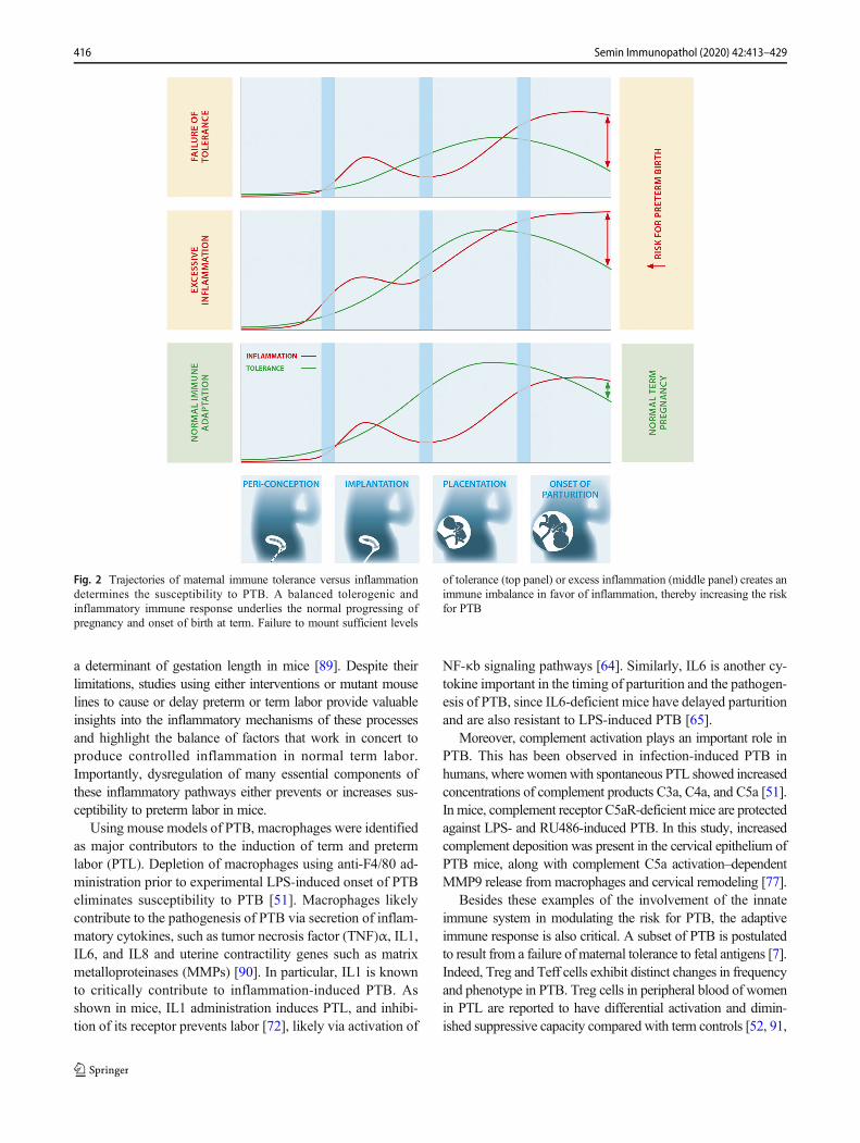
Fig. 2 Trajectories of maternal immune tolerance versus inflammationdetermines the susceptibility to PTB. A balanced tolerogenic andinflammatory immune response underlies the normal progressing ofpregnancy and onset of birth at term. Failure to mount sufficient levels
of tolerance (top panel) or excess inflammation (middle panel) creates animmune imbalance in favor of inflammation, thereby increasing the riskfor PTB
416 Semin Immunopathol (2020) 42:413–429
92]. Normal human labor is associated with a decline in Tregcell function and reciprocal activation of Teff cells which arenormally controlled by Treg cells [92].
A decrease in Treg cell number and/or function in pregnancypathologies is typically associated with an increase in Tcon/Teff
activation. This holds true for labor, as T cells with an activatedmemory phenotype are increased in the choriodecidua (fetalmembranes) of women with spontaneous labor at term [93].Effector memory CD4+ and CD8+ T cells are also enriched inthe decidua in spontaneous PTL, suggesting the inflammatorypotential of these cells is greater in PTL women [73]. CD4+ andCD8+ T cells expressing “exhausted” (PD-1+TIM-3+CTLA4−LAG-3−) and “senescent” (KLRG-1+CD57+) mem-ory and effector phenotypes were identified in the decidua ofwomen with term and preterm labor [66]. Notably, the propor-tions of exhausted CD4+ T cells increased in the deciduaparietalis with increasing gestational age but declined in the de-cidua basalis of women who underwent PTL with placental in-flammation. As TNFα and interferon (IFN)γ production couldbe induced ex vivo in the exhausted T cells, these cells mayrestore their effector function upon placental inflammation [66].
T and B cell–deficient Rag1−/− mice have provided impor-tant clues for the role of T cells in PTB. These mice show anincreased susceptibility to LPS-induced PTB, which may bemediated bymacrophage activation. However, adoptive trans-fer of CD4+ T cells at mid-gestation conferred resistance toLPS-induced PTB, as these cells seem to rapidly differentiateinto Treg cells [79]. Conversely, activation of Teff cells usinganti-CD3 causes PTB via upregulation of local and systemicpro-inflammatory responses such as IL6 and IFNγ [73].Interestingly, in utero adoptive transfer of CD4+ and CD8+
Teff cells causes late fetal resorption dependent on TNFαand IFNγ. These cytokines also cause uterine contractilityin vitro. However, it is unclear whether these Teff cells causePTB if administered later or systemically [54].
Together, these studies suggest that activated memory Tcells and Teff cells may participate in mediating inflammatoryprocesses in normal term labor, whereas Treg cells may act tominimize the inflammatory potential or premature activationof exhausted and memory Teff cells. However, the moleculardetails of these maternal T cell interactions implicated in nor-mal and PTL are yet to be defined. One potential pathwaycould involve IL10, as Treg cells are capable of producingthis anti-inflammatory cytokine. IL10 has been shown to pre-vent PTB, as IL10 deficiency or administration of IL10-neutralizing antibodies increases susceptibility to PTB in mice[67]. As global IL10 deficiency led to a decrease in inflam-matory cytokine gene expression in the uterus and placenta,IL10 may act to regulate excessive inflammatory responsesimplicated in labor. Recent work demonstrates that TLR4 sig-naling in decidual endothelial cells at term may cause IL10induction in perivascular stromal cells via NF-κb activation ofIL6 and STAT3. This could be a mechanism to preserve thehomeostatic immune balance under inflammation, which maybe perturbed in PTB [84].
Emerging data arising from humans and mice indicate thatthe fetal environment in PTB is also characterized by inflam-mation, along with the priming of fetal T cells against
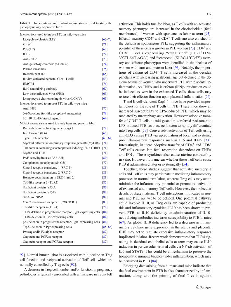
Table 1 Interventions and mutant mouse strains used to study thepathophysiology of preterm birth
Interventions used to induce PTL in wild-type mice
Lipopolysaccharide (LPS) [63–70]
E. coli [71]
Poly(I:C) [63]
IL1β [72]
Anti-CD3e [73]
Anti-galactosylceramide (a-GalCer) [74]
Plasma exosomes [75]
Recombinant IL6 [65]
In vitro activated neonatal CD4+ T cells [53]
HMGB1 [76]
IL10 neutralizing antibody [67]
Low dose influenza virus (PR8) [63]
Lymphocytic choriomeningitis virus (LCMV) [63]
Interventions used to prevent PTL in wild-type mice
Anti-F480 [77]
(+)-Naloxone (toll-like receptor-4 antagonist) [78]
101.10 (IL-1R-biased ligand) [64]
Mutant mouse strains used to study term and preterm labor
Recombination activating gene (Rag) 1 [79]
Interleukin 6 (IL6) [65]
Type I IFN receptor [63, 64]
Myeloid differentiation primary-response gene 88 (MyD88) [71]
TIR domain-containing adaptor protein-inducing IFNβ (TRIF) [71]
Myd88 and TRIF [71]
PAF acetylhydrolase (PAF-AH) [80]
Complement (anaphylatoxin C5a) [77]
Steroid receptor coactivase 1 (SRC-1) [81]
Steroid receptor coactivase 2 (SRC-2) [81]
Heterozygous mutation in SRC-1 and 2 [81]
Toll-like receptor 3 (TLR2) [82]
Surfactant protein (SP)-A [82]
Surfactant protein (SP)-D [82]
SP-A and SP-D [82]
CXC3 chemokine receptor 1 (CXC3CR1) [83]
Toll-like receptor 4 (TLR4) [70]
TLR4 deletion in progesterone receptor (Pgr)–expressing cells [84]
TLR4 deletion in Tie2-expressing cells [84]
p53 deletion in progesterone receptor (Pgr)–expressing cells [84]
Trp53 deletion in Pgr-expressing cells [85, 86]
Prostaglandin F2 alpha receptor [87]
Oxytocin and PGF2α receptor [87]
Oxytocin receptor and PGF2α receptor [87]
417Semin Immunopathol (2020) 42:413–429
maternal antigens [54]. Future studies are now needed to con-firm if fetal inflammation is causal to maternal inflammationand related PTB, or a consequence of the maternal spill-overof inflammatory cytokines.
NKT cells, an innate-like subset of T cells with specializedfunctions, are also implicated in PTB. Maternal NKT cellsrecognize CD1-restricted lipid antigens, which are expressedby fetal trophoblast cells, and are thought to play an immuno-regulatory role in pregnancy [94]. Interestingly, depletion ofinvariant NKT cells reduces the rate of LPS-induced PTB inmice and administration of an antibody specific for NKT cells(α-GalCer) in late gestation causes PTB via activation ofCD4+ T cells, macrophages, neutrophils, and DCs in themyometrium/decidua [68, 74, 95].
Inflammatory signaling pathways in termand preterm labor
A crucial component of inflammatory immune responsesis the engagement of toll-like receptors (TLRs) whichtrigger signaling pathways leading to secretion of cyto-kines and chemokines by innate immune cells. TLRs are
an evolutionarily conserved class of pattern recognitionreceptor (PRR) that recognize pathogen-associated molec-ular patterns (PAMPS) derived from microorganisms, anddamage-associated molecular patterns (DAMPS) releasedfrom immune cells and stressed and dying cells. TLRactivation is an essential initiating component of the in-flammatory pathway that leads to both normal and pre-term labor (Fig. 3).
Most research on the role of TLRs in PTB has focused onTLR4 which binds to a specific range of PAMP and DAMPligands including bacterial LPS. TLR4 is activated in the uter-us in both normal and PTB and is essential for on-time partu-rition in mice, controlling uterine activation and onset of labor[70]. Pregnant TLR4−/− mice deliver an average of 13 h laterthan controls and their offspring exhibit reduced viability [70].Furthermore, inhibition of TLR4 signaling with the TLR4-antagonist (+)-naloxone following intrauterine administrationofE. coli suppressed the inflammatory cascade and effectivelyprevented against PTB [78]. Recently, TLR4 expression bydecidual endothelial cells, and not immune cells, was identi-fied to be a key for initiating this response, since mice withendothelial-specific TLR4 deletion are resistant to LPS-induced PTB [84]. In humans, maternal single nucleotide
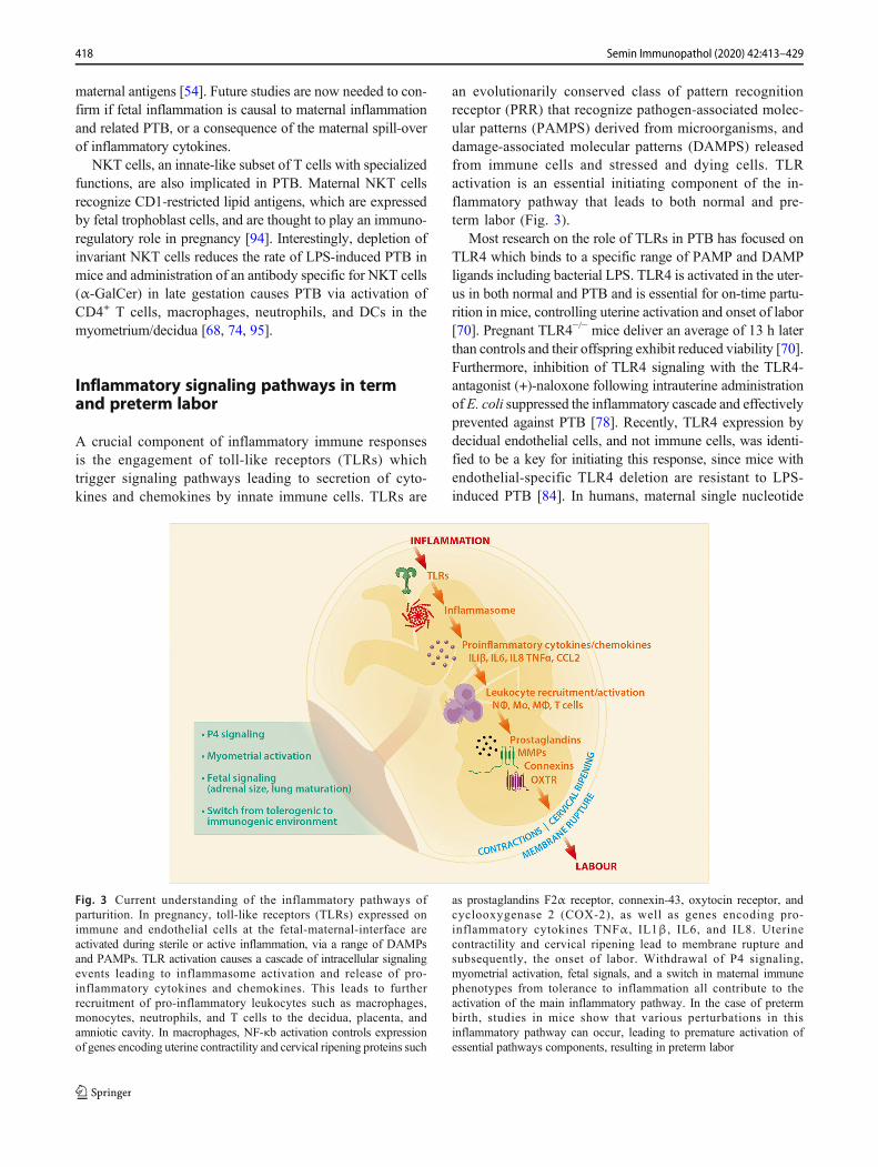
Fig. 3 Current understanding of the inflammatory pathways ofparturition. In pregnancy, toll-like receptors (TLRs) expressed onimmune and endothelial cells at the fetal-maternal-interface areactivated during sterile or active inflammation, via a range of DAMPsand PAMPs. TLR activation causes a cascade of intracellular signalingevents leading to inflammasome activation and release of pro-inflammatory cytokines and chemokines. This leads to furtherrecruitment of pro-inflammatory leukocytes such as macrophages,monocytes, neutrophils, and T cells to the decidua, placenta, andamniotic cavity. In macrophages, NF-κb activation controls expressionof genes encoding uterine contractility and cervical ripening proteins such
as prostaglandins F2α receptor, connexin-43, oxytocin receptor, andcyclooxygenase 2 (COX-2), as well as genes encoding pro-inflammatory cytokines TNFα, IL1β, IL6, and IL8. Uterinecontractility and cervical ripening lead to membrane rupture andsubsequently, the onset of labor. Withdrawal of P4 signaling,myometrial activation, fetal signals, and a switch in maternal immunephenotypes from tolerance to inflammation all contribute to theactivation of the main inflammatory pathway. In the case of pretermbirth, studies in mice show that various perturbations in thisinflammatory pathway can occur, leading to premature activation ofessential pathways components, resulting in preterm labor
418 Semin Immunopathol (2020) 42:413–429
polymorphisms (SNPs) in the TLR4 gene are associated withearly preterm delivery before gestational week 32 [96].Downstream of TLR4 and its coreceptor CD14 is MyD88-dependent and independent (TRIF-dependent) signaling path-ways, that each control specific inflammatory gene expres-sion. In mice, PTB is dependent on MyD88, since MyD88−/− mice are completely protected against E. coli–induced PTB[71].
In addition to TLR4, other TLRs likely mediate timing ofbirth, such as TLR2 [82, 97]. In addition to maternal TLRexpression, fetal TLR expression may have involvement inthe timing of birth as polymorphic fetal TLR4 and TLR2alleles were associated with prematurity [98, 99].
TLR activation subsequently leads to the activation of theinflammasome and the perpetuation of additional cytokineand chemokine production in the placenta and decidua, suchas IL8, CCL2, IL1β, and IL6 [100]. TLR activation also ini-tiates the recruitment of immune cells, production of prosta-glandins, and MMPs, leading to the activation of cervicalripening and uterine contractions (Fig. 3). Also, the influx ofimmune cells such as macrophages to the amniotic cavity ispivotal for the onset of labor, as it induces the production ofinflammatory mediators such as NF-κb. NF-κb is a key me-diator of the inflammatory cascade leading to labor, since itdirectly binds to the promoters of genes that cause uterinecontractility such as PTGFR (prostaglandin F2α receptor),GJA1 (connexin 43), OXTR (oxytocin receptor), and PTGS2(cyclooxygenase 2; COX-2), as well as genes encoding pro-inflammatory cytokines TNFα, IL1β, IL6, and IL8 [90].Taken together, PRRs, and specifically TLRs and their down-stream signaling molecules, are key players in triggering thetiming of birth at term and preterm.
The DAMPs and PAMPs which bind to PRRs also playessential roles as the “messengers” to initiate the inflammatorycascade leading to term and preterm labor. TLRs in pregnancyare activated during sterile inflammation, and in the absenceof active infection, via DAMPs [101]. The most studiedDAMPs in preterm and term labor are high-mobility groupbox 1 (HMGB1), fetal cfDNA, and platelet-activating factor(PAF). Oxidative stress and cellular senescence of the fetalamnion and chorion may trigger human parturition throughthe release of DAMPs. In senescent cells, DAMPs relocatefrom the nucleus to the cytosol where they can be secretedas alarmins and drive the inflammatory parturition cascade[102]. Intra-amniotic administration of HMGB1, an alarminthat induces inflammation upon tissue damage, causes pre-term labor in mice [76] and is elevated in the amniotic fluidof women who underwent PTL, independent of intra-amnioticinfection status [103]. HMGB1 was found to be primarilyexpressed by amnion epithelial cells, myofibroblasts, neutro-phils, and macrophages [103], and incubation ofchorioamniotic membranes with HMGB1 leads to release ofpro-inflammatory IL1β and IL6 [104]. cfDNA, a common
activator of TLRs, was found to be released upon cell deathto cause TLR signaling in the placenta [105]. The inflamma-tory phospholipid PAF is another DAMP critical for labor. Itis increased in the amniotic fluid of women who deliver pre-term [106–108], and its administration induces PTB in micevia TLR signaling in macrophages [80, 108, 109]. Alarminsreleased from cells upon tissue stress can also bind to PRRs,and concentrations of alarmins IL1α and S11 family proteinscalgranulin A and calgranulin C were increased in amnioticfluid during sterile intra-amniotic inflammation [110]. OtherPRRs such as NRLP3 and NOD2 activate inflammasomes,which likely play a role in initiating spontaneous labor at termand preterm labor, since the activation of components of thispathway causes the secretion of IL1" in chorioamniotic mem-branes in normal labor at term [100]. Placental PRR expres-sion mediates their functions as a key barrier capable of rec-ognizing and responding to microbes and stress.
Perturbations affecting the placenta could therefore affectthe placental response to PAMPs and DAMPs and have animpact on pregnancy outcome. In summary, DAMPs play keyroles in initiating labor at term and are implicated in the causeof PTL; however, their origin (fetal versus maternal), triggers,and regulation are not well understood.
Fetal-placental and vaginal microbiomesin PTB
Recent research endeavors aimed to identify the impact of themicrobiome in modulating the risk for PTB [111]. Features ofthe vaginal microbiome have been shown to determine therisk for PTB. For example, in women of African ancestry,dramatic changes in the diversity of the microbiota were ob-served in early pregnancy, with an increased prevalence ofmultiple vagitypes and dysbiosis [61]. Several dysbioticvagitypes could be associated with PTB, likely caused by anearly pregnancy increase in pro-inflammatory cytokines invaginal fluid, such as the chemoattractant CXCL10 [60].Interestingly, preconception administration of antibiotics towomen with previous preterm delivery did not reduce pretermbirth rate and may be associated with earlier delivery anddecreased birth weight [112], suggesting that complex host-microbiome dynamics from the outset of pregnancy are likelyessential for optimal pregnancy success [113]. The molecularmechanisms of these interactions will be important to uncoverin future research efforts. In contrast to the vaginal microbiota,there is still a great deal of ambiguity as to the question ofwhether the intrauterine environment is sterile or non-sterile.16S rRNA sequencing and culture of bacterial species isolatedfrom the female reproductive tract revealed microbial commu-nities in the cervix, uterus, fallopian tubes, and peritoneal fluidthat were diverse and distinct compared with the vagina andcervix, which were Lactobacillus dominated [114].
419Semin Immunopathol (2020) 42:413–429
Examination of microbiota from the endometrial fluid of in-fertile women undergoing in vitro fertilization (IVF) revealedan association between non-Lactobacillus-dominated endo-metrial microbiota and decreases in implantation, pregnancy,and live birth rates [115]. Several mechanisms have been pro-posed to limit bacterial invasion to the uterus from the lowergenital tract, and these may operate during pregnancy to en-sure a tolerogenic environment is maintained for pregnancysuccess. Bacterial invasion of the uterus and amniotic cavityfound in subsets of pregnant women could theoretically causeinflammation and disruption of the immune adaptation topregnancy, predisposing to PTB [113]. Microorganisms sim-ilar to the vaginal microbiota can also be isolated from theplacenta [116]. However, others have rejected this concept,showing contaminants were responsible for the detection ofmost bacterial species in the placenta [117]. Given these find-ings, there is still debate as to whether the uterus and placentaharbor a microbiome or not. Hence, it is still unknown if askew of the intrauterine microbiome—if it indeed exists—may modulate the risk for PTB.
The role of maternal progesterone in PTB
Hormone levels have also been extensively studied in order tounderstand the pathogenesis of PTB. Adequate progesterone(P4) levels are known to be essential for the establishment andprogression of normal pregnancy. Hence, it is not surprisingthat labor is associated with a P4 withdrawal in mice andfunctional progesterone withdrawal in women [118, 119].The P4 decline appears essential for labor to occur, as theadministration of P4 to mice in late gestation extends the ges-tation length and prevents labor. Similarly, mice lacking theP4-metabolizing enzyme 20αHSD show prolonged gestation[120]. Conversely, treatment with the progesterone antagonistRU486 causes PTB in mice and is associated with increaseddecidual PGE2 and IL6 concentrations [121]. P4 applicationis also protective against LPS-induced PTB in mice via pre-vention of inflammation-induced cervical remodeling, cervi-cal macrophage infiltration, and MMP9 expression that pre-cedes PTB, possibly through the disruption of complementsignaling on macrophages [77]. P4 is known as a strong im-munomodulator, it induces stable Treg cells, arrests DC in atolerogenic state, and suppresses inflammatory responses[122–124]. P4 signaling withdrawal may therefore be onemechanism of immune modulation leading to PTB. The P4decline in late gestation may be triggered by immune path-ways, as macrophages have been shown to regulate P4 pro-duction in the ovary in mice [125]. However, it should benoted that PTB in mice can also occur in the absence of P4withdrawal, for instance, with intrauterine administration ofbacteria [126]; multiple, independent immune and hormonalmechanisms are likely in place.
Myometrial and decidual clocks in PTB
The decline in P4 signaling induces a switch of themyometrium from a quiescent to a contractile state at labor[127]. However, evidence places the establishment ofmyometrial quiescence also at the start of gestation, as itmay be regulated by estrogen and P4 hormone receptor sig-naling [128]. Besides the myometrium, the endometrium anddecidua are also proposed to be responsible for the timing ofbirth via induction of a decidual “clock.” This decidual clockmediates mechanisms of fetal-maternal tolerance in earlypregnancy and wanes over time with advancing gestationalage, leading to a withdrawal of active suppression or inductionof inflammatory signals [10]. The induction of decidual qui-escence is an active process occurring in early pregnancy,caused by transcriptional silencing in decidual stromal cells(DSCs) which recruit the repressive histone mark H3K27me3for epigenetic regulation of hundreds of genes, causing sup-pression of type 1 immunity and wound healing response[129]. Again, P4 signaling from the outset of gestation maybe essential to decidual quiescence and maintenance of ananti-inflammatory immune environment needed to last forthe duration of pregnancy, until labor at term.
Fetal origins of the timing of birth
There are several lines of evidence to support that the fetusitself is involved in initiating signals of parturition, such as thesecretion of hormones (cortisol, estrogen, placentalcorticotropin-releasing hormone (CRH)) and surfactant pro-tein A (SP-A) [127]. The fetal adrenal gland is responsiblefor fetal cortisol production. Notably, placental CRH secretionmay provide the first signal for cortisol stimulation [130].Dysregulation or early activation of these processes may beimplicated in the pathogenesis of PTB. Interestingly, SP-A, amajor lung surfactant protein secreted by the maturing fetallung when the capacity to sustain air breathing is developed,was identified in mice as a signal for parturition through itsaction on fetal macrophages [131]. SP-A binds to TLR4 onhuman and mouse macrophages and causes TLR4-dependentinflammatory cytokine expression [132]. SP-A was detectedin the amniotic fluid at term pregnancy and stimulated IL1"and NF-κb expression in amniotic fluid–derived macrophagesin vitro. Intra-amniotic injection of SP-A in mice additionallycaused preterm delivery within 24 h [131]. Expression of ste-roid receptor coactivators SRC-1 and SRC-2, regulators ofSP-A transcription, was determined to be key for the initiationof labor, and additionally key for the expression of PAF andlysophosphatidylcholine acyltransferase-1 (LPCAT1), whichcatalyzes the synthesis of PAF, in the fetal lung. As discussed,PAF initiates TLR signaling in mice and administration ofPAF causes PTB [80, 109]. However, whether pathological
420 Semin Immunopathol (2020) 42:413–429
activation of PAF signaling in PTB arises from the fetus iscurrently unknown.
In addition to fetal hormone- and SP-A-dependent modu-lation of labor and PTB, the fetal immune system has longbeen recognized to also have involvement in preterm birth[133]. The theory that the fetal immune system is immaturehas been robustly disproven, as neonatal T cells have the po-tential for activation [31, 54, 134]. Additionally, qualitativedistinctions of immune responses between neonates andadults are in place. These include CD71+ erythroid cells,which are uniquely present in neonates and have been pro-posed to suppress microbial colonization-related inflamma-tion. Also, IL8 (CXCL8) drives major T cell effector functionin human newborns, as it allows for the activation of antimi-crobial neutrophils and γδ T cells [135]. Importantly, thesemechanisms may already instruct fetal immunity prior to birthduring pregnancy and their dysregulation could be involved inthe pathogenesis of PTB [53, 54, 134].
Interestingly, fetal macrophages appear to play a key role ininitiating inflammatory cascade at parturition, as amniotic flu-id fetal macrophages were shown to migrate to the uterus atterm in response to increasing SP-A levels and engage withvia TLR2 signaling [82, 131]. In neonates, polymorphicTLR4 and TLR2 alleles are associated with prematurity [98,99]. Therefore, TLR signaling in fetal macrophages may beimportant in the pathogenesis of PTB. However, the pheno-type of fetal macrophages seems to be tightly controlled toavoid inappropriate inflammation that may cause early activa-tion of the inflammatory cascade leading to parturition [134]and failure of this control may result in an increased risk forPTB.
Insights arising from comparative cord blood analyses re-vealed distinct differences between term and preterm borninfants [54]. Preterm cord blood was characterized by higherconcentrations of inflammatory cytokines, increased activa-t i o n o f d e n d r i t i c c e l l s , a n d c e n t r a l m emo r y(CCR7+CD45RA−) T cells with a Th1 phenotype. However,it needs to be confirmed if these differences are involved in thepathogenesis of PTB, or merely result from the less advancedimmune ontogeny in preterm born neonates.
Emerging data arising from humans and mice also indicatethat the fetal environment in PTB is also characterized byinflammation, along with the priming of fetal T cells againstmaternal antigens [54]. Activated fetal T cells are also foundin the amniotic cavity and are increased in cases withchorioamnionitis [53]. Future studies are needed to confirmif fetal inflammation is the cause of the maternal inflammationand related PTB, or the consequence of the maternal spill-overof inflammatory cytokines.
The above-mentioned population of immunosuppressiveCD71+ erythroid cells, which allows for microbial coloniza-tion upon birth, may play a functional role before birththrough constraining fetal T cell activation. This is supported
by observations upon depletion of CD71+ cells in splenocytesof neonatal mice, which caused robust cytokine productionupon stimulation in vitro [134, 136].
The potential role of FMC and MMC in PTB
The earlier introduced population of FMC cells may too rep-resent a pathway through which the fetus modulates the timeof labor. FMC, which increase over the course of gestationand peaking at term [32, 34], may be a key source of antigenthat drives the accumulation of maternal-fetal antigen-specificTreg cells in pregnancy, hereby mediating fetal-maternal tol-erance and maintenance of pregnancy [31]. Whether dysreg-ulated FMC, e.g., resulting from fetal inflammation, are im-plicated in PTB is currently unknown. When FMCs weremeasured in maternal blood of term and PTL pregnancies,no quantitative differences were found [54], and insight onaltered functions are still unknown.
As detectable amounts of MMC cells are transferred acrossthe placenta to the fetus during pregnancy, there is a possibil-ity for these cells to play a role in initiating the timing of birth.Indeed, increased MMC with T or B cells or myeloid cellphenotypes were found in mouse fetuses from mothers matedallogeneically and treated with LPS in late gestation [69]. In ahuman study, increased fetal DC and T cell activation in cordblood of preterm infants was associated with an increase inMMCs compared with term cord blood. A correlation be-tweenMMCs and central memory (CM) T cells was observed,leading to the hypothesis that the MMCs could be the sourceof maternal antigens to prime the alloreactive fetal T cells thatare implicated in the induction of PTL [54]. However, as cau-sality has not been shown, it needs to be addressed in futurestudies whether maternal cells trafficking to the fetus are theactual drivers of preterm birth and which mechanism is driv-ing this phenomenon. One study addressed the impact ofMMC cells on reproductive outcome in mice and describesthat T cells specific for the NIMA on MMC mitigate repro-ductive fitness in the next generation [36]. Therefore, MMCcell transmission during gestation may play a role in the healthand pregnancy trajectory of the subsequent offspring, includ-ing modulating their susceptibility to pregnancy complica-tions such as PTB. However, this benefit may be restrictedto pregnancies of the female filial generation where the pater-nal antigen specificity is overlapping between mating partnerand father and future studies are needed to confirm this repro-ductive advantage irrespective of the parental antigen speci-ficity. A study in humans also addressed the topic of MMC-mediated reproductive outcome and could identify that thepresence of MMC cells from a woman’s own mother is asso-ciated with healthy pregnancy outcome, whereas no MMCcells could be detected in women diagnosed with preeclamp-sia, albeit specificity to NIMA could not be addressed [137].
421Semin Immunopathol (2020) 42:413–429
To date, the role of cross-generational MMC has not yet beenmeasured in women with PTL. However, this is an interestingprospect since women born preterm have an increased risk forPTB themselves [138].
Non-cellular mediators and their potentialrole in PTB: fetal cfRNA, cfDNA, and exosomes
Fetal cell-free (cf)DNA has been investigated as a potentialtrigger of PTB. It is well known that fetal cfDNA, whichincreases steadily throughout normal pregnancy, is associatedwith labor [139]. cfDNA originates from the placenta via celldeath andmay act as a DAMP to initiate signals of labor [105].Fetal cfDNA binds to TLR9 and induces NF-κb-dependentinflammatory effects [140]. Recently, several cfRNA tran-scripts, which have also been associated with immune, pla-cental, or developmental function, can predict gestational agewith comparable accuracy to ultrasound. Interestingly, cfRNAtranscripts are enriched in women who delivered preterm [38].The functional significance of these RNA transcripts in the
pathogenesis of PTB is currently unknown. While they mayrepresent novel biomarkers for the detection of PTB, under-standing the molecular basis for their upregulation will beimportant for understanding the root causes of PTB.
Similarly, placental exosomes are detected at increasedconcentrations and have distinct compositions in pregnancycomplications such as preeclampsia, IUGR, and PTB [43,141]. Exosomes may mediate inflammatory signaling be-tween fetal and maternal compartments in response to signalssuch as infection and stress. Exosomes are also detected inamniotic fluid and may play a role in labor induction, provid-ing inflammatory signaling molecules [141]. Exosomes iden-tified in human amniotic fluid showed distinct protein profilesin spontaneous PTB [141] and were shown to be enriched forinflammatory cytokines in women with intra-amniotic inflam-mation [50, 110]. Notably, DAMPs contained in fetalexosomes are released into maternal tissues and can induceinflammatory signaling pathways [102, 142].
Maternal exosomes must also be considered in initiatingparturition. Human trophoblast explants internalizemacrophage-derived exosomes, inducing secretion of pro-
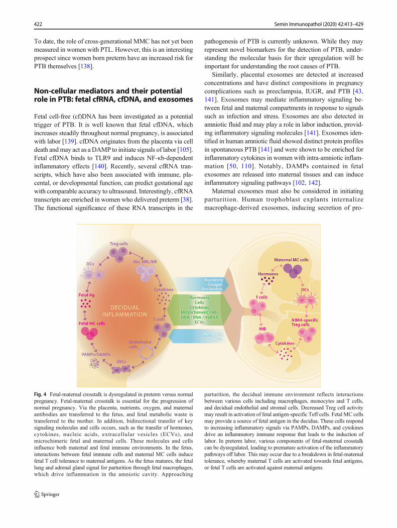
Fig. 4 Fetal-maternal crosstalk is dysregulated in preterm versus normalpregnancy. Fetal-maternal crosstalk is essential for the progression ofnormal pregnancy. Via the placenta, nutrients, oxygen, and maternalantibodies are transferred to the fetus, and fetal metabolic waste istransferred to the mother. In addition, bidirectional transfer of keysignaling molecules and cells occurs, such as the transfer of hormones,cytokines, nucleic acids, extracellular vesicles (ECVs), andmicrochimeric fetal and maternal cells. These molecules and cellsinfluence both maternal and fetal immune environments. In the fetus,interactions between fetal immune cells and maternal MC cells inducefetal T cell tolerance to maternal antigens. As the fetus matures, the fetallung and adrenal gland signal for parturition through fetal macrophages,which drive inflammation in the amniotic cavity. Approaching
parturition, the decidual immune environment reflects interactionsbetween various cells including macrophages, monocytes and T cells,and decidual endothelial and stromal cells. Decreased Treg cell activitymay result in activation of fetal antigen-specific Teff cells. Fetal MC cellsmay provide a source of fetal antigen in the decidua. These cells respondto increasing inflammatory signals via PAMPs, DAMPs, and cytokinesdrive an inflammatory immune response that leads to the induction oflabor. In preterm labor, various components of fetal-maternal crosstalkcan be dysregulated, leading to premature activation of the inflammatorypathways off labor. This may occur due to a breakdown in fetal-maternaltolerance, whereby maternal T cells are activated towards fetal antigens,or fetal T cells are activated against maternal antigens
422 Semin Immunopathol (2020) 42:413–429
inflammatory cytokines IL6, IL8, IL10, and IL12 [49]. Asmacrophages infected with intracellular bacteria releasePAMP-containing exosomes, which activate naïve T cellsand macrophages [143, 144], this may provide a mechanismfor the induction of decidual inflammation in infection-relatedPTB. Thus, macrophage-derived exosomes may traffic to andbe taken up by the placenta to deliver key signals facilitatingimmunomodulatory pathways of parturition. Whetherexosomes containing DAMPs are also delivered to the placen-ta in the case of sterile inflammation in term and pretermpregnancies is currently unknown.
Clinical developments aiming to identifybiomarker and therapeutic avenues
PTB is the leading cause of morbidity and mortality world-wide. Despite decades of research on the topic, the underlyingetiology of PTB is still largely unknown. Multiple fetal-maternal signaling pathways exist and provide key communi-cation between mother and fetus during pregnancy. This com-plexity clearly hampers the identification of the multilayeredprocesses implicated in the immunopathogenesis of PTB(Fig. 4).
To date, therapeutic options for PTB are sparse. Currenttreatments to prevent suspected preterm birth include P4 sup-plementation and progestin prophylaxis. Other studies aimedat PTB prevention by targeting immune mediators. Here, theTLR4-antagonist (+)-naloxone, which is the non-opioid iso-mer of the opioid receptor antagonist (−)-naloxone, was testedin mice, where it suppressed the inflammatory cascade andprevent preterm birth [78]. Another immune-based therapeu-tic approach preventing PTB in mice targeted IL1" by using anovel IL1 receptor biased ligand [64]. Given the importanceof IL10 mediating anti-inflammatory actions in the decidua,boosting IL10 in the decidua may represent one future strategyto combat PTB [67, 84]. The development of therapies aimingto increase Treg cells is also considered [145].
Conclusion
Despite its essential nature, a detailed understanding of themolecular mechanisms underlying normal labor induction isonly slowly emerging. PTB is a syndrome hypothesized toresult from pathological activation of the normal physiologicalprocesses that lead to labor [7]. Yet most of our understandingof the pathways involved in labor originates from studies in-vestigating single pathways or mediators by comparing pre-term and term pregnancies in humans and various animalmodels. As multiomic assessments are now underway to eval-uate the plausibility of immunological findings [146], thepower of biological signatures predictive of preterm birth
may soon be within reach and will inform novel approachesof early prediction and therapeutic interventions. Hence, un-derstanding of molecular mechanisms will likely experiencerapid developments in the near future, hereby enabling theidentification of women at risk and the development of earlyprevention strategies of PTB.
Funding Information Open Access funding provided by Projekt DEAL.Writing of this article and reference to own original data was supported bya research grant provided by the German Research Foundation (KFO296,AR232/25-2).
Compliance with ethical standards
Conflict of interest The authors declare that they have no conflict ofinterest.
Open Access This article is licensed under a Creative CommonsAttribution 4.0 International License, which permits use, sharing, adap-tation, distribution and reproduction in any medium or format, as long asyou give appropriate credit to the original author(s) and the source, pro-vide a link to the Creative Commons licence, and indicate if changes weremade. The images or other third party material in this article are includedin the article's Creative Commons licence, unless indicated otherwise in acredit line to the material. If material is not included in the article'sCreative Commons licence and your intended use is not permitted bystatutory regulation or exceeds the permitted use, you will need to obtainpermission directly from the copyright holder. To view a copy of thislicence, visit http://creativecommons.org/licenses/by/4.0/.
References
1. Blencowe H, Cousens S, Chou D, Oestergaard M, Say L, MollerAB, Kinney M, Lawn J (2013) Born too soon: the global epide-miology of 15 million preterm births. Reprod Health 10 Suppl 1:S2. https://doi.org/10.1186/1742-4755-10-s1-s2
2. Markopoulou P, Papanikolaou E, Analytis A, Zoumakis E,Siahanidou T (2019) Preterm birth as a risk factor for metabolicsyndrome and cardiovascular disease in adult life: a systematicreview and meta-analysis. J Pediatr 210:69–80.e65. https://doi.org/10.1016/j.jpeds.2019.02.041
3. Arpino C, Compagnone E, Montanaro M, Cacciatore D, Luca A,Cerulli A, Girolamo S, Curatolo P (2010) Preterm birth andneurodevelopmental outcome: a review. Childs Nerv Syst 26:1139–1149. https://doi.org/10.1007/s00381-010-1125-y
4. Goedicke-Fritz S, Härtel C, Krasteva-Christ G, Kopp MV, MeyerS, Zemlin M (2017) Preterm birth affects the risk of developingimmune-mediated diseases. Front Immunol 8(1266). https://doi.org/10.3389/fimmu.2017.01266
5. Sonnenschein-van der Voort AM, Arends LR, de Jongste JC,Annesi-Maesano I, Arshad SH, Barros H, Basterrechea M,Bisgaard H, Chatzi L, Corpeleijn E, Correia S, Craig LC,Devereux G, Dogaru C, Dostal M, Duchen K, Eggesbo M, vander Ent CK, Fantini MP, Forastiere F, Frey U, Gehring U, Gori D,van der Gugten AC, Hanke W, Henderson AJ, Heude B, IniguezC, Inskip HM,Keil T, Kelleher CC, KogevinasM,Kreiner-MollerE, Kuehni CE, Kupers LK, Lancz K, Larsen PS, Lau S,Ludvigsson J, Mommers M, Nybo Andersen AM, PalkovicovaL, Pike KC, Pizzi C, Polanska K, Porta D, Richiardi L, Roberts G,Schmidt A, Sram RJ, Sunyer J, Thijs C, Torrent M, Viljoen K,
423Semin Immunopathol (2020) 42:413–429
Wijga AH, Vrijheid M, Jaddoe VW, Duijts L (2014) Pretermbirth, infant weight gain, and childhood asthma risk: a meta-analysis of 147,000 European children. J Allergy Clin Immunol133(5):1317–1329. https://doi.org/10.1016/j.jaci.2013.12.1082
6. Moster D, Lie RT, Markestad T (2008) Long-term medical andsocial consequences of preterm birth. N Engl J Med 359(3):262–273. https://doi.org/10.1056/NEJMoa0706475
7. Romero R, Dey SK, Fisher SJ (2014) Preterm labor: one syn-drome, many causes. Science 345(6198):760–765. https://doi.org/10.1126/science.1251816
8. Purisch SE, Gyamfi-Bannerman C (2017) Epidemiology of pre-term birth. Semin Perinatol 41(7):387–391. https://doi.org/10.1053/j.semperi.2017.07.009
9. Zhang G, Feenstra B, Bacelis J, Liu X, Muglia LM, Juodakis J,Miller DE, Litterman N, Jiang PP, Russell L, Hinds DA, Hu Y,Weirauch MT, Chen X, Chavan AR, Wagner GP, Pavlicev M,Nnamani MC, Maziarz J, Karjalainen MK, Ramet M, Sengpiel V,Geller F, Boyd HA, Palotie A, Momany A, Bedell B, RyckmanKK, Huusko JM, Forney CR, Kottyan LC, Hallman M, TeramoK, Nohr EA, Davey Smith G, MelbyeM, Jacobsson B, Muglia LJ(2017) Genetic associations with gestational duration and sponta-neous preterm birth. N Engl J Med 377(12):1156–1167. https://doi.org/10.1056/NEJMoa1612665
10. Norwitz ER, Bonney EA, Snegovskikh VV, Williams MA,Phillippe M, Park JS, Abrahams VM (2015) Molecular regulationof parturition: the role of the decidual clock. Cold Spring HarbPerspect Med 5(11):a023143. https://doi.org/10.1101/cshperspect.a023143
11. Romero R, Espinoza J, Goncalves LF, Kusanovic JP, Friel LA,Nien JK (2006) Inflammation in preterm and term labour anddelivery. Semin Fetal Neonatal Med 11(5):317–326. https://doi.org/10.1016/j.siny.2006.05.001
12. Mohamed SA, Thota C, Browne PC, Diamond MP, Al-Hendy A(2014) Why is preterm birth stubbornly higher in African-Americans? Obstet Gynecol Int J 1(3):00019. https://doi.org/10.15406/ogij.2014.01.00019
13. Meuleman T, Lashley LELO, Dekkers OM, van Lith JMM, ClaasFHJ, Bloemenkamp KWM (2015) HLA associations and HLAsharing in recurrent miscarriage: a systematic review and meta-analysis. Hum Immunol 76(5):362–373. https://doi.org/10.1016/j.humimm.2015.02.004
14. Robertson SA, Moldenhauer LM (2014) Immunological determi-nants of implantation success. Int J Dev Biol 58(2–4):205–217.https://doi.org/10.1387/ijdb.140096sr
15. Arck PC, Hecher K (2013) Fetomaternal immune cross-talk andits consequences for maternal and offspring’s health. Nat Med19(5):548–556. https://doi.org/10.1038/nm.3160
16. Deshmukh H, Way SS (2019) Immunological basis for recurrentfetal loss and pregnancy complications. Annu Rev Pathol 14(1):185–210. https://doi.org/10.1146/annurev-pathmechdis-012418-012743
17. Robertson SA, Care AS, Moldenhauer LM (2018) Regulatory Tcells in embryo implantation and the immune response to preg-nancy. J Clin Invest 128(10):4224–4235. https://doi.org/10.1172/jci122182
18. Jiang TT, Chaturvedi V, Ertelt JM, Kinder JM, Clark DR, ValentAM, Xin L, Way SS (2014) Regulatory T cells: new keys forfurther unlocking the enigma of fetal tolerance and pregnancycomplications. J Immunol 192(11):4949–4956. https://doi.org/10.4049/jimmunol.1400498
19. Trowsdale J, Betz AG (2006) Mother’s little helpers: mechanismsof maternal-fetal tolerance. Nat Immunol 7(3):241–246. https://doi.org/10.1038/ni1317
20. Ferreira LMR, Meissner TB, Tilburgs T, Strominger JL(2017) HLA-G: at the interface of maternal-fetal tolerance.
Trends Immunol 38(4):272–286. https://doi.org/10.1016/j.it.2017.01.009
21. Jones RL, Stoikos C, Findlay JK, Salamonsen LA (2006) TGF-beta superfamily expression and actions in the endometrium andplacenta. Reproduction 132(2):217–232. https://doi.org/10.1530/rep.1.01076
22. Guerin LR,Moldenhauer LM, Prins JR, Bromfield JJ, Hayball JD,Robertson SA (2011) Seminal fluid regulates accumulation ofFOXP3+ regulatory T cells in the preimplantation mouse uterusthrough expanding the FOXP3+ cell pool and CCL19-mediatedrecruitment. Biol Reprod 85(2):397–408. https://doi.org/10.1095/biolreprod.110.088591
23. Aluvihare VR, Kallikourdis M, Betz AG (2004) Regulatory Tcells mediate maternal tolerance to the fetus. Nat Immunol 5(3):266–271. https://doi.org/10.1038/ni1037
24. Samstein RM, Josefowicz SZ, Arvey A, Treuting PM, RudenskyAY (2012) Extrathymic generation of regulatory T cells in placen-tal mammals mitigates maternal-fetal conflict. Cell 150(1):29–38.https://doi.org/10.1016/j.cell.2012.05.031
25. Rowe JH, Ertelt JM, Xin L, Way SS (2012) Pregnancy imprintsregulatory memory that sustains anergy to fetal antigen. Nature490(7418):102–106. https://doi.org/10.1038/nature11462
26. Chen T, Darrasse-Jeze G, Bergot AS, Courau T, Churlaud G,Valdivia K, Strominger JL, Ruocco MG, Chaouat G, KlatzmannD (2013) Self-specific memory regulatory T cells protect embryosat implantation in mice. J Immunol 191(5):2273–2281. https://doi.org/10.4049/jimmunol.1202413
27. Moldenhauer LM, Diener KR, Thring DM, Brown MP, HayballJD, Robertson SA (2009) Cross-presentation ofmale seminal fluidantigens elicits T cell activation to initiate the female immuneresponse to pregnancy. J Immunol 182(12):8080–8093. https://doi.org/10.4049/jimmunol.0804018
28. Fallarino F, Grohmann U, Hwang KW, Orabona C, Vacca C,Bianchi R, Belladonna ML, Fioretti MC, Alegre M-L, Puccetti P(2003)Modulation of tryptophan catabolism by regulatory T cells.Nat Immunol 4(12):1206–1212. https://doi.org/10.1038/ni1003
29. Kalekar LA, Schmiel SE, Nandiwada SL, LamWY, Barsness LO,Zhang N, Stritesky GL, Malhotra D, Pauken KE, Linehan JL,O’Sullivan MG, Fife BT, Hogquist KA, Jenkins MK, MuellerDL (2016) CD4(+) T cell anergy prevents autoimmunity and gen-erates regulatory T cell precursors. Nat Immunol 17(3):304–314.https://doi.org/10.1038/ni.3331
30. Svensson-Arvelund J, Mehta RB, Lindau R, Mirrasekhian E,Rodriguez-Martinez H, Berg G, Lash GE, Jenmalm MC,Ernerudh J (2015) The human fetal placenta promotes toleranceagainst the semiallogeneic fetus by inducing regulatory T cells andhomeostatic M2 macrophages. J Immunol 194(4):1534–1544.https://doi.org/10.4049/jimmunol.1401536
31. Kinder JM, Stelzer IA, Arck PC, Way SS (2017) Immunologicalimplications of pregnancy-induced microchimerism. Nat RevImmunol 17(8):483–494. https://doi.org/10.1038/nri.2017.38
32. Ariga H, Ohto H, BuschMP, Imamura S, Watson R, ReedW, LeeTH (2001) Kinetics of fetal cellular and cell-free DNA in thematernal circulation during and after pregnancy: implications fornoninvasive prenatal diagnosis. Transfusion 41(12):1524–1530.https://doi.org/10.1046/j.1537-2995.2001.41121524.x
33. Walknowska J, Conte FA, Grumbach MM (1969) Practical andtheoretical implications of fetal-maternal lymphocyte transfer.Lancet 1(7606):1119–1122. https://doi.org/10.1016/s0140-6736(69)91642-0
34. Fujiki Y, Johnson KL, Tighiouart H, Peter I, Bianchi DW (2008)Fetomaternal trafficking in the mouse increases as delivery ap-proaches and is highest in the maternal lung. Biol Reprod 79(5):841–848. https://doi.org/10.1095/biolreprod.108.068973
35. Mold JE, Michaelsson J, Burt TD, Muench MO, Beckerman KP,Busch MP, Lee TH, Nixon DF, McCune JM (2008) Maternal
424 Semin Immunopathol (2020) 42:413–429
alloantigens promote the development of tolerogenic fetal regula-tory T cells in utero. Science 322(5907):1562–1565. https://doi.org/10.1126/science.1164511
36. Kinder JM, Jiang TT, Ertelt JM, Xin L, Strong BS, Shaaban AF,Way SS (2015) Cross-generational reproductive fitness enforcedbymicrochimeric maternal cells. Cell 162(3):505–515. https://doi.org/10.1016/j.cell.2015.07.006
37. KohW, PanW, Gawad C, Fan HC, Kerchner GA,Wyss-Coray T,Blumenfeld YJ, El-Sayed YY, Quake SR (2014) Noninvasivein vivo monitoring of tissue-specific global gene expression inhumans. Proc Natl Acad Sci 111(20):7361–7366. https://doi.org/10.1073/pnas.1405528111
38. Ngo TTM, Moufarrej MN, Rasmussen MH, Camunas-Soler J,Pan W, Okamoto J, Neff NF, Liu K, Wong RJ, Downes K,Tibshirani R, Shaw GM, Skotte L, Stevenson DK, Biggio JR,Elovitz MA, Melbye M, Quake SR (2018) Noninvasive bloodtests for fetal development predict gestational age and pretermdelivery. Science 360(6393):1133–1136. https://doi.org/10.1126/science.aar3819
39. Poon LLM, Leung TN, Lau TK, LoYMD (2000) Presence of fetalRNA in maternal plasma. Clin Chem 46(11):1832–1834
40. Maron JL, Johnson KL, Slonim D, Lai CQ, Ramoni M, AlterovitzG, Jarrah Z, Yang Z, Bianchi DW (2007) Gene expression analy-sis in pregnant women and their infants identifies unique fetalbiomarkers that circulate in maternal blood. J Clin Invest117(10):3007–3019. https://doi.org/10.1172/jci29959
41. van Wijk IJ, de Hoon AC, Jurhawan R, Tjoa ML, Griffioen S,Mulders MAM, van Vugt JMG, Oudejans CBM (2000) Detectionof apoptotic fetal cells in plasma of pregnant women. Clin Chem46(5):729–731
42. Lo YM, Corbetta N, Chamberlain PF, Rai V, Sargent IL, RedmanCW, Wainscoat JS (1997) Presence of fetal DNA in maternalplasma and serum. Lancet 350(9076):485–487. https://doi.org/10.1016/s0140-6736(97)02174-0
43. Mitchell MD, Peiris HN, Kobayashi M, Koh YQ, Duncombe G,Illanes SE, Rice GE, Salomon C (2015) Placental exosomes innormal and complicated pregnancy. Am J Obstet Gynecol 213(4Suppl):S173–S181. https://doi.org/10.1016/j.ajog.2015.07.001
44. Sarker S, Scholz-Romero K, Perez A, Illanes SE, Mitchell MD,Rice GE, Salomon C (2014) Placenta-derived exosomes continu-ously increase in maternal circulation over the first trimester ofpregnancy. J Transl Med 12:204. https://doi.org/10.1186/1479-5876-12-204
45. Salomon C, Torres MJ, Kobayashi M, Scholz-Romero K,Sobrevia L, Dobierzewska A, Illanes SE, Mitchell MD, Rice GE(2014) A gestational profile of placental exosomes in maternalplasma and their effects on endothelial cell migration. PLoS One9(6):e98667. https://doi.org/10.1371/journal.pone.0098667
46. Desrochers LM, Bordeleau F, Reinhart-King CA, Cerione RA,Antonyak MA (2016) Microvesicles provide a mechanism forintercellular communication by embryonic stem cells during em-bryo implantation. Nat Commun 7:11958. https://doi.org/10.1038/ncomms11958
47. Luo SS, Ishibashi O, Ishikawa G, Ishikawa T, Katayama A,Mishima T, Takizawa T, Shigihara T, Goto T, Izumi A,Ohkuchi A, Matsubara S, Takeshita T, Takizawa T (2009)Human villous trophoblasts express and secrete placenta-specificmicroRNAs into maternal circulation via exosomes. Biol Reprod81(4):717–729. https://doi.org/10.1095/biolreprod.108.075481
48. Ouyang Y, Mouillet JF, Coyne CB, Sadovsky Y (2014) Review:placenta-specific microRNAs in exosomes - good things come innano-packages. Placenta 35(Suppl):S69–S73. https://doi.org/10.1016/j.placenta.2013.11.002
49. Holder B, Jones T, Sancho Shimizu V, Rice TF, Donaldson B,Bouqueau M, Forbes K, Kampmann B (2016) Macrophageexosomes induce placental inflammatory cytokines: a novel mode
of maternal-placental messaging. Traffic 17(2):168–178. https://doi.org/10.1111/tra.12352
50. Romero R, Grivel JC, Tarca AL, Chaemsaithong P, Xu Z,Fitzgerald W, Hassan SS, Chaiworapongsa T, Margolis L(2015) Evidence of perturbations of the cytokine network in pre-term labor. Am J Obstet Gynecol 213(6):836.e831–836.e818.https://doi.org/10.1016/j.ajog.2015.07.037
51. Soto E, Romero R, Richani K, Yoon BH, Chaiworapongsa T,Vaisbuch E, Mittal P, Erez O, Gotsch F, Mazor M, KusanovicJP (2009) Evidence for complement activation in the amnioticfluid of women with spontaneous preterm labor and intra-amniotic infection. J Matern Fetal Neonatal Med 22(11):983–992. https://doi.org/10.3109/14767050902994747
52. Schober L, Radnai D, Schmitt E, Mahnke K, Sohn C, Steinborn A(2012) Term and preterm labor: decreased suppressive activityand changes in composition of the regulatory T-cell pool.Immunol Cell Biol 90(10):935–944. https://doi.org/10.1038/icb.2012.33
53. Gomez-Lopez N, Romero R, Xu Y, Miller D, Arenas-HernandezM, Garcia-Flores V, Panaitescu B, Galaz J, Hsu CD, Para R, BerrySM (2019) Fetal T cell activation in the amniotic cavity duringpreterm labor: a potential mechanism for a subset of idiopathicpreterm birth. J Immunol 203(7):1793–1807. https://doi.org/10.4049/jimmunol.1900621
54. Frascoli M, Coniglio L, Witt R, Jeanty C, Fleck-Derderian S,Myers DE, Lee TH, Keating S, Busch MP, Norris PJ, Tang Q,Cruz G, Barcellos LF, Gomez-Lopez N, Romero R, MacKenzieTC (2018) Alloreactive fetal T cells promote uterine contractilityin preterm labor via IFN-gamma and TNF-alpha. Sci Transl Med10(438). https://doi.org/10.1126/scitranslmed.aan2263
55. Elovitz MA, Mrinalini C (2004) Animal models of preterm birth.Trends Endocrinol Metab 15(10):479–487. https://doi.org/10.1016/j.tem.2004.10.009
56. Brosens I, Pijnenborg R, Vercruysse L, Romero R (2011) The"great obstetrical syndromes" are associated with disorders ofdeep placentation. Am J Obstet Gynecol 204(3):193–201.https://doi.org/10.1016/j.ajog.2010.08.009
57. Romero R, Kusanovic JP, Chaiworapongsa T, Hassan SS (2011)Placental bed disorders in preterm labor, preterm PROM, sponta-neous abortion and abruptio placentae. Best Pract Res Clin ObstetGynaecol 25(3):313–327. https://doi.org/10.1016/j.bpobgyn.2011.02.006
58. Cao-Lei L, Laplante DP, King S (2016) Prenatal maternal stressand epigenetics: review of the human research. CurrMol Biol Rep2(1):16–25. https://doi.org/10.1007/s40610-016-0030-x
59. Bashiri A, Halper KI, Orvieto R (2018) Recurrent implantationfailure-update overview on etiology, diagnosis, treatment and fu-ture directions. Reprod Biol Endocrinol 16(1):121–121. https://doi.org/10.1186/s12958-018-0414-2
60. Fettweis JM, Serrano MG, Brooks JP, Edwards DJ, Girerd PH,Parikh HI, Huang B, Arodz TJ, Edupuganti L, Glascock AL, Xu J,Jimenez NR, Vivadelli SC, Fong SS, Sheth NU, Jean S, Lee V,Bokhari YA, Lara AM, Mistry SD, Duckworth RA, Bradley SP,Koparde VN, Orenda XV, Milton SH, Rozycki SK, MatveyevAV, Wright ML, Huzurbazar SV, Jackson EM, Smirnova E,Korlach J, Tsai Y-C, Dickinson MR, Brooks JL, Drake JI,Chaffin DO, Sexton AL, Gravett MG, Rubens CE, WijesooriyaNR, Hendricks-Muñoz KD, Jefferson KK, Strauss JF, Buck GA(2019) The vaginal microbiome and preterm birth. NatMed 25(6):1012–1021. https://doi.org/10.1038/s41591-019-0450-2
61. Serrano MG, Parikh HI, Brooks JP, Edwards DJ, Arodz TJ,Edupuganti L, Huang B, Girerd PH, Bokhari YA, Bradley SP,Brooks JL, Dickinson MR, Drake JI, Duckworth RA, Fong SS,Glascock AL, Jean S, Jimenez NR, Khoury J, Koparde VN, LaraAM, Lee V, Matveyev AV, Milton SH, Mistry SD, Rozycki SK,Sheth NU, Smirnova E, Vivadelli SC, Wijesooriya NR, Xu J, Xu
425Semin Immunopathol (2020) 42:413–429
P, Chaffin DO, Sexton AL, Gravett MG, Rubens CE, Hendricks-Muñoz KD, Jefferson KK, Strauss JF, Fettweis JM, Buck GA(2019) Racioethnic diversity in the dynamics of the vaginalmicrobiome during pregnancy. Nat Med 25(6):1001–1011.https://doi.org/10.1038/s41591-019-0465-8
62. Gargano JW, Holzman CB, Senagore PK, Reuss ML, Pathak DR,Williams MA, Fisher R (2010) Evidence of placental haemor-rhage and preterm delivery. BJOG Int J Obstet Gynaecol 117(4):445–455. https://doi.org/10.1111/j.1471-0528.2009.02472.x
63. Cappelletti M, Presicce P, LawsonMJ, Chaturvedi V, StankiewiczTE, Vanoni S, Harley ITW, McAlees JW, Giles DA, Moreno-Fernandez ME, Rueda CM, Senthamaraikannan P, Sun X, KarnsR, Hoebe K, Janssen EM, Karp CL, Hildeman DA, Hogan SP,Kallapur SG, Chougnet CA, Way SS, Divanovic S (2017) Type Iinterferons regulate susceptibility to inflammation-induced pre-term birth. JCI Insight 2(5). https://doi.org/10.1172/jci.insight.91288
64. Nadeau-Vallee M, Quiniou C, Palacios J, Hou X, Erfani A,Madaan A, Sanchez M, Leimert K, Boudreault A, Duhamel F,Rivera JC, Zhu T, Noueihed B, Robertson SA, Ni X, OlsonDM, Lubell W, Girard S, Chemtob S (2015) Novel noncompeti-tive IL-1 receptor-biased ligand prevents infection- andinflammation-induced preterm birth. J Immunol 195(7):3402–3415. https://doi.org/10.4049/jimmunol.1500758
65. Robertson SA, Christiaens I, Dorian CL, Zaragoza DB, Care AS,Banks AM, Olson DM (2010) Interleukin-6 is an essential deter-minant of on-time parturition in the mouse. Endocrinology 151(8):3996–4006. https://doi.org/10.1210/en.2010-0063
66. SlutskyR, Romero R,XuY,Galaz J,Miller D, DoneB, Tarca AL,Gregor S, Hassan SS, Leng Y, Gomez-Lopez N (2019) Exhaustedand senescent T cells at the maternal-fetal Interface in preterm andterm labor. J Immunol Res 2019:3128010. https://doi.org/10.1155/2019/3128010
67. Robertson SA, Skinner RJ, Care AS (2006) Essential role for IL-10 in resistance to lipopolysaccharide-induced preterm labor inmice. J Immunol 177(7):4888–4896. https://doi.org/10.4049/jimmunol.177.7.4888
68. St Louis D, Romero R, Plazyo O, Arenas-Hernandez M,Panaitescu B, Xu Y, Milovic T, Xu Z, Bhatti G, Mi QS, DrewloS, Tarca AL, Hassan SS, Gomez-Lopez N (2016) Invariant NKTcell activation induces late preterm birth that is attenuated byrosiglitazone. J Immunol 196(3):1044–1059. https://doi.org/10.4049/jimmunol.1501962
69. WegorzewskaM, Le T, Tang Q, MacKenzie TC (2014) Increasedmaternal T cell microchimerism in the allogeneic fetus duringLPS-induced preterm labor in mice. Chimerism 5(3–4):68–74.https://doi.org/10.1080/19381956.2014.1002703
70. Wahid HH, Dorian CL, Chin PY, Hutchinson MR, Rice KC,Olson DM, Moldenhauer LM, Robertson SA (2015) Toll-likereceptor 4 is an essential upstream regulator of on-time parturitionand perinatal viability in mice. Endocrinology 156(10):3828–3841. https://doi.org/10.1210/en.2015-1089
71. Filipovich Y, Lu SJ, Akira S, Hirsch E (2009) The adaptor proteinMyD88 is essential for E coli-induced preterm delivery in mice.Am J Obstet Gynecol 200(1):93.e91–93.e98. https://doi.org/10.1016/j.ajog.2008.08.038
72. Romero R, Tartakovsky B (1992) The natural interleukin-1 recep-tor antagonist prevents interleukin-1-induced preterm delivery inmice. Am J Obstet Gynecol 167(4 Pt 1):1041–1045. https://doi.org/10.1016/s0002-9378(12)80035-4
73. Arenas-Hernandez M, Romero R, Xu Y, Panaitescu B, Garcia-Flores V, Miller D, Ahn H, Done B, Hassan SS, Hsu CD, TarcaAL, Sanchez-Torres C, Gomez-Lopez N (2019) Effector and ac-tivated T cells induce preterm labor and birth that is prevented bytreatment with progesterone. J Immunol 202(9):2585–2608.https://doi.org/10.4049/jimmunol.1801350
74. Boyson JE, Nagarkatti N, Nizam L, Exley MA, Strominger JL(2006) Gestation stage-dependent mechanisms of invariant naturalkiller T cell-mediated pregnancy loss. Proc Natl Acad Sci U S A103(12):4580–4585. https://doi.org/10.1073/pnas.0511025103
75. Sheller-Miller S, Trivedi J, Yellon SM, Menon R (2019)Exosomes cause preterm birth in mice: evidence for paracrinesignaling in pregnancy. Sci Rep 9(1):608. https://doi.org/10.1038/s41598-018-37002-x
76. Gomez-Lopez N, Romero R, Plazyo O, Panaitescu B, FurcronAE, Miller D, Roumayah T, Flom E, Hassan SS (2016) Intra-amniotic administration of HMGB1 induces spontaneous pretermlabor and birth. Am J Reprod Immunol 75(1):3–7. https://doi.org/10.1111/aji.12443
77. Gonzalez JM, Franzke CW, Yang F, Romero R, Girardi G (2011)Complement activation triggers metalloproteinases release induc-ing cervical remodeling and preterm birth in mice. Am J Pathol179(2):838–849. https://doi.org/10.1016/j.ajpath.2011.04.024
78. Chin PY, Dorian CL, Hutchinson MR, Olson DM, Rice KC,Moldenhauer LM, Robertson SA (2016) Novel toll-like recep-tor-4 antagonist (+)-naloxone protects mice from inflammation-induced preterm birth. Sci Rep 6:36112. https://doi.org/10.1038/srep36112
79. Bizargity P, Del Rio R, Phillippe M, Teuscher C, Bonney EA(2009) Resistance to lipopolysaccharide-induced preterm deliverymediated by regulatory T cell function in mice. Biol Reprod 80(5):874–881. https://doi.org/10.1095/biolreprod.108.074294
80. Agrawal V, JaiswalMK, Ilievski V, BeamanKD, Jilling T, HirschE (2014) Platelet-activating factor: a role in preterm delivery andan essential interaction with toll-like receptor signaling in mice.Biol Reprod 91(5):119. https://doi.org/10.1095/biolreprod.113.116012
81. Gao L, Rabbitt EH, Condon JC, Renthal NE, Johnston JM,Mitsche MA, Chambon P, Xu J, O’Malley BW, Mendelson CR(2015) Steroid receptor coactivators 1 and 2 mediate fetal-to-maternal signaling that initiates parturition. J Clin Invest 125(7):2808–2824. https://doi.org/10.1172/JCI78544
82. Montalbano AP, Hawgood S, Mendelson CR (2013) Mice defi-cient in surfactant protein A (SP-A) and SP-D or in TLR2manifestdelayed parturition and decreased expression of inflammatory andcontractile genes. Endocrinology 154(1):483–498. https://doi.org/10.1210/en.2012-1797
83. Mizoguchi M, Ishida Y, Nosaka M, Kimura A, Kuninaka Y,Yahata T, Nanjo S, Toujima S, Minami S, Ino K, Mukaida N,Kondo T (2018) Prevention of lipopolysaccharide-induced pre-term labor by the lack of CX3CL1-CX3CR1 interaction in mice.PLoS One 13(11):e0207085–e0207085. https://doi.org/10.1371/journal.pone.0207085
84. DengW, Yuan J, Cha J, Sun X, Bartos A, Yagita H, Hirota Y, DeySK (2019) Endothelial cells in the decidual bed are potential ther-apeutic targets for preterm birth prevention. Cell Rep 27(6):1755–1768.e1754. https://doi.org/10.1016/j.celrep.2019.04.049
85. Hirota Y, Cha J, Yoshie M, Daikoku T, Dey SK (2011)Heightened uterine mammalian target of rapamycin complex 1(mTORC1) signaling provokes preterm birth in mice. Proc NatlAcad Sci U S A 108(44):18073–18078. https://doi.org/10.1073/pnas.1108180108
86. Cha J, Bartos A, Egashira M, Haraguchi H, Saito-Fujita T,Leishman E, Bradshaw H, Dey SK, Hirota Y (2013)Combinatory approaches prevent preterm birth profoundly exac-erbated by gene-environment interactions. J Clin Invest 123(9):4063–4075. https://doi.org/10.1172/JCI70098
87. Yoshida M, Takayanagi Y, Ichino-Yamashita A, Sato K,Sugimoto Y, Kimura T, Nishimori K (2019) Functional hierarchyof uterotonics required for successful parturition in mice.Endocrinology 160(12):2800–2810. https://doi.org/10.1210/en.2019-00499
426 Semin Immunopathol (2020) 42:413–429
88. McCarthy R, Martin-Fairey C, Sojka DK, Herzog ED, JungheimES, Stout MJ, Fay JC, Mahendroo M, Reese J, Herington JL,Plosa EJ, Shelton EL, England SK (2018) Mouse models of pre-term birth: suggested assessment and reporting guidelines. BiolReprod 99(5):922–937. https://doi.org/10.1093/biolre/ioy109
89. Murray SA, Morgan JL, Kane C, Sharma Y, Heffner CS, Lake J,Donahue LR (2010) Mouse gestation length is genetically deter-mined. PLoS One 5(8):e12418. https://doi.org/10.1371/journal.pone.0012418
90. Lindström TM, Bennett PR (2005) The role of nuclear factorkappa B in human labour. Reproduction 130(5):569. https://doi.org/10.1530/rep.1.00197
91. Kisielewicz A, SchaierM, Schmitt E, Hug F, Haensch GM,MeuerS, Zeier M, Sohn C, Steinborn A (2010) A distinct subset of HLA-DR+-regulatory T cells is involved in the induction of pretermlabor during pregnancy and in the induction of organ rejectionafter transplantation. Clin Immunol 137(2):209–220. https://doi.org/10.1016/j.clim.2010.07.008
92. Shah NM, Edey LF, Imami N, JohnsonMR (2020) Human labouris associated with altered regulatory T cell function and maternalimmune activation. Clin Exp Immunol 199(2):182–200. https://doi.org/10.1111/cei.13384
93. Gomez-Lopez N, Vega-Sanchez R, Castillo-CastrejonM, RomeroR, Cubeiro-Arreola K, Vadillo-Ortega F (2013) Evidence for arole for the adaptive immune response in human term parturition.Am J Reprod Immunol 69(3):212–230. https://doi.org/10.1111/aji.12074
94. Boyson JE, Rybalov B, Koopman LA, Exley M, Balk SP, RackeFK, Schatz F, Masch R, Wilson SB, Strominger JL (2002) CD1dand invariant NKT cells at the human maternal-fetal interface.Proc Natl Acad Sci U S A 99(21):13741–13746. https://doi.org/10.1073/pnas.162491699
95. Li LP, Fang YC, Dong GF, Lin Y, Saito S (2012) Depletion ofinvariant NKT cells reduces inflammation-induced preterm deliv-ery in mice. J Immunol 188(9):4681–4689. https://doi.org/10.4049/jimmunol.1102628
96. Liassides C, Papadopoulos A, Siristatidis C, Damoraki G,Liassidou A, Chrelias C, Kassanos D, Giamarellos-BourboulisEJ (2019) Single nucleotide polymorphisms of toll-like receptor-4 and of autophagy-related gene 16 like-1 gene for predispositionof premature delivery: a prospective study. Medicine 98(40):e17313. https://doi.org/10.1097/md.0000000000017313
97. Patni S, Wynen LP, Seager AL, Morgan G, White JO, ThorntonCA (2009) Expression and activity of toll-like receptors 1–9 in thehuman term placenta and changes associated with labor at term.Biol Reprod 80(2):243–248. https://doi.org/10.1095/biolreprod.108.069252
98. Krediet TG, Wiertsema SP, Vossers MJ, Hoeks SBEA, Fleer A,Ruven HJT, Rijkers GT (2007) Toll-like receptor 2 polymorphismis associated with preterm birth. Pediatr Res 62(4):474–476.https://doi.org/10.1203/PDR.0b013e31813c9401
99. Lorenz E, HallmanM,Marttila R, Haataja R, Schwartz DA (2002)Association between the Asp299Gly polymorphisms in the toll-like receptor 4 and premature births in the Finnish population.Pediatr Res 52(3):373–376. https://doi.org/10.1203/00006450-200209000-00011
100. Romero R, Xu Y, Plazyo O, Chaemsaithong P, ChaiworapongsaT, Unkel R, Than NG, Chiang PJ, Dong Z, Xu Z, Tarca AL,Abrahams VM, Hassan SS, Yeo L, Gomez-Lopez N (2018) Arole for the Inflammasome in spontaneous labor at term. Am JReprod Immunol 79(6):e12440. https://doi.org/10.1111/aji.12440
101. Nadeau-Vallée M, Obari D, Palacios J, Brien M-È, Duval C,Chemtob S, Girard S (2016) Sterile inflammation and pregnancycomplications: a review. Reproduction 152(6):R277. https://doi.org/10.1530/rep-16-0453
102. Menon R (2019) Initiation of human parturition: signaling fromsenescent fetal tissues via extracellular vesicle mediated paracrinemechanism. Obstet Gynecol Sci 62(4):199–211. https://doi.org/10.5468/ogs.2019.62.4.199
103. Romero R, Chaiworapongsa T, Alpay Savasan Z, Xu Y, HusseinY, Dong Z, Kusanovic JP, Kim CJ, Hassan SS (2011) Damage-associated molecular patterns (DAMPs) in preterm labor with in-tact membranes and preterm PROM: a study of the alarminHMGB1. J Matern Fetal Neonatal Med 24(12):1444–1455.https://doi.org/10.3109/14767058.2011.591460
104. Plazyo O, Romero R, Unkel R, Balancio A, Mial TN, Xu Y, DongZ, Hassan SS, Gomez-Lopez N (2016) HMGB1 induces an in-flammatory response in the chorioamniotic membranes that is par-tially mediated by the inflammasome1. Biol Reprod 95(6). https://doi.org/10.1095/biolreprod.116.144139
105. Boeckel SR, Davidson DJ, Norman JE, Stock SJ (2018) Cell-freefetal DNA and spontaneous preterm birth. Reproduction 155(3):R137. https://doi.org/10.1530/rep-17-0619
106. Silver RK, Caplan MS, Kelly AM (1992) Amniotic fluid platelet-activating factor (PAF) is elevated in patients with tocolytic failureand preterm delivery. Prostaglandins 43(2):181–187. https://doi.org/10.1016/0090-6980(92)90085-8
107. Hoffman DR, Romero R, Johnston JM (1990) Detection ofplatelet-activating factor in amniotic fluid of complicated preg-nancies. Am J Obstet Gynecol 162(2):525–528. https://doi.org/10.1016/0002-9378(90)90423-5
108. Zhu YP, Hoffman DR, Hwang SB, Miyaura S, Johnston JM(1991) Prolongation of parturition in the pregnant rat followingtreatment with a platelet activating factor receptor antagonist. BiolReprod 44(1):39–42. https://doi.org/10.1095/biolreprod44.1.39
109. Elovitz MA, Wang Z, Chien EK, Rychlik DF, Phillippe M (2003)A new model for inflammation-induced preterm birth: the role ofplatelet-activating factor and toll-like receptor-4. Am J Pathol163(5):2103–2111. https://doi.org/10.1016/s0002-9440(10)63567-5
110. Bhatti G, Romero R, Rice GE, Fitzgerald W, Pacora P, Gomez-Lopez N, Kavdia M, Tarca AL, Margolis L (2020)Compartmentalized profiling of amniotic fluid cytokines in wom-en with preterm labor. PLoS One 15(1):e0227881–e0227881.https://doi.org/10.1371/journal.pone.0227881
111. Bayar E, Bennett PR, ChanD, Sykes L,MacIntyre DA (2020) Thepregnancy microbiome and preterm birth. Seminars inImmunopathology. https://doi.org/10.1007/s00281-020-00817-w
112. Andrews WW, Goldenberg RL, Hauth JC, Cliver SP, Copper R,ConnerM (2006) Interconceptional antibiotics to prevent spontaneouspreterm birth: a randomized clinical trial. Am J Obstet Gynecol194(3):617–623. https://doi.org/10.1016/j.ajog.2005.11.049
113. Espinoza J, Erez O, Romero R (2006) Preconceptional antibiotictreatment to prevent preterm birth in women with a previous pre-term delivery. Am J Obstet Gynecol 194(3):630–637. https://doi.org/10.1016/j.ajog.2005.11.050
114. Chen C, Song X,WeiW, ZhongH, Dai J, Lan Z, Li F, Yu X, FengQ,Wang Z, Xie H, Chen X, Zeng C,Wen B, Zeng L, Du H, TangH, Xu C, Xia Y, Xia H, Yang H,Wang J, Wang J, Madsen L, BrixS, Kristiansen K, Xu X, Li J, Wu R, Jia H (2017) The microbiotacontinuum along the female reproductive tract and its relation touterine-related diseases. Nat Commun 8(1):875. https://doi.org/10.1038/s41467-017-00901-0
115. Moreno I, Codoñer FM, Vilella F, Valbuena D, Martinez-Blanch JF,Jimenez-Almazán J, Alonso R, Alamá P, Remohí J, Pellicer A,RamonD, SimonC (2016) Evidence that the endometrial microbiotahas an effect on implantation success or failure. Am JObstet Gynecol215(6):684–703. https://doi.org/10.1016/j.ajog.2016.09.075
116. Aagaard K, Ma J, Antony KM, Ganu R, Petrosino J, Versalovic J(2014) The placenta harbors a uniquemicrobiome. Sci Transl Med
427Semin Immunopathol (2020) 42:413–429
6(237):237ra265–237ra265. https://doi.org/10.1126/scitranslmed.3008599
117. de Goffau MC, Lager S, Sovio U, Gaccioli F, Cook E, Peacock SJ,Parkhill J, Charnock-Jones DS, Smith GCS (2019) Human placentahas no microbiome but can contain potential pathogens. Nature572(7769):329–334. https://doi.org/10.1038/s41586-019-1451-5
118. Nadeem L, Shynlova O, Matysiak-Zablocki E, Mesiano S, DongX, Lye S (2016) Molecular evidence of functional progesteronewithdrawal in human myometrium. Nat Commun 7:11565.https://doi.org/10.1038/ncomms11565
119. MURR SM, STABENFELDT GH, BRADFORD GE,GESCHWIND II (1974) Plasma progesterone during pregnancyin the mouse1. Endocrinology 94(4):1209–1211. https://doi.org/10.1210/endo-94-4-1209
120. Piekorz RP, Gingras S, Hoffmeyer A, Ihle JN,Weinstein Y (2005)Regulation of progesterone levels during pregnancy and parturi-tion by signal transducer and activator of transcription 5 and20alpha-hydroxysteroid dehydrogenase. Mol Endocrinol 19(2):431–440. https://doi.org/10.1210/me.2004-0302
121. Dudley DJ, Branch DW, Edwin SS, Mitchell MD (1996)Induction of preterm birth in mice by RU486. Biol Reprod55(5):992–995. https://doi.org/10.1095/biolreprod55.5.992
122. Lee JH, Lydon JP, Kim CH (2012) Progesterone suppresses themTOR pathway and promotes generation of induced regulatory Tcells with increased stability. Eur J Immunol 42(10):2683–2696.https://doi.org/10.1002/eji.201142317
123. Hierweger AM, Engler JB, Friese MA, Reichardt HM, Lydon J,DeMayo F, Mittrücker H-W, Arck PC (2019) Progesterone mod-ulates the T-cell response via glucocorticoid receptor-dependentpathways. Am J Reprod Immunol 81(2):e13084. https://doi.org/10.1111/aji.13084
124. Thiele K, Hierweger AM, Riquelme JIA, Solano ME, Lydon JP,Arck PC (2019) Impaired progesterone-responsiveness ofCD11c(+) dendritic cells affects the generation of CD4(+) regula-tory T cells and is associated with intrauterine growth restriction inmice. Front Endocrinol (Lausanne) 10:96. https://doi.org/10.3389/fendo.2019.00096
125. Care AS, Diener KR, Jasper MJ, Brown HM, Ingman WV,Robertson SA (2013) Macrophages regulate corpus luteum devel-opment during embryo implantation in mice. J Clin Invest 123(8):3472–3487. https://doi.org/10.1172/JCI60561
126. Hirsch E, Muhle R (2002) Intrauterine bacterial inoculation in-duces labor in the mouse by mechanisms other than progesteronewithdrawal. Biol Reprod 67(4):1337–1341. https://doi.org/10.1095/biolreprod67.4.1337
127. Mendelson CR, Gao L,Montalbano AP (2019) Multifactorial reg-ulation of myometrial contractility during pregnancy and parturi-tion. Front Endocrinol 10(714). https://doi.org/10.3389/fendo.2019.00714
128. Anamthathmakula P, Kyathanahalli C, Ingles J, Hassan SS,Condon JC, Jeyasuria P (2019) Estrogen receptor alpha isoformERdelta7 in myometrium modulates uterine quiescence duringpregnancy. EBioMedicine 39:520–530. https://doi.org/10.1016/j.ebiom.2018.11.038
129. Nancy P, Siewiera J, Rizzuto G, Tagliani E, Osokine I, ManandharP, Dolgalev I, Clementi C, Tsirigos A, Erlebacher A (2018)H3K27me3 dynamics dictate evolving uterine states in pregnancyand parturition. J Clin Invest 128(1):233–247. https://doi.org/10.1172/JCI95937
130. Torricelli M, Giovannelli A, Leucci E, De Falco G, Reis FM,Imperatore A, Florio P, Petraglia F (2007) Labor (term and pre-term) is associated with changes in the placental mRNA expres-sion of corticotrophin-releasing factor. Reprod Sci 14(3):241–245.https://doi.org/10.1177/1933719107300971
131. Condon JC, Jeyasuria P, Faust JM, Mendelson CR (2004)Surfactant protein secreted by the maturing mouse fetal lung acts
as a hormone that signals the initiation of parturition. Proc NatlAcad Sci U S A 101(14):4978–4983. https://doi.org/10.1073/pnas.0401124101
132. Guillot L, Balloy V, McCormack FX, Golenbock DT, ChignardM, Si-Tahar M (2002) Cutting edge: the immunostimulatory ac-tivity of the lung surfactant protein-A involves toll-like receptor 4.J Immunol 168(12):5989–5992. https://doi.org/10.4049/jimmunol.168.12.5989
133. Berry SM, Romero R, Gomez R, Puder KS, Ghezzi F, Cotton DB,Bianchi DW (1995) Premature parturition is characterized by inutero activation of the fetal immune system. Am J Obstet Gynecol173(4):1315–1320. https://doi.org/10.1016/0002-9378(95)91378-5
134. Elahi S, Ertelt JM, Kinder JM, Jiang TT, Zhang X, Xin L,Chaturvedi V, Strong BS, Qualls JE, Steinbrecher KA, KalfaTA, Shaaban AF, Way SS (2013) Immunosuppressive CD71+erythroid cells compromise neonatal host defence against infec-tion. Nature 504(7478):158–162. https://doi.org/10.1038/nature12675
135. Gibbons D, Fleming P, Virasami A, Michel M-L, Sebire NJ,Costeloe K, Carr R, Klein N, Hayday A (2014) Interleukin-8(CXCL8) production is a signatory T cell effector function ofhuman newborn infants. Nat Med 20(10):1206–1210. https://doi.org/10.1038/nm.3670
136. Delyea C, Bozorgmehr N, Koleva P, Dunsmore G, Shahbaz S,Huang V, Elahi S (2018) CD71(+) erythroid suppressor cells pro-mote fetomaternal tolerance through arginase-2 and PDL-1. JImmunol 200(12):4044–4058. https://doi.org/10.4049/jimmunol.1800113
137. Gammill HS, Adams Waldorf KM, Aydelotte TM, Lucas J,Leisenring WM, Lambert NC, Nelson JL (2011) Pregnancy,microchimerism, and the maternal grandmother. PLoS One 6(8):e24101–e24101. https://doi.org/10.1371/journal.pone.0024101
138. Boyd HA, Poulsen G, Wohlfahrt J, Murray JC, Feenstra B,Melbye M (2009) Maternal contributions to preterm delivery.Am J Epidemiol 170(11):1358–1364. https://doi.org/10.1093/aje/kwp324
139. Lo Y, Zhang J, Leung T, Lau T, Chang A, Hjelm N (1999) Rapidclearance of fetal DNA from maternal plasma. Am J Hum Genet64:218–224. https://doi.org/10.1086/302205
140. Scharfe-Nugent A, Corr S, Carpenter S, Keogh L, Doyle B,MartinC, Fitzgerald K, Daly S, O’Leary J, O’Neill L (2012) TLR9 pro-vokes inflammation in response to fetal DNA: mechanism for fetalloss in preterm birth and preeclampsia. J Immunol 188:5706–5712
141. Dixon CL, Sheller-Miller S, Saade GR, Fortunato SJ, Lai A,Palma C, Guanzon D, Salomon C, Menon R (2018) Amnioticfluid exosome proteomic profile exhibits unique pathways of termand preterm labor. Endocrinology 159(5):2229–2240. https://doi.org/10.1210/en.2018-00073
142. Sheller-Miller S, Urrabaz-Garza R, Saade G, Menon R (2017)Damage-associated molecular pattern markers HMGB1 and cell-free fetal telomere fragments in oxidative-stressed amnion epithe-lial cell-derived exosomes. J Reprod Immunol 123:3–11. https://doi.org/10.1016/j.jri.2017.08.003
143. Bhatnagar S, Shinagawa K, Castellino FJ, Schorey JS (2007)Exosomes released from macrophages infected with intracellularpathogens stimulate a proinflammatory response in vitro andin vivo. Blood 110(9):3234–3244. https://doi.org/10.1182/blood-2007-03-079152
144. Giri PK, Schorey JS (2008) Exosomes derived from M. BovisBCG infected macrophages activate antigen-specific CD4+ andCD8+ T cells in vitro and in vivo. PLoS One 3(6):e2461. https://doi.org/10.1371/journal.pone.0002461
145. Robertson SA, Green ES, Care AS, Moldenhauer LM, Prins JR,Hull ML, Barry SC, Dekker G (2019) Therapeutic potential ofregulatory T cells in preeclampsia-opportunities and challenges.
428 Semin Immunopathol (2020) 42:413–429

Front Immunol 10:478. https://doi.org/10.3389/fimmu.2019.00478
146. Peterson LS, Stelzer IA, Tsai AS, Ghaemi MS, Han X, Ando K,Winn VD, Martinez NR, Contrepois K, Moufarrej MN, Quake S,Relman DA, Snyder MP, Shaw GM, Stevenson DK, Wong RJ,Arck P, Angst MS, Aghaeepour N, Gaudilliere B (2020)
Multiomic immune clockworks of pregnancy. SeminImmunopathol. https://doi.org/10.1007/s00281-019-00772-1
Publisher’s note Springer Nature remains neutral with regard to jurisdic-tional claims in published maps and institutional affiliations.
429Semin Immunopathol (2020) 42:413–429