pathology dr. yasmin altaf momin dr. dipti patil dr
TRANSCRIPT

ORIGINAL RESEARCH PAPER
HISTOPATHOLOGICAL STUDY OF LUNG LESIONS IN AUTOPSY CASES.
Dr. Yasmin Altaf Momin
Associate Professor Pathology Government Medical College, MIRAJ, Maharashtra PIN; 416410
Dr. Dipti Patil Speciality Medical Officer Pathology Kamla Nehru Hospital Pune, Maharashtra.
Dr. Shivaputra S. Suladhal*
Assistant Professor Pathology Government Medical College, MIRAJ Maharashtra. *Corresponding Author
INTRODUCTION Lung lesions form an important cause of morbidity as well as mortality. The lungs are vulnerable to a wide range of inflammatory, hemodynamic, neoplastic and other lesions.1 Although lung cancer heads among the list of mortality worldwide accounting for 19.3% with similar increased incidence in India (9.3%), chronic lung diseases fare far more in morbidity.2,3 Also lungs are almost always involved secondarily by terminal events of cardiovascular diseases.4 A goal directed autopsy remains a vital component and hence this present study was undertaken for the study and evaluation of various lung diseases.
MATERIALS AND METHODSIn this study, lung specimens from 300 autopsy cases were studied over a period of 2 years from August 2014 to July 2016. An autopsy is performed in deaths that may have medical and legal issues. Autopsies done in our institution include cases of road / railway accidents, burns, drowning and poisoning. We also receive formalin preserved viscera from medicolegal cases of prison death and viscera from Primary Health Centers of nearby places. The specimens received in pieces or in toto were fixed in 10% formalin. Gross examination included recording of weight, assessment of the size, color change and texture. Photography of lesions were taken. Multiple longitudinal cuts were made to ensure maximum exposure and fixation. Microscopic study was done from representative tissue bits.
Routine tissue processing technique was done and paraffin blocks were prepared. Tissue sections of 3-5 microns thickness were cut. These sections were stained with routine Hematoxylin & Eosin stains. Special stains viz. (Z-N, PAS, and GMNS) were employed whenever needed. The stained sections were mounted with cover slips using DPX. The slides thus prepared, were studied under light microscope and diagnosis was rendered correlating the gross & microscopic findings.
RESULTS:A total number of 312 lung specimens were received in our department. In 12 cases, lung was completely autolysed hence were excluded. Observations were tabulated according to the patient age, sex and pathological lesions. Out of 300 cases studied, lung lesions were noted in 270 cases. Distributions of various lesions encountered were tabulated accordingly (table1). Distribution of lesions according to age (table2) and gender was depicted. Wide age ranges from new-born up to 83 years was observed. However, maximum number of cases (85 cases) belonged to young adults within the age range of 21 to 30 years. Number of males affected was much higher than females; yielding a male: female ratio of 1.4:1. 130 cases of pulmonary oedema comprising 48.14% and 111 cases of intra-alveolar haemorrhage
comprising 41.1% ranked amongst the various benign pathological lesions.
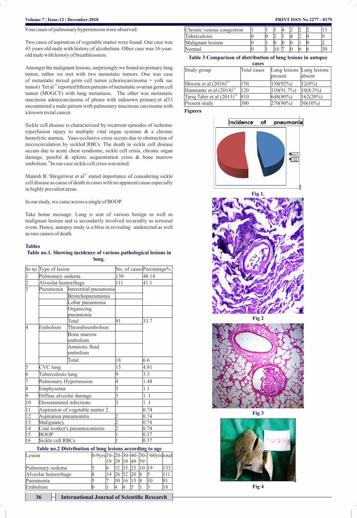
Pneumonia constituted the second major benign lesion comprising 33.7%. Incidence of interstitial pneumonia and bronchopneumonia was higher as depicted in the pie-chart (fig.1). Bacterial pneumonia has two patterns: lobular bronchopneumonia and lobar pneumonia.5 Microscopically bronchioles and peribronchiolar alveoli are filled with inflammatory exudate as was seen in our case.
Embolic episodes were next common findings noted in 18 cases comprising 6.6%. Pulmonary embolism is a complication principally in patients who are suffering from cardiac disease or cancer, or who are immobilized, those with hip fracture being at highest risk. Hypercoagulable states, either primary or secondary are frequent risk factors. Indwelling central venous lines can be a nidus for right atrial thrombus.6 Grossly lung pieces appeared unremarkable however microscopic evidence of marrow embolism with fat and haemopoietic tissue was found (fig2). We also found one case each, of amniotic embolism and air embolism.
Incidence of Chronic venous congestion (CVC) was 4.81 %. Tuberculosis is a systemic disease caused by Mycobacterium tuberculosis (M. Tb).6,7 The organisms are usually found extracellularly within the debris of the necrotic granulomas. Typical from atypical mycobacterium cannot be distinguished on the basis of their microscopic appearance. Acid fast stain brings out typical organisms. However, for definitive differentiation culture and/or polymerase chain reaction is necessary.6,7 We found military tubercles in lung showing epithelioid granulomas with central caseation, Langhan's type of giant cell, surrounded by mantle of lymphocytes. Aspiration as well as aspiration pneumonia can lead to choking syndrome. Two cases of aspiration of vegetable matter were found. One case had a history of alcoholism, aspiration of gastric contents could be the culprit in this case. Other case was a 16-year-old male with history of breathlessness. Microscopic examination showed plugging of bronchioles with vegetable matter (fig3).
Fetal distress initiates vigorous respiratory movement leading to aspiration of amniotic fluid containing vernix caseosa, epithelial cells, meconium and blood which blocks small airways. Pathogenic bacteria may accompany and pneumonitis may ensue.8 One case of aspiration pneumonitis was found in a newborn female baby and other was 12 hours old female. Grossly, lungs were unremarkable in both cases. Microscopically, alveoli were filled with squames, keratin, few neutrophils and macrophages. Single case of acute respiratory distress
INTERNATIONAL JOURNAL OF SCIENTIFIC RESEARCH
Pathology
ABSTRACTWe conducted prospective, descriptive type of study in 300 consecutive autopsy cases over a span of two years. Out of 300 autopsy cases, lung lesions were encountered in 270 cases. benign lesions outnumbered malignant lesions. Pulmonary oedema was the most frequently encountered lesion followed by alveolar hemorrhage both of which usually represent the terminal end stage disease. Infectious causes like pneumonia and tuberculosis were the next culprits. Tuberculosis is a notorious infection not only in Indian community, but also worldwide. It is especially important due to its association with AIDS. Rare cases of disseminated infection with Cryptococcosis, candidiasis, microfilaremia as well as sickle cell disease can be a cause of death.
KEYWORDSAutopsy, Pulmonary oedema, alveolar hemorrhage. Pg2
34 International Journal of Scientific Research
Volume-7 | Issue-12 | December-2018 | PRINT ISSN No 2277 - 8179

Volume-7 | Issue-12 | December-2018
was found in a 25-year-old female with 8 months pregnancy and history of breathlessness. Microscopically, there was evidence of hyaline membrane disease, with accumulation of neutrophils, macrophages and red blood cells.
Pulmonary hypertension was seen in four cases(1.48%). Histologic findings of arterial intimal fibrosis and medial thickness with arteriolar occlusion leading to onion skin appearance9 as was seen in our case. Emphysema was seen in 3 cases (1.1 %). Emphysema is defined as an abnormal permanent enlargement of airspaces distal to terminal bronchioles with destruction of alveolar walls without obvious fibrosis.8. Gross appreciation of bullae is better than microscopic evidence. We appreciated distended multiple tiny bullae on the surface of lower lobe(fig4). Microscopically, abnormally large alveoli were seen separated by thin septae which appear to be floating or protrude blindly into alveolar spaces.
Other rare lesions encountered were single cases of disseminated cryptococcosis, disseminated microfilaremia, and disseminated candidiasis.
Cryptococcal infection was observed as opportunistic infection in a 42-year-old male, a known case of HIV infection with abdominal and CNS Tuberculosis. Microscopically disseminated cryptococcal infection involving lung, liver, spleen & kidney was found.
We encountered an interesting autopsy case of disseminated microfilaremia in a 30-year-old unknown male with no clinical details. Grossly, the organs were unremarkable. Microscopically, there was extensive tissue damage with eosinophils, micro abscess and fibrin- rich inflammatory exudate in the lung, heart, liver, spleen & kidneys with the presence of microfilariae(fig5).
Another case of disseminated infection – candidiasis was seen in 21 years PNC female who died with postpartum hemorrhage. The alveoli were studded with yeast and pseudo-hyphae morphologically consistent with candidiasis.
Lung cancer remains one of the leading causes of cancer mortality worldwide.10,11 Literature study has stated a strong association between lung cancer and cigarette smoking. 80 to 85% of cancer
12mortality is directly attributable to smoking.
In our study no, primary lung cancer was seen. All cases were secondary tumors(0.74%) with a known primary in one case and unknown primary in other. The case with unknown primary was a 60-year male with mucinous adenocarcinoma in pleura. Grossly, the pleural surface showed multiple tiny irregular nodules, largest measuring 3x2x0.5cm. Histologic examination showed mucinous adenocarcinoma. The other case was a known case of carcinoma ovary in a young 15-year-old female. We found metastatic mixed germ cell tumor in lungs. Grossly, cut section of both lungs showed hemorrhagic dark brown appearance. Microscopically, metastatic mixed germ cell tumor (Yolk sac tumor +Choriocarcinoma) involving both lungs showing tumor cells arranged in microcystic, glandular and papillary pattern. At places, syncytiotrophoblastic and cytotrophoblastic cells were seen arranged in biphasic plexiform pattern(fig6).
Two cases (0.74%) of Coal workers' pneumoconiosis (CWP) were found. Coal being an important global commodity, mining of coal is important. Despite improvements in exposure assessment, ventilation controls and the existence of protective government regulations, coal miners are still at risk for respiratory diseases and their associated morbidity and mortality.13 Microscopically, there was presence of carbon laden macrophages, forming macules. BOOP is a chronic lung disease. Patients present with respiratory distress.6,14 Chronic inflammatory cells and plugs of loose, matrix rich connective tissue (Masson bodies) are distributed throughout the terminal bronchi, bronchioles, alveolar ducts and alveolar spaces.14 Alveoli show foamy cells, inflammatory cells and cellular debris.
Sickling of red blood cells was evident in one case. A young 25-year-old female, G4P3L2A1 with 32-33 weeks' pregnancy induced hypertension and pre-eclampsia had HELLP syndrome with IUD with severely deranged liver function tests. She vaginally delivered a still
born female baby & died the very next day. Grossly, cut surface of lungs was dark brown to black. Microscopically, blood vessels of all organs revealed sickled RBCs (fig7).
DISCUSSION Out of 300 cases studied, lesions were noted in 270(90%) cases, our findings are comparable with study of Selvambigai G. et al15 Hanmante R.D et al,16 Kalpana Mangal et al.17(table no.3) Male preponderance was noted. Male affection was 58.33% while female affection was 41.67 %. In our study, the most common histopathological lesion found was pulmonary edema, which is similar to the study by Hanmante R D et
19 al16, Uday Shankar et al18 and Soeiro AM et al
Amongst the Infectious diseases, pneumonia headed the list and accounted for 30.33 % which is similar to the findings of UdayShankar et al.18 The relative higher frequency of pneumonia cases in our setup might be due lack of knowledge or ignorance. were affected by pneumonia. It is estimated that 10% of the cases of symptomatic pulmonary thromboembolism (PTE) will result in death within the first hour after the onset of symptoms. Patients who do not succumb during the acute phase usually present nonspecific symptoms.20 In our study incidence of pulmonary thromboembolism is 4.6%. Hanmante et al (2014)16 found incidence of 2.5%. Bone-marrow embolism may occur during convulsions which impart shock to the skeleton or may follow bone fracture dislodging fragments of haemopoietic tissue into the veins by
21the increased pressure. Bone-marrow embolism was seen in 3 cases (1%). In one of the case, deceased was 65-year-old male, who died immediately after knee joint replacement. Schenken and Coleman22 stating similar case postulated that, fragments of marrow are forced into the veins mechanically by the wooden bone screws. In our case it is tempting to accuse similar theory.21,22 Amniotic fluid embolism usually present as sudden, unexpected maternal death that may occur during labour or postpartum period.23 In present study, one case of amniotic fluid embolism was found in a postpartum death of a 30-year-old female.
Incidence of chronic venous congestion was 4.3%. These findings are 24 close to findings of Shweta et al.
Tuberculosis is a treatable disease; its fatality is attributed to its association with HIV infection cases. Prateek Rastogi et al25 found that tuberculosis was the single most important cause of sudden unexpected death involving the respiratory system. Tuberculosis of lung was seen in 3% cases in our study which is similar with Selvam et al26 Soeiro AM et al19 and Bal MS et al.27 With HIV infection on the rise, a high suspicion of concurrent multiple opportunistic infections should be kept in mind.٢٧٬٢٨
We came across a case of disseminated cryptococcosis in a 42-year-old male, a known case of HIV with abdominal and CNS Tuberculosis. Microscopic examination of CSF or India ink preparation highlights the organism. A definitive diagnosis is made on culture. Cryptococcal antigen assay is helpful.29 Prognosis is grave without treatment, as was observed in our case. W. bancrofti is a nematode causing lymphatic filariasis though tropics and subtropics. The adult worm inhabits the lymphatics and produces lymphangitis and lymphadenitis, & the female produces sheathed microfilariae which circulate in the peripheral blood. Unlike lymphatic disease syndromes, the extra lymphatic manifestations of Bancroftian filariasis are not caused by the adult worm per se, but by diffusible products or by immune complexes.30 In our case, the extensive inflammatory responses due to disseminated microfilarial infection may have resulted in toxemia which together with pulmonary thromboembolism could have led to demise. Kirti Gupta et al30reported a similar case of disseminated microfilaremia.
Candida species are important opportunistic pathogens that can invade the vascular space giving rise to disseminated candidiasis in kidneys, liver, spleen, retina and occasionally the heart and brain.31 We found a similar case of disseminated candidiasis involving lungs, heart and spleen.
International Journal of Scientific Research 35
PRINT ISSN No 2277 - 8179
Four cases of pulmonary hypertension were observed.
Two cases of aspiration of vegetable matter were found. One case was 43 years old male with history of alcoholism. Other case was 16-year-old male with history of breathlessness. Amongst the malignant lesions, surprisingly we found no primary lung tumor, rather we met with two metastatic tumors. One was case of metastatic mixed germ cell tumor (choriocarcinoma + yolk sac
32tumor) Yet al reported fifteen patients of metastatic ovarian germ cell tumor (MOGCT) with lung metastasis. The other was metastatic mucinous adenocarcinoma of pleura with unknown primary.et al33 encountered a male patient with pulmonary mucinous carcinoma with a known rectal cancer.
Sickle cell disease is characterized by recurrent episodes of ischemic reperfusion injury to multiple vital organ systems & a chronic hemolytic anemia. Vaso-occlusive crisis occurs due to obstruction of microcirculation by sickled RBCs. The death in sickle cell disease occurs due to acute chest syndrome, sickle cell crisis, chronic organ damage, painful & splenic sequestration crisis & bone marrow
34 embolism. In our case sickle cell crisis was noted.
34Manish B. Shrigiriwar et al stated importance of considering sickle cell disease as cause of death in cases with no apparent cause especially in highly prevalent areas.
In our study, we came across a single of BOOP.
Take home message: Lung is seat of various benign as well as malignant lesions and is secondarily involved invariably as terminal event. Hence, autopsy study is a bliss in revealing undetected as well as rare causes of death.
TablesTable no.1. Showing incidence of various pathological lesions in
lung.
Table no.2 Distribution of lung lesions according to age
Table 3 Comparison of distribution of lung lesions in autopsy cases
Figures
Fig 1.
Fig 2
Fig 3
Fig 4
Volume-7 | Issue-12 | December-2018
Sr no Type of lesion No. of cases Percentage%
1 Pulmonary oedema 130 48.14 2 Alveolar hemorrhage 111 41.13 Pneumonia Interstitial pneumonia
Bronchopneumonia Lobar pneumonia Organizing pneumonia Total 91 33.7
4 Embolism Thromboembolism
Bone marrow embolism Amniotic fluid embolism
Total 18 6.6
5 CVC lung 13 4.81
6 Tuberculosis lung 9 3.3
7 Pulmonary Hypertension 4 1.48
8 Emphysema 3 1.1
9 Diffuse alveolar damage 3 1 .1
10 Disseminated infections 3 1 .1
11 Aspiration of vegetable matter 2 0.74 12 Aspiration pneumonitis 2 0.74 13 Malignancy 2 0.74 14 Coal worker's pneumoconiosis 2 0.74 15 BOOP 1 0.37 16 Sickle cell RBCs 1 0.37
Chronic venous congestion 1 1 1 4 2 2 2 13 Tuberculosis 0 0 2 1 4 2 0 9 Malignant lesions 0 1 0 0 0 1 0 2 Normal 0 1 10 7 0 6 6 30
Study group Total cases Lung lesions present
Lung lesions absent
61Shweta et al (2016) 150 138(92%) 12(8%) 54 Hanmante et al (2014) 120 110(91.7%) 10(8.3%)
59Tariq Tahir et al (2013) 810 648(80%) 162(20%) Present study 300 270(90%) 30(10%)
36 International Journal of Scientific Research
Lesion 0-9yrs 10-19
20-29
30-39
40-49
50-59
>60yrs total
Pulmonary oedema 5 6 32 33 25 10 19 133 Alveolar hemorrhage 6 14 26 32 20 8 5 111 Pneumonia 5 7 30 16 15 8 10 91 Embolism 0 1 4 4 5 1 3 18
PRINT ISSN No 2277 - 8179

Fig 5
Fig 5
Fig 6
REFERENCES1. Nihon Hoigaku Zasshi.Some findings of the lung in medicolegal autopsy cases.1994
Dec;48(6):379-94. 2. D. Behera, T. Balamugesh. Lung cancer in India. Ind journal of chest diseases and allied
sciences 2004;6:269-82.3. P.S. Malik, V. Raina. Lung cancer. Present trends and emerging concepts. The Ind Jour
of Medical Research 2015;5(1):141-.4. Mohanty MK, Singh B, Arun M. et al. Autopsy: The Changing Trends. International
Journal of Medical Toxicology and Forensic Medicine. 2011;1(1):17-23.5. Murray JF. Pulmonary edema: pathophysiology and diagnosis. Int J Tuber Lung Dis.
2011;15(2):155-160. 6. Husain AN. The lung. In: Kumar V, Abbas AK, Aster JC. Robbins and Cotran pathologic
basis of disease. 9th edition. Saunders-an imprint of Elsevier ;2014. Chapter 15 p.669-711.
7. Juan Rosai, Rosai and Ackerman's Surgical Pathology. 10th edition, Elsevier;2011. Chapter 7, Lung and pleura. p.348-400.
8. Hogg JC, Senior RM. Chronic obstructive pulmonary disease Ch 2: Pathology and biochemistry of emphysema. Thorax 2002;57:830-34.
9. Philip T. Cagle. Color atlas and text of pulmonary pathology. 2nd edition, Lippincott Williams & Wilkins;2008. Chapter 43, Sarcoidosis.pg243.
10. Davidson MR., Gazdar AF, Clarke BE. The pivotal role of pathology in the management of lung cancer. J Thora Dis2013;5(S5): S463-78.
11. Doll R, Hill AB. Smoking and carcinoma of the lung; preliminary report. BMJ 1950;2(4682):739-48.
12. Ridge CA, McErlane AM, Ginsberg MS. Epidemiology of Lung Cancer. Semin Intervent Radiol 2013;30:93-98.
13. Costabel U, Guzman J, Teschler H. Bronchiolitis obliterans with organizing pneumonia. Thorax1995;50(1):S59-64.
14. Mills, Stacey E, Carter D, Greenson JK Reuter VE, Stoler MH. Sternberg's diagnostic surgical pathology. 5th edition, Lippincott Williams & Wilkins; 2010. Chapter 25, Nonneoplastic pulmonary diseases.p.996-1047.
15. Selvambigai G, Amudhavalli S, Chakravarthi DCD, Ravi S. Histopathological study of lung in autopsy cases. Int J Res Med Sci.2016 Nov;4(11):4816-19.
16. Hanmante R. D., Chavan Y. H., Mulay P. S., Suvernakar S.V, Deshpande S.A. Histopathological patterns of lung lesions in autopsy cases. Int Jour of advances in health sciences 2014;1(1):15-19.
17. Mangal K, Dhakar P, Yadav A, et al. Magnitude of pulmonary diseases - incidentally diagnosed on autopsy at largest hospital & medical college of Rajasthan. Int J Cur Res Rev. 2016 ;8(8):37-43.
18. Udayashankar SK, Shashikala P, Kavita GU, Deepti Pruthvi. Histomorphogical Pattern of Lung in Medicolegal Autopsies. Int Jour of Sci and Res July 2015;4(7):1937-39.
19. Soeiro AM, Ruppert AD, Canzian M, Parra ER, Farhat C, Capelozzi VL. Demographic, etiological, and histological pulmonary analysis of patients with acute respiratory failure: a study of 19 years of autopsies. Clinics.2011;66(7):1193-97.
20. Bok Yoo, Hugo Hyung et al. Clinicopathological findings in pulmonary thromboembolism: a 24-year autopsy study. J. bras. Pneumol.2004;30(5):426-433.
21. Tierney RBH, Bone-marrow embolism,J.ciin.Path;1952;5:63-66.22. Schenken, J.R., Coleman, F.C. Bone Marrow and Fat Embolism Following Fracture of
the Femur.Am.J. Surg.1943;61:126.23. AP Patra, KK Shaha, AP Rayamane, Review of 24 cases of Maternal Deaths from
Amniotic-Fluid Embolism: in Correlation with Clinical and Histopathological Findings, Sch. Acad. J. Biosci 2013;1(3):85-89.
24. Shweta, Deepti Mahajan, Vidhu Mahajan, P. Angmo. Histopathological pattern in lung autopsy in government medical college Jammu. Journal of Evolution of Medical and Dental Sciences 2015;Nov4:15694-96.
25. Rastogi P, Kanchan T, Menezes RG. Sudden unexpected deaths due to tuberculosis: An autopsy-based study. Journal of Foren Med & Toxi 2011July-Dec;28(2):81-84.
26. Silva, R. ThamilSelvi, P.M. Subramaniam, Vijayanath.V, Prevalence of common disease in lungs and liver; a histopathological study. Journal of pharmaceutical and biomedical sciences 2011;12(12):1-5.
27. Bal MS, Sethi PS, Suri AK, et al. Histopathological pattern in lung autopsies. JPAFMAT 2008;8(2):29-31.
28. KP Akakpo, SE Quayson and M Lartey. Disseminated cryptococcosis in a patient with HIV/AIDS at a teaching hospital. SAGE Open Medical Case Reports; 2014:1-4.
29. RM Saldanha Dominic,HV Prashanth,Shalini Shenoy, and Diagnostic Value of Latex Agglutination in Cryptococcal Meningitis. J Lab Physicians.2009Jul Dec;1(2):67–68.
30. Gupta K, Saikia UN, Bhatia P, Garg M, Wanchu A, Disseminated Microfilaremia Associated with Lung Cyst and Empyema: An Autopsy Report, Korean J Parasitol. March 2009;47(1):49-52.
31. Thorn JL, Gilchrist KB, Sobonya RE, Gaur NK, Lipke PN, Klotz SAC. Postmortem candidemia: marker of disseminated Disease, J Clin Pathol. 2010 April;63(4):337-40.
32. Clinical analysis of 15 cases of malignant ovarian germ cell tumors with lung metastasis.Zhonghua Fu Chan KeZaZhi. 2012 Jan;47(1):40-4.
33. Yoshino, N., Kubokura, H., Yamauchi, S. et al. Jpn J Thorac Cardiovasc Surg 2006;54:328-31.
34. Shrigiriwar MB, Ghormade PS, Tingne CV. Case Report Death due to Sickle Cell Anaemia: Autopsy Diagnosis. J Indian Acad Forensic Med.2013;35(4):383-85.
Volume-7 | Issue-12 | December-2018
International Journal of Scientific Research 37
PRINT ISSN No 2277 - 8179