pathology of germ cell tumors of the testis library/main nav/research and clinical... · pathology...
TRANSCRIPT
November/December 2004, Vol. 11, No. 6374 Cancer Control
origin, has been noted over the second half of the 20thcentury.1 Germ cell tumors occur at all ages. Congenitalmalformation of the male genitalia, prenatal risk factors,nonspecific and specific exposures in adulthood,and maleinfertility have all been associated with the etiology ofgerm cell tumors. Over half of germ cell tumors consist ofmore than one cell type, requiring appropriate samplingfor the correct diagnosis and correlation with the serumtumor markers (human chorionic gonadotropin [hCG]and α-fetoprotein [AFP]). The most recent WHO histolog-ic classification of testis tumors2 is similar to the 1998 clas-sification,3 differing most significantly with respect to theclassification of teratomas and polyembryomas (Table 1).Since mature and immature teratomas have the samegenetic changes and biologic potential specific for the pre-pubertal and postpubertal patient, teratomas are no longersubclassified as mature and immature. Polyembryoma was
Pathology of Germ Cell Tumors of the TestisIsabell A. Sesterhenn, MD, and Charles J. Davis, Jr, MD
Background: An increasing incidence of testis tumors has been noted over the second half of the 20th century.
Congenital malformation of the male genitalia, prenatal risk factors, nonspecific and specific exposures in
adulthood, and male infertility have all been associated with the etiology of germ cell tumors.
Methods: The histologic classification, pathology, and current concepts of testicular germ cell tumors are reviewed.
Results: Germ cell tumors occur at all ages. The tumors are identified as pure form (those of one histologic type)
and mixed form (more than one histologic type). Over half of germ cell tumors consist of more than one cell type,
requiring appropriate sampling for the correct diagnosis and correlation with the serum tumor markers. Burned-out
germ cell tumors may occur in patients with metastatic disease with no gross evidence of a testicular tumor.
Conclusions: Appropriate management of testis tumors relies on accurate pathology and classification of
these tumors.
The pathology and current concepts
of classification of testicular
germ cell tumors are reviewed.
Michael Mahany. Black Bear at Wibel Mining Camp. Photograph. Anchorage,Alaska.
From the Department of Genitourinary Pathology at the Armed ForcesInstitute of Pathology, Washington, DC.
Submitted May 26, 2004; accepted August 16, 2004.
Address correspondence to Isabell Sesterhenn, MD, Armed Forces Institute of Pathology, Genitourinary Pathology, 14th Street and Alaska Avenue NW, Building 54 #2090, Washington, DC 20306.E-mail: [email protected]
No significant relationship exists between the authors and the compa-nies/organizations whose products or services may be referenced inthis article.
Abbreviations used in this paper: hCG = human chorionicgonadotropin, AFP = α-fetoprotein, hPL = human placental lactogen,H&E = hematoxylin-eosin.
Introduction
The majority of testis tumors originate from the germ cell,which is the principal cell type of the testis. An increasingincidence of testis tumors,particularly in men of European
November/December 2004, Vol. 11, No. 6 Cancer Control 375
once classified with tumors of one histologic type; how-ever, in recognition of the different cell types present inpolyembryoma, it is now classified with tumors of morethan one histologic type. The tumors are staged accordingto the TNM classification (Table 2).4,5
Precursor Lesions
Several terms are used to refer to intratubular malignantgerm cells, including intratubular germ cell neoplasia-unclassified, carcinoma in situ, and intratubular preinvasivetumor. In the postpubertal patient, they are seen in approx-imately 82% of testes harboring a germ cell tumor.6 How-ever, they are rarely present in prepubertal children.7-12
In testicular biopsies for infertility, intratubular malignantgerm cells are seen in 0.3% to 1.8% of cases.13
There are no specific macroscopic features for theselesions. Histologically, the malignant germ cells are largewith abundant pale, glycogen-rich cytoplasm. The irregu-larly outlined nuclei are enlarged, with one or two promi-nent nucleoli (Fig 1). They are located at the periphery ofthe seminiferous tubules between residual Sertoli cells.They often have a segmental distribution, and they canextend into the rete. At the site of invasion into the adja-cent stroma, they often elicit a lymphocytic response.By immunohistology, placental alkaline phosphatase(PLAP) can be identified in up to 99% of cases with mem-branous and/or cytoplasmic reaction (Fig 2).6,13-15 CD117a(c-kit)16,17 shows a membranous reaction. They are strong-ly positive for OCT3/4 (POU5F1), a marker of pluripotentstem cells.18 Intratubular malignant germ cells appear tohave a similar appearance in all germ cell tumor types
with PLAP and CD117a, but other antibodies such as 43-9F19,20 and TRA 1-6021,22 indicate different immunopheno-types adjacent to different germ cell tumor types. Gener-ally, the malignant germ cells are similar to the associatedgerm cell tumor type with respect to their DNA ploidy.23-
25 They do not show an increased number of isochromo-some 12p [i(12p)] until they become invasive.26-28
Tumors of One Histologic Type
SeminomaSeminomas comprise 35% to 70% of germ cell tumors,depending on the patient population (Table 3). Thetumor is most commonly seen in patients between 30 and50 years of age, and the testis is usually enlarged. The cutsurface of seminomas is usually grayish-white, bulging,and glistening (Fig 3). Histologically, the tumor cells arelarge with abundant pale or amphophilic cytoplasm,depending on the glycogen content. The nuclei have acoarse chromatin distribution with prominent nucleoli.These cells occur in sheets, lobules, or columns. The sup-porting stroma shows varying amounts of lymphocyticinfiltrate or granulomatous reaction (Figs 4–6). Both areprominent in approximately 20% of cases. This reactionapparently represents a host response. Immunohisto-chemically, the tumor cells react with antibodies to PLAPand CD117a (Table 4).6,29,30 OCT4, a marker of pluripo-tent cells, is demonstrable in the nucleus of seminomacells.18,31 Other markers include VASA32 and CD143.33
The cytokeratins are occasionally positive.34
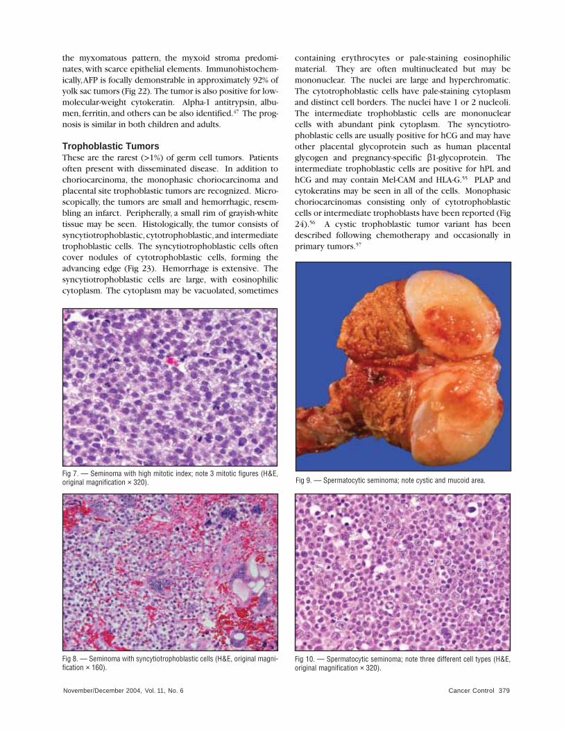
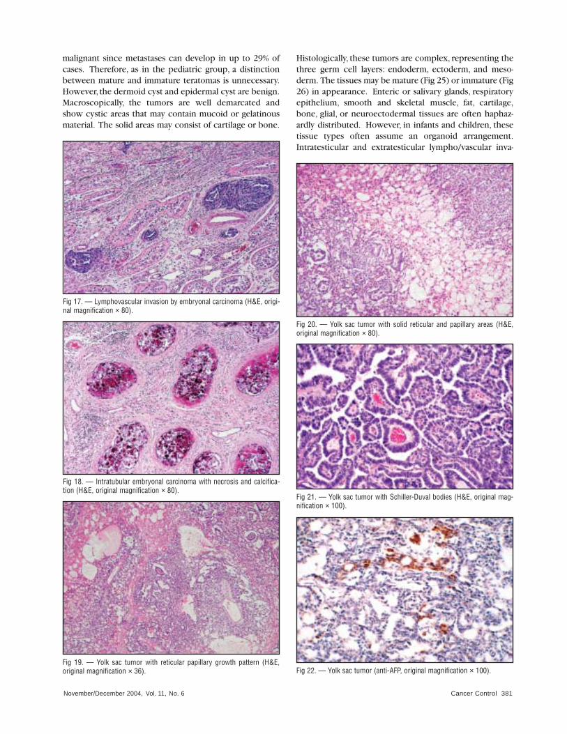
Variants of SeminomaA number of seminomas show increased mitotic activity,averaging 3 mitoses per high-power fields throughout thetumor (Fig 7). In addition, these seminomas often showmore cellular atypia and pleomorphism. These tumorsmay express CD30, which has been interpreted by someas evidence of early differentiation toward embryonal car-cinoma. This type of seminoma usually presents at a high-er stage.35,36 Others have questioned the significance ofthese findings in seminoma. When examined by immuno-histochemistry, approximately 25% of seminomas containsyncytiotrophoblastic giant cells compared with 7% whenexamined only by hematoxylin-eosin (H&E) staining.37
These are usually multinucleated cells with vacuolatedcytoplasm, or they are flattened cells that are associatedwith vascular spaces (Fig 8). They react positive for hCGand other placental glycoproteins such as human placen-tal lactogen (hPL) and pregnancy-specific β1-glycopro-tein.37,38 This type of tumor is often associated with ele-vated serum levels of hCG.
Spermatocytic SeminomaThis germ cell tumor accounts for approximately 1.2% to4.5% of germ cell tumors.39,40 Spermatocytic seminoma is
Germ Cell TumorsIntratubular germ cell neoplasia, unclassifiedOther types
Tumors of One Histologic Type (Pure Forms)SeminomaSeminoma with syncytiotrophoblastic cellsSpermatocytic seminomaEmbryonal carcinomaYolk sac tumorTrophoblastic tumorsChoriocarcinomaTrophoblastic neoplasms other than choriocarcinomaMonophasic choriocarcinomaPlacental site trophoblastic tumorTeratomaDermoid cyst Monodermal teratomaTeratoma with somatic type malignancies
Tumors of More Than One Histologic Type (Mixed Forms)Mixed embryonal carcinoma and teratomaMixed teratoma and seminomaChoriocarcinoma and teratoma/embryonal carcinomaOthers
Table 1. — Histologic Classification of Testis Tumors
near diploid and medium cells have an intermediate value.The edematous stroma is generally devoid of a lymphocyticinfiltrate. Lakes of pink proteinaceous material can be seen.
The adjacent seminiferous tubules often showintratubular spermatocytic seminoma (Fig 11). The typicalintratubular germ cell neoplasia unclassified is not present.About 20% of spermatocytic seminomas contain PLAP inisolated or small clusters of cells.39 CD117a (c-kit) can alsobe demonstrated (I.A.S., unpublished data, 2004). AFP andhCG are not demonstrable. Most spermatocytic seminomasreact diffusely with antibodies to VASA.32 NY-ESO-1 hasbeen demonstrated in spermatocytic seminoma but not inother germ cell tumor types.41 Cytokeratins are usually seenin a dot-like pattern. Spermatocytic seminoma has a good
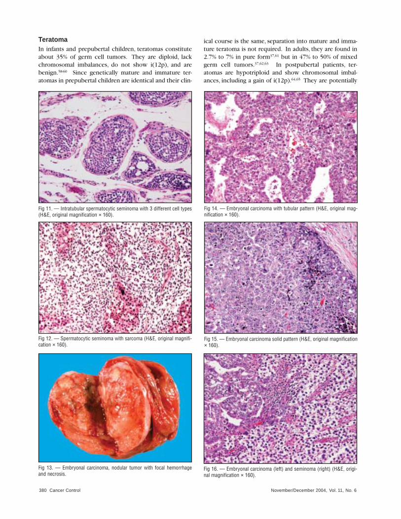
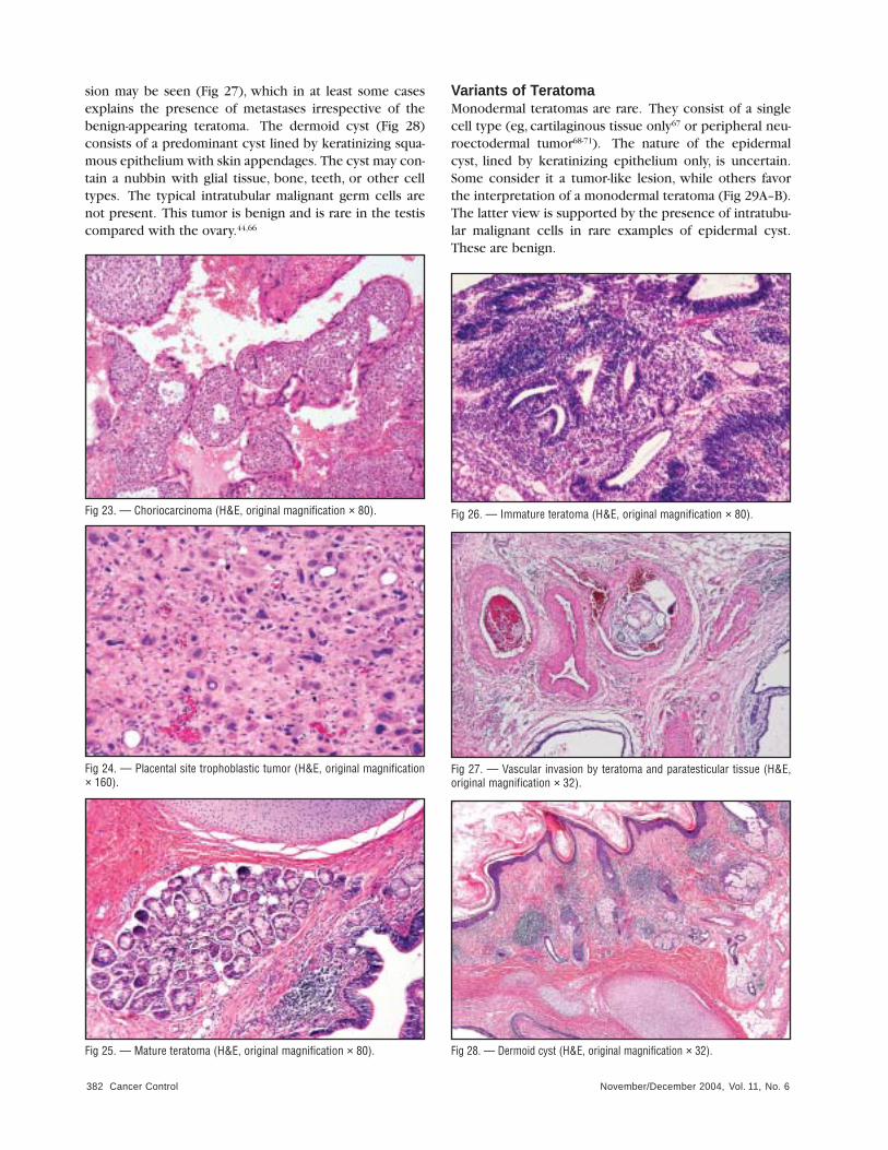
usually seen in men over 40 years of age. Approximately 5%of patients have bilateral disease.39 Spermatocytic semino-ma develops only in the testis,unlike other germ cell tumortypes that also involve the ovary, retroperitoneum, medi-astinum, and other sites. Grossly, the tumors are usuallylarge. The cut surface is yellowish, soft, and mucoid, withcystic or spongy areas (Fig 9). Microscopically, the tumorconsists of three cell types: large mononucleated or multi-nucleated cells with abundant cytoplasm, intermediatecells, and small cells with hyperchromatic nuclei (Fig 10).The nuclei of the large and intermediate cells have a char-acteristic filamentous chromatin distribution. Mitosesoccur frequently. Using cytophotometry, large cells haveDNA values up to 42C, whereas small cells are diploid or
November/December 2004, Vol. 11, No. 6376 Cancer Control
T – Primary TumorExcept for pTis and pT4, where radical orchiectomy is not always necessary for classification purposes, the extent of the primary tumor is classified after radical orchiectomy; see pT. In other circumstances, TX is used if no radical orchiectomy has been performed
N – Regional Lymph NodesNX Regional lymph nodes cannot be assessedN0 No regional lymph node metastasisN1 Metastasis with a lymph node mass 2 cm or less in greatest
dimension or multiple lymph nodes, none more than 2 cm in greatest dimension
N2 Metastasis with a lymph node mass more than 2 cm but not more than 5 cm in greatest dimension, or multiple lymph nodes, any one mass morethan 2 cm but not more than 5 cm in greatest dimension
N3 Metastasis with a lymph node mass more than 5 cm in greatest dimension
M – Distant MetastasisMX Distant metastasis cannot be assessedM0 No distant metastasisM1 Distant metastasisM1a Non-regional lymph node(s) or lungM1b Other sites
pTNM Pathological ClassificationpT – Primary Tumor
pTX Primary tumor cannot be assessed (see T–Primary Tumor, above)pT0 No evidence of primary tumor (eg, histologic scar in testis)pTis Intratubular germ cell neoplasia (carcinoma in situ)pT1 Tumor limited to testis and epididymis without vascular/
lymphatic invasion; tumor may invade tunica albuginea but not tunica vaginalis
pT2 Tumor limited to testis and epididymis with vascular/lymphatic invasion, or tumor extending through tunica albuginea with involvement of tunica vaginalis
pT3 Tumor invades spermatic cord with or without vascular/lymphatic invasion
pT4 Tumor invades scrotum with or without vascular/lymphatic invasion
pN – Regional Lymph NodespNX Regional lymph nodes cannot be assessedpN0 No regional lymph node metastasispN1 Metastasis with a lymph node mass 2 cm or less in greatest
dimension and 5 or fewer positive nodes, none more than 2 cm in greatest dimension
Table 2. — TNM Classification of Tumors of the Testis
pTNM Pathological Classification (continued)pN2 Metastasis with a lymph node mass more than 2 cm but
not more than 5 cm in greatest dimension; or more than 5 nodes positive, none more than 5 cm; or evidence of extranodal extension of tumor
pN3 Metastasis with a lymph node mass more than 5 cm in greatest dimension
S – Serum Tumor MarkersSX Marker studies not available or not performedS0 Marker study levels within normal limitsS1 LDH <1.5 × N* and hCG (mIU/mL) <5,000 and
AFP (ng/mL) <1,000S2 LDH 1.5–10 × N* or hCG (mIU/mL) 5,000–50,000 or
AFP (ng/mL) 1,000–10,000S3 LDH >10 × N* or hCG (mIU/mL) >50,000 or
AFP (ng/mL) >10,000
*N = upper limit of normal for the LDH assay.
Stage GroupingStage 0: pTis N0 M0 S0, SXStage I: pT1–4 N0 M0 SXStage IA: pT1 N0 M0 S0Stage IB: pT2 N0 M0 S0
pT3 N0 M0 S0pT4 N0 M0 S0
Stage IS: Any pT/TX N0 M0 S1–3Stage II: Any pT/TX N1–3 M0 SXStage IIA: Any pT/TX N1 M0 S0
Any pT/TX N1 M0 S1Stage IIB: Any pT/TX N2 M0 S0
Any pT/TX N2 M0 S1Stage IIC: Any pT/TX N3 M0 S0
Any pT/TX N3 M0 S1Stage III: Any pT/TX Any N M1, M1a SXStage IIIA: Any pT/TX Any N M1, M1a S0
Any pT/TX Any N M1, M1a S1Stage IIIB: Any pT/TX N1–3 M0 S2
Any pT/TX Any N M1, M1a S2Stage IIIC: Any pT/TX N1–3 M0 S3
Any pT/TX Any N M1, M1a S3Any pT/TX Any N M1b Any S
Used with the permission of the American Joint Committee on Cancer(AJCC), Chicago, Illinois. The original source for this material is the AJCCCancer Staging Manual, Sixth Edition (2002) published by Springer-Ver-lag New York, www.springer-ny.com.
November/December 2004, Vol. 11, No. 6 Cancer Control 377
prognosis. Only one fully documented case of a metastasiz-ing spermatocytic seminoma has been reported.42
Variants of Spermatocytic SeminomaSpermatocytic seminoma is occasionally associated with asarcoma. Macroscopically, these tumors are large with avariegated appearance. Generally, the sarcoma is undiffer-entiated, but in some cases it may be differentiated, eg,rhabdomyosarcoma or chondrosarcoma.39,43-46 The recog-nizable foci of spermatocytic seminoma often showmarked nuclear pleomorphism (Fig 12). The sarcomametastasizes extensively.
Embryonal CarcinomaIn pure form, this tumor comprises 3% to 4% of germ celltumors. However, it is present in approximately 40% of
tumors of more than one histologic type. It is seen mostoften in men between 20 and 40 years of age and lessoften in adolescents (15 to 20 years of age). It has notbeen described in the prepubertal testes. In pure form, itis not associated with elevated serum levels of AFP. Gross-ly, the tumors are usually small and located close to therete. The cut surface is grayish-white with foci of hemor-rhage and necrosis. They are not encapsulated (Fig 13).Microscopically, the cells are large and embryonic inappearance and have pale, amphophilic or eosinophiliccytoplasm. The cell borders are ill-defined, and there isfrequent nuclear overlap. The nuclei are vesicular, with asee-through appearance. Nucleoli are prominent andmitoses are common. The growth patterns include papil-lary, acinar, tubular, and solid forms (Figs 14–15). The stro-ma is variable and may consist of loose, immature cells. Inthe solid pattern, degenerating cells may mimic syncy-tiotrophoblastic cells (Fig 15). These findings are con-trary to those in seminoma, which consists of large cellswith well-defined cell borders (Fig 16). Vascular and lym-phatic invasion (Fig 17) and infiltration of paratesticulartissue and the epididymis are common. The adjacent sem-iniferous tubules may show intratubular embryonal carci-
Fig 2. — Intratubular malignant germ cells with membranous and cytoplasmic staining (anti-PLAP, original magnification × 80).
Fig 3. — Seminoma with homogeneous pale cut surface.Fig 1. — Intratubular malignant germ cells (H&E, original magnification × 160).
Germ Cell Tumor Adults Infants/Children(%) (%)
Pure seminoma 26.9 2.5Seminoma + SCT 8.1Spermatocytic seminoma 2.4ECA 3.1YST 2.4 58–82Teratoma 2.7 14–38Choriocarcinoma 0.1Intratubular malignant germ cells 0.6ECA + YST + teratoma + SCT 14.3ECA + YST + teratoma + seminoma + SCT 7.4ECA + YST + teratoma 4.7YST + teratoma 2.5 0.85ECA + teratoma + teratocarcinoma 1.4Teratoma + seminoma 0.45Other combinations 24.0
ECA = embryonal carcinomaYST = yolk sac tumorSCT = syncytiotrophoblastsAdapted from Mostofi FK, Sesterhenn IA, Davis CJ Jr. Immunopathology of germ cell tumors of the testis. Semin Diagn Pathol. 1987;4:320-341.Copyright 1987, with permission from Elsevier.
Table 3. — Frequency of Various Histologic Types of Germ Cell Tumors in Adults

November/December 2004, Vol. 11, No. 6378 Cancer Control
noma with extensive degenerative change and calcifica-tion (Fig 18). AFP is seen in approximately 13% of casesin isolated or small clusters of cells that appear to beinsufficient in number to cause a measurable elevation ofserum AFP,47,48 and hPL can also be found.37,48 OCT4 hasbeen demonstrated in most embryonal carcinomas con-sistent with its pluripotentiality.18,31 The presence ofCD30 is common49 and cytokeratins are often demonstra-ble, but vimentin and epithelial membrane antigen (EMA)are not. Syncytiotrophoblastic cells producing hCG canbe found scattered throughout the tumor. These cells mayalso be positive for pregnancy-specific β1-glycoprotein.37
Patients with pure embryonal carcinoma or embryonalcarcinoma exceeding 40% in a mixed germ cell tumorwith vascular and/or lymphatic invasion often present inadvanced stage.50-52
Yolk Sac TumorYolk sac tumor is the most common germ cell tumor ininfants and children, accounting for approximately 65% ofgerm cells tumors.53 It is seen in about 2.4% of adultpatients, but in tumors of more than one histologic type, it
is seen in 42% of cases. On gross examination, the tumor issoft, homogenous, grayish-yellow, and not encapsulated.Microscopically, the yolk sac tumor has at least 10 differentpatterns, which may explain the difficulties recognizingyolk sac tumor elements in the mixed germ cell tumor. Thecommon pattern is the reticular or microcystic pattern (Fig19). The cells are small, ranging from cuboidal to flattenedendothelial in appearance. The nuclei are of variable sizes.Mitoses are frequent, and hyaline globules are common.The solid pattern consists of cells that are smaller thanseminoma cells (Fig 20). They form sheets or nodularaggregates with occasional cystic structures. If these cellsare eosinophilic and resemble hepatocytes, they are of thehepatoid pattern, which is invariably positive for AFP. Aglandular-alveolar pattern is not uncommon. The presenceof immature glands resembling teratoma but without otherteratomatous components is interpreted as the enteric pat-tern. The endodermal sinus pattern is characterized bypapillary structures with a central fibrovascular core cov-ered by a layer of cuboidal tumor cells. These structuresare known as Schiller-Duval bodies54 (Fig 21). Thepolyvesicular vitelline pattern consists of cysts of varyingsizes separated by edematous,cellular,or fibrous stroma. In
Germ Cell Tumors AntibodiesIntratubular malignant germ cell PLAP, CD117a, (intratubular germ cell neoplasia, OCT4, 43-9F, TRA1-60unclassified)
Seminoma PLAP, CD117a, OCT4, VASA, cytokeratins
Spermatocytic seminoma VASA, NY-ESO-1, cytokeratins (PLAP,* CD117a*)
Embryonal carcinoma AFP,* OCT4, CD30, cytokeratins, PLAP, hPL
Yolk sac tumor AFP, PLAP, cytokeratins, AAT, albumin, ferritin
Trophoblastic tumors hCG, hPL, SP1, PLAP, Mel-CAM, HLA-G, cytokeratins
Teratoma AFP,** PLAP, and markers specificfor the different tissue types
* rare cells** intestinal-like glands and hepatoid cells
Table 4. — Immunohistochemistry of Germ Cell Tumors
Fig 4. — Seminoma (H&E, original magnification × 80).
Fig 5. — Seminoma with marked lymphocytic infiltrate (H&E, original mag-nification × 80).
Fig 6. — Seminoma with granulomatous stroma (H&E, original magnifica-tion × 100).
November/December 2004, Vol. 11, No. 6 Cancer Control 379
Fig 7. — Seminoma with high mitotic index; note 3 mitotic figures (H&E,original magnification × 320).
Fig 8. — Seminoma with syncytiotrophoblastic cells (H&E, original magni-fication × 160).
Fig 9. — Spermatocytic seminoma; note cystic and mucoid area.
Fig 10. — Spermatocytic seminoma; note three different cell types (H&E,original magnification × 320).
the myxomatous pattern, the myxoid stroma predomi-nates, with scarce epithelial elements. Immunohistochem-ically,AFP is focally demonstrable in approximately 92% ofyolk sac tumors (Fig 22). The tumor is also positive for low-molecular-weight cytokeratin. Alpha-1 antitrypsin, albu-men, ferritin, and others can be also identified.47 The prog-nosis is similar in both children and adults.
Trophoblastic TumorsThese are the rarest (>1%) of germ cell tumors. Patientsoften present with disseminated disease. In addition tochoriocarcinoma, the monophasic choriocarcinoma andplacental site trophoblastic tumors are recognized. Micro-scopically, the tumors are small and hemorrhagic, resem-bling an infarct. Peripherally, a small rim of grayish-whitetissue may be seen. Histologically, the tumor consists ofsyncytiotrophoblastic,cytotrophoblastic, and intermediatetrophoblastic cells. The syncytiotrophoblastic cells oftencover nodules of cytotrophoblastic cells, forming theadvancing edge (Fig 23). Hemorrhage is extensive. Thesyncytiotrophoblastic cells are large, with eosinophiliccytoplasm. The cytoplasm may be vacuolated, sometimes
containing erythrocytes or pale-staining eosinophilicmaterial. They are often multinucleated but may bemononuclear. The nuclei are large and hyperchromatic.The cytotrophoblastic cells have pale-staining cytoplasmand distinct cell borders. The nuclei have 1 or 2 nucleoli.The intermediate trophoblastic cells are mononuclearcells with abundant pink cytoplasm. The syncytiotro-phoblastic cells are usually positive for hCG and may haveother placental glycoprotein such as human placentalglycogen and pregnancy-specific β1-glycoprotein. Theintermediate trophoblastic cells are positive for hPL andhCG and may contain Mel-CAM and HLA-G.55 PLAP andcytokeratins may be seen in all of the cells. Monophasicchoriocarcinomas consisting only of cytotrophoblasticcells or intermediate trophoblasts have been reported (Fig24).56 A cystic trophoblastic tumor variant has beendescribed following chemotherapy and occasionally inprimary tumors.57
November/December 2004, Vol. 11, No. 6380 Cancer Control
TeratomaIn infants and prepubertal children, teratomas constituteabout 35% of germ cell tumors. They are diploid, lackchromosomal imbalances, do not show i(12p), and arebenign.58-60 Since genetically mature and immature ter-atomas in prepubertal children are identical and their clin-
ical course is the same, separation into mature and imma-ture teratoma is not required. In adults, they are found in2.7% to 7% in pure form37,61 but in 47% to 50% of mixedgerm cell tumors.37,62,63 In postpubertal patients, ter-atomas are hypotriploid and show chromosomal imbal-ances, including a gain of i(12p).64,65 They are potentially
Fig 11. — Intratubular spermatocytic seminoma with 3 different cell types(H&E, original magnification × 160).
Fig 12. — Spermatocytic seminoma with sarcoma (H&E, original magnifi-cation × 160).
Fig 13. — Embryonal carcinoma, nodular tumor with focal hemorrhageand necrosis.
Fig 14. — Embryonal carcinoma with tubular pattern (H&E, original mag-nification × 160).
Fig 15. — Embryonal carcinoma solid pattern (H&E, original magnification× 160).
Fig 16. — Embryonal carcinoma (left) and seminoma (right) (H&E, origi-nal magnification × 160).
November/December 2004, Vol. 11, No. 6 Cancer Control 381
malignant since metastases can develop in up to 29% ofcases. Therefore, as in the pediatric group, a distinctionbetween mature and immature teratomas is unnecessary.However, the dermoid cyst and epidermal cyst are benign.Macroscopically, the tumors are well demarcated andshow cystic areas that may contain mucoid or gelatinousmaterial. The solid areas may consist of cartilage or bone.
Histologically, these tumors are complex, representing thethree germ cell layers: endoderm, ectoderm, and meso-derm. The tissues may be mature (Fig 25) or immature (Fig26) in appearance. Enteric or salivary glands, respiratoryepithelium, smooth and skeletal muscle, fat, cartilage,bone, glial, or neuroectodermal tissues are often haphaz-ardly distributed. However, in infants and children, thesetissue types often assume an organoid arrangement.Intratesticular and extratesticular lympho/vascular inva-
Fig 17. — Lymphovascular invasion by embryonal carcinoma (H&E, origi-nal magnification × 80).
Fig 18. — Intratubular embryonal carcinoma with necrosis and calcifica-tion (H&E, original magnification × 80).
Fig 19. — Yolk sac tumor with reticular papillary growth pattern (H&E,original magnification × 36).
Fig 20. — Yolk sac tumor with solid reticular and papillary areas (H&E,original magnification × 80).
Fig 21. — Yolk sac tumor with Schiller-Duval bodies (H&E, original mag-nification × 100).
Fig 22. — Yolk sac tumor (anti-AFP, original magnification × 100).
November/December 2004, Vol. 11, No. 6382 Cancer Control
sion may be seen (Fig 27), which in at least some casesexplains the presence of metastases irrespective of thebenign-appearing teratoma. The dermoid cyst (Fig 28)consists of a predominant cyst lined by keratinizing squa-mous epithelium with skin appendages. The cyst may con-tain a nubbin with glial tissue, bone, teeth, or other celltypes. The typical intratubular malignant germ cells arenot present. This tumor is benign and is rare in the testiscompared with the ovary.44,66
Variants of TeratomaMonodermal teratomas are rare. They consist of a singlecell type (eg, cartilaginous tissue only67 or peripheral neu-roectodermal tumor68-71). The nature of the epidermalcyst, lined by keratinizing epithelium only, is uncertain.Some consider it a tumor-like lesion, while others favorthe interpretation of a monodermal teratoma (Fig 29A–B).The latter view is supported by the presence of intratubu-lar malignant cells in rare examples of epidermal cyst.These are benign.
Fig 24. — Placental site trophoblastic tumor (H&E, original magnification× 160).
Fig 23. — Choriocarcinoma (H&E, original magnification × 80).
Fig 25. — Mature teratoma (H&E, original magnification × 80).
Fig 26. — Immature teratoma (H&E, original magnification × 80).
Fig 27. — Vascular invasion by teratoma and paratesticular tissue (H&E,original magnification × 32).
Fig 28. — Dermoid cyst (H&E, original magnification × 32).
November/December 2004, Vol. 11, No. 6 Cancer Control 383
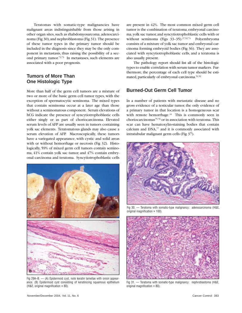
Teratomas with somatic-type malignancies havemalignant areas indistinguishable from those arising inother organ sites, such as rhabdomyosarcoma, adenocarci-noma (Fig 30),and nephroblastoma (Fig 31). The presenceof these tumor types in the primary tumor should beincluded in the diagnosis since they may be the only com-ponent in metastasis, thus raising the possibility of a sec-ond primary tumor.72,73 In metastases, such elements areassociated with a poor prognosis.
Tumors of More Than One Histologic Type
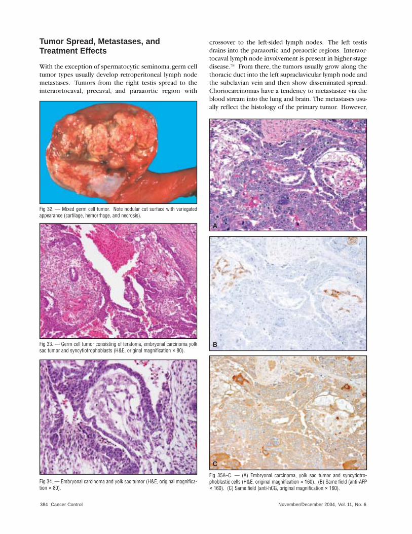
More than half of the germ cell tumors are a mixture oftwo or more of the basic germ cell tumor types, with theexception of spermatocytic seminoma. The mixed typesthat contain seminoma occur at a later age than thosewithout a seminomatous component. Serum elevations ofhCG indicate the presence of syncytiotrophoblastic cellseither singly or as part of choriocarcinoma. Elevatedserum levels of AFP are usually seen in tumors containingyolk sac elements. Teratomatous glands may also cause aserum elevation of AFP. Macroscopically, these tumorshave a variegated appearance, with cystic and solid areaswith or without hemorrhage or necrosis (Fig 32). Histo-logically, 59% of mixed germ cell tumors contain semino-ma, 41% contain yolk sac tumor, and 47% contain embry-onal carcinoma and teratoma. Syncytiotrophoblastic cells
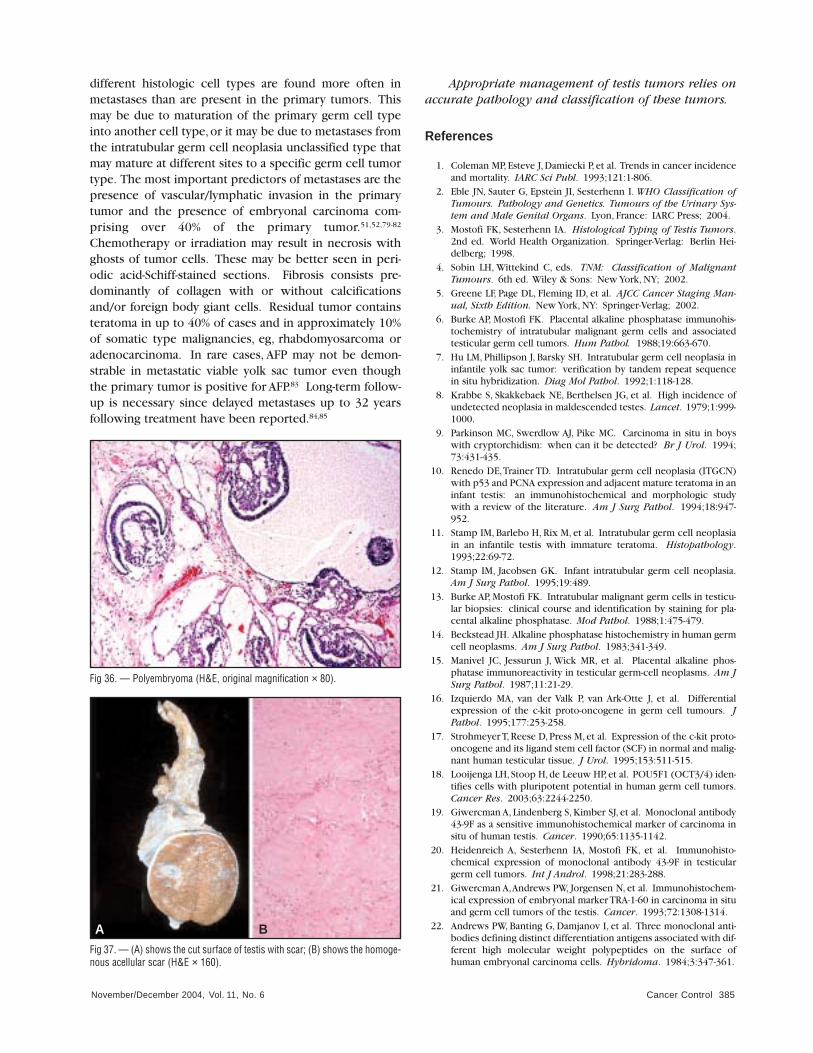
are present in 42%. The most common mixed germ celltumor is the combination of teratoma, embryonal carcino-ma,yolk sac tumor,and syncytiotrophoblastic cells with orwithout seminoma (Figs 33–35).37,52,74 Polyembryomaconsists of a mixture of yolk sac tumor and embryonal car-cinoma forming embryoid bodies (Fig 36). They are asso-ciated with syncytiotrophoblastic cells, and a teratoma isalso usually present.
The pathology report should list all of the histologictypes to enable correlation with serum tumor markers. Fur-thermore, the percentage of each cell type should be esti-mated, particularly of embryonal carcinoma.51,52
Burned-Out Germ Cell Tumor
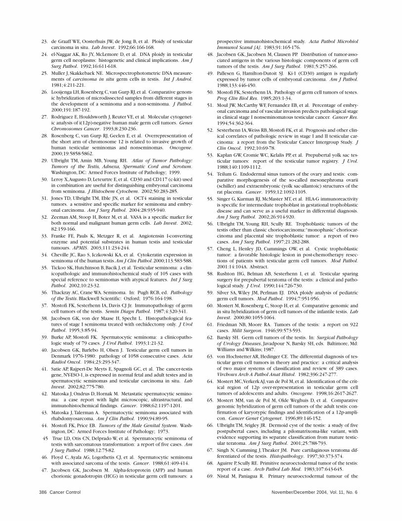
In a number of patients with metastatic disease and nogross evidence of a testicular tumor, the only evidence ofa primary tumor in that location is a homogeneous scarwith remote hemorrhage.44 This is commonly seen inchoriocarcinomas75,76 or in association with teratoma. Thisscar can have hematoxylin-staining bodies that containcalcium and DNA,77 and it is commonly associated withintratubular malignant germ cells (Fig 37).
Fig 29A–B. — (A) Epidermoid cyst, note keratin lamellae with onion appear-ance. (B) Epidermoid cyst consisting of keratinizing squamous epithelium(H&E, original magnification × 80).
A
B
Fig 30. — Teratoma with somatic-type malignancy: adenocarcinoma (H&E,original magnification × 100).
Fig 31. — Teratoma with somatic-type malignancy: nephroblastoma (H&E,original magnification × 80).
November/December 2004, Vol. 11, No. 6384 Cancer Control
Tumor Spread, Metastases, and Treatment Effects
With the exception of spermatocytic seminoma, germ celltumor types usually develop retroperitoneal lymph nodemetastases. Tumors from the right testis spread to theinteraortocaval, precaval, and paraaortic region with
crossover to the left-sided lymph nodes. The left testisdrains into the paraaortic and preaortic regions. Interaor-tocaval lymph node involvement is present in higher-stagedisease.78 From there, the tumors usually grow along thethoracic duct into the left supraclavicular lymph node andthe subclavian vein and then show disseminated spread.Choriocarcinomas have a tendency to metastasize via theblood stream into the lung and brain. The metastases usu-ally reflect the histology of the primary tumor. However,
Fig 35A–C. — (A) Embryonal carcinoma, yolk sac tumor and syncytiotro-phoblastic cells (H&E, original magnification × 160). (B) Same field (anti-AFP× 160). (C) Same field (anti-hCG, original magnification × 160).
A
B
C
Fig 34. — Embryonal carcinoma and yolk sac tumor (H&E, original magnifica-tion × 80).
Fig 32. — Mixed germ cell tumor. Note nodular cut surface with variegatedappearance (cartilage, hemorrhage, and necrosis).
Fig 33. — Germ cell tumor consisting of teratoma, embryonal carcinoma yolksac tumor and syncytiotrophoblasts (H&E, original magnification × 80).
November/December 2004, Vol. 11, No. 6 Cancer Control 385
different histologic cell types are found more often in metastases than are present in the primary tumors. Thismay be due to maturation of the primary germ cell typeinto another cell type, or it may be due to metastases fromthe intratubular germ cell neoplasia unclassified type thatmay mature at different sites to a specific germ cell tumortype. The most important predictors of metastases are thepresence of vascular/lymphatic invasion in the primarytumor and the presence of embryonal carcinoma com-prising over 40% of the primary tumor.51,52,79-82
Chemotherapy or irradiation may result in necrosis withghosts of tumor cells. These may be better seen in peri-odic acid-Schiff-stained sections. Fibrosis consists pre-dominantly of collagen with or without calcificationsand/or foreign body giant cells. Residual tumor containsteratoma in up to 40% of cases and in approximately 10%of somatic type malignancies, eg, rhabdomyosarcoma oradenocarcinoma. In rare cases, AFP may not be demon-strable in metastatic viable yolk sac tumor even thoughthe primary tumor is positive for AFP.83 Long-term follow-up is necessary since delayed metastases up to 32 yearsfollowing treatment have been reported.84,85
Appropriate management of testis tumors relies onaccurate pathology and classification of these tumors.
References
1. Coleman MP, Esteve J, Damiecki P, et al. Trends in cancer incidenceand mortality. IARC Sci Publ. 1993;121:1-806.
2. Eble JN, Sauter G, Epstein JI, Sesterhenn I. WHO Classification ofTumours. Pathology and Genetics. Tumours of the Urinary Sys-tem and Male Genital Organs. Lyon, France: IARC Press; 2004.
3. Mostofi FK, Sesterhenn IA. Histological Typing of Testis Tumors.2nd ed. World Health Organization. Springer-Verlag: Berlin Hei-delberg; 1998.
4. Sobin LH, Wittekind C, eds. TNM: Classification of MalignantTumours. 6th ed. Wiley & Sons: New York, NY; 2002.
5. Greene LF, Page DL, Fleming ID, et al. AJCC Cancer Staging Man-ual, Sixth Edition. New York, NY: Springer-Verlag; 2002.
6. Burke AP, Mostofi FK. Placental alkaline phosphatase immunohis-tochemistry of intratubular malignant germ cells and associatedtesticular germ cell tumors. Hum Pathol. 1988;19:663-670.
7. Hu LM, Phillipson J, Barsky SH. Intratubular germ cell neoplasia ininfantile yolk sac tumor: verification by tandem repeat sequencein situ hybridization. Diag Mol Pathol. 1992;1:118-128.
8. Krabbe S, Skakkebaek NE, Berthelsen JG, et al. High incidence ofundetected neoplasia in maldescended testes. Lancet. 1979;1:999-1000.
9. Parkinson MC, Swerdlow AJ, Pike MC. Carcinoma in situ in boyswith cryptorchidism: when can it be detected? Br J Urol. 1994;73:431-435.
10. Renedo DE,Trainer TD. Intratubular germ cell neoplasia (ITGCN)with p53 and PCNA expression and adjacent mature teratoma in aninfant testis: an immunohistochemical and morphologic studywith a review of the literature. Am J Surg Pathol. 1994;18:947-952.
11. Stamp IM, Barlebo H, Rix M, et al. Intratubular germ cell neoplasiain an infantile testis with immature teratoma. Histopathology.1993;22:69-72.
12. Stamp IM, Jacobsen GK. Infant intratubular germ cell neoplasia.Am J Surg Pathol. 1995;19:489.
13. Burke AP, Mostofi FK. Intratubular malignant germ cells in testicu-lar biopsies: clinical course and identification by staining for pla-cental alkaline phosphatase. Mod Pathol. 1988;1:475-479.
14. Beckstead JH. Alkaline phosphatase histochemistry in human germcell neoplasms. Am J Surg Pathol. 1983;341-349.
15. Manivel JC, Jessurun J, Wick MR, et al. Placental alkaline phos-phatase immunoreactivity in testicular germ-cell neoplasms. Am JSurg Pathol. 1987;11:21-29.
16. Izquierdo MA, van der Valk P, van Ark-Otte J, et al. Differentialexpression of the c-kit proto-oncogene in germ cell tumours. JPathol. 1995;177:253-258.
17. Strohmeyer T, Reese D, Press M, et al. Expression of the c-kit proto-oncogene and its ligand stem cell factor (SCF) in normal and malig-nant human testicular tissue. J Urol. 1995;153:511-515.
18. Looijenga LH,Stoop H,de Leeuw HP,et al. POU5F1 (OCT3/4) iden-tifies cells with pluripotent potential in human germ cell tumors.Cancer Res. 2003;63:2244-2250.
19. Giwercman A, Lindenberg S, Kimber SJ, et al. Monoclonal antibody43-9F as a sensitive immunohistochemical marker of carcinoma insitu of human testis. Cancer. 1990;65:1135-1142.
20. Heidenreich A, Sesterhenn IA, Mostofi FK, et al. Immunohisto-chemical expression of monoclonal antibody 43-9F in testiculargerm cell tumors. Int J Androl. 1998;21:283-288.
21. Giwercman A,Andrews PW, Jorgensen N, et al. Immunohistochem-ical expression of embryonal marker TRA-1-60 in carcinoma in situand germ cell tumors of the testis. Cancer. 1993;72:1308-1314.
22. Andrews PW, Banting G, Damjanov I, et al. Three monoclonal anti-bodies defining distinct differentiation antigens associated with dif-ferent high molecular weight polypeptides on the surface ofhuman embryonal carcinoma cells. Hybridoma. 1984;3:347-361.
Fig 37. — (A) shows the cut surface of testis with scar; (B) shows the homoge-nous acellular scar (H&E × 160).
A B
Fig 36. — Polyembryoma (H&E, original magnification × 80).
November/December 2004, Vol. 11, No. 6386 Cancer Control
23. de Graaff WE, Oosterhuis JW, de Jong B, et al. Ploidy of testicularcarcinoma in situ. Lab Invest. 1992;66:166-168.
24. el-Naggar AK, Ro JY, McLemore D, et al. DNA ploidy in testiculargerm cell neoplasms: histogenetic and clinical implications. Am JSurg Pathol. 1992;16:611-618.
25. Muller J, Skakkebaek NE. Microspectrophotometric DNA measure-ments of carcinoma in situ germ cells in testis. Int J Androl.1981;4:211-221.
26. Looijenga LH,Rosenberg C,van Gurp RJ,et al. Comparative genom-ic hybridization of microdissected samples from different stages inthe development of a seminoma and a non-seminoma. J Pathol.2000;191:187-192.
27. Rodriguez E, Houldsworth J, Reuter VE, et al. Molecular cytogenet-ic analysis of i(12p)-negative human male germ cell tumors. GenesChromosomes Cancer. 1993;8:230-236.
28. Rosenberg C, van Gurp RJ, Geelen E, et al. Overrepresentation ofthe short arm of chromosome 12 is related to invasive growth ofhuman testicular seminomas and nonseminomas. Oncogene.2000;19:5858-5862.
29. Ulbright TM, Amin MB, Young RH. Atlas of Tumor Pathology:Tumors of the Testis, Adnexa, Spermatic Cord and Scrotum.Washington, DC: Armed Forces Institute of Pathology; 1999.
30. Leroy X,Augusto D,Leteurtre E, et al. CD30 and CD117 (c-kit) usedin combination are useful for distinguishing embryonal carcinomafrom seminoma. J Histochem Cytochem. 2002;50:283-285.
31. Jones TD, Ulbright TM, Eble JN, et al. OCT4 staining in testiculartumors. a sensitive and specific marker for seminoma and embry-onal carcinoma. Am J Surg Pathol. 2004:28:935-940.
32. Zeeman AM, Stoop H, Boter M, et al. VASA is a specific marker forboth normal and malignant human germ cells. Lab Invest. 2002;82:159-166.
33. Franke FE, Pauls K, Metzger R, et al. Angiotensin I-convertingenzyme and potential substrates in human testis and testiculartumours. APMIS. 2003;111:234-244.
34. Cheville JC, Rao S, Iczkowski KA, et al. Cytokeratin expression inseminoma of the human testis.Am J Clin Pathol.2000;113:583-588.
35. Tickoo SK,Hutchinson B,Bacik J, et al. Testicular seminoma: a clin-icopathologic and immunohistochemical study of 105 cases withspecial reference to seminomas with atypical features. Int J SurgPathol. 2002;10:23-32.
36. Thackray AC, Crane WA. Seminoma. In: Pugh RCB, ed. Pathologyof the Testis. Blackwell Scientific: Oxford; 1976:164-198.
37. Mostofi FK, Sesterhenn IA, Davis CJ Jr. Immunopathology of germcell tumors of the testis. Semin Diagn Pathol. 1987;4:320-341.
38. Jacobsen GK, von der Maase H, Specht L. Histopathological fea-tures of stage I seminoma treated with orchidectomy only. J UrolPathol. 1995;3:85-94.
39. Burke AP, Mostofi FK. Spermatocytic seminoma: a clinicopatho-logic study of 79 cases. J Urol Pathol. 1993;1:21-32.
40. Jacobsen GK, Barlebo H, Olsen J. Testicular germ cell tumors inDenmark 1976-1980: pathology of 1058 consecutive cases. ActaRadiol Oncol. 1984;23:293-347.
41. Satie AP, Rajpert-De Meyts E, Spagnoli GC, et al. The cancer-testisgene, NY-ESO-1, is expressed in normal fetal and adult testes and inspermatocytic seminomas and testicular carcinoma in situ. LabInvest. 2002;82:775-780.
42. Matoska J, Ondrus D, Hornak M. Metastatic spermatocytic semino-ma: a case report with light microscopic, ultrastructural, andimmunohistochemical findings. Cancer. 1988;62:1197-1201.
43. Matoska J, Talerman A. Spermatocytic seminoma associated withrhabdomyosarcoma. Am J Clin Pathol. 1990;94:89-95.
44. Mostofi FK, Price EB. Tumors of the Male Genital System. Wash-ington, DC: Armed Forces Institute of Pathology; 1973.
45 True LD, Otis CN, Delprado W, et al. Spermatocytic seminoma oftestis with sarcomatous transformation: a report of five cases. AmJ Surg Pathol. 1988;12:75-82.
46. Floyd C, Ayala AG, Logothetis CJ, et al. Spermatocytic seminomawith associated sarcoma of the testis. Cancer. 1988;61:409-414.
47. Jacobsen GK, Jacobsen M. Alpha-fetoprotein (AFP) and humanchorionic gonadotropin (HCG) in testicular germ cell tumours: a
prospective immunohistochemical study. Acta Pathol MicrobiolImmunol Scand [A]. 1983;91:165-176.
48. Jacobsen GK, Jacobsen M, Clausen PP. Distribution of tumor-asso-ciated antigens in the various histologic components of germ celltumors of the testis. Am J Surg Pathol. 1981;5:257-266.
49. Pallesen G, Hamilton-Dutoit SJ. Ki-1 (CD30) antigen is regularlyexpressed by tumor cells of embryonal carcinoma. Am J Pathol.1988;133:446-450.
50. Mostofi FK,Sesterhenn IA. Pathology of germ cell tumors of testes.Prog Clin Biol Res. 1985;203:1-34.
51. Moul JW, McCarthy WF, Fernandez EB, et al. Percentage of embry-onal carcinoma and of vascular invasion predicts pathological stagein clinical stage I nonseminomatous testicular cancer. Cancer Res.1994;54:362-364.
52. Sesterhenn IA,Weiss RB,Mostofi FK,et al. Prognosis and other clin-ical correlates of pathologic review in stage I and II testicular car-cinoma: a report from the Testicular Cancer Intergroup Study. JClin Oncol. 1992;10:69-78.
53. Kaplan GW, Cromie WC, Kelalis PP, et al. Prepubertal yolk sac tes-ticular tumors: report of the testicular tumor registry. J Urol.1988;140:1109-1112.
54. Teilum G. Endodermal sinus tumors of the ovary and testis: com-parative morphogenesis of the so-called mesonephroma ovarii(schiller) and extraembryonic (yolk sac-allantoic) structures of therat placenta. Cancer. 1959;12:1092-1105.
55. Singer G, Kurman RJ, McMaster MT, et al. HLA-G immunoreactivityis specific for intermediate trophoblast in gestational trophoblasticdisease and can serve as a useful marker in differential diagnosis.Am J Surg Pathol. 2002;26:914-920.
56. Ulbright TM, Young RH, Scully RE. Trophoblastic tumors of thetestis other than classic choriocarcinoma:“monophasic” choriocar-cinoma and placental site trophoblastic tumor: a report of twocases. Am J Surg Pathol. 1997;21:282-288.
57. Cheng L, Henley JD, Cummings OW, et al. Cystic trophoblastictumor: a favorable histologic lesion in post-chemotherapy resec-tions of patients with testicular germ cell tumors. Mod Pathol.2001:14:104A. Abstract.
58. Rushton HG, Belman AB, Sesterhenn I, et al. Testicular sparingsurgery for prepubertal teratoma of the testis: a clinical and patho-logical study. J Urol. 1990;144:726-730.
59. Silver SA, Wiley JM, Perlman EJ. DNA ploidy analysis of pediatricgerm cell tumors. Mod Pathol. 1994;7:951-956.
60. Mostert M, Rosenberg C, Stoop H, et al. Comparative genomic andin situ hybridization of germ cell tumors of the infantile testis. LabInvest. 2000;80:1055-1064.
61. Friedman NB, Moore RA. Tumors of the testis: a report on 922cases. Milit Surgeon. 1946;99:573-593.
62. Barsky SH. Germ cell tumors of the testis. In: Surgical Pathologyof Urology Diseases, Javadpour N, Barsky SH, eds. Baltimore, Md:Williams and Wilkins; 1987.
63. von Hochstetter AR, Hedinger CE. The differential diagnosis of tes-ticular germ cell tumors in theory and practice: a critical analysisof two major systems of classification and review of 389 cases.Virchows Arch A Pathol Anat Histol. 1982;396:247-277.
64. Mostert MC,Verkerk AJ,van de Pol M,et al. Identification of the crit-ical region of 12p over-representation in testicular germ celltumors of adolescents and adults. Oncogene. 1998;16:2617-2627.
65. Mostert MM, van de Pol M, Olde Weghuis D, et al. Comparativegenomic hybridization of germ cell tumors of the adult testis: con-firmation of karyotypic findings and identification of a 12p-ampli-con. Cancer Genet Cytogenet. 1996;89:146-152.
66. Ulbright TM, Srigley JR. Dermoid cyst of the testis: a study of fivepostpubertal cases, including a pilomatrixoma-like variant, withevidence supporting its separate classification from mature testic-ular teratoma. Am J Surg Pathol. 2001;25:788-793.
67. Singh N, Cumming J,Theaker JM. Pure cartilaginous teratoma dif-ferentiated of the testis. Histopathology. 1997;30:373-374.
68. Aguirre P,Scully RE. Primitive neuroectodermal tumor of the testis:report of a case. Arch Pathol Lab Med. 1983;107:643-645.
69. Nistal M, Paniagua R. Primary neuroectodermal tumour of the
November/December 2004, Vol. 11, No. 6 Cancer Control 387
testis. Histopathology. 1985;9:1351-1359.70. Nocks BN, Dann JA. Primitive neuroectodermal tumor (immature
teratoma) of testis. Urology. 1983;22:543-544.71. Young RH, Scully RE. Testicular Tumors. Chicago, Ill: ASCP Press;
1990.72. Motzer RJ, Amsterdam A, Prieto V, et al. Teratoma with malignant
transformation: diverse malignant histologies arising in men withgerm cell tumors. J Urol. 1998;159:133-138.
73. Ahmed T, Bosl GJ, Hajdu SI. Teratoma with malignant transforma-tion in germ cell tumors in men. Cancer. 1985;56:860-863.
74. Mostofi FK. Pathology of germ cell tumors of testis: a progressreport. Cancer. 1980;45:1735-1754.
75. Lopez JI,Angulo JC. Burned-out tumour of the testis presenting asretroperitoneal choriocarcinoma. Int Urol Nephrol. 1994;26:549-553.
76. Rottinto A, Debellis H. Extragenital chorioma: its relation to tera-toid vestiges in the testicles. Arch Pathol. 1944;37:78-80.
77. Azzopardi JG, Mostofi FK,Theiss EA. Lesions of testes observed incertain patients with widespread choriocarcinoma and relatedtumors: the significance and genesis of hematoxylin-staining bod-ies in human testis. Am J Pathol. 1961;38:207-225.
78. Donohue JP,Zachary JM,Maynard BR. Distribution of nodal metas-tases in nonseminomatous testis cancer. J Urol. 1982;128:315-320.
79. Borge N, Fossa SD. Late relapses of testicular cancer: a review.Cancer J. 1990;3:53-55.
80. Roth BJ, Greist A, Kubilis PS, et al. Cisplatin-based combinationchemotherapy for disseminated germ cell tumors: long-term fol-low-up. J Clin Oncol. 1988;6:1239-1247.
81. Freedman LS, Parkinson MC, Jones WG, et al. Histopathology in theprediction of relapse of patients with stage I testicular teratomatreated by orchidectomy alone. Lancet. 1987;2:294-298.
82. Fung CY, Kalish LA, Brodsky GL, et al. Stage I nonseminomatousgerm cell testicular tumor: prediction of metastatic potential byprimary histopathology. J Clin Oncol. l988;6:1467-1473.
83. Mostofi FK. Histological change ostensibly induced by therapy inthe metastasis of germ cell tumors of testis. Prog Clin Biol Res.1985;203:47-60.
84. Blanke CD,Delgalvis SC,Nichols GR. Late recurrence of seminoma.South Med J. 1997;90:653-655.
85. Baniel J, Foster RS, Gonin R, et al. Late relapse of testicular cancer.J Clin Oncol. 1995;13:1170-176.