patient interactions
DESCRIPTION
Patient Interactions. 2010 FINAL. Patient Interactions. ______________ ______________ ______________ ______________ ______________. Interaction in the body begin at the atomic level _______________ _______________ _______________ _______________ _______________. - PowerPoint PPT PresentationTRANSCRIPT

11
Patient InteractionsPatient Interactions
2010 2010
FINALFINAL
2
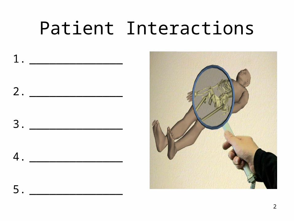
Patient Interactions
1. ______________
2. ______________
3. ______________
4. ______________
5. ______________
3
4
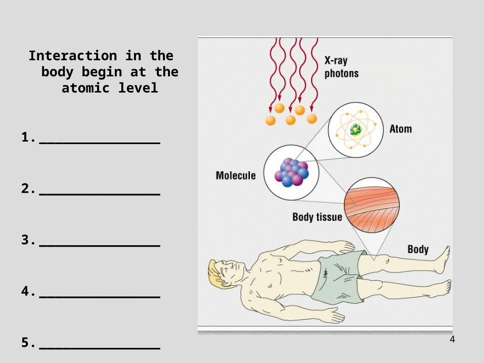
Interaction in the body begin at the atomic
level
1. _______________
2. _______________
3. _______________
4. _______________
5. _______________

5
X-ray photons can change cells
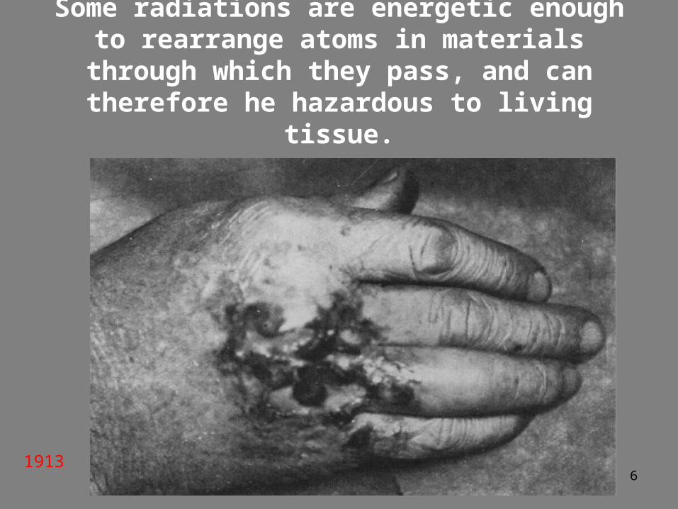
6
Some radiations are energetic enough to rearrange atoms in materials through which
they pass, and can therefore he hazardous to living tissue.
1913
7
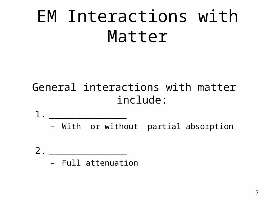
EM Interactions with Matter
General interactions with matter include:1. ______________
– With or without partial absorption
2. ______________ – Full attenuation
8
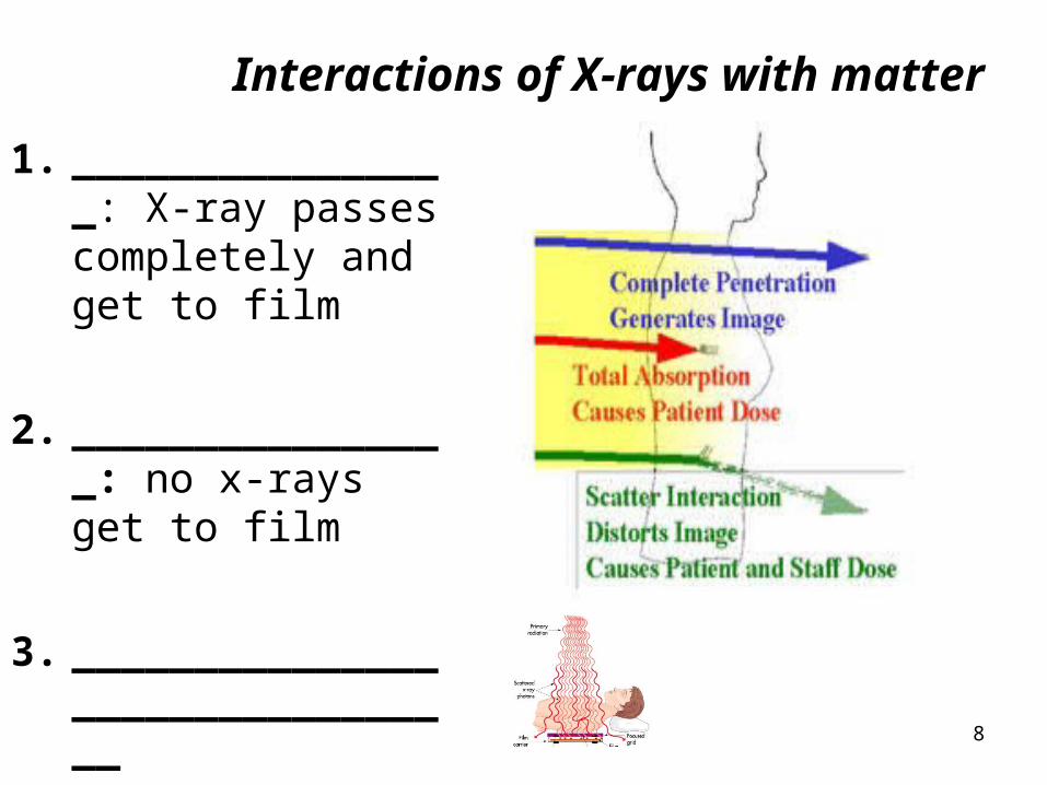
Interactions of X-rays with matter
1. ________________: X-ray passes completely and get to film
2. ________________: no x-rays get to film
3. ________________________________
9
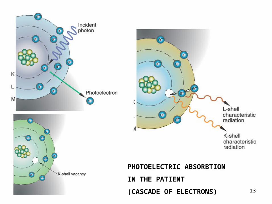
Photoelectric effect
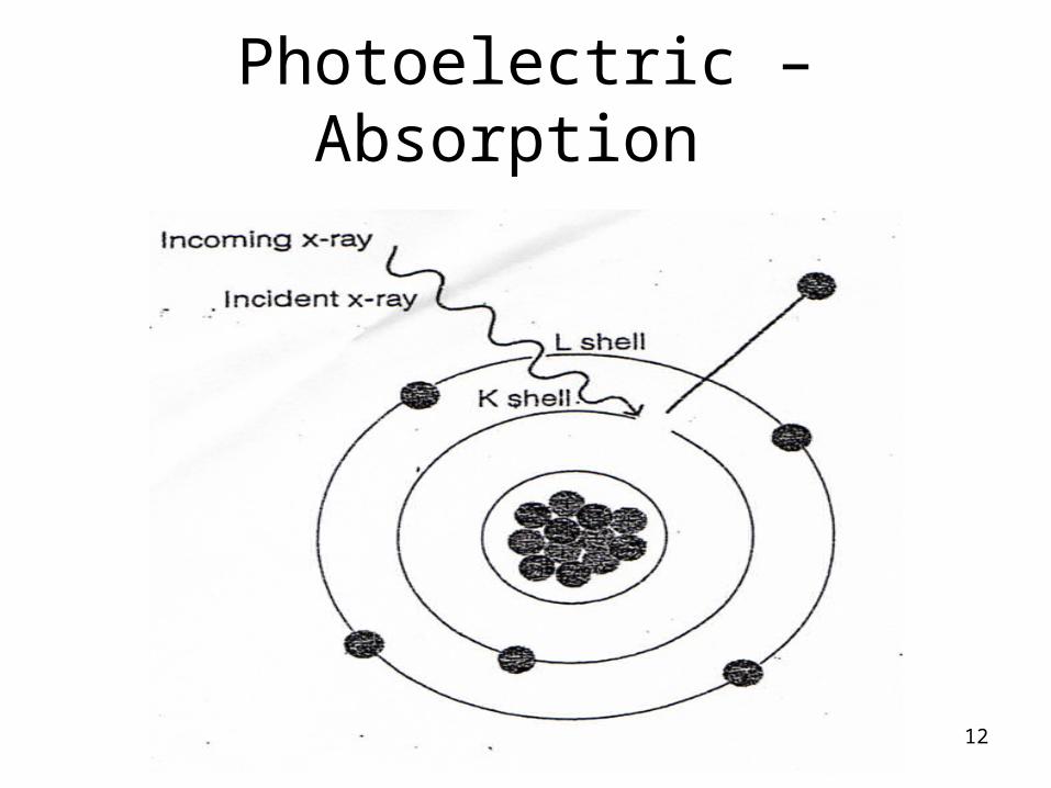
1. Low energy (low kVp) x-ray photon ejects inner shell electron (energy absorbed)
2. Leaving an orbital vacancy. As vacancy is filled a photon is produced
3. More likely to occur in absorbers of high atomic number (eg, bone, positive contrast media)
4. Contributes significantly to patient dose,
5. As all the photon energy is absorbed by the patient (and for the latter reason, is responsible for the production of short-scale contrast).

10
FIG. 9–3 Photoelectric absorption interaction.
(Modified from Carlton RC, Adler AM: Principles of radiographic imaging, an art and a science, ed 4, Thomson Delmar Learning, 2006, Albany, NY. Reprinted with permission of Delmar Learning, a division of Thomson Learning: http://www.thomsonrights.com.
Fax 800-730-2215.)

11CASCADE
12
Photoelectric – Absorption
13
PHOTOELECTRIC ABSORBTION
IN THE PATIENT
(CASCADE OF ELECTRONS)

14
• PHOTOELECTRIC
ABSORBTION
IS WHAT GIVES US
THE CONTRAST
ON THE FILM

15
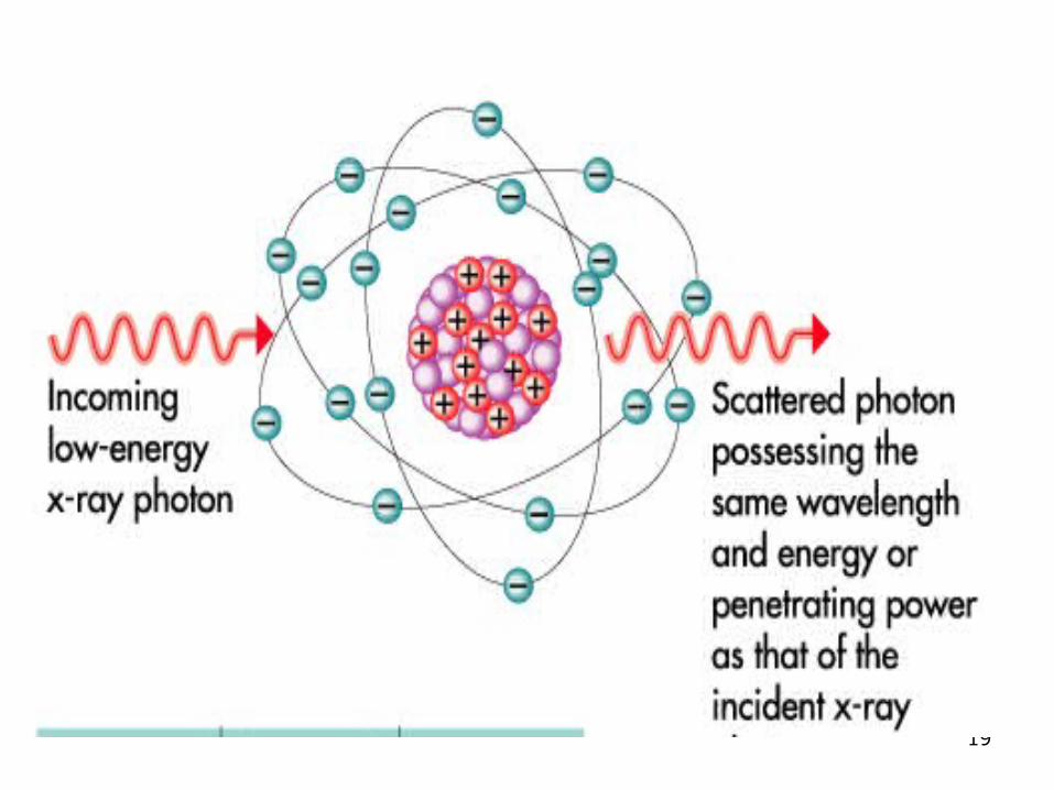
8 p+ + 8e- = neutral atom
1. Incoming photons form tube
2. Pass by the electrons in the patient
3. Do not interact with e–
4. Causes them to vibrate- releasing smnall amounts of heat
CLASSICAL SCATTER IN PATIENT
16
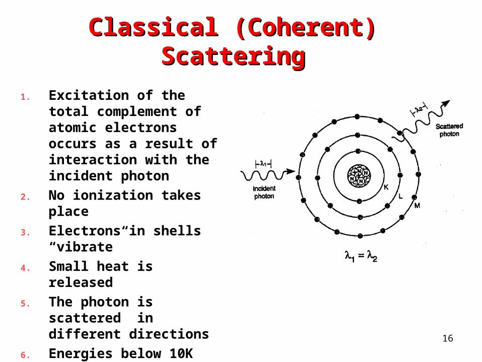
Classical (Coherent) ScatteringClassical (Coherent) Scattering
1. Excitation of the total complement of atomic electrons occurs as a result of interaction with the incident photon
2. No ionization takes place
3. Electrons in shells “vibrate”
4. Small heat is released
5. The photon is scattered in different directions
6. Energies below 10K keV

17
Coherent / Classical Scatter

18
Classic Coherent Scatter
19
20
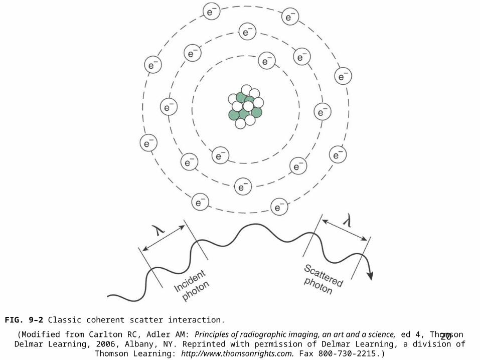
FIG. 9–2 Classic coherent scatter interaction.
(Modified from Carlton RC, Adler AM: Principles of radiographic imaging, an art and a science, ed 4, Thomson Delmar Learning, 2006, Albany, NY. Reprinted with permission of Delmar Learning, a division of Thomson Learning: http://www.thomsonrights.com.
Fax 800-730-2215.)

21
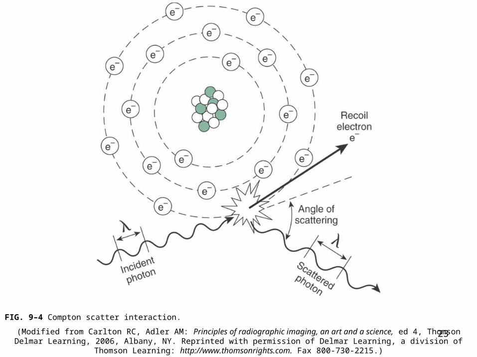
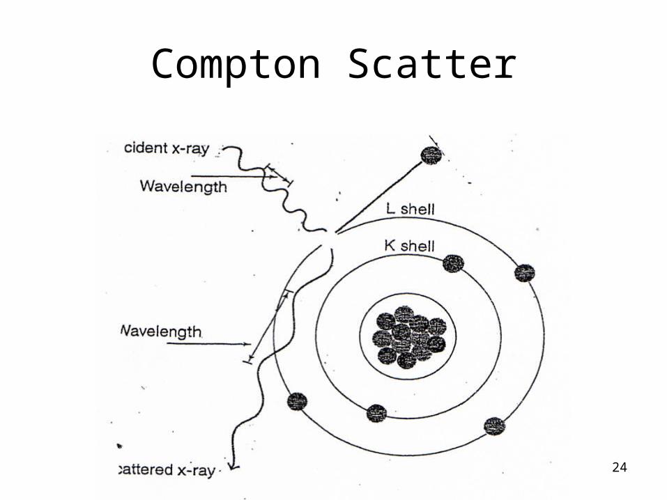
Compton scatter1. High energy (high kVp) x-ray photon ejects an
outer shell electron. 2. Energy is divided between scattered photon
and the compton electron (ejected e-)3. Scattered photon has sufficient energy to exit
body. 4. Since the scattered photon exits the body, it
does not pose a radiation hazard to the patient.
5. Can increase film fog (reduces contrast)6. Radiation hazard to personnel

22
23
FIG. 9–4 Compton scatter interaction.
(Modified from Carlton RC, Adler AM: Principles of radiographic imaging, an art and a science, ed 4, Thomson Delmar Learning, 2006, Albany, NY. Reprinted with permission of Delmar Learning, a division of Thomson Learning: http://www.thomsonrights.com.
Fax 800-730-2215.)
24
Compton Scatter
25
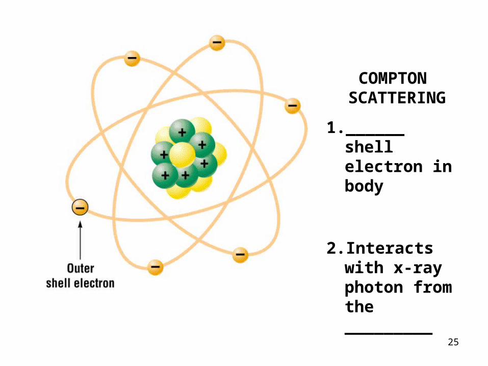
COMPTON SCATTERING
1. ______ shell electron in body
2. Interacts with x-ray photon from the _________
26

27
(WAVY LINE IN = ________ MUST BE INTERACTION IN THE BODY)
28
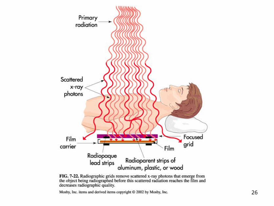
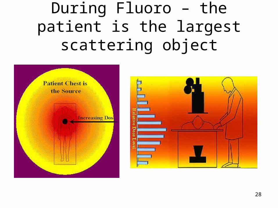
During Fluoro – the patient is the largest scattering object

29
XXXXX
30
Differential Absorbtion
• Results from the differences between xrays being abosorbed and those transmitted to the image receptor
1. ____________________________
2. ____________________________
3. ____________________________
31
Compton and Differential Absorbtion
1. Provides ____ useful info to the image
2. Produces image ________• dulling of the image • NOT representing ___________ information
3. At ____________ energies
32
Photoelectric and Differential Absorbtion
1. Provides _________________ information
2. X-rays do not reach film because they are __________________
3. ______ energies (more differential absorbtion)
4. Gives us the ______________ on our image

33
No interactions with Image Receptor and Differential
Absorbtion
1. No interaction
2. Usually ____________ kVp
3. Goes ______________ body
4. Hits ____________ ________________
5. Usually represents areas of __________• _____atomic numbers
6. Results in __________ areas on the film
34
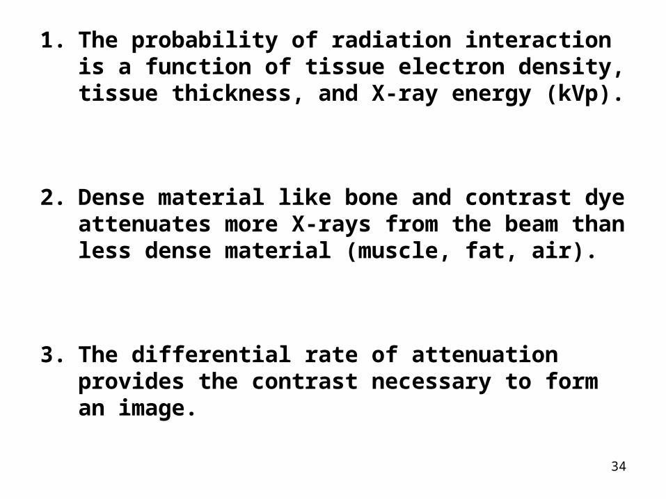
1. The probability of radiation interaction is a function of tissue electron density, tissue thickness, and X-ray energy (kVp).
2. Dense material like bone and contrast dye attenuates more X-rays from the beam than less dense material (muscle, fat, air).
3. The differential rate of attenuation provides the contrast necessary to form an image.
35

36
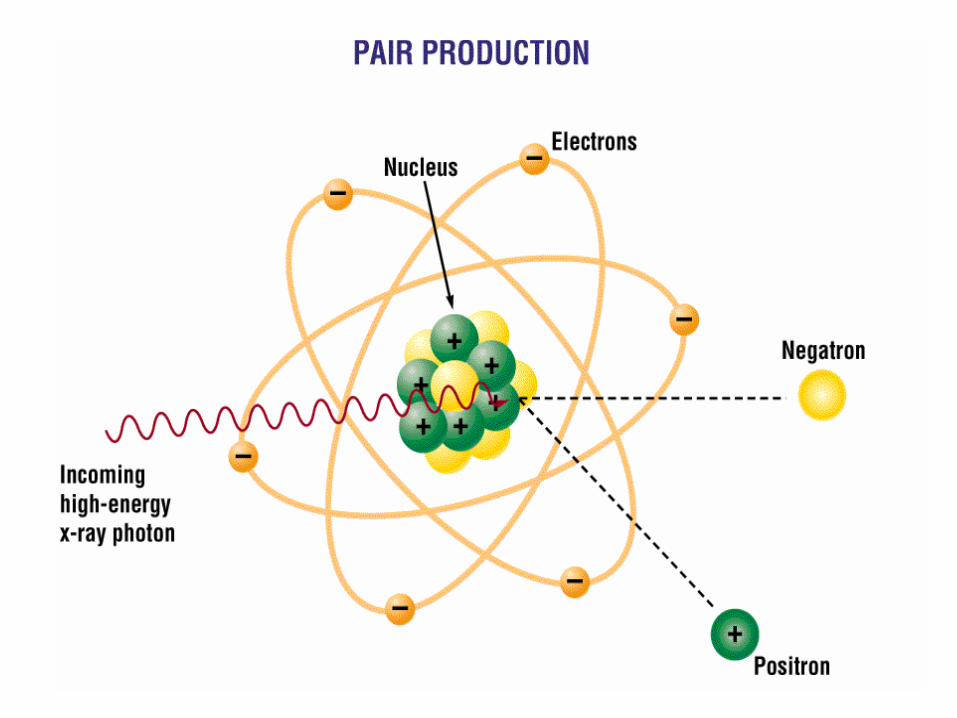
Pair Production

37
FIG. 9–5 Pair production interaction.
(Modified from Carlton RC, Adler AM: Principles of radiographic imaging, an art and a science, ed 4, Thomson Delmar Learning, 2006, Albany, NY. Reprinted with permission of Delmar Learning, a division of Thomson Learning: http://www.thomsonrights.com.
Fax 800-730-2215.)

38
Photodisintegration

39
FIG. 9–6 Photodisintegration interaction.
(Modified from Carlton RC, Adler AM: Principles of radiographic imaging, an art and a science, ed 4, Thomson Delmar Learning, 2006, Albany, NY. Reprinted with permission of Delmar Learning, a division of Thomson Learning: http://www.thomsonrights.com.
Fax 800-730-2215.)
40
Remember….When reviewing diagrams
What is coming in (e or photon?
Where is it occurring (the tube or body?)
Keep practicing – you will get it