pediatric hem/onc: when to go on a zebra hunt on the boards · case 1 hpi: 3 y/o male with a 6 week...
TRANSCRIPT

Pediatric Hem/Onc: When to Go on a Zebra Hunt on the Boards
Terrie Tristan Flatt, DO Hematology/Oncology Children’s Mercy Hospital Kansas City, MO

General Information
Pediatric cancer is rare yet it accounts for the leading cause of disease related death in children less than 15 y/o

A 2-year-old girl presents for evaluation of fussiness, low-grade fever, and what her parents describe as "growing pains." On physical examination, you palpate a nontender mass deep in the right periumbilical area and note mild purple discoloration of the eyelids. Of the following, the MOST likely diagnosis is A. hepatoblastoma B. Hirschsprung disease C. intussusception D. neuroblastoma E. Wilms tumor

You identify a right sided mass in a 2 m/o female on routine heath supervision visit. The mass is firm but not tender to palpation. US reveals a mass pushing the right kidney downward. The most likely diagnosis is: A. hepatoblastoma B. Adrenocortical carcinoma C. Neuroblastoma D. Rhabdomyosarcoma E. Wilm’s tumor

A parent brings in her 4 y/o son for you to evaluate a mass that she discovered while bathing him. He has remained active and not been ill. You palpate a large, non-tender mass in RUQ of the abdomen. His BP is 130/70 and HR is 80. UA reveals microscopic hematuria.
The most likely diagnosis is: A. Hepatoblastoma B. Wilm’s tumor C. Renal cell CA D. Neuroblastoma E. Pheochromocytoma

The parents of a child who was diagnosed at birth with Beckwith-Wiedemann syndrome bring in the baby for his 2-month evaluation. They ask about future health problems and his prognosis now that his omphalocele has been repaired. Of the following, the child is MOST at risk for A. acute lymphocytic leukemia B. astrocytoma C. Hodgkin disease D. rhabdomyosarcoma E. Wilms tumor
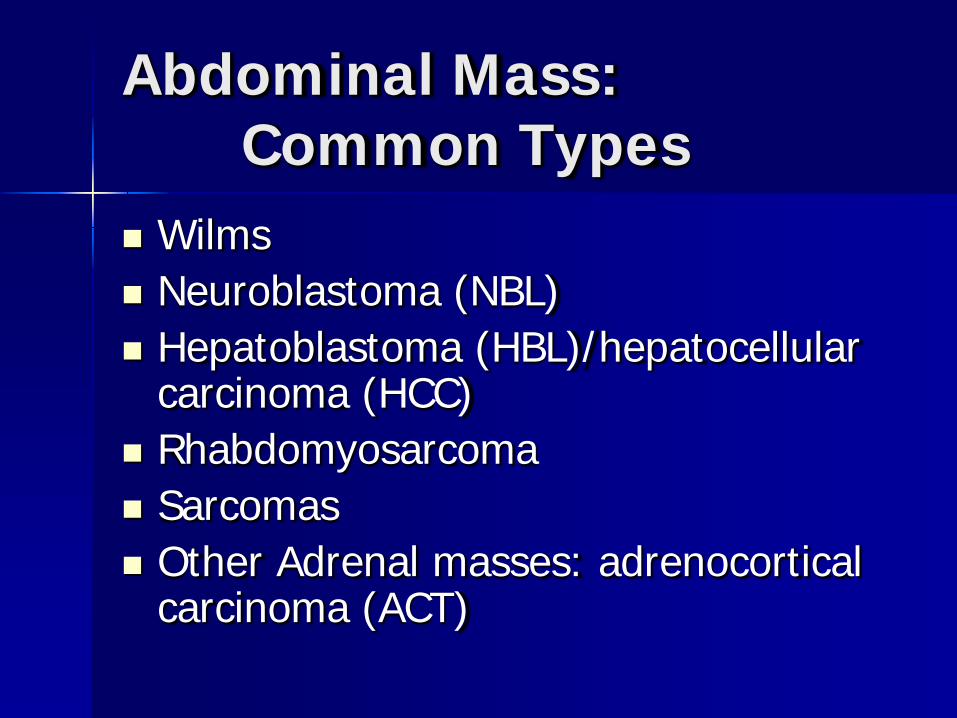
Abdominal Mass: Common Types Wilms Neuroblastoma (NBL) Hepatoblastoma (HBL)/hepatocellular
carcinoma (HCC) Rhabdomyosarcoma Sarcomas Other Adrenal masses: adrenocortical
carcinoma (ACT)

Abdominal Mass: Clinical Presentation Chronic constipation Chronic diarrhea Hypertension Chronically distended abdomen Hormonal changes--virilization, acne, malar
rash, etc (germinoma and ardrenocortical carcinoma)
Intussusception

Abdominal Masses By Age
Newborns: – NBL, mesoblastic nephroma, HBL
Up to age 3: – NBL, Wilms, HBL, rhabdomyosarcoma
Child 3-11: – NBL, Wilms (3-5), lymphoma, HCC, rhabdomyosarcoma
Teenager/young adult – Lymphoma, HCC, rhabdomyosarcoma, adrenal , ovarian
masses (germ cell tumors)
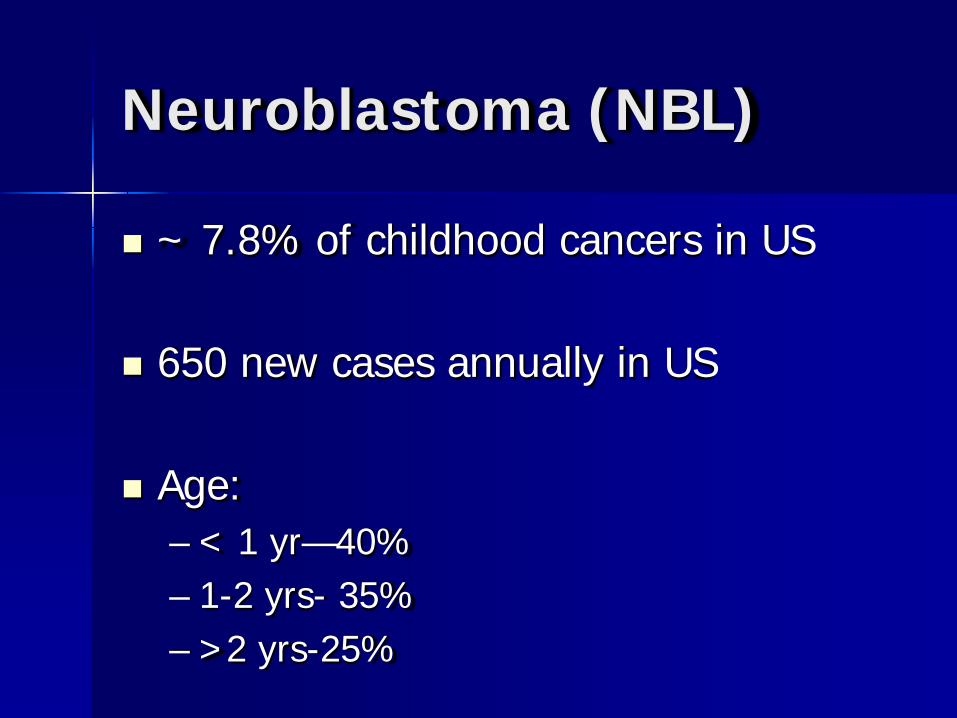
Neuroblastoma (NBL)
~ 7.8% of childhood cancers in US
650 new cases annually in US
Age: – < 1 yr—40% – 1-2 yrs- 35% – >2 yrs-25%

NBL: Clinical Presentation
Symptoms associated with bone marrow failure—bruising, fever, infection, limp, weight loss. 50% of patients present with advanced disease to bone marrow and bone.
2/3 of all patients present with asymptomatic abdominal mass

NBL: Clinical Presentation
Other: – Skin nodules (blueberry muffin) – Liver mets – Bone mets (pathological fractures) – Periorbital bruising known as raccoon eyes
Paraneoplastic syndromes: – Myoclonus and opsoclonus (2%) – Intractable/chronic diarrhea(rare)
HTN and other SNS associated symptoms
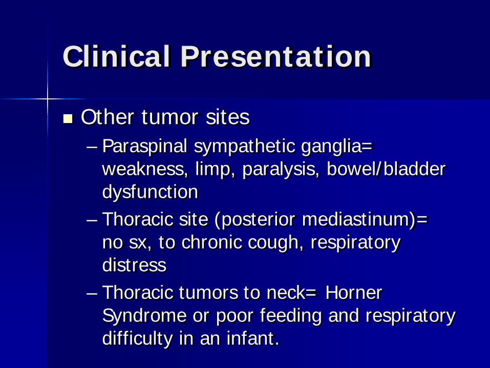
Clinical Presentation
Other tumor sites – Paraspinal sympathetic ganglia=
weakness, limp, paralysis, bowel/bladder dysfunction
– Thoracic site (posterior mediastinum)= no sx, to chronic cough, respiratory distress
– Thoracic tumors to neck= Horner Syndrome or poor feeding and respiratory difficulty in an infant.

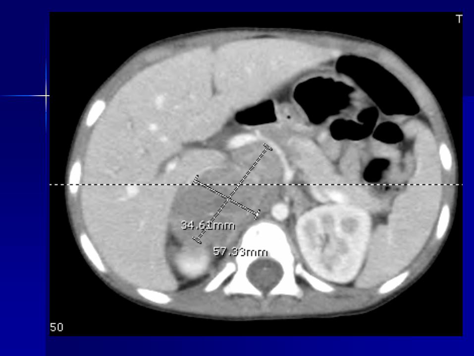
Case 1
HPI: 3 y/o male with a 6 week h/o fatigue, orbital swelling, mild peri-orbital discoloration, limp, and fever. Treated for allergic rhinitis and otitis media with 3 different antibiotics and no response
CT scan obtained to evaluate for sinusitis
Diagnosis: Stage 4 Neuroblastoma


Wilms Tumor
Fifth most common pediatric malignancy – Most common renal tumor in children
~ 0.8 cases per 100,000 persons. – 500 each year in the United States – 6% of cases involving both kidneys
The mean age at diagnosis is 3.5 years and is common b/t 3 to 5 yrs of age

Wilms Tumor
The most common feature at presentation is a painless abdominal mass.
Abdominal pain occurs in only 30-40% of cases.
Other signs and symptoms include hypertension, fever from tumor necrosis, hematuria, and anemia.

Wilms Tumor & Syndromes
Beckwith-Wiedemann syndrome – Macroglossia – Organomegaly (liver, kidneys, pancreas, heart – Omphalocele, umbilical hernia or diastasis recti – Hemihypertrophy – Chrom 11p, increased production IGF-2
Denys-Drash (11p13) – Wilms tumor, – Pseudohermaphroditism – Glomerulopathy

Wilms Tumor & Syndromes
WAGR Syndrome – Wilms' tumor – Aniridia (lack or defect of the iris) – Genitourinary Anomalies (gonadal
dysgenesis, hypospadias, cryptorchidism, duplication of collecting system )
– Mental Retardation – Deletion of chromosome 11p13 – Tumor develops at a younger age – Increased incidence of bilateral tumor

Wilms Tumor & Syndromes
Trisomy 18—Edward syndrome

Surveillance: Beckwith-Wiedemann syndrome
Abdominal US every 3-4 months until 8 yrs of age
Alpha-fetal protein level every 3-4 months until 4 yrs of age
**Vary from center to center


A 4 y/o make presents with several “masses” over the skull (see XR). The lesion was biopsied and the following finding was present (see photo). Of the following, the child is MOST at risk for A. Ewing’s Sarcoma B. Langerhan’s cell histiocytosis C. Osteosarcoma D. Aneurysmal bone cyst E. Wiskott-Aldrich Syndrome

Ma a aaa a aa aaa a a a a Haa aaa a a aa a aa

LCH Ba a a Wa a a a
– Stain CD1a & S-100 positive
– Birbeck Granules

Osteosarcoma
The incidence is 400 cases per year (4.8 cases per million persons <20 y)
The incidence increases steadily with age; a relatively dramatic increase in adolescence corresponds with the growth spurt.
The incidence is slightly higher in African Americans than in
Caucasians

Osteosarcoma
Location – femur (42%), with 75% of tumors in the distal femur
– tibia (19%), with 80% of tumors in the proximal tibia – humerus (10%), with 90% of tumors in the proximal
humerus – jaw (8%) and pelvis (8%) – ALWAYS think METHAPHYSEAL portion of long bones

Osteosarcoma
Radiographic Buzz Words – characteristic Codman triangle

Osteosarcoma
Radiographic Buzz Words – Sunburst lesion: hair-on-end appearance
when new bone is laid down perpendicular to cortex along Sharpey’s fibers

Ewing Sarcoma
The annual incidence of Ewing sarcoma family tumors from birth to age 20 years is 2.9 cases per million
Incidence peaks in the late teenage years (64%) in 2nd decade of life
The incidence in whites is at least 9 times higher than that in
blacks
Ewing Sarcoma
Patients usually present with pain. Patients often have a palpable mass. Patients with lesions of the long bones
can present with a pathologic fracture. Back pain may indicate a paraspinal,
retroperitoneal, or deep pelvic tumor.

Ewing Sarcoma: Buzz Words
Translocation t(11;22) or one of a series of related translocations occurs in more than 95% of Ewing sarcoma
Radiographic Buzz words – Onion Skin lesion (laying down of lamellar bone) – THINK DIAPHYSEAL

Aneurysmal bone cyst
Painless swelling ABCs most commonly affect the long, tubular bones, followed
spine/flat bones
70-86% occurring in patients younger than 20 years

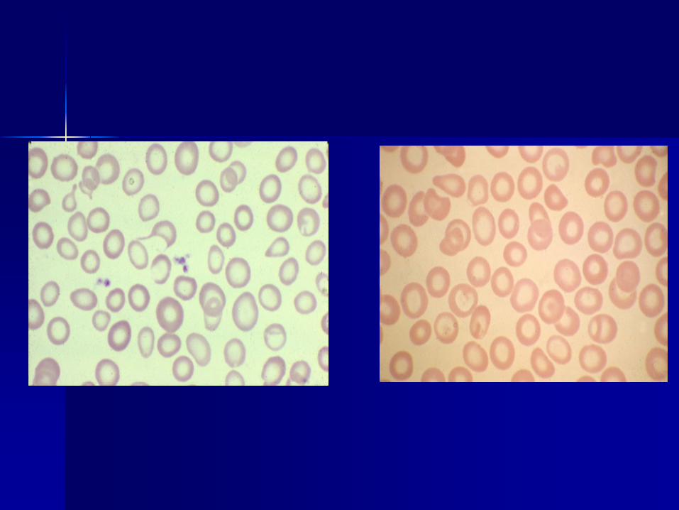
15 m/o female of Southeast Asian ancestry presents for well child exam. She is well appearing and exam is unremarkable. CBC is significant for Hgb 8, MCV 66, RBC 6.5 million and RDW 13. All other cell lines are normal. She drinks 16oz of cow’s milk/day.


What is the next step in management a) Iron supplementation b) Osmotic fragility test c) Hgb Electrophoresis d) G6PD levels

Parameter Iron Deficiency Anemia
Thalassemia Trait
MCV Low Low
RDW High NL
RBC Low Normal to High
Hb Electrophoresis
β-Thal Trait: Elevated HbA2
α-Thal Trait: Low HbA2
FEP Elevated NL
Ferritin Low NL
Serum Iron Low NL
Transferrin sat. Low NL
Mentzer index (MCV/RBC) >13.5 <11.5
Iron Deficiency vs. Thalassemia

A 3 y/o Caucasian male presents with jaundice and fatigue. Splenomegaly is present on examination. Hgb is 6.3, MCV 77, and MCHC is 39 (elevated). He had significant jaundice at birth as well anemia that resolved. There is a family history of anemia and cholecystectomy.


How is this disorder most often inherited: – a) autosomal recessive – b) autosomal dominant – c) X-linked – d) never inherited

A 3 y/o male presents to his PCP with new onset bruising and “rash” on his neck. He has h/o recent URI. He is well appearing and examination is only remarkable for petechiae on neck and bruising to shins and arms. CBC is remarkable for platelet count of 6000 and all other cell lines are normal. Blood chemistries, ESR and uric acid are normal. No family history of blood disorders or deaths from cancer or infection before 40 years of age.


What is the most likely diagnosis: – a) Acute lymphoblastic leukemia – b) Immune thrombocytopenia purpura – c) Fanconi anemia – d) Wiskott Aldridge syndrome


The most common cause of bacteremia in a patient with the above blood smear is: – a) Salmonella – b) Staphylococcous areus – c) Strep. Pneumoniae – d) Pseudomonas species

An 18 m/o boy presents with pallor, decreased activity and appetite but he always tends to be a picky eater. He has been limping and complaining of leg pain for 2 weeks. On exam you note bruising to the legs, arms and one on his back. He is ill-appearing but non-toxic. He is sallow appearing, oral mucosa is pale, conjunctivae are pale and he has several small mouth sores. He has several 1-2 cm hard nodes in the inguinal area. His HR is 170.
The most likely diagnosis is: A. ITP B. Child abuse C. ALL D. iron-deficiency anemia E. Transient erythroblastopenia of childhood

A 12-year-old girl presents to the emergency department with nausea, vomiting, and abdominal pain of 1 month's duration. Physical examination reveals a large, smooth mass encompassing almost the entire lower abdomen. Computed tomography scan confirms a mass, and biopsy documents Burkitt lymphoma. She immediately begins receiving chemotherapy, and 12 hours later she develops the classic electrolyte and urinary findings consistent with tumor lysis syndrome (TLS). Of the following, the laboratory findings MOST consistent with TLS are A. Serum Potassium: Elevated; Serum Phosphorous: Elevated; Serum lactate dehydrogenase:Normal; Serum sodium: Elevated B. Serum Potassium: Elevated; Serum Phosphorous: Normal; Serum lactate dehydrogenase:Elevated; Serum sodium: Normal C. Serum Potassium: Normal; Serum Phosphorous: Elevated; Serum lactate dehydrogenase:Elevated; Serum sodium: Elevated D. Serum Potassium: Normal; Serum Phosphorous: Normal; Serum lactate dehydrogenase:Elevated; Serum sodium: Normal E. Serum Potassium: Elevated; Serum Phosphorous: Elevated; Serum lactate dehydrogenase:Elevated; Serum sodium: Normal

Tumor Lysis
Definition: Break down (lysis) of fragile tumor cells that contain increased quantities of potassium, phosphorous, and protein (uric acid).

Risk for Tumor Lysis
***Burkitt’s Lymphoma Leukemia, esp lymphoblastic AML is less associated with this Lymphoma High WBC with high blast count Large spleen, liver, bulky nodes

Tumor Lysis
Lab Findings: – K↑, Phos ↑, Uric Acid ↑ – Ca↓ to normal – LDH ↑

Treatment
IV Hydration (1.5 to 2 x maintenance fluids WITHOUT K+).
Allopurinol or Rasburicase for uric acid Phosphorous binders if Phos extremely elevated Some use NaHCO3 in IVF but must exercise caution
b/c if ca x phos ratio is >50, then it will increase the risk of precipitation and phos will often continue to rise. HCO3 is most beneficial with high uric acid as it makes it more soluble for excretion. HCO3 decreases the solubility of Phos

“When you hear hoof beats beneath your window, think ponies for there are far fewer zebras in our presence.”
HOWEVER, there will always be zebras when you hunt for them.