periodontal case presentation - 1 › pdf › case-1-emsworth-26.… · periodontal case...
TRANSCRIPT

P E R I O D O N T A L C A S E P R E S E N T A T I O N - 1
Overview A 32 year-old patient presented with generalized aggressive periodontitis.
Treatment included non-surgical therapy with adjunctive antibiotics and surgical
treatment.
Treatment aims On presentation our patient’s main concern was the recent change in the
position of her upper left central incisor. She was keen to prevent any further
movement of this tooth. Retaining her natural teeth was a key aim for treatment.
Clinical examination Clinical examination noted profuse bleeding on probing, deep periodontal
probing depths mainly associated with molar and upper incisor teeth,
generalised mobility and furcation involvement UL6.
Upper occlusal intra-oral view

g
Lower occlusal intra-oral view
Special tests
Long cone periapical radiographs UR2,1 & UL1,2
Long cone periapical radiographs of molar teeth
LR6 and LL6 gave a positive response to EPT and endo-frost.

Diagnosis A diagnosis of generalized aggressive periodontitis and pathological tooth
migration associated with UL1 and UL2 was established.
Initial treatment plan (Phase 1) An initial treatment plan was established:
� Oral hygiene advice
� Full mouth supra-gingival scale
� Extraction of UR8 and UL8
� Root surface debridement under local anaesthetic of all sites
5mm or more with adjunctive systemic antibiotics
� Periodontal reassessment
Our patient was aware that initial therapy would affect the appearance of her
teeth by potentially increasing recession and the prominence of anterior ‘black
triangles’. She requested initial treatment be completed but would consider
further cosmetic treatment to the upper anterior teeth in the future if needed.
Reassessment following phase 1 treatment At reassessment our patient reported:
� that her gums felt better
� no bleeding on brushing
� that the UL1 seemed to have moved back into a
position closer to its original position
Intra-oral anterior view at presentation
Intra-oral anterior view following phase 1 treatment

Blue = periodontal chart following phase 1 treatment
At this stage further treatment options were discussed with our patient:
� Accept residual deep pockets and enter a supportive periodontal
therapy program. The aim would be to reduce the rate of any
further loss of attachment.
� Periodontal surgery. The aim would be to reduce pocket depths,
improve access for plaque control and improve the periodontal
prognosis of these teeth.
Our patient requested periodontal surgery.
Treatment Plan – Phase 2 � Further root surface debridement UR7,6,4
� Occlusal adjustment UR7
� Pocket reduction surgery UL6,7, LR6, LL6
� Composite build-up UL2 and UL1 to reduce the appearance of the
black triangle
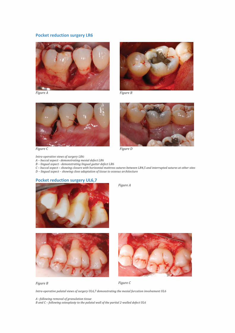
Pocket reduction surgery LR6
Figure A
Figure B
Figure C
Figure D
Intra-operative views of surgery LR6: A – buccal aspect - demonstrating mesial defect LR6 B – lingual aspect - demonstrating lingual gutter defect LR6 C – buccal aspect – showing closure with horizontal mattress sutures between LR4,5 and interrupted sutures at other sites D – lingual aspect – showing close adaptation of tissue to osseous architecture
Pocket reduction surgery UL6,7
Figure A
Figure B
Figure C
Intra-operative palatal views of surgery UL6,7 demonstrating the mesial furcation involvement UL6
A –following removal of granulation tissue B and C – following osteoplasty to the palatal wall of the partial 2-walled defect UL6

Reassessment following phase 2 treatment
Blue = Periodontal chart following phase 1 treatment
Green = Periodontal chart following phase 2 treatment
Overall outcome Our patient was very pleased with the outcome of treatment. She had no
bleeding on brushing and noted that her gums felt firmer. She is much happier
with the position of her UL1.
On presentation probing depths of 5mm and above were recorded at 46% of
sites, now less that 1% of sites have such probing depths.
Overall our patient has shown an excellent response to treatment and enters a
supportive periodontal therapy programme involving shared care with her
general dental practitioner.

Pre-treatment anterior view
Post-treatment anterior view
Pre-treatment lateral views
Post-treatment lateral views

Pre-treatment upper occlusal view
Post-treatment upper occlusal view