peripheral organs of dengue fatal cases present … laboratory of biotechnology and physiology ......
TRANSCRIPT
RESEARCH ARTICLE
Peripheral Organs of Dengue Fatal Cases
Present Strong Pro-Inflammatory Response
with Participation of IFN-Gamma-, TNF-Alpha-
and RANTES-Producing Cells
Tiago F. Povoa1☯, Edson R. A. Oliveira2☯, Carlos. A. Basılio-de-Oliveira3, Gerard
J. Nuovo4,5, Vera L. A. Chagas6, Natalia G. Salomão7, Ester M. Mota8, Marciano V. Paes7*
1 Laboratory of Biotechnology and Physiology of Viral Infections, Oswaldo Cruz Institute, Oswaldo Cruz
Foundation, Rio de Janeiro, Brazil, 2 Laboratory of Molecular Modeling, Federal University of Rio de Janeiro,
Rio de Janeiro, Brazil, 3 Pathological Anatomy, Gaffree Guinle Hospital, University of Rio de Janeiro, Rio de
Janeiro, Brazil, 4 Ohio State University Comprehensive Cancer Center, Columbus, Ohio, United States of
America, 5 Phylogeny Inc, Powell, Ohio, United States of America, 6 Pathological Anatomy, Clementino
Fraga Filho University Hospital, Rio de Janeiro, Brazil, 7 Interdisciplinary Laboratory of Medical Research,
Oswaldo Cruz Institute, Oswaldo Cruz Foundation, Rio de Janeiro, Brazil, 8 Laboratory of Pathology,
Oswaldo Cruz Institute, Oswaldo Cruz Foundation, Rio de Janeiro, Brazil
☯ These authors contributed equally to this work.
Abstract
Dengue disease is an acute viral illness caused by dengue virus (DENV) that can progress
to hemorrhagic stages leading to about 20000 deaths every year worldwide. Despite many
clinical investigations regarding dengue, the immunopathogenic process by which infected
patients evolve to the severe forms is not fully understood. Apart from differences in viru-
lence and the antibody cross reactivity that can potentially augment virus replication, imbal-
anced cellular immunity is also seen as a major concern in the establishment of severe
dengue. In this context, the investigation of cellular immunity and its products in dengue
fatal cases may provide valuable data to help revealing dengue immunopathogenesis.
Here, based in four dengue fatal cases infected by the serotype 3 in Brazil, different periph-
eral organs (livers, lungs and kidneys) were studied to evaluate the presence of cell infil-
trates and the patterns of local cytokine response. The overall scenario of the studied cases
revealed a considerable systemic involvement of infection with mononuclear cells targeted
to all of the evaluated organs, as measured by immunohistochemistry (IHC). Quantification
of cytokine-expressing cells in peripheral tissues was also performed to characterize the
ongoing inflammatory process by the severe stage of the disease. Increased levels of IFN-
γ- and TNF-α-expressing cells in liver, lung and kidney samples of post-mortem subjects
evidenced a strong pro-inflammatory induction in these tissues. The presence of increased
RANTES-producing cell numbers in all analyzed organs suggested a possible link between
the clinical status and altered vascular permeability. Co-staining of DENV RNA and IFN-γ or
TNF-α using in situ hibridization and IHC confirmed the virus-specific trigger of the pro-
inflammatory response. Taken together, this work provided additional evidences that cor-
roborated with the traditional theories regarding the “cytokine storm” and the occurrence of
PLOS ONE | DOI:10.1371/journal.pone.0168973 December 22, 2016 1 / 19
a1111111111
a1111111111
a1111111111
a1111111111
a1111111111
OPENACCESS
Citation: Povoa TF, Oliveira ERA, Basılio-de-Oliveira
CA, Nuovo GJ, Chagas VLA, Salomão NG, et al.
(2016) Peripheral Organs of Dengue Fatal Cases
Present Strong Pro-Inflammatory Response with
Participation of IFN-Gamma-, TNF-Alpha- and
RANTES-Producing Cells. PLoS ONE 11(12):
e0168973. doi:10.1371/journal.pone.0168973
Editor: Dong-Yan Jin, University of Hong Kong,
HONG KONG
Received: June 24, 2016
Accepted: December 10, 2016
Published: December 22, 2016
Copyright: © 2016 Povoa et al. This is an open
access article distributed under the terms of the
Creative Commons Attribution License, which
permits unrestricted use, distribution, and
reproduction in any medium, provided the original
author and source are credited.
Data Availability Statement: All relevant data are
within the paper and its Supporting Information
files.
Funding: This work was funded by the FundacãoCarlos Chagas Filho de Amparo à Pesquisa do
Estado do Rio de Janeiro (FAPERJ, Grant: E-26/
110.511/2014) www.faperj.br to MVP. The funder
had no role in study design, data collection and
analysis, decision to publish, or preparation of the
manuscript.

uneven cellular immunity in response to DENV as major reasons for progress to severe
disease.
Introduction
Dengue is considered the most important mosquito-borne viral disease due to its clinical rele-
vance and rapid spread, nowadays putting at risk about half of the world’s population [1]. The
etiologic agent, dengue virus (DENV), is distributed as four distinct serotypes (DENV1 to
DENV4) and infections can result in a mild flu-like acute illness known as dengue fever (DF)
[2]. From an epidemiological view, it is estimated that 390 million dengue infections occur
each year, of which nearly 25% are symptomatic [3]. While most patients naturally recover
from the non-severe clinical DF course, a small proportion evolves to severe disease, mostly
characterized by plasma leakage and hemorrhagic manifestations (namely dengue shock syn-
drome—DSS and dengue hemorrhagic fever—DHF) [2, 4]. Despite the relevant mortality
rates derived from dengue complications (arround 20000 deaths each year) [5], the elucidation
of the pathogenic process by which infected patients evolve to the severe forms is still an ongo-
ing challenge. Apart from the relationship between social determinants of health and dengue
fatal cases, biological factors such as distinct virulence levels among virus strains and host
immunity have been considered as key elements to drive patients to severe stages [6, 7]. Dis-
ease complications triggered by DENV enhanced infections assisted by previously-formed
opsonizing antibodies, were related to altered T cell activation and cytokine production in sec-
ondary infections [8–10]. Yet, concerning a host primary response environment, other
unknown factors could also play a role in triggering severe dengue. Classical DF symptoms,
such as fever and headaches, usually match with high viremia levels, but interestingly the
severe forms of dengue (DSS/DHF), when manifested, occur after virus clearance. This obser-
vation has raised concerns about the association of severe dengue with immonopathological
mechanisms [11, 12].
In this context, the investigation of post-mortem severe dengue cases may represent a valu-
able tool for a better understanding of the immune scenario during a terminal stage. Addition-
ally, a search for evidences regarding cell migration and cytokine production in peripheral
tissues may also provide new insights about possible underpinning immune mechanisms
linked to the development of severe forms. In a previous report of our laboratory, peripheral
organs such as livers, lungs and kidneys of four dengue cases that died from DENV-3 were
histopathologically and ultrastructurally screened [13]. Aside from virus detection in unusual
sites such as hepatocytes and type II pneumocytes, all studied organs presented lesions that
corresponded to severe dengue cases. In this work, the same post-mortem samples were object
of study for investigation of the cellular immune response and its products. Immunohisto-
chemical analysis revealed a systemic involvement of infection with mononuclear cells targeted
to all of the analyzed tissues. Assessment of local cytokine response showed increased levels of
IFN-γ- and TNF-α-expressing cells in livers, lungs and kidneys that evidenced a consistent
pro-inflammatory induction in these tissues. Co-expression of DENV RNA and IFN-γ or
TNF-α by Kupffer cells confirmed the specific DENV induction over the cytokine production,
as found by in situ hibridization and IHC. Furthermore, an indicative of altered vascular per-
meability found in all analyzed organs was also suggested due to the presence of increased lev-
els of local RANTES-producing cells.
Immunopathological Evidences in Dengue Fatal Cases
PLOS ONE | DOI:10.1371/journal.pone.0168973 December 22, 2016 2 / 19
Competing Interests: The authors have declared
that no competing interests exist.

Ultimately, this work brought additional evidences that the effect of the uneven cellular
immunity in response to DENV can contribute to disease severity. Given the limited numbers
of reports concerning investigation of post-mortem samples from dengue severe cases, this
work importantly contributes to narrowing the gaps of dengue immunopathogenesis.
Materials and Methods
Ethical procedures
All procedures performed during this work were approved by the Ethics Committee of the
Oswaldo Cruz Foundation/FIOCRUZ, with the number CAEE: 47525115.3.0000.5248 for
studies with dengue fatal cases and controls. The use of tissue samples informed consent was
verbally provided by family members to the responsible physician Dr. Carlos Alberto Basıllio
de Oliveira by the time of the necropsy. This consent procedure was approved by the ethics
committee.
Human fatal cases
The human tissues analyzed in this study (livers, lungs and kidneys) were obtained from four
dengue fatal cases that occurred during a Brazilian outbreak of DENV-3 in 2002 in Rio de
Janeiro. All patients died with a clinical diagnosis of severe dengue with infections confirmed
by positive serum IgM antibodies. The four negative controls, from both sexes and ranging
from 40 to 60 year old, were non-dengue and did not present any other infectious disease.
More information about these cases can be found in a previous report of our laboratory [13].
Briefly:
Case 1. A 63-year-old male patient that developed a sudden onset of headache, myalgia,
anorexia and abdominal pain. A few days later the patient presented diarrhea, thrombocytope-
nia (platelet 79.000/mm3) and hemoconcentration (hematocrit 59%). The case eventually
evolved to shock with severe pulmonary congestion followed by death with a clinical diagnosis
of dengue hemorrhagic fever.
Case 2. A 21-year-old female patient who experienced fever, myalgia and headache with
progression to metrorrhagia, nausea, vomiting and diarrhea. The patient also presented severe
leukopenia and thrombocytopenia (platelet 10.000/mm3). During hospitalization, the case pro-
gressed to respiratory failure, followed by evolution of multiple organ failure and refractory
shock.
Case 3. A 41-year-old female presenting fever, weakness, abdominal pain, leukocytosis,
hematocrit of 48% and fluid in the abdominal cavity. The patient was diagnosed with dengue
hemorrhagic fever and died from an acute pulmonary edema.
Case 4. A 61-year-old female that manifested classical dengue symptoms (fever, myalgia,
vomiting and diarrhea). The patient evolved to severe clinics and died from acute pulmonary
edema with sudden cardiac arrest.
Histopathological analysis
Tissue samples from the human necropsies were fixed in formalin (10%), blocked in paraffin
resin, cut in 4 μm, deparaffinized in xylene and rehydrated with alcohol, as described previ-
ously [14]. Sections were stained with hematoxylin and eosin (H.E.) for histological examina-
tion and visualized under a Nikon ECLIPSE E600 microscope.
Immunopathological Evidences in Dengue Fatal Cases
PLOS ONE | DOI:10.1371/journal.pone.0168973 December 22, 2016 3 / 19

Immunohistochemical procedure
For immunohistochemical studies, the paraffin-embedded tissues were cut (sections of 4 μm),
deparaffinized in xylene and rehydrated with alcohol. Antigen retrieval was performed by
heating the tissue in the presence of citrate buffer [15]. Tissues were blocked for endogenous
peroxidase with 3% hydrogen peroxidase in methanol and rinsed in Tris-HCl (pH 7.4). To
reduce non-specific binding, sections were incubated for 30 min at room temperature. Sam-
ples were then incubated over-night at 4˚C with anti-human antibodies that recognize CD4
(Spring Bioscence, CA, USA), CD8 (DAKOCytomation, CA, USA), CD20 (Biocare Medical,
CA, USA), CD68 (Biocare Medical, CA, USA), IFNγ (Abbiotec, CA, USA), TNFα (Abbiotec,
CA, USA), IL-10 (Abbiotec, CA, USA), RANTES/CCL5 (Santa Cruz Biotechnology, CA, USA)
or TGF-β (Abbiotec, CA, USA). The next day, sections were incubated with rabbit anti-mouse
IgG, a secondary antibody horseradish peroxidase (HRP) conjugate (Spring Bioscience, CA,
USA), for 30 min at room temperature. For negative control of the immunohistochemical
reactions, samples were incubated only with the secondary HRP-conjugated antibody. Reac-
tions were revealed with diaminobenzidine (Dako, CA, USA) as a chromogen and the sections
were counterstained in Meyer’s hematoxylin (Dako). Finally, samples were examined under a
Nikon ECLIPSE E600 microscope.
Quantification of positive cells by immunohistochemistry
Slides were evaluated using a Nikon ECLIPSE E600 microscope with a coupled Cool SNAP-
Procf Color camera. For each specific antibody, 50 images (fields) were randomly acquired at
400x magnification using the software Image Pro version 4.5. After collecting the frames, posi-
tive cells were quantified in each of the 50 fields in every organ and the median of positive cell
number was determined. All analyzes were accomplished in a blind test without prior knowl-
edge of the studied groups. After quantification, frames exhibited in figures were selected as to
be more informative according to specific areas in the analyzed tissues. S1 Table contains the
raw data.
In Situ Hybridization
The assessment of DENV in liver sections was performed by in situ hybridization using a
digoxigenin-tagged probe (5’-TGACCATCATGGACCTCCA-3’) which anneals within the
negative strand of the DENV RNA genome, as previously described [13]. Briefly, paraffin-
embedded sections of tissues were deparaffinized and digested with pepsin (1.3 mg/ ml) for 4
min at room temperature. Tissues were incubated with the probe cocktail at 60˚C for 5 min
and then kept overnight at 37˚C for denaturation and hybridization, respectively. Next, sam-
ples were washed with 0.2 x SSC and 2% bovine serum albumin at 55˚C for 5 min. The probe-
target complexes were revealed by the activity of alkaline phosphatase conjugated to anti-
digoxigenin.
Co-staining of DENV RNA and pro-inflammatory cytokines
Co-staining of virus and IFNγ or TNFα were performed, as previously described [13], using
in situ hybridization and immunohistochemistry. The dengue case 2 was considered for this
analysis. Briefly, the DENV probe was first tagged with 59 digoxigenin and locked nucleic
acid (LNA) modified (Exiqon). Resulting complexes were visualized using an antidigoxi-
genin-alkaline phosphates conjugate and nitro-blue tetrazolium and 5-bromo-4-chloro-
39-indolyphosphate as the chromogen. Detection of IFNγ or TNFα was then performed by
immunohistochemistry (anti-IFNγ antibody—ABCAM ab133566-rabbit and anti-TNFα—
Immunopathological Evidences in Dengue Fatal Cases
PLOS ONE | DOI:10.1371/journal.pone.0168973 December 22, 2016 4 / 19

ABCAM ab6671-rabbit) using Leica Bond Max automated platform (Leica Biosystems) and
DAB as the chromogen. No counterstain was done. Data were analyzed by the computer
based Nuance system (Caliper Life Sciences, Hopkinton, MA, USA) which separates the dif-
ferent chromogenic signals, converts them to fluorescent-based signals and combine them to
determine co-expression.
Statistical analyses
Data were analyzed with GraphPad prism software v5.1 (La Jolla, USA) using non-parametric
statistical tests. Significant differences between analyzed groups (controls and DENV-patients)
were determined using Mann-Whitney test with �p< 0.05.
Results
The liver as a target of immune-mediated mechanisms in dengue fatal
cases
The liver is considered as an important target for DENV infection and is the most common
organ to be involved in the disease. Hepatic alterations are key characteristics found in DENV
cases. As observed in biopsies and autopsies of previously reported fatal cases [16], hepatocytes
and Kupffer cells are described as important targets during DENV infection [17, 18]. For this
reason, liver samples of the four DENV-3 fatal cases were first considered for our evaluations.
Histopathological studies of all samples showed diffuse mononuclear cell infiltrates,
mainly around the portal space (Fig 1 panel a). Detection of CD68+ cells revealed the presence
of hyperplasic Kupffer cells and/or circulating macrophages (Fig 1 panel b), although quantifi-
cation analysis did not show statistical difference when comparing dengue to control samples
(Fig 1 panel c). Yet, we detected numerous CD4+ (Fig 1 panel d), and CD8+ (Fig 1 panel f) T
cells placed mainly in sinusoidal capillaries. Quantification of these cell populations showed a
significant increase in the number of T lymphocytes present in liver samples of dengue cases
when compared to controls (about 5 and 3 fold, respectively—Fig 1 panels e and g).
In order to qualify the ongoing inflammatory process in the hepatic tissue, we also investi-
gated the cytokine production by the mononuclear cell types found in the liver. In this case,
cells expressing TNF-α, IFN-γ, IL-10, TGF-β and RANTES were considered for quantification.
We observed a great number of cells producing these cytokines in the midzonal area and, to a
lesser extent, in other hepatic areas. Production of TNF-α was detected by Kupffer cells and
monocytes, mainly in the sinusoidal capillaries (Fig 2 panel a), while detection of IFN-γ was
found mostly in lymphocytes, Kupffer cells and monocytes (Fig 2 panels b and c). In sinusoidal
capillaries, we also detected groups of cells with an anti-inflammatory profile, such as IL-
10-expressing monocytes and lymphocytes (Fig 2 panel d) and TGF-β-expressing macro-
phages and Kupffer cells (Fig 2 panel h). The chemokine RANTES/CCL5 was detected mainly
in endothelium and Kupffer cells (Fig 2 panels i and j). Quantification of cells producing the
inflammatory cytokines TNF-α, IFN-γ and RANTES/CCL5 revealed a significant increase (4-,
4.5- and 3-fold, respectively) in dengue group compared to control (Fig 2 panels e, f and l),
while the number of cells expressing the anti-inflammatory cytokines IL-10 and TGF-β did
not change significantly in either group (Fig 2 panels g and k).
Histopathological analysis and cytokine profile present in the lungs of
dengue fatal cases
Previous studies in our laboratory revealed that lung tissues from fatal dengue cases showed
severe damages as represented by diffuse areas of hemorrhage and edema [13]. Here, we
Immunopathological Evidences in Dengue Fatal Cases
PLOS ONE | DOI:10.1371/journal.pone.0168973 December 22, 2016 5 / 19

aimed to investigate a possible contribution of an exacerbated pro-inflammatory response that
could be related to this local tissue impairment.
After analysis of lung sections of dengue cases we observed a diffuse mononuclear infiltrate
in alveolar septa and edema areas (Fig 3 panel a), thus, indicating that this tissue could also be
targeted by immune mechanisms triggered by infection. The immunohistochemical assay
revealed the presence of macrophages (CD68+ cells) (Fig 3 panel b) in alveolar septa, CD4+
(Fig 3 panel d) and CD8+ T cells (Fig 3 panel f) mainly in the blood vessels. The quantification
analysis revealed a 4-fold increase of lymphocytes in the dengue group when compared to
non-dengue samples (Fig 3 panels e and g), whereas the number of CD68+ was not statistically
different from the controls (Fig 3 panel c).
We next aimed to identify the local cytokine profile in the lungs to characterize the ongoing
inflammatory process in fatal cases of severe dengue. The evaluation of cytokine-expressing
cells in the tissues of the dengue group exhibited pro- and anti-inflammatory profiles occur-
ring simultaneously, what revealed an atypical elicited immunity. TNF-α, which is an impor-
tant cytokine related to dengue pathogenesis, was detected mainly in alveolar macrophages.
(Fig 4 panel a). Increased numbers of TNF-α- (Fig 4 panel d), IFN-γ- (Fig 4 panels b and e),
IL-10- (Fig 4 panels c and f) and TGF-β- (Fig 4 panels g and i) expressing cells such as macro-
phages and lymphocytes were characterized in the tissues of dengue cases, when compared to
controls. Vascular permeability impairment was also addressed in dengue cases by the pres-
ence of several RANTES-expressing endothelial cells and alveolar macrophages in the
Fig 1. Characterization of cell subpopulations in liver tissues from DENV-3 fatal cases. Sections were stained with (a) H.E. or (b, d, f)
incubated with specific antibodies in immunohistochemical assays. (a) Liver of a representative dengue case showing diffuse mononuclear
infiltrates around the portal space; (b) detection of hyperplasic Kupffer cells (CD68+) observed mainly in sinusoidal capillaries of the dengue
cases; (c) quantification of CD68+ cells in dengue cases and controls (non-dengue cases); (d) detection of CD4+ cells manly in portal space
of the dengue cases; (e) quantification of CD4+ cells in controls and dengue cases; (f) CD8+ cells detected mainly in portal space; (g)
quantification of CD8+ cells. MI—mononuclear cell infiltrates. Asterisks indicate differences that are statistically significant between dengue
cases and control groups, (*p < 0.05). Staining controls are shown in S1 Fig.
doi:10.1371/journal.pone.0168973.g001
Immunopathological Evidences in Dengue Fatal Cases
PLOS ONE | DOI:10.1371/journal.pone.0168973 December 22, 2016 6 / 19

perivascular space (Fig 4 panel h). The quantification of these subpopulations was found to be
increased in dengue cases when compared to controls (Fig 4 panel j).
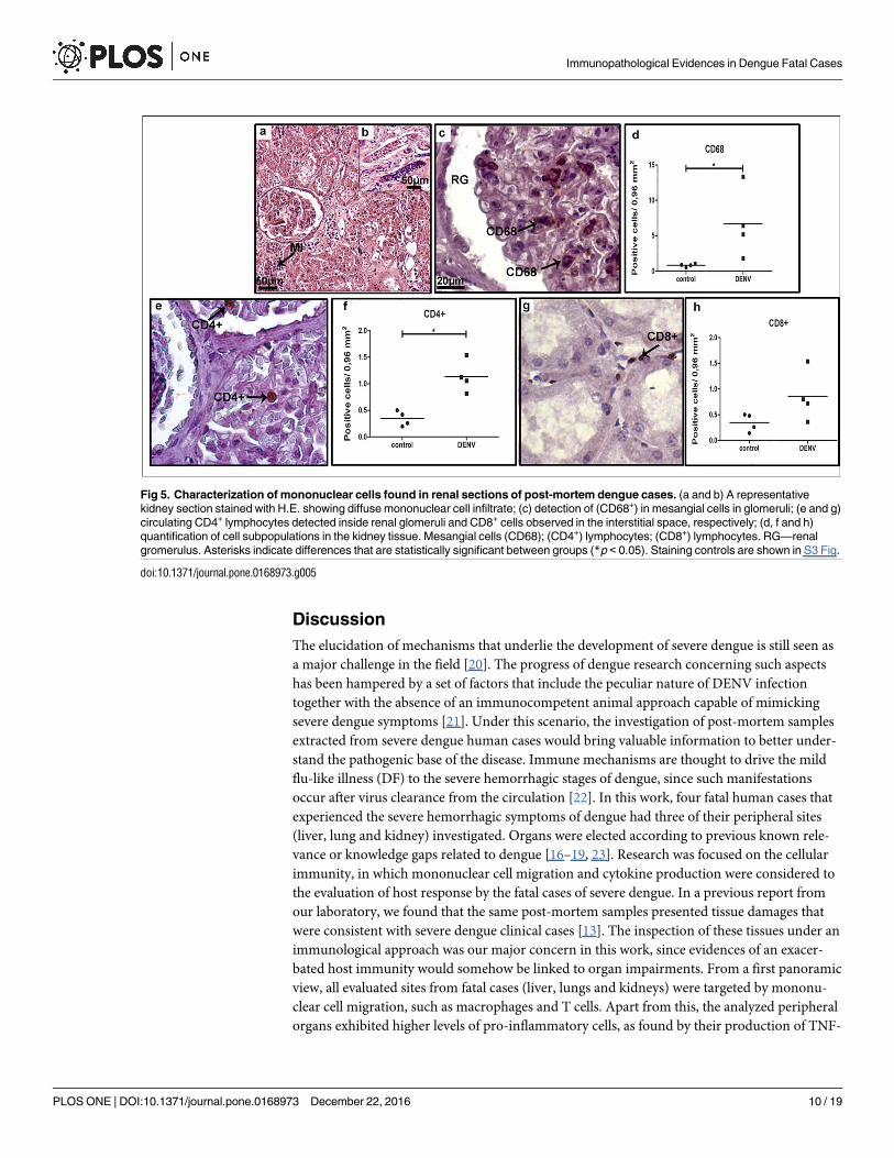
Kidneys in severe dengue are targeted by pro-inflammatory cells
Kidney involvement with dengue virus infection is being recognized, since the prevalence of
proteinuria and hematuria has been reported as high as 70-80% [19]. Mechanisms that may
drive kidney complications in dengue are not clear but possibly result from indirect pathways
via host immunity. Due to these knowledge gaps, renal sections extracted from post-mortem
dengue cases were also considered for investigation.
Tissue analysis of severe cases revealed a diffuse mononuclear infiltrate, more pronounced
in cortical and medullar regions (Fig 5 panels a and b, respectively). CD68+ cells were detected
mainly in mesangial cells (monocyte or smooth muscle origin, responsible for filtration, struc-
tural support, and phagocytosis), located in the glomerulus (Fig 5 panel c). The quantification
Fig 2. Detection of cytokine-producing cells in liver tissues. (a) Detection of TNF-α in macrophages and Kupffer cells in sinusoidal
capillaries; (b and c) detection of INF-α in Kupffer cells and lymphocytes; (d) production of anti-inflammatory cytokines (IL-10 in monocytes
and lymphocytes circulating in sinusoidal capillaries and TGFβ detected in macrophages inside the portal space; (h) TGF-β-expressing
macrophages and Kupffer cells in sinusoidal capillaries; (i and j) detection of RANTES in endothelium and Kupffer cells; (e, f, g, k and l)
quantification of the number of cells expressing these cytokines in the hepatic tissue. Monocytes (Mo); Macrophages (Mϕ); Kupffer cells
(KC); lymphocyte (Ly) and sinusoidal endothelium cells (E). Asterisks indicate differences that are statistically significant between groups
(*p < 0.05). Staining controls are shown in S1 Fig.
doi:10.1371/journal.pone.0168973.g002
Immunopathological Evidences in Dengue Fatal Cases
PLOS ONE | DOI:10.1371/journal.pone.0168973 December 22, 2016 7 / 19

of CD68+ cells revealed an 8-fold increment of this population in dengue cases when compared
to non-dengue patients (Fig 5 panel d). The number of CD4+ T cell lymphocytes, located pri-
marily within the renal glomerulus, was also found to be increased in dengue group renal sec-
tions, when compared to controls (Fig 5 panels e and f). CD8+ T cells were detected mainly in
the medullar zone (Fig 5 panel g) and their quantification showed no statistical difference
between dengue and control groups (Fig 5 panel h).
The evaluation of cytokine profile in renal samples revealed TNF-α being produced mainly
by monocytes/macrophages present in the medullar region (Fig 6 panel a). IFN-γ-producing
cells, such as macrophages, were found in blood vessels located also in the medullar region
(Fig 6 panel b). The anti-inflammatory cytokines IL-10 and TGF-γ were observed mostly in
lymphocytes within the renal glomerulus (Fig 6 panel c) and macrophages (Fig 6 panel g),
while RANTES production was detected mainly in macrophages only (Fig 6 panel h). Quantifi-
cation of cells producing these cytokines revealed a general increase in dengue cases when
compared to controls. The number of cells producing of TNF-α and IFN-γ in dengue cases
was 3- and 5-fold higher, respectively, after comparison with non-dengue cases (Fig 6 panels d
and e). Concerning the number of cells expressing TGF-β (Fig 6 panel i), IL-10 (Fig 6 panel f)
and RANTES (Fig 6 panel j), in dengue cases we noted increments of about 2.5-, 3- and
13-fold, respectively.
DENV-specific induction of pro-inflammatory response
In order to confirm the participation of DENV-infected cells in inducing the local pro-inflam-
matory response, DENV RNA and cytokine production were co-tested in host samples. For
Fig 3. Characterization of mononuclear cell subpopulations in lung tissues collected from DENV-3 fatal cases. Lung tissue
sections stained with (a) H.E. or (b, d and f) incubated with specific antibodies in immunohistochemistry assays. (a) Mononuclear
infiltrates located in interstitial septa; (b) macrophages (CD68+ cells) observed in alveolar capillaries; (d and f) CD4+ and CD8+ T
lymphocytes respectively, found in interstitial infiltrates septa; (c, e and g) quantification of cell subpopulations in the lung tissue.
Macrophage (CD68); Mononuclear infiltrate (MI); edema (E); alveolar macrophage (AM); (CD4+) lymphocytes; (CD8+) lymphocytes.
Asterisks indicate differences that are statistically significant between analyzed groups (*p < 0.05). Staining controls are shown in S2
Fig.
doi:10.1371/journal.pone.0168973.g003
Immunopathological Evidences in Dengue Fatal Cases
PLOS ONE | DOI:10.1371/journal.pone.0168973 December 22, 2016 8 / 19

this evaluation, the liver was elected due to its importance as a target organ in dengue
pathogenesis.
As expected, the light microscopy of a dengue fatal case exhibited mononuclear cell infil-
trates with the presence of Kupffer cells (KC) in the sinusoid capillaries (Fig 7 panels a and b),
while non-dengue control presented regular hepatic structures with resident KCs (Fig 7 panels
i and j). As detected by in situ hybridization and IHC, the dengue case showed many areas of
co-expression in the hepatic tissue considering DENV RNA and IFN-γ or TNF-α (Fig 7 panels
g and h). In this case, Kupffer cells were the main targets of co-staining. The control case pre-
sented no staining for either DENV or the studied cytokines.
These data confirmed, under a qualitative basis, the specific DENV-induction over pro-
inflammatory cytokine response and also addressed the virus spread versus IFN-γ or TNF-αexpression in the liver sample of a dengue fatal case.
Fig 4. Cytokine-producing cells profile in lung tissues of dengue fatal cases. Immunohistochemical analysis of lung sections
collected from dengue fatal cases exhibited (a) TNF-α and (b) INF-γ production in alveolar macrophages; (c) IL-10 detected in
monocytes/macrophages and lymphocytes in alveolar septa; (g) TGF-β produced by numerous macrophages and lymphocytes in the
septa; (h) RANTES detected in endothelium cells and several macrophages in pulmonary tissue; (d-f, i and j) quantification of cytokine-
expressing cells in the lung sections. Monocyte (Mo); Alveolar macrophage (AM); Endothelium (E); Lymphocytes (Ly). Asterisks indicate
differences that are statistically significant between dengue cases and controls (*p < 0.05). Staining controls are shown in S2 Fig.
doi:10.1371/journal.pone.0168973.g004
Immunopathological Evidences in Dengue Fatal Cases
PLOS ONE | DOI:10.1371/journal.pone.0168973 December 22, 2016 9 / 19
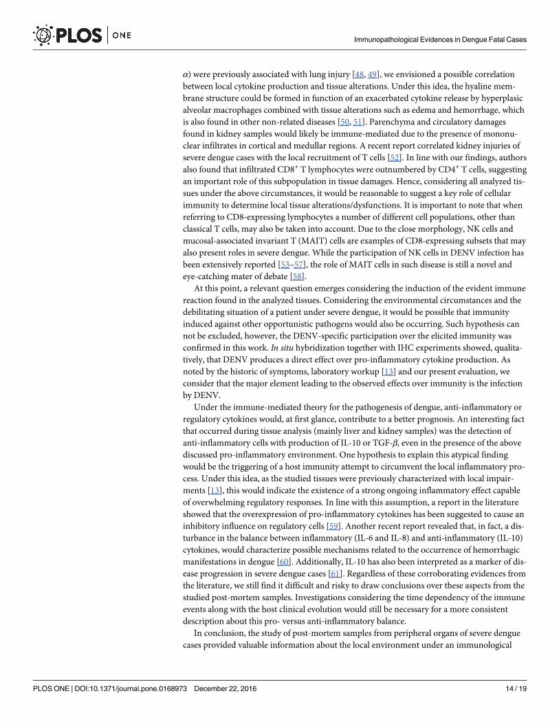
Discussion
The elucidation of mechanisms that underlie the development of severe dengue is still seen as
a major challenge in the field [20]. The progress of dengue research concerning such aspects
has been hampered by a set of factors that include the peculiar nature of DENV infection
together with the absence of an immunocompetent animal approach capable of mimicking
severe dengue symptoms [21]. Under this scenario, the investigation of post-mortem samples
extracted from severe dengue human cases would bring valuable information to better under-
stand the pathogenic base of the disease. Immune mechanisms are thought to drive the mild
flu-like illness (DF) to the severe hemorrhagic stages of dengue, since such manifestations
occur after virus clearance from the circulation [22]. In this work, four fatal human cases that
experienced the severe hemorrhagic symptoms of dengue had three of their peripheral sites
(liver, lung and kidney) investigated. Organs were elected according to previous known rele-
vance or knowledge gaps related to dengue [16–19, 23]. Research was focused on the cellular
immunity, in which mononuclear cell migration and cytokine production were considered to
the evaluation of host response by the fatal cases of severe dengue. In a previous report from
our laboratory, we found that the same post-mortem samples presented tissue damages that
were consistent with severe dengue clinical cases [13]. The inspection of these tissues under an
immunological approach was our major concern in this work, since evidences of an exacer-
bated host immunity would somehow be linked to organ impairments. From a first panoramic
view, all evaluated sites from fatal cases (liver, lungs and kidneys) were targeted by mononu-
clear cell migration, such as macrophages and T cells. Apart from this, the analyzed peripheral
organs exhibited higher levels of pro-inflammatory cells, as found by their production of TNF-
Fig 5. Characterization of mononuclear cells found in renal sections of post-mortem dengue cases. (a and b) A representative
kidney section stained with H.E. showing diffuse mononuclear cell infiltrate; (c) detection of (CD68+) in mesangial cells in glomeruli; (e and g)
circulating CD4+ lymphocytes detected inside renal glomeruli and CD8+ cells observed in the interstitial space, respectively; (d, f and h)
quantification of cell subpopulations in the kidney tissue. Mesangial cells (CD68); (CD4+) lymphocytes; (CD8+) lymphocytes. RG—renal
gromerulus. Asterisks indicate differences that are statistically significant between groups (*p < 0.05). Staining controls are shown in S3 Fig.
doi:10.1371/journal.pone.0168973.g005
Immunopathological Evidences in Dengue Fatal Cases
PLOS ONE | DOI:10.1371/journal.pone.0168973 December 22, 2016 10 / 19

α, IFN-γ and RANTES. Those observations strongly supported the proposed theories claiming
that exaggerated [24, 25] or misdirected [8, 26–28] T-cell responses would eventually lead the
host to severe clinical stages.
Numerous reports describe the release of different cytokines and soluble receptors during
dengue infection [25, 29, 30], which has also been associated to the unfavorable disease out-
come [31]. In this work, increased levels of RANTES-producing endothelial cells may have
contributed to the occurrence of cell infiltrates, since this chemokine signals for cell movement
from the bloodstream into tissues [32, 33]. Therefore, that would infer a close connection
between RANTES and the altered vascular permeability events related to severe dengue. In
this case, higher RANTES production and secretion would favor plasma leakage and lympho-
cyte cell infiltration into the liver, lungs and kidneys, hence, potentially mediating the inflam-
matory response found in these peripheral organs. Among all chemokines, RANTES is
Fig 6. Cytokine-producing cells in renal tissues of dengue cases. (a) TNF-α detected in monocytes located in blood vessels; (b)
Production of INF-γ observed in circulating macrophages and lymphocytes located in the interstitial space; (c) IL-10 found in endothelial
cells of glomerulus; (g) TGF-β production by lymphocytes present mainly inside renal glomeruli; (h) RANTES detected in macrophages
and lymphocytes in the interstitial renal space; (d-f, i and j) quantification of the number of cells expressing the above cytokines.
Macrophages (Mϕ); Lymphocytes (Ly). Asterisks indicate differences that are statistically significant between analyzed groups
(*p < 0.05). Staining controls are shown in S3 Fig.
doi:10.1371/journal.pone.0168973.g006
Immunopathological Evidences in Dengue Fatal Cases
PLOS ONE | DOI:10.1371/journal.pone.0168973 December 22, 2016 11 / 19

Fig 7. Co-expression of DENV and pro-inflammatory cytokines in the liver. Liver samples of dengue
fatal case 2 were processed for in situ hybridization and IHC procedures. DENV was detected by a probe that
aneals to a conserved sequence within the viral RNA negative strand. IFN-γ and TNF-αwere assessed by
immunohistochemistry assay. Probe-target complexes were revealed by alkaline phosphatase activity and
cytokines were identified by standard DAB reactions. The chromogenic signals were converted to fluorescent-
Immunopathological Evidences in Dengue Fatal Cases
PLOS ONE | DOI:10.1371/journal.pone.0168973 December 22, 2016 12 / 19

particularly associated with viral infections [34]. RANTES, also known as CCL5, is an early
expressed chemokine induced by pattern recognition receptors, but can also be induced by
TNF-α and IFN-γ at late stages of infection [35], that is the case of a terminal dengue situation.
The discussion about chemokine signaling and its effects over infectious diseases can some-
times be controversial in the literature. Together with other chemotactic effectors (CXCL9/10/
11) and immune cell response-modulating cytokines (IL-6, IL-7 and BAFF), RANTES has
been associated with immune enhancement and increased vascular permeability following
dengue virus infection [36, 37]. Conversely, in vitro experiments revealed that hantavirus can
infect human lung microvascular endothelial cells (HMVEC-Ls) and stimulate secretion of
RANTES by these cells without increasing vascular permeability [38]. Together, these observa-
tions still arise skeptical thoughts that lead to a more careful discussion concerning a direct
link between RANTES and vascular permeability enhancement in dengue.
The higher amounts of TNF-α-producing cells characterized in the post-mortem samples
were in line with reports in the literature. TNF-α is considered as a major pro-inflammatory
mediator in dengue infections since its activity has been linked to the immunopathogenesis of
the disease [28, 39]. Apart from the existing drawbacks regarding animal models to reproduc-
ing dengue, reports showed that the inhibition of TNF-α by administrating specific antibodies
was associated with reduced severity [39, 40]. Although the TNF-α inhibition assay was key to
infer its importance in severe dengue, in practical terms, targeting TNF with antibody or
receptor antagonists for treating human diseases is controversial. In other diseases with immu-
nological basis, not all patients were helped despite the clinical effectiveness of anti-TNF
aproaches. This fact, perhaps, reflected the existence of distinct underlying mechanisms that
drive the symptoms apart from the TNF network [41].
Along with TNF-α, IFN-γ was also found to be increased in terms of production by infil-
trated mononuclear cells in peripheral tissues of the studied fatal dengue cases. Hence, the
present work showed INF-γ as a pro-inflammatory element that may contribute to tissue dis-
tress, also representing an in situ evidence of disease severity. In a recent report, metabolomics
was adopted to screen dengue-induced metabolites in 116 dengue patients (60 presenting DF
and 56 with severe dengue). It was found that circulating IFN-γ combined with serotonin lev-
els provided accurate early prognosis of severe dengue, thus revealing its importance and an
additional clinical usage of this pro-inflammatory cytokine to assess severity [42].
The mononuclear cell migration that targeted the studied tissues were also proposed to be
correlated with local impairments. In liver samples, CD4+ T cells, Kupffer cells and monocytes
were characterized near hepatocellular necrosis and steatosis in the presence of pro-inflamma-
tory cytokines, IFN-γ and TNF-α. Additional alterations on hepatocytes, such as nuclear vacu-
olar degeneration and the presence of swollen mitochondria, based on preceeding studies,
suggested an ongoing mechanism of apoptotic cell death possibly mediated by the cytokine
environment [13, 43–47]. In our previous report, the lung scenario of the studied dengue fatal
cases was marked by a peculiar histopathological evidence. The presence of septum thickening
with an increase of cellularity characterized a hyaline membrane formation possibly due to
dengue shock syndrome [13]. As the activity of pro-inflammatory cytokines (IFN-γ and TNF-
based signals using a computer based Nuance system. Representative images are shown: (a, b) Light
microscopy of sinusiodal areas of dengue case stained for DENV RNA and IFN-γ or TNF-α, respectively. (c,
d) Expression of DENV RNA (blue) in samples stained with IFN-γ or TNF-α, respectively. (e, f) Expression of
IFN-γ or TNF-α (red) in in samples stained with DENV RNA, respectively. (g, h) Merged signals of DENV RNA
and IFN-γ or TNF-αwhere co-expression is exhibited in yellow, respectively. (i, j) Light microscopy of
sinusiodal areas of a non-dengue case stained for DENV RNA and IFN-γ or TNF-α, respectively. (k, l) Comtrol
samples showing DENV and IFN-γ or TNF-α fluorescent signals, respectively. KC—Kupffer cells.
doi:10.1371/journal.pone.0168973.g007
Immunopathological Evidences in Dengue Fatal Cases
PLOS ONE | DOI:10.1371/journal.pone.0168973 December 22, 2016 13 / 19
α) were previously associated with lung injury [48, 49], we envisioned a possible correlation
between local cytokine production and tissue alterations. Under this idea, the hyaline mem-
brane structure could be formed in function of an exacerbated cytokine release by hyperplasic
alveolar macrophages combined with tissue alterations such as edema and hemorrhage, which
is also found in other non-related diseases [50, 51]. Parenchyma and circulatory damages
found in kidney samples would likely be immune-mediated due to the presence of mononu-
clear infiltrates in cortical and medullar regions. A recent report correlated kidney injuries of
severe dengue cases with the local recruitment of T cells [52]. In line with our findings, authors
also found that infiltrated CD8+ T lymphocytes were outnumbered by CD4+ T cells, suggesting
an important role of this subpopulation in tissue damages. Hence, considering all analyzed tis-
sues under the above circumstances, it would be reasonable to suggest a key role of cellular
immunity to determine local tissue alterations/dysfunctions. It is important to note that when
referring to CD8-expressing lymphocytes a number of different cell populations, other than
classical T cells, may also be taken into account. Due to the close morphology, NK cells and
mucosal-associated invariant T (MAIT) cells are examples of CD8-expressing subsets that may
also present roles in severe dengue. While the participation of NK cells in DENV infection has
been extensively reported [53–57], the role of MAIT cells in such disease is still a novel and
eye-catching mater of debate [58].
At this point, a relevant question emerges considering the induction of the evident immune
reaction found in the analyzed tissues. Considering the environmental circumstances and the
debilitating situation of a patient under severe dengue, it would be possible that immunity
induced against other opportunistic pathogens would also be occurring. Such hypothesis can
not be excluded, however, the DENV-specific participation over the elicited immunity was
confirmed in this work. In situ hybridization together with IHC experiments showed, qualita-
tively, that DENV produces a direct effect over pro-inflammatory cytokine production. As
noted by the historic of symptoms, laboratory workup [13] and our present evaluation, we
consider that the major element leading to the observed effects over immunity is the infection
by DENV.
Under the immune-mediated theory for the pathogenesis of dengue, anti-inflammatory or
regulatory cytokines would, at first glance, contribute to a better prognosis. An interesting fact
that occurred during tissue analysis (mainly liver and kidney samples) was the detection of
anti-inflammatory cells with production of IL-10 or TGF-β, even in the presence of the above
discussed pro-inflammatory environment. One hypothesis to explain this atypical finding
would be the triggering of a host immunity attempt to circumvent the local inflammatory pro-
cess. Under this idea, as the studied tissues were previously characterized with local impair-
ments [13], this would indicate the existence of a strong ongoing inflammatory effect capable
of overwhelming regulatory responses. In line with this assumption, a report in the literature
showed that the overexpression of pro-inflammatory cytokines has been suggested to cause an
inhibitory influence on regulatory cells [59]. Another recent report revealed that, in fact, a dis-
turbance in the balance between inflammatory (IL-6 and IL-8) and anti-inflammatory (IL-10)
cytokines, would characterize possible mechanisms related to the occurrence of hemorrhagic
manifestations in dengue [60]. Additionally, IL-10 has also been interpreted as a marker of dis-
ease progression in severe dengue cases [61]. Regardless of these corroborating evidences from
the literature, we still find it difficult and risky to draw conclusions over these aspects from the
studied post-mortem samples. Investigations considering the time dependency of the immune
events along with the host clinical evolution would still be necessary for a more consistent
description about this pro- versus anti-inflammatory balance.
In conclusion, the study of post-mortem samples from peripheral organs of severe dengue
cases provided valuable information about the local environment under an immunological
Immunopathological Evidences in Dengue Fatal Cases
PLOS ONE | DOI:10.1371/journal.pone.0168973 December 22, 2016 14 / 19

approach. The existence of a strong ongoing pro-inflammatory response was suggested to be
occurring in liver and manly in lung and kidney samples. The presence of mononuclear cell
infiltrates, higher counts of pro-inflammatory cells (as found by the production of TNF-α,
IFN-γ and RANTES) and the apparent outrun of inflammation over anti-inflammatory ele-
ments (such as IL-10 and TGF-β) were the major evidences for such characterization. Apart
from many other known hypotheses for the determination of severe dengue cases [20], this
work provided additional evidences that supported the cellular immune-mediated theories,
hence, contributing to a better understanding of dengue pathogenesis.
Supporting Information
S1 Fig. Liver tissue controls of immunohistochemistry assays. Histological sections of a
non-dengue case organ showing regular structures and preserved parenchyma. Slides were
stained with anti-CD68 (a), anti-CD4 (b), anti-CD8 (c), anti-TNFα (d), anti-IFNγ (e), anti-IL-
10 (f), anti-TGFβ (g) and anti-RANTES (h).
(TIF)
S2 Fig. Lung tissue controls of immunohistochemistry assays. Histological sections of a
non-dengue case organ showing regular structures and preserved parenchyma. Slides were
stained with anti-CD68 (a), anti-CD4 (b), anti-CD8 (c), anti-TNFα (d), anti-IFNγ (e), anti-IL-
10 (f), anti-TGFβ (g) and anti-RANTES (h).
(TIF)
S3 Fig. Kidney tissue controls of immunohistochemistry assays. Histological sections of a
non-dengue case organ showing regular structures and preserved parenchyma. Slides were
stained with anti-CD68 (a), anti-CD4 (b), anti-CD8 (c), anti-TNFα (d), anti-IFNγ (e), anti-IL-
10 (f), anti-TGFβ (g) and anti-RANTES (h).
(TIF)
S1 Table. Quantification of positive cells by immunohistochemistry.
(XLSX)
Acknowledgments
We thank Rodrigo Mexas (Service of Production and Image Treatment/IOC/Fiocruz) and
Geraldo (Gaffree Hospital-UNIRIO) for technical assistance in the production of slides for the
immunohistochemical assays and FAPERJ for financial support (grant number: E-26/110.511/
2014). We also acknowledge Dr. Ada Alves from Laboratory of Biotechnology and Physiology
of Viral Infections (LABIFIV-Fiocruz) for the environment where experiments were set up.
Author Contributions
Conceptualization: MVP ERAO TFP.
Data curation: MVP TFP GJN NGS.
Formal analysis: MVP TFP ERAO.
Funding acquisition: MVP.
Investigation: MVP TFP ERAO GJN NGS EMM.
Methodology: MVP ERAO TFP GJN.
Project administration: MVP TFP ERAO.
Immunopathological Evidences in Dengue Fatal Cases
PLOS ONE | DOI:10.1371/journal.pone.0168973 December 22, 2016 15 / 19

Resources: MVP CAB GJN VLAC EMM.
Supervision: MVP ERAO TFP.
Validation: MVP ERAO TFP.
Visualization: TFP ERAO MVP.
Writing – original draft: ERAO.
Writing – review & editing: ERAO MVP.
References1. WHO: Global strategy for dengue prevention and control 2012-2020. World Health Organization. 2012.
Available from: www.who.int
2. WHO: Dengue: Guidelines for diagnosis, treatment, prevention and control. World Health Organization.
2009. Available from: www.who.int
3. Bhatt S, Gething PW, Brady OJ, Messina JP, Farlow AW, Moyes CL, et al. The global distribution and
burden of dengue. Nature. 2013; 496:504–507. doi: 10.1038/nature12060 PMID: 23563266
4. Halstead SB, Cohen SN. Dengue hemorrhagic fever at 60 Years: early evolution of concepts of causa-
tion and treatment. Microbiol Mol Biol Rev. 2015; 79:281–291. doi: 10.1128/MMBR.00009-15 PMID:
26085471
5. Guzman MG, Halstead SB, Artsob H, Buchy P, Farrar J, Gubler DJ, et al. Dengue: a continuing global
threat. Nat Rev Microbiol. 2010; 8:S7–16. doi: 10.1038/nrmicro2460 PMID: 21079655
6. Carabali M, Hernandez LM, Arauz MJ, Villar LA, Ridde V. Why are people with dengue dying? A scop-
ing review of determinants for dengue mortality. BMC Infect Dis. 2015; 15:301–315. doi: 10.1186/
s12879-015-1058-x PMID: 26223700
7. Huy NT, Van Giang T, Thuy DH, Kikuchi M, Hien TT, Zamora J, et al. Factors associated with dengue
shock syndrome: a systematic review and meta-analysis. PLoS Negl Trop Dis. 2013; 7:e2412. doi: 10.
1371/journal.pntd.0002412 PMID: 24086778
8. Pang T, Cardosa MJ, Guzman MG. Of cascades and perfect storms: the immunopathogenesis of den-
gue haemorrhagic fever-dengue shock syndrome (DHF/DSS). Immunol Cell Biol. 2007; 85:43–45. doi:
10.1038/sj.icb.7100008 PMID: 17130899
9. Nielsen DG. The relationship of interacting immunological components in dengue pathogenesis. Virol J.
2009; 6:211. doi: 10.1186/1743-422X-6-211 PMID: 19941667
10. Halstead SB. Dengue Antibody-Dependent Enhancement: Knowns and Unknowns. Microbiol Spectr.
2014; 2. doi: 10.1128/microbiolspec.AID-0022-2014 PMID: 26104444
11. Green S, Rothman A. Immunopathological mechanisms in dengue and dengue hemorrhagic fever.
Curr Opin Infect Dis. 2006; 19:429–36. doi: 10.1097/01.qco.0000244047.31135.fa PMID: 16940865
12. Pawitan JA. Dengue virus infection: predictors for severe dengue. Acta Med Indones. 2011; 43:129–35.
PMID: 21785176
13. Povoa TF, Alves AM, Oliveira CA, Nuovo GJ, Chagas VL, Paes MV. The pathology of severe dengue in
multiple organs of human fatal cases: histopathology, ultrastructure and virus replication. PLoS One.
2014; 9:e83386. doi: 10.1371/journal.pone.0083386
14. Paes MV, Lenzi HL, Nogueira AC, Nuovo GJ, Pinhão AT, Mota EM, et al. Hepatic damage associated
with dengue-2 virus replication in liver cells of BALB/c mice. Lab Invest. 2009; 89:1140–1151. doi: 10.
1038/labinvest.2009.83 PMID: 19721415
15. Braga EL, Moura P, Pinto LM, Ignacio SR, Oliveira MJ, Cordeiro MT, et al. Detection of circulant tumor
necrosis factor-alpha, soluble tumor necrosis factor p75 and interferon-gamma in Brazilian patients with
dengue fever and dengue hemorrhagic fever. Mem Inst Oswaldo Cruz. 2001; 96:229–32. doi: 10.1590/
S0074-02762001000200015 PMID: 11285501
16. Huerre MR, Lan NT, Marianneau P, Hue NB, Khun H, Hung NT, et al. Liver histopathology and biologi-
cal correlates in five cases of fatal dengue fever in Vietnamese children. Virchows Arch. 2001;
438:107–115. PMID: 11253111
17. Seneviratne SL, Malavige GN, de Silva HJ. Pathogenesis of liver involvement during dengue viral infec-
tions. Trans R Soc Trop Med Hyg. 2006; 100:608–614. doi: 10.1016/j.trstmh.2005.10.007 PMID:
16483623
Immunopathological Evidences in Dengue Fatal Cases
PLOS ONE | DOI:10.1371/journal.pone.0168973 December 22, 2016 16 / 19

18. Samanta J, Sharma V. Dengue and its effects on liver. World J Clin Cases. 2015; 3:125–131. doi: 10.
12998/wjcc.v3.i2.125 PMID: 25685758
19. Vachvanichsanong P, Thisyakorn U, Thisyakorn C. Dengue hemorrhagic fever and the kidney. Arch
Virol. 2016; 161:771–778. doi: 10.1007/s00705-015-2727-1 PMID: 26699788
20. Halstead SB. Pathogenesis of Dengue: Dawn of a New Era. F1000Res. 2015; 4. pii: F1000 Faculty
Rev-1353. doi: 10.12688/f1000research.7024.1 PMID: 26918141
21. Chan KW, Watanabe S, Kavishna R, Alonso S, Vasudevan SG. Animal models for studying dengue
pathogenesis and therapy. Antiviral Res. 2015; 123:5–14. doi: 10.1016/j.antiviral.2015.08.013 PMID:
26304704
22. John DV, Lin YS, Perng GC. Biomarkers of severe dengue disease—a review. J Biomed Sci. 2015;
22:83. doi: 10.1186/s12929-015-0191-6 PMID: 26462910
23. Rodrigues RS, Brum AL, Paes MV, Povoa TF, Basilio-de-Oliveira CA, Marchiori E, et al. Lung in den-
gue: computed tomography findings. PLoS One. 2014; 9:e96313. doi: 10.1371/journal.pone.0096313
PMID: 24836605
24. Mathew A, Kurane I, Green S, Stephens HA, Vaughn DW, Kalayanarooj S, et al. Predominance of
HLA-restricted cytotoxic T-lymphocyte responses to serotype-cross-reactive epitopes on nonstructural
proteins following natural secondary dengue virus infection. J Virol. 1998; 72:3999–4004. PMID:
9557687
25. Rothman AL, Ennis FA. Immunopathogenesis of Dengue hemorrhagic fever. Virology. 1999; 257:1–6.
doi: 10.1006/viro.1999.9656 PMID: 10208914
26. Mongkolsapaya J, Dejnirattisai W, Xu XN, Vasanawathana S, Tangthawornchaikul N, Chairunsri A,
et al. Original antigenic sin and apoptosis in the pathogenesis of dengue hemorrhagic fever. Nat Med.
2003; 9:921–927. doi: 10.1038/nm887 PMID: 12808447
27. Rothman AL. Cellular immunology of sequential dengue virus infection and its role in disease pathogen-
esis. Curr Top Microbiol Immunol. 2010; 338:83–98. doi: 10.1007/978-3-642-02215-9_7 PMID:
19802580
28. Rothman AL. Immunity to dengue virus: a tale of original antigenic sin and tropical cytokine storms. Nat
Rev Immunol. 2011; 11:532–43. doi: 10.1038/nri3014 PMID: 21760609
29. Hober D, Poli L, Roblin B, Gestas P, Chungue E, Granic G, et al. Serum levels of tumor necrosis factor-
alpha (TNF-alpha), interleukin-6 (IL-6), and interleukin-1 beta (IL-1 beta) in dengue-infected patients.
Am J Trop Med Hyg. 1993; 48:324–331. PMID: 8470771
30. Green S, Vaughn DW, Kalayanarooj S, Nimmannitya S, Suntayakorn S, Nisalak A, et al. Early immune
activation in acute dengue illness is related to development of plasma leakage and disease severity. J
Infect Dis. 1999; 179:755–762. doi: 10.1086/314680 PMID: 10068569
31. Chaturvedi UC, Agarwal R, Elbishbishi EA, Mustafa AS. Cytokine cascade in dengue hemorrhagic
fever: implications for pathogenesis. FEMS Immunol Med Microbiol. 2000; 28:183–188. doi: 10.1111/j.
1574-695X.2000.tb01474.x PMID: 10865168
32. Ebert LM, Schaerli P, Moser B. Chemokine-mediated control of T cell traffic in lymphoid and peripheral
tissues. Mol Immunol. 2005; 42:799–809. doi: 10.1016/j.molimm.2004.06.040 PMID: 15829268
33. de-Oliveira-Pinto LM, Marinho CF, Povoa TF, de Azeredo EL, de Souza LA, Barbosa LD, et al. Regula-
tion of inflammatory chemokine receptors on blood T cells associated to the circulating versus liver che-
mokines in dengue fever. PLoS One. 2012; 7:e38527. doi: 10.1371/journal.pone.0038527 PMID:
22815692
34. Glass WG, Rosenberg HF, Murphy PM. Chemokine regulation of inflammation during acute viral infec-
tion. Curr Opin Allergy Clin Immunol. 2003; 3:467–473. doi: 10.1097/00130832-200312000-00008
PMID: 14612671
35. Grandvaux N, Servant MJ, tenOever B, Sen GC, Balachandran S, Barber GN, et al. Transcriptional pro-
filing of interferon regulatory factor 3 target genes: direct involvement in the regulation of interferon-stim-
ulated genes. J Virol. 2002; 76:5532–5539. doi: 10.1128/JVI.76.11.5532-5539.2002 PMID: 11991981
36. Dalrymple NA, Mackow ER. Endothelial cells elicit immune-enhancing responses to dengue virus infec-
tion. J Virol. 2012; 86:6408–15. doi: 10.1128/JVI.00213-12 PMID: 22496214
37. Chen J, Ng MM, Chu JJ. Molecular profiling of T-helper immune genes during dengue virus infection.
Virol J. 2008; 5:165. doi: 10.1186/1743-422X-5-165 PMID: 19117515
38. Sundstrom JB, McMullan LK, Spiropoulou CF, Hooper WC, Ansari AA, Peters CJ, et al. Hantavirus
infection induces the expression of RANTES and IP-10 without causing increased permeability in
human lung microvascular endothelial cells. J Virol. 2001; 75:6070–6085. doi: 10.1128/JVI.75.13.6070-
6085.2001 PMID: 11390609
Immunopathological Evidences in Dengue Fatal Cases
PLOS ONE | DOI:10.1371/journal.pone.0168973 December 22, 2016 17 / 19
39. Shresta S, Sharar KL, Prigozhin DM, Beatty PR, Harris E. Murine model for dengue virus-induced lethal
disease with increased vascular permeability. J Virol. 2006; 80:10208–10217. doi: 10.1128/JVI.00062-
06 PMID: 17005698
40. Atrasheuskaya A, Petzelbauer P, Fredeking TM, Ignatyev G. Anti-TNF antibody treatment reduces
mortality in experimental dengue virus infection. FEMS Immunol Med Microbiol. 2003; 35:33–42. doi:
10.1111/j.1574-695X.2003.tb00646.x PMID: 12589955
41. Croft M, Benedict CA, Ware CF. Clinical targeting of the TNF and TNFR superfamilies. Nat Rev Drug
Discov. 2013; 12:147–68. doi: 10.1038/nrd3930 PMID: 23334208
42. Cui L, Lee YH, Thein TL, Fang J, Pang J, Ooi EE, et al. Serum Metabolomics Reveals Serotonin as a
Predictor of Severe Dengue in the Early Phase of Dengue Fever. PLoS Negl Trop Dis. 2016; 10(4):
e0004607. doi: 10.1371/journal.pntd.0004607 PMID: 27055163
43. Ding WX, Yin XM. Dissection of the multiple mechanisms of TNF-alpha-induced apoptosis in liver injury.
J Cell Mol Med. 2004; 8:445–454. doi: 10.1111/j.1582-4934.2004.tb00469.x PMID: 15601573
44. Quaresma JA, Barros VL, Pagliari C, Fernandes ER, Guedes F, Takakura CF, et al. Revisiting the liver
in human yellow fever: virus-induced apoptosis in hepatocytes associated with TGF-beta, TNF-alpha
and NK cells activity. Virology. 2006; 345:22–30. doi: 10.1016/j.virol.2005.09.058 PMID: 16278000
45. Rimkunas VM, Graham MJ, Crooke RM, Liscum L. TNF-alpha plays a role in hepatocyte apoptosis in
Niemann-Pick type C liver disease. J Lipid Res. 2009; 50:327–333. doi: 10.1194/jlr.M800415-JLR200
PMID: 18815434
46. Quaresma JA, Pagliari C, Medeiros DB, Duarte MI, Vasconcelos PF. Immunity and immune response,
pathology and pathologic changes: progress and challenges in the immunopathology of yellow fever.
Rev Med Virol. 2013; 23:305–318. doi: 10.1002/rmv.1752 PMID: 23873723
47. Rowan PJ, Dunn NJ, El-Serag HB, Kunik ME. Views of hepatitis C virus patients delayed from treatment
for psychiatric reasons. J Viral Hepat. 2007; 14:883–889. PMID: 18070292
48. Bhargava R, Altmann CJ, Andres-Hernando A, Webb RG, Okamura K, Yang Y, et al. Acute lung injury
and acute kidney injury are established by four hours in experimental sepsis and are improved with pre,
but not post, sepsis administration of TNF-α antibodies. PLoS One. 2013 Nov 12; 8(11):e79037. doi:
10.1371/journal.pone.0079037 PMID: 24265742
49. Spender LC, Hussell T, Openshaw PJ. Abundant IFN-gamma production by local T cells in respiratory
syncytial virus-induced eosinophilic lung disease. J Gen Virol. 1998; 79:1751–1758. doi: 10.1099/0022-
1317-79-7-1751 PMID: 9680139
50. Kwong KY, Jones CA, Cayabyab R, Lecart C, Stotts CL, Randhawa I, et al. Differential regulation of IL-
8 by IL-1beta and TNFalpha in hyaline membrane disease. J Clin Immunol. 1998; 18:71–80. doi: 10.
1023/A:1023244005765 PMID: 9475356
51. Groneck P, Gotze-Speer B, Oppermann M, Eiffert H, Speer CP. Association of pulmonary inflammation
and increased microvascular permeability during the development of bronchopulmonary dysplasia: a
sequential analysis of inflammatory mediators in respiratory fluids of high-risk preterm neonates. Pedi-
atrics. 1994; 93:712–718. PMID: 8165067
52. Pagliari C, Simões Quaresma JA, Kanashiro-Galo L, de Carvalho LV, Vitoria WO, da Silva WL, et al.
Human kidney damage in fatal dengue hemorrhagic fever results of glomeruli injury mainly induced by
IL17. J Clin Virol. 2016; 75:16–20. doi: 10.1016/j.jcv.2015.12.005 PMID: 26741825
53. Lim DS, Yawata N, Selva KJ, Li N, Tsai CY, Yeong LH, et al. The combination of type I IFN, TNF-α, and
cell surface receptor engagement with dendritic cells enables NK cells to overcome immune evasion by
dengue virus. J Immunol. 2014; 193:5065–5075. doi: 10.4049/jimmunol.1302240 PMID: 25320280
54. Townsley E, O’Connor G, Cosgrove C, Woda M, Co M, Thomas SJ, et al. Interaction of a dengue virus
NS1-derived peptide with the inhibitory receptor KIR3DL1 on natural killer cells. Clin Exp Immunol.
2016; 183:419–430. doi: 10.1111/cei.12722 PMID: 26439909
55. Petitdemange C, Wauquier N, Devilliers H, Yssel H, Mombo I, Caron M, et al. Longitudinal Analysis of
Natural Killer Cells in Dengue Virus-Infected Patients in Comparison to Chikungunya and Chikungunya/
Dengue Virus-Infected Patients. PLoS Negl Trop Dis. 2016; 10:e0004499. doi: 10.1371/journal.pntd.
0004499 PMID: 26938618
56. Petitdemange C, Wauquier N, Rey J, Hervier B, Leroy E, Vieillard V. Control of acute dengue virus
infection by natural killer cells. Front Immunol. 2014; 5:209. doi: 10.3389/fimmu.2014.00209 PMID:
24860571
57. Beltran D, Lopez-Vergès S. NK Cells during Dengue Disease and Their Recognition of Dengue Virus-
Infected cells. Front Immunol. 2014; 5:192. doi: 10.3389/fimmu.2014.00192 PMID: 24829565
58. van Wilgenburg B, Scherwitzl I, Hutchinson EC, Leng T, Kurioka A, Kulicke C, et al. MAIT cells are acti-
vated during human viral infections. Nat Commun. 2016; 7:11653. doi: 10.1038/ncomms11653 PMID:
27337592
Immunopathological Evidences in Dengue Fatal Cases
PLOS ONE | DOI:10.1371/journal.pone.0168973 December 22, 2016 18 / 19

59. Tillu H, Tripathy AS, Reshmi PV, Cecilia D. Altered profile of regulatory T cells and associated cytokines
in mild and moderate dengue. Eur J Clin Microbiol Infect Dis. 2016; 35:453–461. doi: 10.1007/s10096-
015-2561-0 PMID: 26861813
60. Iani FC, Caldas S, Duarte MM, Cury AL, Cecılio AB, Costa PA, et al. Dengue Patients with Early Hemor-
rhagic Manifestations Lose Coordinate Expression of the Anti-Inflammatory Cytokine IL-10 with the
Inflammatory Cytokines IL-6 and IL-8. Am J Trop Med Hyg. 2016; 95:193–200. doi: 10.4269/ajtmh.15-
0537 PMID: 27139443
61. Tauseef A, Umar N, Sabir S, Akmal A, Sajjad S, Zulfiqar S. Interleukin-10 as a Marker of Disease Pro-
gression in Dengue Hemorrhagic Fever. J Coll Physicians Surg Pak. 2016; 26:187–90. PMID:
26975948
Immunopathological Evidences in Dengue Fatal Cases
PLOS ONE | DOI:10.1371/journal.pone.0168973 December 22, 2016 19 / 19