pertanika j. sci. & technol. 25 (s): 41 - 52 (2017 ... papers/jst vol. 25 (s) apr....
TRANSCRIPT
Pertanika J. Sci. & Technol. 25 (S): 41 - 52 (2017)
SCIENCE & TECHNOLOGYJournal homepage: http://www.pertanika.upm.edu.my/
ISSN: 0128-7680 © 2017 Universiti Putra Malaysia Press.
ARTICLE INFO
Article history:Received: 25 October 2016Accepted: 17 March 2017
E-mail addresses: [email protected] (Wan Mazlina Md Saad),[email protected] (Mohd Khairul Amran Mohammad),[email protected] (Muhamad Idham Mohamed),[email protected] (Hairil Rashmizal Abdul Razak) *Corresponding Author
Enhancement of Oxidative DNA Damage and Alteration of p53, Bax, and Bcl-2 Protein Expressions Following Low Dose Radiation Exposure
Wan Mazlina Md Saad1*, Mohd Khairul Amran Mohammad1, Muhamad Idham Mohamed1 and Hairil Rashmizal Abdul Razak2 1Department of Medical Laboratory Technology, Faculty of Health Sciences, Universiti Teknologi MARA (UiTM) Puncak Alam, 42300, Puncak Alam, Selangor, Malaysia2Department of Medical Imaging, Faculty of Health Sciences, Universiti Teknologi MARA (UiTM) Puncak Alam, 42300 Puncak Alam, Selangor, Malaysia
ABSTRACT
This animal modelling study aimed to investigate the effects of LDR exposure on cellular ROS production, oxidative DNA damage, and alteration of cellular ultrastructure and apoptosis-related protein expressions. Ten male ICR mice were randomly divided into two groups consisting of control (Cx) and radiation (Rx) groups. On day 29 of post-acclimatisation, mice underwent total body irradiation with 100 µGy X-ray. Liver and lung tissues were assessed for the levels of cellular ROS production and Apurinic/Apyrimidinic sites generation. Ultrastructural alteration was detected using TEM, alteration of p53, Bax, and Bcl-2 expressions was determined by western blotting. Results showed that exposure to LDR significantly increased the levels of cellular ROS and AP sites in mice. Ultrastructure of the nucleus in Rx showed nuclear blebbing and structural changes in morphology that indicate cell death. Meanwhile, p53, Bax, and Bcl-2 proteins increased in expressions and altered the balance of Bax/Bcl-2 ratio. These findings may postulate that LDR exposure may enhance oxidative DNA damage and alter expression of apoptosis-related proteins.
Keywords: Bax, Bcl-2, Low Dose radiation (LDR), oxidative DNA damage, p53
INTRODUCTION
Deleterious effects of high dose radiation (HDR) exposure on human health have been widely studied compared to low dose radiation (LDR) exposure (Zielinski et al., 2009; Ponzinibbio et al., 2010). Medical radiation professions, including radiologists, dentists,
Wan Mazlina Md Saad, Mohd Khairul Amran Mohammad, Muhamad Idham Mohamed andHairil Rashmizal Abdul Razak
42 Pertanika J. Sci. & Technol. 25 (S): 41 - 52 (2017)
nurses and radiographers are among individuals who are mainly exposed to LDR employed in diagnostic imaging modalities, including diagnostic X-ray, computed tomography (CT), and nuclear medicine scans. These individuals are the largest occupations cohort that are exposed to ionising radiation (Zielinski et al., 2009) and thus, studies on effects of LDIR and counter mechanism are warranted. Ionising radiation (IR) has some unique characteristics as a carcinogenic and mutagenic agent which cause several impacts on human health depending on exposed and absorbed dose, duration of exposure and time interval after exposure, and susceptibility of tissues to IR (Mohamed et al., 2014; Klaunig, Kamendulis, & Hocevar, 2010).
IR can directly disrupt atomic structures as being absorbed by living cells, producing chemical and biological changes (Azzam, Jay-Gerin, & Pain, 2012) and causing damage to important macromolecules. Total body irradiation may contribute to multiple organ dysfunctions caused by oxidative stress resulting from overproduction of ROS (Gultekin et al., 2013). Thus, this results in oxidative damage as they act on biomolecules including DNA, lipids and proteins (Cardozo-Pelaez et al., 2000). A recent study by Mohamed et al. (2014) and Zakaria et al. (2014) revealed that total body irradiation of 100 µGy of X-ray (LDR) induced oxidative stress and inflammatory response in male ICR mice in radiation group. As IR is being absorbed by living cells, it may induce more than 100 different DNA lesions produced through direct and/or indirect effects that contribute to structural damage of DNA molecules (Martin et al., 2010; Shackelford, Kaufmann, & Paules, 2000). These lesions include single- and double-strand breaks, oxidised bases, DNA protein cross-links and abasic sites (Sudprasert, Navasumrit, & Ruchirawat, 2006). The rising levels of DNA damage as the results of radiation exposure activate several mechanisms including DNA repair mechanism and cell cycle arrest, or apoptosis. However, failure in DNA repair mechanism in preventing any potentially mutagenic and carcinogenic outcomes from DNA damage lesions may lead to activation of apoptosis mechanism (Paz-Elizur et al., 2008).
Apoptosis or programmed cell death is a prevailing cell destructive mechanism responsible in eliminating damaged cells that have been exposed to mutagenic and carcinogenic agents that are capable in contributing development of cancer (Verheij & Bartelink, 2000). The control and regulation of apoptosis mechanism are highly complex and sophisticated, which involve molecular events that occur through Bcl-2 family of proteins (Elmore, 2007). The family of Bcl-2 proteins can be either pro-apoptotic (e.g., Bax, Bcl-Xs, Bak, Bad, Bik, Bid, Blk) or anti-apoptotic (e.g., Bcl-2, Bcl-Xl, Bcl-w, BAG) (Hoetelmans et al., 2000). In addition, these proteins have significant role as they may determine if the cell executes to apoptosis or aborts the process (Elmore, 2007). Apoptosis may be initiated through activation of p53-dependent mechanisms due to accumulation of DNA damage, which alters the ratio of Bax/Bcl-2 protein expressions (Verheij & Bartelink, 2000; Xu et al., 2008). The ratio of Bax/Bcl-2 protein expressions increases during the induction of apoptosis (Lee et al., 1999). Likewise, the internal vital organs were chosen as they are vulnerable to the radiation, even in lower dosage. Thus, this study aimed to investigate the effects of LDR exposure on cellular ROS production, oxidative DNA damage (AP sites), and alteration of cellular ultrastructure and apoptosis-related protein expressions.

Low Dose Ionising Radiation-Induced DNA Damage
43Pertanika J. Sci. & Technol. 25 (S): 41 - 52 (2017)
METHOD
Chemicals
In Vitro ROS/RNS Assay Kit (Green Fluorescence) and Oxidative DNA Damage Quantification Kit (AP Sites) were procured from Cell Biolabs Inc., whereas Invisorb® Spin Tissue Mini Kit from Stratec Molecular. Polyclonal anti-mouse Bcl-2 antibodies, monoclonal anti-mouse Bax antibodies, polyclonal anti-mouse p53 antibodies, GAPDH loading control antibodies and ECL Chemiluminescence Detection Kit were from Thermo Scientific Pierce.
Animals and Irradiation
The experimental protocols were conducted with the approval of the Committee of Animal Research and Ethics (UiTM CARE No.38/2014), Universiti Teknologi MARA. Ten four-week-old male ICR mice, each weighing 30 grams, were obtained from Laboratory Animal Facility and Management (LAFAM), UiTM Selangor. The animals underwent acclimatisation period for 28 days, and on day 29, a total of ten mice were randomly divided into two groups consisting of control (Cx) and radiation (Rx) groups. Mice from Rx were total body irradiated with single fractionated of 100 µGy X-ray under Philips Bucky DIAGNOST X-ray Machine. Mice were sacrificed by cervical dislocation within 12 hours following irradiation.
Determination of Cellular ROS Production
Cellular ROS production was quantified using OxiSelect in Vitro ROS/RNS Assay Kit (Green Fluorescence). Lung and liver tissues were homogenised with 20mg/mL ice-cold potassium PBS prior to centrifugation at 10,000 g for 5 minutes (4°C). Tissue lysates were collected and assayed directly. The Fluorescence signal was read using fluorescence plate reader at 480 nm excitation and 530 nm emission.
Determination of Oxidative DNA Damage (AP Sites) Production
Genomic DNA of lung and liver were isolated with Invisorb® Spin Tissue Mini Kit. Oxidative DNA Damage Quantification Kit (AP Sites) was used to quantitate AP sites. The Aldehyde Reactive Probe (ARP) that reacts specifically with an aldehyde group on the open ring form of AP sites (ARP-derived DNA) was detected with Streptavidin-Enzyme Conjugate. Quantity of the AP sites in samples was determined using POLARstar Omega Reader at 450 nm by comparing standard curve of predetermined AP sites.
Alteration of p53, Bax, and Bcl-2 Proteins Expression
Sample Preparation. Frozen tissues were resuspended in Radio Immuno Precipitation Assay (RIPA) buffer. Tissues were homogenised in 20 mg/mL cold RIPA buffer containing 10 µL EDTA-free protease inhibitor cocktail on ice, and then kept for 30 min prior to centrifugation at 10,000 g (4°C) for 20 min. Supernatant was recovered and kept in -80°C prior to electrophoresis.
Wan Mazlina Md Saad, Mohd Khairul Amran Mohammad, Muhamad Idham Mohamed andHairil Rashmizal Abdul Razak
44 Pertanika J. Sci. & Technol. 25 (S): 41 - 52 (2017)
Protein Level Determination. Protein concentration in each sample was determined using Quick StartTM Bradford Protein Assay (Bio-Rad) according to the manufacturer’s protocol.
Western Blotting Techniques. Aliquots from supernatant containing 50 µg proteins were mixed with 5X Laemmli sample buffer at ratio of 1:5 (v/v). Samples were boiled for 5 minutes at 95°C and subjected to 10% SDS-PAGE, and transferred to a PVDF membrane. Membranes will be blocked at RT for 2 hours in blocking buffer containing 10% skimmed milk (Merck) prior to incubation with anti-p53 (1:500 dilution), anti-Bcl-2 (1:500 dilution) or anti-Bax (1:500 dilution) primary antibodies in blocking buffer for two hours in RT. Next, the membranes were washed in 1X TBST (50 mmol/L Tris-HCl, pH 7.6, 150 mmol/L NaCl, 0.1% Tween 20) three times (10 minutes each) and incubated with HRP conjugated secondary antibody (1:20 000 dilution) for 2 hours at RT. The membranes were washed three times in TBST (10 minutes each) and exposed to ECL chemiluminescence reagents for 5 minutes. Blots were then exposed to X-ray film and processed by (brand automated film processor) for radiographic detection of the bands. The autoradiograms were scanned and protein bands were quantified by densitometry using ImageJ (version 1.45 k). To verify equal protein loading and transfer, the blots were probed for GAPDH using an anti-GAPDH antibody (1:5000 dilution). Thereafter, the same protocol was followed, except that the incubation time of the primary antibody was reduced to 2 hours.
Ultrastructural Alteration. Freshly dissected liver tissues were washed with cold PBS, followed by fixation in 4% glutaraldehyde. Tissues were then washed with sodium cacodylate buffer and post-fixed with 1% osmium tetroxide in 0.1M cacodylate buffer, followed by dehydrating through graded alcohols and rinsing in propylene oxide before embedding in epoxy resin. Ultrathin sections were stained with uranyl acetate and then examined under FEI TECHNAI G2 TEM.
Statistical Analysis. Statistical analysis was performed by using Software Package for Statistical Analysis version 18.0; ANOVA, followed by Tukey test. Statistically significant was judged at 0.05.
RESULTS
Induction of Cellular ROS Production Following LDR Exposure
Figure 1 refers to the mean value of cellular ROS production in liver and lung tissues measured in DCF (nM). In Rx, exposure to LDR significantly increased the production of cellular ROS in liver compared to Cx (P = 0.01). The mean value of cellular ROS production in liver tissues of Cx and Rx was 16,243 ± 997 nM DCF and 18,154 ± 803 nM DCF, respectively. A similar result was obtained in the lung, but there was no significant increase in Rx (16,594 ± 885 nM DCF) compared to Cx (15,247 ± 760 nM DCF) groups.

Low Dose Ionising Radiation-Induced DNA Damage
45Pertanika J. Sci. & Technol. 25 (S): 41 - 52 (2017)
cellular ROS in liver compared to Cx (P = 0.01). The mean value of cellular ROS production
in liver tissues of Cx and Rx was 16,243 ± 997 nM DCF and 18,154 ± 803 nM DCF,
respectively. A similar result was obtained in the lung, but there was no significant increase
in Rx (16,594 ± 885 nM DCF) compared to Cx (15,247 ± 760 nM DCF) groups.
Figure 1. LDR-induced cellular ROS production. The bar chart shows the levels of cellular ROS
production in liver and lung tissues of Cx and Rx. Values are expressed as mean ± SEM (n=5).
*Significant difference between Rx and Cx (P < 0.05)
Induction of Oxidative DNA Damage (AP Sites) Generation Following LDR Exposure
The induction of oxidative DNA damage generation (AP sites) in mice tissues is shown in
Figure 2. The number of AP sites generated per 105 base pairs in liver tissues of Rx showed a
significant increment compared to Cx with P = 0.03. The mean numbers of noncoding AP
sites per 105 base pairs generated in Rx and Cx were 33.37 ± 0.94 and 27.84 ± 1.65,
respectively. Meanwhile, there was a significant increment in the number of AP sites
generated per 105 base pairs in the lung tissues of Rx (34.98 ± 0.58) compared to Cx (29.02 ±
2.20) with P = 0.001 (Figure 2).
Figure 1. LDR-induced cellular ROS production. The bar chart shows the levels of cellular ROS production in liver and lung tissues of Cx and Rx. Values are expressed as mean ± SEM (n=5). *Significant difference between Rx and Cx (P < 0.05)
Induction of Oxidative DNA Damage (AP Sites) Generation Following LDR Exposure
The induction of oxidative DNA damage generation (AP sites) in mice tissues is shown in Figure 2. The number of AP sites generated per 105 base pairs in liver tissues of Rx showed a significant increment compared to Cx with P = 0.03. The mean numbers of noncoding AP sites per 105 base pairs generated in Rx and Cx were 33.37 ± 0.94 and 27.84 ± 1.65, respectively. Meanwhile, there was a significant increment in the number of AP sites generated per 105 base pairs in the lung tissues of Rx (34.98 ± 0.58) compared to Cx (29.02 ± 2.20) with P = 0.001 (Figure 2).
Figure 2. LDR-induced oxidative DNA damage (AP sites) production. The bar chart shows the levels
of AP sites production in the liver and lung tissues of Cx and Rx. Values were expressed as mean ±
SEM (n=5). *Significant difference between Rx and Cx (P < 0.05)
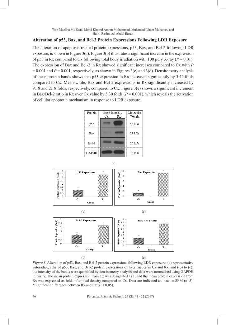
Alteration of p53, Bax, and Bcl-2 Protein Expressions Following LDR Exposure
The alteration of apoptosis-related protein expressions, p53, Bax, and Bcl-2 following LDR
exposure, is shown in Figure 3(a). Figure 3(b) illustrates a significant increase in the
expression of p53 in Rx compared to Cx following total body irradiation with 100 µGy X-ray
(P = 0.01). The expression of Bax and Bcl-2 in Rx showed significant increases compared to
Cx with P = 0.001 and P = 0.001, respectively, as shown in Figures 3(c) and 3(d).
Densitometry analysis of these protein bands shows that p53 expression in Rx increased
significantly by 3.42 folds compared to Cx. Meanswhile, Bax and Bcl-2 expressions in Rx
significantly increased by 9.18 and 2.18 folds, respectively, compared to Cx. Figure 3(e)
shows a significant increment in Bax/Bcl-2 ratio in Rx over Cx value by 3.30 folds (P =
0.001), which reveals the activation of cellular apoptotic mechanism in response to LDR
exposure.
Figure 2. LDR-induced oxidative DNA damage (AP sites) production. The bar chart shows the levels of AP sites production in the liver and lung tissues of Cx and Rx. Values were expressed as mean ± SEM (n=5). *Significant difference between Rx and Cx (P < 0.05)
Wan Mazlina Md Saad, Mohd Khairul Amran Mohammad, Muhamad Idham Mohamed andHairil Rashmizal Abdul Razak
46 Pertanika J. Sci. & Technol. 25 (S): 41 - 52 (2017)
Alteration of p53, Bax, and Bcl-2 Protein Expressions Following LDR Exposure
The alteration of apoptosis-related protein expressions, p53, Bax, and Bcl-2 following LDR exposure, is shown in Figure 3(a). Figure 3(b) illustrates a significant increase in the expression of p53 in Rx compared to Cx following total body irradiation with 100 µGy X-ray (P = 0.01). The expression of Bax and Bcl-2 in Rx showed significant increases compared to Cx with P = 0.001 and P = 0.001, respectively, as shown in Figures 3(c) and 3(d). Densitometry analysis of these protein bands shows that p53 expression in Rx increased significantly by 3.42 folds compared to Cx. Meanswhile, Bax and Bcl-2 expressions in Rx significantly increased by 9.18 and 2.18 folds, respectively, compared to Cx. Figure 3(e) shows a significant increment in Bax/Bcl-2 ratio in Rx over Cx value by 3.30 folds (P = 0.001), which reveals the activation of cellular apoptotic mechanism in response to LDR exposure.
(a)
(b) (c)
(d) (e)
Figure 3. Alteration of p53, Bax, and Bcl-2 protein expressions following LDR exposure: (a)
representative autoradiographs of p53, Bax, and Bcl-2 protein expressions of liver tissues in Cx and
Rx; and ((b) to (e)) the intensity of the bands were quantified by densitometry analysis and data were
normalised using GAPDH intensity. The mean protein expression from Cx was designated as 1, and
the mean protein expression from Rx was expressed as folds of optical density compared to Cx. Data
are indicated as mean ± SEM (n=5). *Significant difference between Rx and Cx (P < 0.05).
Figure 3. Alteration of p53, Bax, and Bcl-2 protein expressions following LDR exposure: (a) representative autoradiographs of p53, Bax, and Bcl-2 protein expressions of liver tissues in Cx and Rx; and ((b) to (e)) the intensity of the bands were quantified by densitometry analysis and data were normalised using GAPDH intensity. The mean protein expression from Cx was designated as 1, and the mean protein expression from Rx was expressed as folds of optical density compared to Cx. Data are indicated as mean ± SEM (n=5). *Significant difference between Rx and Cx (P < 0.05).

Low Dose Ionising Radiation-Induced DNA Damage
47Pertanika J. Sci. & Technol. 25 (S): 41 - 52 (2017)
Ultrastructural Alteration of Nucleus in Liver Tissues Following LDR Exposure
Severe alterations of cellular ultrastructure of nucleus were observed in the liver tissues of Rx compared to Cx (Figure 4). Figure 4(b) reveals morphologic changes of the nucleus related to apoptosis in the liver tissues of Rx. The nuclear membrane was shrunken and lost its common shape. The presence of condensed and peripheralised chromatin (X) in the nucleus of Rx is considered as a morphological characteristic of cell death through apoptotic mechanism following total body exposure to LDR.
Ultrastructural Alteration of Nucleus in Liver Tissues Following LDR Exposure
Severe alterations of cellular ultrastructure of nucleus were observed in the liver tissues of Rx
compared to Cx (Figure 4). Figure 4(b) reveals morphologic changes of the nucleus related to
apoptosis in the liver tissues of Rx. The nuclear membrane was shrunken and lost its common
shape. The presence of condensed and peripheralised chromatin (X) in the nucleus of Rx is
considered as a morphological characteristic of cell death through apoptotic mechanism
following total body exposure to LDR.
(a) (b)
Figure 4. Ultrastructure alterations of nucleus in liver tissues following LDR exposure. (a) A normal
ultrastructure of nucleus in liver tissues from Cx. (b) An altered ultrastructure of nucleus induced by
LDR in liver tissues of Rx. This nucleus is characterised by the changes on the nuclear morphology.
Nuclear membrane shrunken and lost common shape (arrow) and the present of chromatin
condensation (X) in the perinuclear area (A and B: 6000×).
DISCUSSION
Figure 4. Ultrastructure alterations of nucleus in liver tissues following LDR exposure. (a) A normal ultrastructure of nucleus in liver tissues from Cx. (b) An altered ultrastructure of nucleus induced by LDR in liver tissues of Rx. This nucleus is characterised by the changes on the nuclear morphology. Nuclear membrane shrunken and lost common shape (arrow) and the present of chromatin condensation (X) in the perinuclear area (A and B: 6000×)
DISCUSSION
Exposure to ionising radiation may lead to cellular damage through direct and/or indirect mechanisms (Cai, Koropatnick, & Cherian, 2001; Srinivasan et al., 2014). However, exposure to occupational sources of radiation frequently involves LDR (Ponzinibbio et al., 2010), which may contribute to overproduction of ROS under aerobic conditions (Cai et al., 2001). The results of this study are clinically relevance to radiation workers as they are continuously exposed to the low dose radiation from imaging modalities. As such, a single fractionated exposure was used as the baseline for a further research which is currently being conducted in assessing multiple exposures of low dose radiation in clinical setting. Results presented in Figure 1 demonstrate a significant production of ROS in the liver tissues of Rx following total body irradiation with LDR compared to Cx. This is in agreement with a study conducted by Park et al. (2001) which reveals significant increases of ROS production in lungs of male mice after a single dose of 10 Gy total-body irradiation with X-ray. It is possible to suggest that this was mainly due to the increased production in cellular ROS through radiolysis of water induced by LDR exposure, that in turn attacked cellular macromolecules including DNA through indirect mechanisms (Azzam et al., 2012).
Wan Mazlina Md Saad, Mohd Khairul Amran Mohammad, Muhamad Idham Mohamed andHairil Rashmizal Abdul Razak
48 Pertanika J. Sci. & Technol. 25 (S): 41 - 52 (2017)
Sites of missing bases termed as abasic sites, or apurinic/apyrimidinic (AP) sites, are continuously being generated by DNA following exposure to endogenous and exogenous sources including radiation which may lead to oxidative DNA damage (Mohammad et al., 2014). Referring to Figure 2, the number of non-coding AP sites generated in the liver and lung tissues of Rx showed significant increment compared to Cx. The present study seems to be consistent with a previous in vivo study by Pogribny et al. (2005) which reveals the accumulation of DNA damage measured as the levels of γH2AX foci in thymus tissues of mice following exposed to fractionated LDR. In addition, Ponzinibbio et al. (2010) found that CHO and MRC-5 cells treated with LDR (50 mSv) significantly induced DNA damage, as measured by alkaline comet assay compared to the control cell lines. These findings may suggest that exposure to LDR causes damage to DNA structure directly and/or indirectly through free radicals attack producing several oxidative DNA lesions including AP sites (Sudprasert et al., 2006).
p53 protein closely related to apoptosis mechanism, which remained at low state under normal condition and dramatically elevated in levels following accumulation of DNA damage (Xu et al., 2008). The present findings showed significant increment on expression of p53 in Rx by 3.42 folds compared to Cx (Figure 3(b)). Increased expression of p53 following total body irradiation with LDR in this study consistent with earlier findings by Wang et al., (1996), which revealed significant accumulation of p53 within 24 hours in adrenal glands, pancreas, thymus, skin, lungs, bone marrow and liver of C57BL/6N mice irradiated with 25 cGy and 50 cGy. An in vitro study conducted by Furlong et al. (2013) also supports the present findings, which demonstrates up-regulation of p53 in human keratinocyte cell line (HaCat) irradiated with a series of LDR. A possible explanation for the current finding is that LDR leads to accumulation of DNA damage, which in turn raises the expression of p53. Kastan et al. (1991), and Fei and El-deiry (2003) pointed out that the expression of p53 rises dramatically as the levels of DNA damage increase, thus activating p53 to either induce cell cycle arrest coupled with DNA damage repair or lead to cell death through apoptosis.
Damaged cells induced by ionising radiation are regulated by a complex balance in signal transduction pathways between pro-apoptotic (Bax) and anti-apoptotic (Bcl-2) proteins (Hoetelmans et al., 2000). The ratio of Bax/Bcl-2 provides important information whether cells will undergo apoptosis or not (Xu et al., 2008). In the current study, densitometry analysis of protein bands revealed significant increase of Bax and Bcl-2 expressions in Rx compared to Cx. Interestingly, the Bax/Bcl-2 ratio increased by 3.3 folds in Rx over Cx value (Figure 3(e)), which strongly suggests that total body irradiation with LDR may stimulate apoptosis when p53 protein is activated in response to DNA damage, thus altering the balance between ratio of Bax/Bcl-2. These findings are supported by a previous research by Park et al. (2008), which revealed a significant increment in the expression of pro-apoptotic proteins such as p53 and Bax within 24 hours in intestinal tissues of C57BL/6 mice after being irradiated with 2 Gy γ-rays. This result may explain the fact that, in apoptosis, Bax, which is a pro-apoptotic protein-containing p53-binding sites in promoter, has been shown to be regulated in response to p53 accumulation (Xu et al., 2008). Under normal physiological condition, Bax is predominantly present in the cytosol in a soluble form. However, during the activation of apoptosis, Bax becomes a membrane-bound form and cross-link as a homodimer with mitochondria (Brady & Gil-gomez, 1998). This biologically active Bax protein may form ion channels in the
Low Dose Ionising Radiation-Induced DNA Damage
49Pertanika J. Sci. & Technol. 25 (S): 41 - 52 (2017)
mitochondria membrane that allow passive flux of mitochondria apoptogenic proteins such as cytochrome c across intracellular membrane, which leads to cells death through activation of caspases. The regulation of Bax inactivates Bcl-2 through heterodimerisation, thus increases the Bax/Bcl-2 ratio, and cells are susceptible to self-destruction (Basu & Haldar, 1998). The electron micrographs also reveal apoptosis in the nucleus of liver tissues of Rx group compared to normal ultrastructure of nucleus in Cx (Figure 4(b)). Nucleus in Rx shows shrunken and loss of common shape. Another ultrastructural changes observed in Rx after being irradiated with LDR consisting condensation of chromatin in the perinuclear area (X). The appearance of these severe alterations of the nucleus may suggest that the liver cells in Rx underwent apoptosis.
CONCLUSION
The present study provides evidences that LDR exposure may induce oxidative DNA damage through excessive production of cellular ROS and significant increase in the production of AP sites. In addition, exposure to LDR may alter cellular ultrastructure and apoptosis-related proteins. These finding serve as important implications for estimating risks related to LDR exposure among medical radiation professions. It is perhaps best to recommend that study on cell cycle checkpoint and repair mechanisms be conducted for a better understanding of molecular mechanisms in response to LDR exposure.
ACKNOWLEDGEMENTS
The authors gratefully acknowledge the support given by: (1) Faculty of Health Sciences UiTM Puncak Alam; and (2) UiTM Institute for Research Management & Innovation (RAGS:600-RMI/RAGS 5/3 (120/2012)) for funding the study. The authors deeply appreciate: (3) Department of Medical Laboratory Technology; and (4) Department of Medical Imaging, and Imaging Centre (IMACE), Faculty of Pharmacy, for providing the research facilities throughout this study.
REFERENCESAzzam, E. I., Jay-Gerin, J.-P., & Pain, D. (2012). Ionizing radiation-induced metabolic oxidative stress
and prolong cell injury. Cancer Letters, 327(0), 48–60.
Basu, A., & Haldar, S. (1998). The relationship between Bcl2 , Bax and p53 : consequences for cell cycle progression and cell death. Molecular Human Reproduction, 4(12), 1099–1109.
Brady, H. J. M., & Gil-gomez, G. (1998). Molecules in focus Bax . The pro-apoptotic Bcl-2 family member, Bax. The International Journal of Biochemistry and Cell Biology, 30, 647–650.
Cai, L., Koropatnick, J., & Cherian, M. G. (2001). Roles of vitamin C in radiation-induced DNA damage in presence and absence of copper. Chemico-Biological Interaction, 137, 75–88.
Cardozo-Pelaez, F., Brooks, P. J., Stedeford, T., Song, S., & Sanchez-Ramos, J. (2000). DNA damage, repair, and antioxidant systems in brain regions: a correlative study. Free Radical Biology and Medicine, 28(5), 779–85. Retrieved from http://www.ncbi.nlm.nih.gov/pubmed/10754274.
Elmore, S. (2007). Apoptosis: a review of programmed cell death. Toxicologic Pathology, 35(4), 495–516.
Wan Mazlina Md Saad, Mohd Khairul Amran Mohammad, Muhamad Idham Mohamed andHairil Rashmizal Abdul Razak
50 Pertanika J. Sci. & Technol. 25 (S): 41 - 52 (2017)
Fei, P., & El-deiry, W. S. (2003). P53 and radiation responses. Oncogene, 22, 5774–5783.
Furlong, H., Mothersill, C., Lyng, F. M., & Howe, O. (2013). Apoptosis is signalled early by low doses of ionising radiation in a radiation-induced bystander effect. Mutation Research, 741-742(2013), 35–43.
Gultekin, F. A., Bakkal, B. H., Guven, B., Tasdoven, I., Bektas, S., Can, M., & Comert, M. (2013). Effects of ozone oxidative preconditioning on radiation-induced organ damage in rats. Journal of Radiation Research, 54(1), 36–44.
Hoetelmans, R., van Slooten, H. J., Keijzer, R., Erkeland, S., van de Velde, C. J., & Dierendonck, J. H. (2000). Bcl-2 and Bax proteins are present in interphase nuclei of mammalian cells. Cell Death and Differentiation, 7, 384–392.
Kastan, M. B., Onyekwere, O., Sidransky, D., Vogelstein, B., & Craig, R. W. (1991). Participation of p53 Protein in the Cellular Response to DNA Damage Participation of p53 Protein in the Cellular Response to DNA Damage. Cancer Research, 6304–6311.
Klaunig, J. E., Kamendulis, L. M., & Hocevar, B. A. (2010). Oxidative stress and oxidative damage in carcinogenesis. Toxicologic Pathology, 38(1), 96–109.
Lee, J., Hosotani, R., Wada, M., Doi, R., Kosiba, T., Fujimoto, K., & Imamura, M. (1999). Role of Bcl-2 Family Proteins ( Bax , Bcl-2 and Bcl-X ) on Cellular Susceptibility to Radiation in Pancreatic Cancer Cells. European Journal of Cancer, 35(9), 1374–1380.
Martin, L. M., Marples, B., Coffey, M., Lawler, M., Lynch, T. H., Hollywood, D., & Marignol, L. (2010). DNA mismatch repair and the DNA damage response to ionizing radiation: making sense of apparently conflicting data. Cancer Treatment Reviews, 36(7), 518–27.
Mohamed, M. I., Mohammad, M. K. A., Zakaria, A. M., Ghazali, N., Mohamed Isa, M., Abdul Razak, H. R., & Md Saad, W. M. (2014). Induction of Oxidative Stress Following Low Dose Ionizing Radiation in ICR Mice. World Journal of Medical Sciences, 10(2), 198–203.
Mohammad, M. K. A., Mohamed, M. I., Zakaria, A. M., Abdul Razak, H. R., & Md Saad, W. M. (2014). Watermelon (Citrullus lanatus (Thunb.) Matsum. and Nakai) Juice Modulates Oxidative Damage Induced by Low Dose X-Ray in Mice. BioMed Research International, 2014.
Park, E., Lee, N. H., Joo, H.-G., & Jee, Y. (2008). Modulation of apoptosis of eckol against ionizing radiation in mice. Biochemical and Biophysical Research Communications, 372(4), 792–7.
Park, E., Park, J., Kim, Y., Sung, J., Hwang, T., Kim, W., Park, Y. (2001). Role of Oxidative Stress in the Radiation-Induced Lung Pathogenesis in Mice. Journal of Biochemistry and Molecular Biology, 34(6), 544–550.
Paz-Elizur, T., Sevilya, Z., Leitner-Dagan, Y., Elinger, D., Roisman, L. C., & Livneh, Z. (2008). DNA repair of oxidative DNA damage in human carcinogenesis: potential application for cancer risk assessment and prevention. Cancer Letters, 266, 60–72.
Pogribny, I., Koturbash, I., Tryndyak, V., Hudson, D., Stevenson, S. M. L., Sedelnikova, O., Kovalchuk, O. (2005). Fractionated low-dose radiation exposure leads to accumulation of DNA damage and profound alterations in DNA and histone methylation in the murine thymus. Molecular Cancer Research, MCR, 3(10), 553–61.
Ponzinibbio, M. V, Crudeli, C., Peral-García, P., & Seoane, A. (2010). Low-dose radiation employed in diagnostic imaging causes genetic effects in cultured cells. Acta Radiologica (Stockholm, Sweden: 1987), 51(9), 1028–33.

Low Dose Ionising Radiation-Induced DNA Damage
51Pertanika J. Sci. & Technol. 25 (S): 41 - 52 (2017)
Shackelford, R. E., Kaufmann, W. K., & Paules, R. S. (2000). Oxidative stress and cell cycle checkpoint function. Free Radical Biology and Medicine, 28(9), 1387–1404.
Srinivasan, M., Kalpana, K. B., Devipriya, N., & Menon, V. P. (2014). Protective effect of lycopene on whole body irradiation induced liver damage of Swiss albino mice: Pathological evaluation. Biomedicine and Preventive Nutrition, 4(2), 87–94.
Sudprasert, W., Navasumrit, P., & Ruchirawat, M. (2006). Effects of low-dose gamma radiation on DNA damage, chromosomal aberration and expression of repair genes in human blood cells. International Journal of Hygiene and Environmental Health, 209(6), 503–11.
Verheij, M., & Bartelink, H. (2000). Radiation-induced apoptosis. Cell Tissue Research, 301, 133–142.
Wang, X., Matsumoto, H., Takahashi, A., Nakano, T., Okaichi, K., Ihara, M., & Ohnishi, T. (1996). p53 accumulation in the organs of low-dose X-ray-irradiated mice. Cancer Letters, 04(I 996), 79–84.
Xu, J., Lian, L., Wu, C., Wang, X., Fu, W., & Xu, L. (2008). Lead induces oxidative stress, DNA damage and alteration of p53, Bax and Bcl-2 expressions in mice. Food and Chemical Toxicology: An International Journal Published for the British Industrial Biological Research Association, 46(5), 1488–94.
Zakaria, A. M., Ghazali, N., Mohammad, M. K. A., Mohamed, M. I., Mohamed Isa, M., Abdul Razak, H. R., & Md Saad, W. M. (2014). Radioprotective Effect of Watermelon Juice Against Low Dose Ionizing Radiation-Induced Inflammatory Response in Mice. World Journal of Medical Sciences, 10(2), 191–197.
Zielinski, J. M., Garner, M. J., Band, P. R., Krewski, D., Shilnikova, N. S., Jiang, H., & Semenciw, R. (2009). Health outcomes of low-dose ionizing radiation exposure among medical workers: a cohort study of the Canadian national dose registry of radiation workers. International Journal of Occupational Medicine and Environmental Health, 22(2), 149–156.
