phase transitions in cell biology || unexpected linkage between unstirred layers, exclusion zones,...
TRANSCRIPT

Unexpected Linkage Between Unstirred Layers,Exclusion Zones, and Water
Gerald H. Pollack and James Clegg
Abstract We make the case that the unstirred layers of classical physiology arisefrom the influence of surfaces on the structure, and therefore the properties, ofcontiguous water. Traditionally, unstirred layers have been thought to arise merelyout of stagnant volumes adjacent to membranes and other surfaces. These volumeswould have to extend tens to hundreds of micrometers in order to account for the ob-served effects. On the other hand, charged and hydrophilic surfaces have been shownto impact water out to surprising distances, on the order of hundreds of micrometers.We present evidence that it is this water-structuring effect that is responsible for theunstirred layer.
Keywords Unstirred layers · exclusion zones · water structure
1 Unstirred Layers (USLs)
It is generally agreed that unstirred layers (USLs) are more or less static fluid regionsadjacent to membranes, and probably many other surfaces, within which thermalconvection or density gradients do not cause significant mixing (Barry and Dia-mond, 1984). Even when the surrounding fluid is vigorously stirred this seeminglystatic fluid remains, at least to some extent, and there is a relationship between thethickness of USLs (�) and the intensity of stirring of the surrounding fluid (Barryand Diamond, 1984).
USLs are of critical importance in a wide variety of physiological processes, no-tably those involving solute transport, enzyme and other chemical reactions on or nearthe cell surface. Membrane electrochemical potentials may also be affected becauseconcentrations of ions and other solutes adjacent to the plasma membrane are not thesame as those in the bulk, an important point to which we will return. The literatureon USLs is massive, including research in physical chemistry where the USLs havebeen referred to as boundary layers (House, 1974; Barry and Diamond, 1984). These
G.H. PollackDepartment of Bioengineering, Box 355061, University of Washington, Seattle WA, 98195, USAe-mail: [email protected]
G.H. Pollack, W.-C. Chin (eds.), Phase Transitions in Cell Biology,C© Springer Science+Business Media B.V. 2008
143

144 G.H. Pollack, J. Clegg
layers have been implicated in many processes, and in a wide variety of systems suchas the gills of mussels (Wright et al., 1980), the human jejunum (Pappenheimer,2001),monolayer cultures of mammalian cells (Spivak et al., 2006), rainbow-trout embryosand larvae (Ciuhandu et al., 2007) and a giant-celled alga (Kim et al., 2006). We makeno pretence about a thorough review of this literature but will attempt to point outfeatures that are salient to the topics covered in this essay.
A brief examination of the history of USLs seems worthwhile. In an importantearly review, Dainty and House (1966) state that the USL concept was originallydeveloped by Noyes and Whitney in 1897 and further in 1904 by Nernst. However,based on the translation of Wilhelm Pfeffer’s 1877 book, Osmotische Unteruchun-gen (Kepner and Stadelmann, 1985), it appears that Pfeffer described USLs wellbefore those authors. Pfeffer clearly emphasized the critical role played by USLs inhis extensive research on osmosis and related processes. Key papers on USLs in theearly to mid-1900s have been considered or reviewed by Teorell (1936) Ginzbergand Katchalsky (1963) and Dick (1959). The review by Barry and Diamond, (1984)is definitive in our opinion, with excellent overall coverage of USLs. More recently,work from the laboratory of Peter Pohl considers the wide-ranging significance andimplications of USLs and their relation to what is called reaction layers, RLs (seeAntonenko et al., 1996; Pohl et al., 1998). RLs are solution layers adjacent to cellmembranes within which chemical reactions, including enzymatic ones, take place.These layers have been described previously (see Gutnecht and Tosteson,1973;Verkman and Dix, 1984) but particularly by Antonenko et al. (1996). We mentionthem here to draw attention to the important relationship between USLs and RLsand, as we will propose, the importance of water in the operation of both layers.
A key feature of USLs concerns their thickness—the distance from membranesand other surfaces over which mechanical stirring is not effective. An excellentsource of information on USL thickness (�) is the review by Barry and Diamond(1984). Without stirring, � exceeds 300 μm, depending on the nature of the interface,and can even extend over several millimeters from the surface. Despite the mostvigorous stirring, � remains near 10 μm, obviously a huge distance in molecularterms. The view is widely held that the USL is not stationary but a region of slowlaminar flow, parallel to the interface, in which solute transport occurs by diffusionalone (Dainty and House, 1966).
A key question arises: what generates the USL? A logical possibility is that thephysical properties of the USL are somehow different from those of regions beyond.To our knowledge the “structure” of water in such interfacial regions has not beenconsidered in this connection. We wish to raise this possibility, that the influenceof the adjacent “solid” surface, membrane, or otherwise, alters the structure andproperties of the interfacial water in such a way that the USL is generated.
2 Cellular Water
Studies on the properties and structure of water in cells and adjacent to surfaceshave been many, and the topic has had a long and often controversial history. Itis not appropriate here to review that history, except to note certain points. Cell

Unexpected Linkage Between Unstirred Layers, Exclusion Zones, and Water 145
biology seems to have proceeded throughout its history on the assumption that allbut a small fraction (perhaps 5%) of total cell water exhibited the properties of bulkwater, and that the cytoplasm could be treated as an ordinary, although non-ideal,aqueous solution (see Clegg, 1982). Some have objected to that view over the years,notably Gilbert Ling, Walter Drost-Hansen, and Aphanasij Troschin, who proposedthat most and probably all of cell water is different. Those three scientists began theirwork in the 1950s, and two continue their work today (see Pollack et al., 2006). Ahigh point arrived in the late 1960s when both Freeman Cope (1969), and CarltonHazlewood and colleagues (1969), published papers using pulsed NMR technology.They concluded that the structure of most or even all of water in skeletal muscle andbrain was not that of the bulk liquid.
Much attention was given to these results, supporting the positions of Ling,Drost-Hansen and Troschin, but it was not very long until opposition to those viewsarose (see Kolata, 1976). What followed was a contentious period spanning some 10years of claim and counterclaim (considered to some extent in works by Hazlewood,1973; Franks and Mathias, 1982; Clegg and Drost-Hansen, 1991; Drost-Hansen andClegg, 1979; Ling, 2001; Mentre, 2001; Pollack, 2001; Pollack et al., 2006). As ofthis writing research on cell water has slowed greatly, with a few notable exceptions(see articles in Mentre, 2001; Pollack, 2001; Pollack et al., 2006) although interestin the subject continues (for example, Leterrier, 2001; Shepherd, 2006). The versiongiven in textbooks and the general literature is not much different than it was acentury ago: almost all of cell water is considered to be essentially the same as thatin the bulk liquid. But there remains a dissenting group, albeit small, that believesthe last word has yet to be said; we are among that group.
That surfaces alter the structure of water immediately adjacent to them is wellestablished for very short distances, equivalent to two or three molecular layers ofwater. But it is our impression that very few (other than those named above) wouldaccept the possibility that such influences might extend over distances of tens orhundreds of nanometers, let alone micrometers. However, in the sections that followwe will make a case for just that; namely, that the existence and properties of USLsarise from the influence of membranes and other “solid” surfaces on the structure ofwater adjacent to them.
3 Exclusion Zones (EZs)
Motivated independently from unstirred layer considerations, studies of the physicalbehavior of this near-surface aqueous zone were begun several years ago (Zheng andPollack, 2003). These studies were in fact broader than just examining the unstirredlayer next to cells: they dealt with the nature of aqueous regions adjacent to manyhydrophilic surfaces.
A half-century old review of the solid-liquid interface (Henniker, 1949) citedmore than 100 studies showing a surface influence extending into the liquid farbeyond the two or three molecular layers generally implied; and, a later studyshowed that on polished quartz, ordered water could extend out to some 600
146 G.H. Pollack, J. Clegg
molecular layers from the surface (Pashley and Kitchener, 1979). For many decadesDrost-Hansen has mustered evidence that similar distances are involved in a widevariety of surface systems, the adjacent water being referred to as vicinal water (seeDrost-Hansen, 2006). These reports, and many references therein, indicate consid-erably more structuring than has generally been accepted. Hence, positions weretaken on both sides of the issue.
The experimental approach taken by Zheng and Pollack (2003) was to explore thebehavior of colloidal particles next to hydrophilic surfaces. Since it is energeticallyfavorable for colloidal particles and solutes to reside outside of structured regions sothey can more readily hydrate, attempts were made to examine solute partitioning inthe vicinity of interfaces. If such solutes are preferentially excluded from the interfa-cial zone, the presumption was that this zone was likely to be structured differentlyfrom ordinary bulk water.
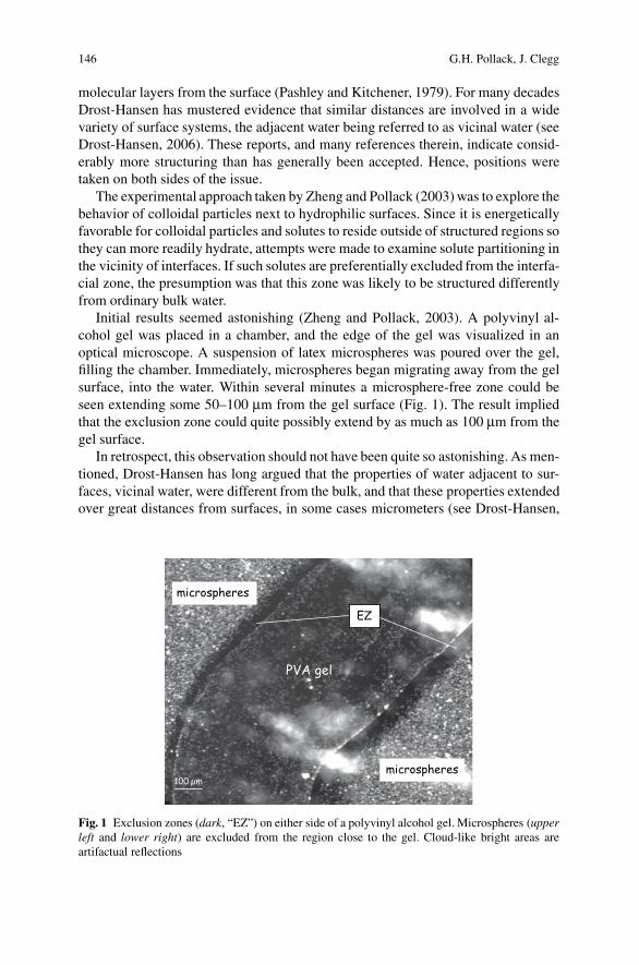
Initial results seemed astonishing (Zheng and Pollack, 2003). A polyvinyl al-cohol gel was placed in a chamber, and the edge of the gel was visualized in anoptical microscope. A suspension of latex microspheres was poured over the gel,filling the chamber. Immediately, microspheres began migrating away from the gelsurface, into the water. Within several minutes a microsphere-free zone could beseen extending some 50–100 μm from the gel surface (Fig. 1). The result impliedthat the exclusion zone could quite possibly extend by as much as 100 μm from thegel surface.
In retrospect, this observation should not have been quite so astonishing. As men-tioned, Drost-Hansen has long argued that the properties of water adjacent to sur-faces, vicinal water, were different from the bulk, and that these properties extendedover great distances from surfaces, in some cases micrometers (see Drost-Hansen,
PVA gel
100 µ m
microspheres
microspheres
EZ
Fig. 1 Exclusion zones (dark, “EZ”) on either side of a polyvinyl alcohol gel. Microspheres (upperleft and lower right) are excluded from the region close to the gel. Cloud-like bright areas areartifactual reflections

Unexpected Linkage Between Unstirred Layers, Exclusion Zones, and Water 147
200 µm
PAA gel
Fig. 2 Exclusion zones (dark) on either side of a polyacrylic acid gel. Microspheres translatedfrom the gel surface appear at the far right. The vertical bright stripe in the middle of the PAA gelis an optical artifact
2006). Further, those observations seemed in concordance with the many results ob-tained during the first half of the previous century, as reviewed by Henniker (1949).But the fact remains that these observations and proposals have had little effect oncurrent views concerning interfacial water.
Even more remarkable than the 100-μm EZ seen with polyvinyl alcohol gels wasthe exclusion zone seen with the more highly charged polyacrylic-acid gels. There,the microsphere-exclusion zone could easily extend from the gel surface by 250 μmor more (Fig. 2).
It was not only microspheres that were excluded from the region next to gels.Many suspended or dissolved species were excluded, ranging in size from particlessuch as sand and silt, to biological entities including bacteria and erythrocytes,down to much smaller species such as proteins and low molecular weight dyes. Allof those were excluded (Zheng et al., 2006). The kinds of surfaces that producedsuch exclusion also ranged broadly. They included a series of hydrogels, variouspolymeric surfaces including Nafion, and biological surfaces including vascular en-dothelium, seaweed, and various plant roots. Muscle, for example, produced an EZof several hundred micrometers (Zheng et al., 2006). Hence, the exclusion phe-nomenon seems rather general, and is not the quirk of a particular solute juxtaposednext to a particular surface.
It was after those experiments had been largely completed that we came uponthe study by Green and Otori (1970) which linked surfaces and microspheres withunstirred layers. Curious about the nature of the unstirred layer around the eyecornea, those investigators exposed the cornea to suspensions of polystyrene latexmicrospheres in much the same manner as we had done more recently with otherhydrophilic surfaces. The results were similar. Green and Otori found microsphere-exclusion zones on the order of 350 μm. They found exclusion not only adjacentto the natural cornea, but also next to artificial contact lenses – results virtually

148 G.H. Pollack, J. Clegg
identical to what we found next to polyNIPAM, the gel used to fabricate contactlenses. Clearly, the results of Green and Otori and the more recent results obtainedin the Pollack laboratory are for all intents and purposes indistinguishable. The un-stirred layer of classical physiology and the solute-exclusion zone seem to be oneand the same.
4 Nature of Unstirred Layers and Exclusion Zones
What is the nature of the water within USLs and EZs? One possibility is that it isidentical to bulk water, and that exclusion merely derives from proximity to surfaces.Because of entropic effects, solutes should be in lower concentrations in regions“close to” surfaces. Such effects are generally considered to extend out to several,or even tens of molecular layers of water. But, here the distances under considerationare far greater: a span of 250–300 μm is equivalent to a stack of about a million watermolecules. Such numbers are far too large to fall into the realm of surface-entropiceffects.
A second possibility is that the water in this zone may be impacted by the surface;i.e., that the surface influences water structure out to distances considerably fartherthan expected. The results described above are fully consistent with that possibility,but they do not prove it. More recent results, on the other hand, lend further supportto the possibility that water in the exclusion zone is physically different from bulkwater.
A significant indication of water-structural differences in the vicinal zone derivesfrom studies using UV-Vis spectrometry (Zheng et al., 2006). Those studies involvedNafion, whose surface generates an appreciable exclusion zone. A sheet of Nafionwas glued to the inside face of one of the four vertical walls of a standard cuvette,which was then filled with distilled, deionized water. The spectrometer was modi-fied such that a narrow slit of light could sample zones parallel to, and at variabledistances from, the Nafion face.
Far from the Nafion surface, the absorption spectrum was indistinguishable fromthat of bulk water. As the narrow sampling window came progressively closer tothe Nafion surface, a new absorption peak at ∼270 nm, could be detected. The peakbecame prominently visible at the exclusion-zone boundary, ∼ 500 μm from theNafion surface. It grew further as the window came closer to the surface, ultimatelydominating the pattern (Fig. 3). Since no absorption peaks at 270 nm are foundin bulk water, the presence of a strong “anomalous” absorption peak implies thatexclusion-zone water differs appreciably from bulk water.
Artifacts in such measurements are possible: namely, some leaching of polymerfrom the Nafion could be responsible for this absorption. Control experiments havebeen carried out to determine whether a similar absorption peak occurs in situationsin which water structuring is anticipated. Indeed, various concentrated salt solutionsand sugar solutions showed similar absorption peaks (Chai et al., 2008). When thesalts were first heat-purified in order to remove volatile hydrocarbon contaminants,

Unexpected Linkage Between Unstirred Layers, Exclusion Zones, and Water 149
Fig. 3 UV-Vis absorption spectrum as a function of distance from the Nafion surface
the absorption peaks grew more intense. Hence, these absorption peaks do not ap-pear to be artifacts, but may be a genuine “signature” of water structure.
Several other physical features of exclusion-zone water differ from their coun-terparts in bulk water. These include shorter NMR-T2 relaxation times, reducedinfrared radiation, and a steep electrical potential gradient (Zheng et al., 2006).Unpublished experiments also show different optical refractive features, increasedviscosity, and higher density.
Taken together, these observations imply that water in the exclusion zone is phys-ically, and perhaps chemically different from ordinary bulk water. In particular, thereduced infrared radiation and shorter T2 values imply that water in USLs and EZsmay be structurally more stable than bulk water—perhaps partially liquid crystallinein nature. In such a case it would be clear why colloids and solutes might be ex-cluded: energetically, they prefer to lodge in the region of bulk water, where theycan more easily hydrate. It also accounts for the cornea and contact lens observationsof Green and Otori (1970), who found that microspheres did not invade the unstirredlayer. Apparently, the microspheres avoided the region because it was structured ina way that did not admit them.
Thus, the unstirred layers of classical physiology appear to be the same as exclu-sion zones found next to many hydrophilic surfaces. Water in this zone is extensivelyimpacted by the surface, and the resulting structure tends to exclude particles and so-lutes. On the other hand, important questions remain. One of the issues to be studiedin more detail is the degree to which ions and small solutes penetrate the exclusionzone. To our knowledge this has not yet been explored directly. Although largersolutes and colloids seem fully excluded from such zones, it is not yet clear whetherthe smallest solutes are fully excluded, partially excluded, or not excluded. Giventhe striking similarities thus far established between unstirred layers and exclusionzones, there is reason to be optimistic that the “limited diffusion” of small solutesthrough the USL may be seen as well in the exclusion zone, but such hypothesesremain to be tested.

150 G.H. Pollack, J. Clegg
5 Ultrastructural Evidence for Exclusion Zones
Mollenhauer and Morre (1978) seem to have been the first to point out that zonesof exclusion (their term) can be detected at the ultrastructural level in eukaryoticcells using transmission electron microscopy. They defined these zones as “differ-entiated regions of cytoplasm in which ribosomes, glycogen and organelles such asmitochondria, plastids or microbodies are scarce or absent.” They also list other cellstructures that are excluded. Our impression is that exclusion zones are consistentlyfound adjacent to various membranes or cytoskeletal structures. These include actinfilaments (Kamitsubo, 1972), and microtubules (Stebbings and Willison, 1973;Stebbings and Hunt, 1982), and they persist even when most of the water hasbeen osmotically removed (Albrecht-Buehler and Bushnell, 1982). Thus, we believethese ultrastructural exclusion zones are comparable to the USLs and EZs we havediscussed in this paper, extending their importance to the in vivo case.
6 Concluding Comments
The USL of classical physiology has long been known and studied. That trail hasgone on with little or no attention given to what generates it. In this essay we supportthe view that the properties and structure of both the USL and EZ arise because ofthe influence of surfaces on the properties and structure of water in its vicinity. Thiswater is somehow altered. The alteration may be profound—enough so that it hasbeen considered to constitute a “fourth phase” of water (Hardy, 1932). The viewthat “boundary water” is altered is supported by evidence briefly summarized here,much of it coming from work done over the last 50 years, indicating that the influ-ence of surfaces extends over large distances, exceeding 100 μm. That contention,standing in opposition to the conventional wisdom, indicates that the role of water inand around cells and tissues is vastly more important than the teachings of currenttextbooks permit. As always, the evidence will prevail, and the future will decidethat outcome.
References
Albrecht-Buehler G, Bushnell A (1982) Reversible compression of the cytoplasm. Exp Cell Res140:173–189
Antonenko YN, Pohl P, Rosenfeld E (1996) Visualization of the reaction layer in the immediatemembrane vicinity. Arch Biochem Biophys 333:225–232
Barry PH, Diamond JM (1984) Effects of unstirred layers on membrane phenomena. Physiol Rev64:763–871
Chai, B-H, Zheng, J-M, Zhao, Q, Pollack, GH (2008) Spectroscopic studies of solutes in aqueoussolution. J. Phys. Chem A 112:2242–2247
Ciuhandu CS, Wright PA, Goldberg JI, Stevens ED (2007) Parameters influencing the dissolvedoxygen in the boundary layer of rainbow trout (Oncorhynchus mykiss) embryos and larvae.J Exp Biol 210:1435–1445

Unexpected Linkage Between Unstirred Layers, Exclusion Zones, and Water 151
Clegg JS (1982) Alternative views on the role of water in cell function. In: Franks F, Mathias SF(ed.) Biophysics of Water. John Wiley and Sons, New York, pp. 365–385
Clegg JS, Drost-Hansen W (1991) On the biochemistry and cell physiology of water. In:Hochachka PW, Mommsen TP (ed.), Biochemistry and Molecular Biology of Fishes, Elsevier,Amsterdam, pp. 1–23
Cope F (1969) Nuclear magnetic resonance evidence using D2O for structured water in muscle andbrain. Biophys J 9:303–319
Dainty J, House CR (1966) “Unstirred layers” in frog skin. J Physiol 182:66–78Dick DAT (1959) Osmotic properties of living cells. Int Rev Cytol 8:387–448Drost-Hansen W (2006) Vicinal hydration of biopolymers: cell biological consequences. In:
Pollack GH, Cameron IL, Wheatley DN (ed.). Water and the Cell. Springer-Verlag, Berlin,pp. 175–217
Drost-Hansen W, Clegg JS (ed.) (1979) Cell Associated Water. Ascademic Press, New York, p. 440Franks F, Mathias SF (ed.) (1982) Biophysics of Water. John Wiley and Sons, New York, p. 400Ginzberg BZ, Katchalsky A (1963) The frictional coefficients of the flows of non-electrolytes
through artificial membranes. J Gen Physiol 47:403–418Green K, Otori T (1970) Direct measurement of membrane unstirred layers. J Physiol 207:93–102Gutknecht, J, Tosteson, DC (1973). Diffusion of weak acids across lipid bilayer membranes: effects
of chemical reactions in the unstirred layers. Science 182:1258–1261Hardy, W (1932) Problems of the boundary state. Phil Trans Roy Soc Lon Ser A 230:1–37Hazlewood CF (ed.) (1973) Physicochemical state of ions and water in living tissues and model
systems. Ann NY Acad Sci 204:5–631Hazlewood CF, Nichols BL, Chamberlain NF (1969) Evidence for the existence of a minimum of
two phases of ordered water in skeletal muscle. Nature 222:747–750Henniker, JC (1949) The depth of the surface zone of a liquid. Rev Mod Phys 21(2):322–341House CR (1974) Water Transport in Cells. Arnold Press, London, p. 276Kamitsubo, E (1972) Motile protoplasmic fibrils in cells of the Characae. Protoplasma 74:53–70Kepner GR, Stadelmann EJ (1985) Translation of Osmotische Untersuchungen. Studien zur
Zellemechanik. Van Nostrand Reinhold, New York, p. 267Kim Y, Ye Q, Reinhardt H, Steudle E (2006) Further quantification of the role of internal stirred
layers during the measurement of transport coefficients in giant internodes of Chara by a newstop-flow technique. J Exp Bot 57:4133–4144
Kolata G (1976) Water structure and ion binding: a role in cell physiology? Science 192:1220–1222Leterrier JF (2001) Water and the cytoskeleton. Cell Mol Biol (Noisy-le-grand) 47:901–923Ling GN (2001) Life at the Cell and Below-Cell Level. Pacific Press, New York, p. 373Mentre P (ed.) (2001) Water in the cell. Cell Mol Biol 47:709–970Mollenhauer HH, Morre DJ (1978) Structural compartmentation of the cytosol: zones of exclusion,
zones of adhesion, cytoskeletal and intercisternal elements. In: Roodyn DB (ed.) SubcellularBiochemistry, vol. 5, Plenum Press, New York, pp. 327–362
Pappenheimer JR (2001) Role of pre-epithelial “unstirred” layers in absorption of nutrients fromthe human jejunum. J Membrane Biol 179:185-204
Pashley RM, Kitchener JA (1979) Surface forces in adsorbed multilayers of water on quartz. J CollInterface Sci 71: 491–500
Pohl P, Saparov SM, Antonenko YN (1998) The size of the unstirred layer as a function of thesolute diffusion coefficient. Biophys J 75:1403–1409
Pollack GH (2001) Cells, Gels and the Engines of Life. Ebner and Sons, Seattle, p. 305Pollack GH, Cameron IL, Wheatley DN (ed.) (2006) Water and the Cell. Springer-Verlag, Berlin,
pp. 165–174.Shepherd VA (2006) The cytomatrix as a cooperative system of macromolecular and water net-
works. Curr Top Dev Biol 75:171–233Spivak CE, Oz M, Beglan CL, Shrager RI (2006) Diffusion delays and unstirred layer effects at
monolayer cultures of Chinese hamster ovary cells. Cell Biochem Biophys 45:43–58Stebbings H and Hunt C (1982) The nature of the clear zone around microtubules. Cell and Tissue
Res 227:609–617

152 G.H. Pollack, J. Clegg
Stebbings H, Willison JHM (1973) Structure of microtubules: a study of freeze-etched and neg-atively stained microtubules from the ovaries of Notonecta. Z. Zellforsch u Mikrosc Anat138(3):387–396
Teorell T (1936) A method of studying conditions within diffusion layers. J Biol Chem113:735–748
Verkman AS, Dix JA (1984) Effect of unstirred layers on binding and reaction kinetocs at a mem-brane surface. Anal Biochem 142:109–116
Wright SH, Becker SA, Stephens GC (1980) Influence of temperature and unstirred layers on thekinetics of glycine transport in isolated gills of Mytilus californianus. J Exp Zool 214:27–35
Zheng, JM, Pollack, GH (2003) Long range forces extending from polymer surfaces. Phys Rev E.:68:031408
Zheng, J-M, Chin, W-C, Khijniak, E, Khijniak, E, Jr, Pollack, GH (2006) Surfaces and InterfacialWater: Evidence that hydrophilic surfaces have long-range impact. Adv. Colloid Interface Sci.127:19–27