photoemission tomography: applications and future developments
TRANSCRIPT

Photoemission Tomography: Applications and
Future Developments
734. WE-Heraeus-Seminar
25 – 28 October 2021 hybrid at the Physikzentrum Bad Honnef,
Germany

Introduction
The Wilhelm und Else Heraeus-Stiftung is a private foundation that supports research and education in science with an emphasis on physics. It is recognized as Germany’s most important private institution funding physics. Some of the activities of the foundation are carried out in close cooperation with the German Physical Society (Deutsche Physikalische Gesellschaft). For detailed information see https://www.we-heraeus-stiftung.de Aims and scope of the 734. WE-Heraeus-Seminar: Angle-resolved ultraviolet photoemission spectroscopy is arguably the most direct method of addressing the (filled) electronic structure critical for the electronic and optical properties of matter. The photoemission tomography technique, a combined experimental/theoretical approach based on interpretation of the photoelectron angular distribution in terms of a one-electron initial state, has been very useful in the characterizations of electronic properties of molecular films from sub-monolayers to multilayers. This includes the unambiguous assignment of emissions to particular molecular orbitals, their reconstruction to real space orbitals, the deconvolution of spectra into individual orbital contributions beyond the limits of energy resolution, the extraction of detailed geometric information, and the precise description of the charge balance and transfer at the interfaces. Despite these successes, the theoretical description of the angular distribution of photoelectrons remains challenging and is – at the current level of theory – presently crude. Moreover, there has been significant progress in momentum space imaging photoemission spectrometers with extensions to the spin- and time-resolved domains, which make present models inadequate.
The seminar will bring together experts and young researchers interested in the application and the future developments of momentum space imaging of matter. The major aim of the seminar is to set photoemission tomography on firm grounds and to identify the directions for future theoretical and experimental investigations.
The scope of the seminar includes but is not limited to
• Photoemission tomography and photoelectron diffraction • Limitations of the plane wave final state approximation • Real space orbital reconstruction and phase retrieval • Theoretical advances: Time dependent DFT, multiple scattering theory • Experimental advances: Imaging spin-and time-resolved detectors, high
harmonics generation etc. • Valence band electronic structure of organic molecular films and 2D materials
.

Introduction
Scientific Organizers: Prof. Peter Puschnig Universität Graz, Austria
E-mail: [email protected]
Prof. Mathias Richter Physikalisch-Technische Bundesanstalt, Germany E-mail: [email protected]
Prof. Stefan Tautz Forschungszentrum Jülich, Germany
E-mail: [email protected] Administrative Organization: Dr. Stefan Jorda Wilhelm und Else Heraeus-Stiftung Elisabeth Nowotka Postfach 15 53 63405 Hanau, Germany
Phone +49 6181 92325-12 Fax +49 6181 92325-15 E-mail [email protected] Internet : www.we-heraeus-stiftung.de Venue: Physikzentrum Hauptstrasse 5 53604 Bad Honnef, Germany
Conference Phone +49 2224 9010-120 Phone +49 2224 9010-113 or -114 or -117 Fax +49 2224 9010-130 E-mail [email protected] Internet www.pbh.de Taxi Phone +49 2224 2222 Registration: Elisabeth Nowotka (WE Heraeus Foundation) at the Physikzentrum, reception office Sunday (16:00 h – 21:00 h) and Monday morning

Program
Sunday, 24 October 2021
17:00 – 20:00 Registration
18:00 BUFFET SUPPER and get-together
19:30 – 19:35 Scientific organizers Welcome words
19:35 – 20:15 Michael Ramsey Kick-off talk
Monday, 25 October 2021
08:00 BREAKFAST
09:00 – 09:45 Ulrich Höfer Time-resolved photoemission orbital tomography of organic interfaces
09:45 – 10:30 Benjamin Stadtmüller Ultrafast dynamics of charge transfer, frenkel, and interlayer excitons in fullerene thin films
10:30 – 11:00 COFFEE BREAK
11:00 – 11:45 Ralph Ernstorfer Mapping of excitons and orbital texture with (tr)ARPES
11:45 – 12:30 Matthijs Jansen Efficient orbital tomography: combining state-of-the-art experimental and mathematical approaches
12:30 – 12:40 Conference Photo (in the front of the lecture hall)
12:40 LUNCH

Program
Monday, 25 October 2021
14:00 – 14:45 Satoshi Kera Anisotropic charge localization upon strong phonon and vibronic couplings
14:45 – 15:30 Christian Metzger Orbital imaging of non-planar molecules at the metal-organic interface
15:30 – 16:00 COFFEE BREAK
16:00 – 16:45 Peter Krüger Multiple scattering effects in photoemission tomography and resonant photoelectron diffraction
16:45 – 17:30 Stephan Kümmel Simulating the photoemission process in real-time
17:30 – 18:15 Umberto De Giovannini
ARPES with TDDFT: Floquet topology in 2D materials
18:15 – 18:30 Stefan Jorda About the Wilhelm and Else Heraeus-Foundation
19:00 HERAEUS DINNER (social event with cold & warm buffet with complimentary drinks)

Program
Tuesday, 26 October 2021
08:00 BREAKFAST
09:00 – 09:45 Christian Tusche Spin-resolving momentum microscopy: towards an 'all-in-one' photoemission experiment
09:45 – 10:30 Vitalyi Feyer Spin-resolved photoemission tomography: direct probe of the spin texture of molecular orbitals
10:30 – 11:00 COFFEE BREAK
11:00 – 11:45 Florian Lackner Photoemission electron microscopy of plasmonic nanoparticles and nanostructures
11:45 – 12:30 Poster flash I
12:30 LUNCH
14:00 – 14:45 Fumihiko Matsui Photoelectron momentum microscope at UVSOR: Resonating valence orbitals by core excitation
14:45 – 15:30 Martin Sterrer Charge transfer at metal/oxide/organic interfaces
15:30 – 16:00 COFFEE BREAK
16:00 – 16:45 Poster flash II
16:55 – 17:55 Poster session online
18:00 – 19:00 DINNER
19:00 – 21:00 Poster session on site

Program
Wednesday, 27 October 2021
08:00 BREAKFAST
09:00 – 09:45 Claudia Draxl
Electronic structure of complex materials: the dilemma of choosing the right method
09:45 – 10:30 Alice Ruini Ab-initio computational spectroscopy applied to graphene nanostructures
10:30 – 11:00 COFFEE BREAK
11:00 – 11:45 Giovanni Zamborlini Charge transfer at organic/metal interface: combining Photoemission Tomography and X-ray absorption spectroscopy
11:45 – 12:30 Simon Moser The Huygens principle of ARPES
12:30 – 12:40 Scientific organizers Concluding remarks
12:40 LUNCH
End of the seminar and departure
NO DINNER for participants leaving on Thursday morning


Posters Poster flash I
Samuel Beaulieu New strategies for probing local orbital and topological properties of solids using ARPES
Wiebke Bennecke Ultrafast time-resolved orbital tomography of optically excited states using time-of-flight momentum microscopy with a HHG light source
Thomas Georg Boné Interplay of organo-metallic interface charge transfer and adsorbate orientation
Dominik Brandstetter kMap.py: A Python program for simulation and data analysis in photoemission tomography
Giovanni Di Santo Ordered assemblies of bis-perylene derivatives on metal single crystals
Anja Haags Reciprocal-space imaging of σ-orbitals for chemical analysis
Ralf Hemm Photoemission tomography of excitonic states in molecular materials by time-resolved two-photon momentum microscopy
Masato Iwasawa Momentum microscopy of highly oriented organic thin films
David Janas Co porphyrins deposited on the passivated Fe(100)-p(1x1)O surface: a photoemission tomography study
Christian Simon Kern Photoemission tomography on the time-domain: Simulation of photoelectron spectroscopy from time-dependent density functional theory
Hans Kirschner Photoemission tomography on photoionization cross sections from s-states of noble gases
Maren Klein 1-1’-Bitetracene – a precursor molecule for a surface-assisted cyclodehydrogenation to peritetracene

Posters Poster flash II
Martin Mitkov Implementation of a polychromatic beamline for time-resolved two-photon momentum microscopy
Alexander Neef Orbital-resolved observation of singlet fission
Kaori Niki Analysis of pump–probe ultrafast photoemission
Jonah Elias Nitschke Molecular level alignment at the NiTPP/O-Cu(100) interface
Felix Otto Restoring the molecular properties: K intercalation of the flexible DBP on Ag(111)
Miriam Raths Tracing orbital images on ultrafast time scales: The PTCDA/Cu(001)-2O-system
Marie Sophie Sättele Heptacene – thin film studies on Ag(110) using photoemission tomography
Maximilian Schaal Enhancement of the contrast of photoelectron momentum maps
Mathias Schwendt Lippmann-Schwinger approach to the final state in photoemission
Nils Weber Michael Merkel
Microscopy with momentum and imaging spin-filter (Au/Ir)
Andreas Windischbacher
On the simulation of different spin states of Ni-containing molecular interfaces
Xiaosheng Yang Momentum-resolved hybridization of molecular and metal states


ARPES with TDDFT: Floquet topology in 2D materials
Umberto De Giovannini1 1Max Planck Institute for the Structure and Dynamics of Matter and Center for Free
Electron Laser Science, 22761 Hamburg, Germany Modifying materials’ properties with laser light is a venue in material design appealing to both theorists and experimentalists that constitutes the main tenant of the emerging field of Floquet engineering. Many proposals in this field focus on manipulating the topological properties of a material with the realization of the Floquet-Haldane model in graphene being probably the most famous example. In this talk I will present our most recent results modeling the underlying physics and the experimental observables in the pursue of Floquet topology. Particular emphasis will be given to ARPES experiments and the real-time dynamical approaches based on TDDFT we employed to model it. References [1] UDG and H. Hübener J. Phys. Mater. 3 012001 (2020) [2] S. A. Sato, J. W. McIver, M. Nuske, P. Tang, G. Jotzu, B. Schulte, H. Hübener,
UDG, L. Mathey, M. A. Sentef, A. Cavalleri, and A. Rubio, PRB 99, 214302 (2019)
[3] S. A. Sato, P. Tang, M. A. Sentef, UDG, H. Hübener, and A. Rubio, New Journal of Physics 21, 093005 (2019)
[4] S. A. Sato, UDG, S. Aeschlimann, I. Gierz , H. Hübener, A. Rubio, J Phys B Atomic Mol Opt Phys (2020)
[5] S. Aeschlimann, S. A. Sato, R. Krause, M. Chávez-Cervantes, UDG, H. Hübener, S. Forti, C. Coletti, K. Hanff, K. Rossnagel, A. Rubio, and I. Gierz, Nano Lett 21, 5028 (2021)
[6] M. Schüler, UDG, H. Hübener, A. Rubio, M. A. Sentef, and P. Werner, Sci Adv 6, eaay2730 (2020)
[7] M. Schüler, UDG, H. Hübener, A. Rubio, M. A. Sentef, T. P. Devereaux, and P. Werner, Phys Rev X 10, 041013 (2020).

Electronic structure of complex materials: the dilemma of choosing the right method
Claudia Draxl Physics department and IRIS Adlershof, Humboldt-Universität zu Berlin, Germany
Complex materials like surface systems, interfaces, and nanostructures are most exciting as they may exhibit properties that are absent in simple systems. This poses, however, challenges for their first-principles description as various interactions may happen on the same energy scale and thus need to be treated on equal footing. A most prominent example are organic-inorganic metal halide perovskites (HaPs) that are governed by the interplay between strong electron-electron interaction and spin-orbit coupling (SOC). By assessing various methodologies for computing their electronic structure, comprising density-functional theory (DFT) and many-body perturbation theory (MBPT), we will demonstrate the difficulties of finding a trustworthy quantitative approach [1] that allows for comparison with photoemission experiments. Moreover, we will show how SOC effects can be understood and tuned in their 2D counterparts [2]. Another critical example are hybrid systems where organic molecules are adsorbed on inorganic surfaces. With the examples of pyrene and pyridine on MoS2, we show that even the level alignment is wrongly predicted by semi-local DFT and changes from type II to type I when including dynamical screening effects. All calculations have been performed with the full-potential all-electron computer package exciting [4] that employs linearized augmented planewave + local-orbital (LAPW+lo) basis sets. References
[1] C. Vona, D. Nabok, and C. Draxl, preprint (2021). [2] B. Maurer, C. Vorwerk, and C. Draxl, preprint (2021). [3] I. Gonzalez Oliva, F. Caruso, P. Pavone, and C. Draxl, preprint (2021). [4] A. Gulans, et al., J. Phys: Condens. Matter 26, 363202 (2014).

Mapping of excitons and orbital texture with (tr)ARPES
S. Dong1, S. Beaulieu1, T. Pincelli1, M. Puppin1,2, J. Maklar1, R.P. Xian1, C.W. Nicholson1,3, M. Dendzik1,4, A. Neef1,
D. Christiansen5, M. Selig5, A. Knorr5, J. Schusser6, M. Schüler7, H. Hübener8, A. Rubio8, J. Minar6, M. Wolf1, L. Rettig1, and
R. Ernstorfer1,5 1Fritz-Haber-Institut der Max-Planck-Gesellschaft, Berlin, Germany
2École polytechnique fédérale de Lausanne, ISIC, Lausanne, Switzerland 3Université de Fribourg, Switzerland
4KTH Royal Institute of Technology, Stockholm, Sweden 5Technische Universität Berlin, Germany
6University of West Bohemia, Pilsen, Czech Republic 7SLAC National Accelerator Laboratory, Menlo Park, USA
8Max Planck Institute for the Structure and Dynamics of Matter, Hamburg, Germany Time- and angle-resolved photoemission spectroscopy (trARPES) provides a quantum-state-resolved picture of the ultrafast dynamics of quasi-particles in non-equilibrium states of matter. Utilizing a high-repetition-rate extreme ultraviolet laser source and a momentum microscope detector [1,2], we obtain four-dimensional data of the formation and scattering of excitons in the layered semiconductor WSe2 [2]. Their signature in trARPES reveals all key properties of the excitons like binding energy, exciton-phonon coupling and the real-space distribution of the many-body wave functions through Fourier transform of the photoelectrons’ momentum distribution [2,3]. We will additionally discuss the retrieval of the orbital texture of a crystal’s electronic band structure from new dichroic observables in ARPES [4,5]. The dependence of the ARPES signal on basic symmetry operations like time-reversal and mirror symmetries provides momentum-resolved information of the orbital character, as demonstrated for measured and simulated ARPES data from WSe2. Finally, we will propose a universal data and meta standard for the realization of ARPES data sharing platforms according to the FAIR principles [6]. References
[1] M. Puppin et al., Rev. Sci. Inst. 90, 23104 (2019); J. Maklar et al., Rev. Sci. Inst. 91, 123112 (2020).
[2] S. Dong et al., Natural Sciences 1, e10010 (2021) [3] D. Christiansen et al., Phys. Rev. B 100, 205401 (2019) [4] S. Beaulieu et al., Phys. Rev. Lett. Journal 125, 216404 (2020) [5] S. Beaulieu et al., npj Quantum Materials, accepted; arXiv:2107.07158 (2021) [6] https://mpes.science/

Spin Resolved Photoemission Tomography: direct probe of the spin texture in molecular orbitals I. Cojocariu, D. Baranowski, C. M. Schneider and V. Feyer
Peter Grünberg Institute (PGI-6), Forschungszentrum Jülich GmbH, Jülich, Germany So far, the magnetic properties of the adsorbed metal-organic complexes are studied with laterally integrated techniques, such as x-ray magnetic circular dichroism (XMCD) and spin-polarized photoelectron spectroscopy (SP-ARPES) [1-2]. Importantly, these methods do not directly probe the spin texture in the molecular orbitals, still demanding theoretical calculations are required to get spin texture information on the individual molecular orbitals. Recently, it has been shown that using the photoemission tomography (PT) approach the phase of the respective wave function, which is a key parameter for the knowledge of the full orbital information, can be derived from the momentum patterns using the circular dichroism angular distribution of photoelectrons or an iterative
procedure providing experimental access to the full quantum mechanical wave function [3-4]. Beyond the phase retrieval, the next milestone in fully exploiting the potential of PT is mapping the spin texture of the molecular orbitals of magnetic molecules. However, putting this into practice is challenging and requires data acquisition with high transmission instruments, high brilliant light sources and, most importantly, high efficient spin detection. The photoelectron microscope at NanoESCA
beamline fully fulfills these requirements. This instrument equipped with high efficiency spin detector can acquire a 2-dimensional momentum maps in the k-PEEM operation mode, with spin resolution, at a given binding energy within a single acquisition (see Figure 1). References
[1] N. Ballav, et al., J. Phys. Chem. Lett., 4, 2303 (2013). [2] S. Lach, et al., Advanced Functional Materials, 22, 989 (2012). [3] M. Wießner et al., Nature Communications, 5, (2014). [4] D. Lüftner, et al., Proc. Natl. Aca. Sci. 111, 605 (2014).

Time-resolved photoemission orbital tomography of organic interfaces
R. Wallauer1, M. Raths2, K. Stallberg1, L. Münster1, D. Brandstetter3, X. Yang2, J. Güdde1, P. Puschnig3, S. Soubatch2, C. Kumpf2,
F. C. Bocquet2, F. S. Tautz2, and U. Höfer1
1Fachbereich Physik, Philipps-Universität Marburg, 35032 Marburg, Germany 2Peter Grünberg Institut (PGI-3), Forschungszentrum Jülich, 52425 Jülich, Germany
3Institut für Physik, Universität Graz, 8010 Graz, Austria
Time-resolved photoemission orbital tomography (tr-POT) combines laser-pump probe
techniques with the recording and analysis of momentum-resolved photoelectron
intensity maps. We apply this powerful, new spectroscopic technique to investigate the
dynamics of charge transfer and exciton formation at molecular interfaces, processes
that play key roles in organic electronic devices. Recent results obtained for the model
system PTCDA/Cu(100)-2O reveal two distinct excitation pathways with visible light.
While the parallel component of the electric field makes a direct HOMO-LUMO
transition in PTCDA, the perpendicular component transfers a substrate electron into
the molecular LUMO. Once excited, the LUMO decays with a lifetime of 250 fs,
independent of the excitation pathway [1]. We will further present preliminary tr-POT
results obtained for an organic donor/acceptor interface and discuss future
perspectives of the method.
References
[1] R. Wallauer et al., Science 371, 1056 (2021)

Efficient orbital tomography: combining state-of-the-art experimental and mathematical approaches
G.S.M. Jansen1, M. Keunecke1, M. Düvel1, C. Möller1, D. Schmitt1, W. Bennecke1, F.J.S. Kappert1, D. Steil1, D.R. Luke2, S. Steil1 and
S. Mathias1 1 I. Physical Institute, University of Göttingen, Göttingen, Germany
2 Institute for Numerical and Applied Mathematics, University of Göttingen, Göttingen, Germany
Photoemission orbital tomography (OT) is a powerful technique which provides in-depth information on molecular quantum mechanics at metal-organic interface. In particular, high-quality real-space molecular orbitals can be reconstructed without input from density functional theory using iterative reconstruction algorithms. Recently, various multi-dimensional photoemission spectroscopy techniques such as time-of-flight momentum microscopy have been developed, which yield exceptionally rich experimental information. Such rich experimental data provide an ideal starting point for OT. Supplied with femtosecond extreme ultraviolet light pulses from high-harmonic generation, the momentum microscope is an ideal platform for both time- and spin-resolved OT. However, such complex experiments also require a step forward in the accurate reconstruction using mathematically well-supported algorithmic innovations. Here, we demonstrate a novel, sparsity-driven iterative reconstruction. By application to both simulated and experimental data from the momentum microscope, we conclude that this significantly reduces the need for prior information while also simplifying the analysis procedure. References
[1] G.S.M. Jansen et al., New Journal of Physics 22 063012 (2020)

Anisotropic charge localization upon strong phonon and vibronic couplings
K. Fukutani and S. Kera 1Institute for Molecular Science, Okazaki 4448585, Japan
Understanding the impacts of weak electronic interaction on the electron delocalization is required to discuss the rich functionalities of organic molecular materials. Moreover, effects of the strong coupling of phonon (collective lattice vibration) and/or local molecular vibration to the electron must be unveiled. Angle-resolved UPS (ARUPS) is known to be a powerful technique to study the electronic structure. The HOMO-band features can offer a wide variety of key information, that is essential to comprehend charge-hopping transport (small-polaron related transport) in the ordered monolayer film [1] as well as to coherent band transport in the molecular single crystal [2,3]. However, the experimental study of fine features in the HOMO state has not been progressed till recently due to difficulty in the sample preparation, damages upon irradiation, and so on. We present recent findings regarding the precise measurements of electronic fine features found for rubrene (C42H28) single crystal by using low-energy-excited and high-resolution ARUPS [4,5]. We concluded that the HOMO band consists of a single MO where Gamma point is a saddle point against the previous theory [6-8] for the orthorhombic crystal structure (Cmca). The precise experiments of the 2D momentum scan in the ARUPS would provide a perspective of designing the organic semiconductor devices. References [1] S. Kera, H. Yamane. N. Ueno, Prog. Surf. Sci. 84,135 (2009). [2] N. Ueno and S. Kera, Prog. Surf. Sci. 83, 490 (2009). [3] Y. Nakayama, S. Kera, and N. Ueno, J. Mater. Chem. C 8, 090 (2020). [4] S. Machida, et al. Phys Rev. Lett. 104,156401 (2010). [5] F. Bussolotti, et al., Nat. Comm. 8, 173 (2017). [6] Y. Li, V. Coropceaunu, J-L. Bredas, J. Phys. Chem. Lett. 3, 3325 (2012). [7] D. A. da Silva Filho, E-G. Kim, J-L. Bredas, Adv. Mater. 17, 1072 (2005). [8] S. Yanagisawa, Y. Morikawa, A. Schindlmayr, Phys. Rev. B 88, 115438 (2013).

Multiple scattering effects in photoemission
tomography and resonant photoelectron diffraction P. Krüger1
1Materials Science Dpt, Grad. Sch. Eng and MCRC, Chiba University, Chiba, Japan [email protected]
In orbital tomography the final state plane wave approximation (PWA) is widely used and has proved successful for π-bonded molecular orbitals. However, it fails to account for some well-established phenomena including perpendicular emission, circular dichroism in angular distribution and the correct photon energy dependence. A more realistic description of the final state wave function is afforded by the distorted wave approach and the multiple scattering method. Here, angle resolved photoemission of organic molecules (N2, pentacene) is calculated with the multiple scattering method and the results are compared with other final state treatments, including the PWA. We show that final scattering has a dramatic effect on the photoemission angular distribution of oriented N2 molecules. For π-bonded molecular orbitals of pentacene, the PWA gives good results in parallel emission geometry. Using multiple scattering theory, we show, however, that there is substantial perpendicular emission and circular dichroism in angular distribution, in sharp contrast to the PWA [1]. In the second part, we present a theory of angle resolved resonant photoemission. We focus on the LMM resonance of transition elements which was found to be useful for the characterization of gap states and magnetic moments. Our method combines an atomic multiplet [2] or cluster calculation for the resonant photoemission process with a multiple scattering of the photoemission final state. We apply the theory to LMM resonant photoemission from a Ni(111) surface. In this system, circular dichroism at forward scattering peaks (the so-called Daimon effect) has recently been observed for the first time in resonant photoelectron diffraction [3]. Our theory reproduces the experimental data very well including the complex dependence of the dichroic effect on the final state multiplet. This is explained by the occurrence of reversed angular transfer from the photon to the photoelectron, via spin-orbital mixing in the 2p-hole intermediate state [4]. References
[1] P. Krüger, J. Phys. Soc. Japan 87, 061007 (2018) [2] P. Krüger, Radiat. Phys. Chem. 175, 108051 (2020) [3] F. Matsui et al, Phys. Rev. B 97, 035424 (2018) [4] P. Krüger, G. Park and F. Matsui, in preparation.

Simulating the photoemission process in real-timeS. Hammon and S. Kümmel
Theoretical Physics IV, University of Bayreuth, Bayreuth, Germany
In this contribution we discuss how the photoemission process can be simulated fromfirst principles using time-dependent density functional theory in real-time [1]. Afterthe system has been excited, the escape of electron density from the system iscomputed on large grids in real space as a function of time. In this way, the plane-wave approximation for the final state can be avoided and effects such as circulardichroism in the angular distribution of photoelectrons and the dependence of theemission on the energy of the photoelectron become readily accessible [2].Furthermore, photoemission from electronically excited systems, e.g., as realized in apump-probe setup, can be calculated [3]. Real-time simulation can thus complementexperimental studies nicely. However, the approach also allows to shed light on themany-particle nature of photoemission. Often, single-particle eigenvalues areinterpreted as (negative) electron removal energies in the spirit of Koopmans’theorem, and one assumes that one electron corresponds to one orbital. This is anapproximation that may work well or not at all, depending on the system studied. Itrests on the assumption that the emitted electron can be well approximated as anindependent particle. We show that real-time simulations allow to go beyond thissingle-particle picture and can capture the many-body nature of photoemissionexcitations [3]: When we study a pump-probe photoemission process in which thefirst excited state of perylene-3,4,9,10-tetracarboxylic dianhydride (PTCDA) istriggered by a first pulse, and photoemission is then initiated by a second pulse, wesee clear signatures of more than one orbital contributing to the emission.
References
[1] M. Dauth and S. Kümmel, Phys. Rev. A 93, 022502 (2016).[2] M. Dauth, M. Graus, I. Schelter, M. Wießner, A. Schöll, F. Reinert, and S.
Kümmel, Phys. Rev. Lett. 117, 183001 (2016).[3] S. Hammon and S. Kümmel, Phys. Rev. A 104, 012815 (2021).
Photoemission Electron Microscopy of Plasmonic Nanoparticles and Nanostructures
F. Lackner,1* T. Jauk,1 A. Schiffmann,1 K. Komatsu,1 H. Ditlbacher,2 J. R. Krenn,2 Z. Pápa,3,4 P. Dombi,3,4 H. Fitzek,5 D. Knez,5 F. Hofer,5
C. Denker,6 J. Walowski,6 M. Münzenberg,6 M. Sterrer,2 M. Schultze,1 and W. E. Ernst1
1Institute of Experimental Physics, Graz University of Technology, Graz, Austria
2Institute of Physics, University of Graz, NAWI Graz, Graz, Austria 3Wigner Research Centre for Physics, Budapest, Hungary
4ELI-ALPS Research Institute, Szeged, Hungary 5Institute for Electron Microscopy and Nanoanalysis & Graz Centre for Electron
Microscopy, Graz University of Technology, Graz, Austria 6Institut für Physik, Universität Greifswald, Greifswald, Germany
*E-Mail: [email protected] This presentation will describe recent results from our NanoESCA laboratory, equipped with an energy-filtered photoemission electron microscope (EF-PEEM). In a recent series of experiments, core@shell Ag@ZnO nanoparticles have been analyzed employing EF-PEEM and scanning transmission electron microscopy. [1] The particles have a diameter of around 6 nm, consisting of a plasmonic metal Ag core surrounded by a transition metal oxide ZnO shell layer. Of particular interest are the plasmonic properties of the sample particles, which are investigated in two-photon photoemission (2PPE) spectroscopy. In larger, micron-sized aluminium and gold nanostructures, surface plasmon polaritons are excited with nano- and femtosecond laser pulses. Plasmonic structures allow for the control of electromagnetic fields at the nanoscale. For the investigated triangular shaped structures, for example, a high photoelectron yield is obtained at the apex, indicating a field-enhancement in this region. The NanoESCA system, thereby, enables an investigation of the photoemitted electrons originating from these structures in both real and momentum space. Beyond the study of plasmonic materials, our ongoing experiments focus on ultrafast dynamics in magnetic thin-films probed by circularly polarized femtosecond laser pulses in a 2PPE pump-probe spectroscopy setup. References
[1] A. Schiffmann, T. Jauk, D. Knez, H. Fitzek, F. Hofer, F. Lackner, and W.E. Ernst, Nano Research, 1-8, (2020)

Figure 1 Fermi surface of Ag(110) surface. Excitation photon energy was 70 eV.
Photoelectron momentum microscope at UVSOR: Resonating valence orbitals by core excitation
F. Matsui1, Y. Okano1, S. Makita1, H. Matsuda1, T. Yano1, E. Nakamura1, S. Suga2,3, K. Fukutani1,4, and S. Kera1,4
1Institute for Molecular Science, Okazaki, Japan, 2Osaka University, Ibaraki, Japan, 3 Forschungszentrum Jülich, Jülich, Germany, 4SOKENDAI, Okazaki, Japan,
Photoelectron micro-spectroscopy and spectro-microscopy are important
approaches for elucidating the local electronic properties of micro- and nono-structures. We have built a new PMM station for 3D momentum-resolved photoelectron spectroscopy with a microscope field of view at the soft X-ray beamline BL6U of UVSOR. The details of the specification evaluation result are described elsewhere. [1,2] In brief, the energy, spatial, and momentum resolutions of the analyzer were estimated to be 20 meV, 50 nm, and 0.012 Å-1, respectively. Samples can be cooled down to 8 K and heated up to 400 K. Figure 1 shows the Fermi surface of Ag(110) surface. The surface state at X’ point is well resolved. We have just started a photoemission tomography experiment with a collaborating group.
Figure 2 shows an example of 3D valence band dispersion imaging by a momentum mode measured over a wide wavenumber range. With a photon energy range up to 800 eV covered by the BL6U, core-level excitation of a variety of important elements including C, N, O and transition metals is possible. Specific atomic sites and electronic states can be selectively characterized by the resonant Auger process. Resonant photoelectron analysis of graphite at C-K absorption edge will be discussed [3]. Resonant momentum-resolved photoelectron spectroscopy is a method unique to this station that opens the door to elemental- and orbital-selective valence band dispersion analysis.
References
[1] F. Matsui, et al., Jpn. J. Appl. Phys. 59, 067001 (2020). [2] S. Makita, et al., e-J. Surf. Sci. Nanotech. 19, 42 (2021). [3] F. Matsui, et al., Jpn. J. Appl. Phys. submitted.
Figure 2. Stereograph of valence band dispersion of graphite crystal surface. A parabolic π band with six Dirac points is imaged.
Orbital Imaging of Non-Planar Molecules at theMetal-Organic Interface
C. Metzge r 1 , M. Graus1, M. Grimm1, G. Zamborlini2,3, V. Feyer2,
M. Schwendt4, D. Lüftner4, P. Puschnig4, A. Schöll1 and F. Reinert1
1 Julius-Maximilians-Universität Würzburg, 97074 Würzburg, Germany2 Forschungszentrum Jülich, 52425 Jülich, Germany
3 Technische Universität Dortmund, 44227 Dortmund, Germany4 Karl-Franzens-Universität Graz, NAWI Graz, 8010 Graz, Austria
We present a method for the calculation of photoemission matrix elements fororganic molecules in a one-step model. The technique is based on the independentatomic center approximation [1,2] and expands previous work on the structural andelectronic analysis of molecular thin films [3,4].In the second part of the talk, simulations within this framework are compared toexperimental ARPES data of an evaporated monolayer of the truly three-dimensionalmolecule buckminsterfullerene (C60) on a silver substrate [5]. Additional simulationswith a free electron final state [6] are performed and also examined in contrast to theexperiment. These calculations compare favorably to the experimental data eventhough the free electron final state was previously expected to be applicable forplanar molecules only [7]. The fundamental conclusions we draw from this study canreadily be transferred to other systems.
References
[1] M. Grimm et al., Physical Review B 98, 195412 (2018)[2] W. D. Grobman, Physical Review B 17, 4573 (1978)[3] S. Hasegawa et al., Physical Review B 48, 2596 (1993)[4] S. Kera et al., Chemical Physics 325, 113 (2006)[5] C. Metzger et al., Physical Review B 101, 165421 (2020)[6] P. Puschnig et al. Science 326, 702 (2009)[7] A. M. Bradshaw et al., New Journal of Physics 17, 013033 (2015)
The Huygens principle of ARPES S. Moser1
1Physikalisches Institut, Universität Würzburg, Germany About 340 years ago, the Dutch physicist Christiaan Huygens proposed that every point on a plane wave front is itself the source of spherical wavelet, and that the secondary wavelets emanating from different points mutually interfere, producing phenomena such as reflection, refraction and diffraction. This fundamental principle of wave mechanics can be readily adapted to the process of angle resolved photoemission spectroscopy (ARPES), where every atomic orbital exposed to a light wave is interpreted as the source of two dipole-allowed Coulomb wavelets, whose mutual interference produces intricate angular distribution patterns of photoelectrons. I will show how this simplistic but intuitive view of ARPES emerges as a natural extension to the free electron final state approximation and demonstrate its predictive power in terms of dichroism other final state effects.

Ab-initio computational spectroscopy applied to graphene nanostructures
D. Prezzi1 and A. Ruini1,2 1Centro S3, CNR–Istituto Nanoscienze, 41125 Modena, Italy b
21Department of Physics, Mathematics, and Informatics, University of Modena and Reggio Emilia, 41125 Modena, Italy
Graphene-based nanostructures, such as graphene nanoribbons (GNRs), offer extraordinary versatility as next-generation semiconducting materials for nanoelectronics and optoelectronics due to their tunable properties, including charge-carrier mobility, optical absorption, and electronic bandgap, which are uniquely defined by their chemical structures. Theit bottom-up fabrication of opened new opportunities to tune their properties by precisely controlling their atomic structure. We adopt first-principles computational schemes - based on both density-functional theory and many-body perturbation theory - to aquire a comprehensive understanding of the electronic [1], optical [2,3] and vibrational (Raman and IR) [4-6] excitations in GNRs as a function of their width, edge shape, functionalization, distortion, and different reaction stage in their fabrication. Our results, often combined to experimental investigations, allow to fingerprint their precise structures, and to physically interpret and predict their spectral features. References [1] Y.B. Hu, P. Xie, M.De Corato, A. Ruini, S. Zhao, F. Meggendorfer, L.A. Straaso, L. Rondin, P. Simon, J. Li, J.J. Finley, M.R. Hansen, J.S. Lauret, E. Molinari, X.L. Feng, J.V. Barth, C.A. Palma, D. Prezzi, K. Mullen, A. Narita, “Bandgap Engineering of Graphene Nanoribbons by Control over Structural Distortion”, J. Am. Chem. Soc.140, 7803-7809 (2018) [2] R. Denk, A. Lodi-Rizzini, S. Wang, M. Hohage, P. Zeppenfeld, J. Cai, R. Fasel, P. Ruffieux, R. F. J. Berger, Z. Chen, A. Narita, X. Feng, K. Müllen, R. Biagi, V. De Renzi, D. Prezzi, D.; A. Ruini, A. Ferretti, “Probing optical excitations in chevron-like armchair graphene nanoribbons”, Nanoscale 9, 18326-18333 (2017). [3] R. Denk, M. Hohage, P. Zeppenfeld, J. M. Cai, C. A. Pignedoli, H. Sode, R. Fasel, X. L. Feng, K. Mullen, S. D. Wang, D. Prezzi, A Ferretti, A. Ruini, E. Molinari, P. Ruffieux, “Exciton-dominated optical response of ultra-narrow graphene nanoribbons”, Nature Communications 5, 4253 (2014). [4] N. Cavani, M. De Corato, A. Ruini, D. Prezzi, E. Molinari, A. Lodi Rizzini, A. Rosi, R. Biagi, V. Corradini, X.Y. Wang, X. L. Feng, A. Narita, K. Mullen, V. De Renzi, “Vibrational signature of the graphene nanoribbon edge structure from high-resolution electron energy-loss spectroscopy”, Nanoscale 12, 19681-19688 (2020) [5] D. Rizzo, D. Prezzi, A. Ruini, V. Nagyte, A. Keerthi, A. Narita, U. Beser, F.G. Xu, Y,Y, Mai, X.L. Feng, K. Mullen, E. Molinari, C. Casiraghi, “Multiwavelength Raman spectroscopy of ultranarrow nanoribbons made by solution-mediated bottom-up approach”, Phys. Rev. B 100, 045406 (2019) [6] Y.B. Hu, P. Xie, M.De Corato, A. Ruini, S. Zhao, F. Meggendorfer, L.A. Straaso, L. Rondin, P. Simon, J. Li, J.J. Finley, M.R. Hansen, J.S. Lauret, E. Molinari, X.L. Feng, J.V. Barth, C.A. Palma, D. Prezzi, K. Mullen, A. Narita, “Bandgap Engineering of Graphene Nanoribbons by Control over Structural Distortion”, J. Am. Chem. Soc.140, 7803-7809 (2018)
Ultrafast Dynamics of Charge Transfer, Frenkel, and Interlayer Excitons in Fullerene Thin Films
Benjamin Stadtmüller1,2 1 Department of Physics and Research Center OPTIMAS, University of
Kaiserslautern, 67663 Kaiserslautern, Germany 2Institut für Physik, Johannes Gutenberg-Universität Mainz, Mainz 55128, Germany
e-mail: [email protected]
One of the crucial milestones to advance the performance of molecular electronic and photonic devices is to obtain a comprehensive understanding of the photo-physics and the excited state dynamics of this class of materials. In contrast to metallic systems, optical excitation of molecular materials does not result in the generation of free charge carriers, but in bound electron-hole pairs called excitons. These excitons can exhibit different degrees of localization, which are typically described in the limit of Frenkel or charge transfer (CT) excitons.
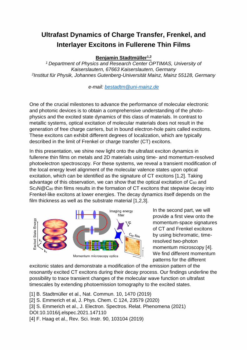
In this presentation, we shine new light onto the ultrafast exciton dynamics in fullerene thin films on metals and 2D materials using time- and momentum-resolved photoelectron spectroscopy. For these systems, we reveal a transient modification of the local energy level alignment of the molecular valence states upon optical excitation, which can be identified as the signature of CT excitons [1,2]. Taking advantage of this observation, we can show that the optical excitation of C60 and Sc3N@C80 thin films results in the formation of CT excitons that stepwise decay into Frenkel-like excitons at lower energies. The decay dynamics itself depends on the film thickness as well as the substrate material [1,2,3].
In the second part, we will provide a first view onto the momentum-space signatures of CT and Frenkel excitons by using bichromatic, time-resolved two-photon momentum microscopy [4]. We find different momentum patterns for the different
excitonic states and demonstrate a modification of the emission pattern of the resonantly excited CT excitons during their decay process. Our findings underline the possibility to trace transient changes of the molecular wave function on ultrafast timescales by extending photoemission tomography to the excited states.
[1] B. Stadtmüller et al., Nat. Commun. 10, 1470 (2019) [2] S. Emmerich et al, J. Phys. Chem. C 124, 23579 (2020) [3] S. Emmerich et al., J. Electron. Spectros. Relat. Phenomena (2021) DOI:10.1016/j.elspec.2021.147110 [4] F. Haag et al., Rev. Sci. Instr. 90, 103104 (2019)

Charge transfer at metal/oxide/organic interfaces M. Sterrer, M. G. Ramsey, P. Puschnig
Institute of Physics, University of Graz, Graz, Austria Charge transfer processes through ultrathin, supported oxide films have received increasing attention in recent years because of the possibility to control the charge state of adsorbates or the direction of catalytic reactions. The main driving force for the occurrence of charge transfer in these systems is the reduction of the substrate work function induced by deposition of the oxide film in combination with an adsorbate of relatively high electron affinity. While previous studies have focused on the charging of metal atoms (e.g. Au) or small molecules (e.g. O2, NO2), we have recently extended these investigations to charge transfer processes into organic molecules. In this contribution, we present results on the adsorption and charging of model organic semiconductors (e.g. pentacene (5A), 2H-tetraphenylporphyrin (2H-TPP) and others) on ultrathin MgO(001) films supported on Ag(001). By combing scanning tunneling microscopy and photoemission spectroscopy and tomography, we identify and quantify charge transfer into the organic monolayer film. In addition, we show that by tuning the work function and/or the MgO thickness it is possible to control: the ratio of charged and neutral species, the number of electrons transferred and, concomitantly, the conformation of the adsorbates. In the case of 2H-TPP, charge transfer also appears to strongly influence the self-metalation of 2H-TPP to Mg-TPP. Thus, our investigations lay the basis for the ultimate control of charge transfer, and the related chemistry, on ultrathin oxide film systems.[1-3] References
[1] L. Egger, M. Hollerer, C. Kern, H. Herrmann, P. Hurdax, A. Haags, X. Yang, A. Gottwald, M. Richter, S. Soubatch, F. S. Tautz, G. Koller, P. Puschnig, M. G. Ramsey, M. Sterrer. Charge-promoted self-metalation of porphyrins on an oxide surface. Angew. Chem. Int. Ed. 60, 5078-5082 (2021).
[2] P. Hurdax, M. Hollerer, L. Egger, G. Koller, X. Yang, A. Haags, S. Soubatch, F. S. Tautz, M. Richter, A. Gottwald, P. Puschnig, M. Sterrer, M. G. Ramsey. Controlling the electronic and physical coupling on dielectric thin films. Beilstein J. Nanotechnol. 11, 1492–1503 (2020).
[3] P. Hurdax, M. Hollerer, P. Puschnig, D. Lüftner, L. Egger, M. G. Ramsey, M. Sterrer. Controlling the charge transfer across dielectric interlayers. Adv. Mater. Interfaces 7, 200592 (2020).

Spin-resolving momentum microscopy:
towards an 'all-in-one' photoemission experimentC. Tusche1 ,2
1Peter Grünberg Institut (PGI-6), Forschungszentrum Jülich, Jülich, Germany2Fakultät für Physik, Universität Duisburg-Essen, Duisburg, Germany
The physical properties of condensed matter systems are largely determined by thedetails of their electronic structure. The concepts of symmetry and topology governthe novel field of quantum materials [1]. Only recently, the comprehensiveexperimental access to the spin-resolved band structure became feasible by spin-resolved momentum microscopy [2]. This novel concept combines high resolutionimaging of the spectral function in two-dimensional (kx, ky) maps of the valenceelectronic structure with an imaging spin filter [3].Spin-resolving momentum microscopy opens a new avenue in photoelectronspectroscopy and paves the way to an “All-in-One” photoemission experiment. Weare able to investigate spin-dependent electronic states and processes in real space(r), reciprocal space (k), as a function of binding energy (E) and time (t). Theaccessible timescales range form several picoseconds down to a few femtoseconds,using pulsed synchrotron radiation [4], free-electron laser [5], and laser-based UVlight sources [6]. This unique combination provides a completely new tool set toinvestigate and disentangle the complex interplay of spin-dependent electronicinteractions and correlations.The comprehensive spin-resolved information of the electronic structure revealsintricate effects of non-local electron correlations in ferromagnets [7] and the mixingof different spin states in topological materials [8]. Moreover, combining strong spin-orbit coupling and magnetism gives rise to complex spin-orbital textures, andtopological phase transitions in the Fermi surface [9].
References
[1] H.L. Meyerheim and C. Tusche, Phys. Status Solidi RRL 12, 1800078 (2018)[2] C. Tusche, A. Krasyuk, J. Kirschner, Ultramicroscopy 159, p. 520 (2015)[3] C. Tusche, et al., Appl. Phys. Lett. 99, 9, 032505 (2011)[4] C. Tusche et al., Appl. Phys. Lett., 108, 261602 (2016)[5] D. Kutnyakhov et al., Rev. Sci. Instrum. 91, 013109 (2020)[6] M. Büscher, R. Adam, C. Tusche, et al., Journal of large-scale research
facilities 6, A138. DOI: 10.17815/jlsrf-6-174 (2020)[7] C. Tusche et al., Nat. Commun. 9, 3727 (2018)[8] Y.-J. Chen, et al., Commun. Phys. 4, 179 (2021)[9] Y.-J. Chen, et al., arXiv:2106.15670 (2021)

Charge transfer at organic/metal interface: combining Photoemission Tomography and X-ray
absorption spectroscopy. Giovanni Zamborlini1
1 TU Dortmund University, Experimental Physics VI, Dortmund, Germany Molecule–substrate interactions play a key role for the spin and charge injection in organic-based devices. Charge transfer at molecule–metal interfaces strongly affect the overall physical and magnetic properties of the system, and ultimately the device performance. In this regard, photoemission tomography (PT) gives an unprecedent insight on the electronic properties of organic/metal interfaces, as it provides direct access to the energy level alignment of the frontier orbitals. However, for tetrapyrrolic compounds, such as porphyrins and phthalocyanines, where a metal is chelated at the macrocycle, PT cannot easily access the electronic structure of their metallic center. On the other hand, X-ray absorption spectroscopy can be used a complementary tool to address the oxidation and the spin state of chelated metal ions as well as to probe the unoccupied electronic states of the tetrapyrrolic complex. In this talk, I will provide a characterization of the interface between Ni-containing tetraphenyl porphyrin (NiTPP) molecules and the copper (100) surface based on PT and XAS. Whereas in the gas-phase the nickel ion is in the formal 2+ oxidation state (Ni(II)), the charge transfer taking place upon the adsorption of NiTPP on the copper surface leads to a stabilization of the Ni(I) species [1] and to a partial filling of the former lowest unoccupied molecular orbitals (LUMOs) up to the LUMO+3 [2,3]. Moreover, the charge transfer changes both spin and oxidation states of the Ni ion from [Ni(II), S=0] to [Ni(I), S=1/2]. The chemically active Ni(I) can be functionalized with nitrogen dioxide allowing selectively tuning the electronic properties of the Ni center, that is changed to [Ni(II), S=1]. While Ni acts as a reversible spin switch, we find that the electronic structure of the macrocycle backbone, where the frontier orbitals are mainly localized, remains unaffected. The presence of the Ni(I) oxidation state at the NiTTP-Cu interface is also responsible for its very high thermal stability [4]. Finally, I will show how the porphyrin film can be decoupled from the Cu surface via insertion of an oxygen layer, restoring the gas-phase Ni(II) oxidations state [5]. These observations make the NiTTP-Cu interface a very intriguing system for applications such as single-atom catalysis and biosensing. References
[1] G. Zamborlini et al., Chem. Commun. 54, 13423 (2018). [2] G. Zamborlini et al., Nat. Commun. 8, 335 (2017). [3] H. M. Sturmeit et al., J. Mater. Chem. C 8, 8876 (2020) [4] H. M. Sturmeit et al., Under Review (2021) [5] I. Cojocariu et al., Appl. Surf. Science 504, 144343 (2020).


New Strategies for Probing Local Orbital and TopologicalProperties of Solids using ARPES
S. Beaulieu1, M. Schüler2, J. Schusser3,4, S. Dong5, T. Pincelli5, M. Dendzik6,J. Maklar5, A. Neef5, H. Ebert7, K. Hricovini8,9, F. Reinert3, T.P. Devereaux2,
J. Braun7, M. Wolf5, L. Rettig5, J. Minar4, and R. Ernstorfer5,10
1Université de Bordeaux - CNRS - CEA, CELIA, UMR5107, F33405 Talence, France2Stanford Institute for Materials and Energy Sciences (SIMES), SLAC National
Accelerator Laboratory, Menlo Park, CA 94025, USA3Experimentelle Physik VII and Würzburg-Dresden Cluster of Excellence ct.qmat,
Universität Würzburg, Würzburg, Germany4New Technologies-Research Center, University of West Bohemia, 30614 Pilsen,
Czech Republic5Fritz Haber Institute of the Max Planck Society, Faradayweg 4-6, 14195 Berlin,
Germany6Department of Applied Physics, KTH Royal Institute of Technology, Hannes Alfvens
vag 12, 114 19 Stockholm, Sweden7Department Chemie, Ludwig-Maximilians-Universität München, Butenandtstrasse
11, 81377 München, Germany8Laboratoire de Physique des Matériaux et Surfaces, CY Cergy Paris Université,
95031 Cergy-Pontoise, France9LIDYL, CEA, CNRS, Université Paris-Saclay, CEA Saclay, F-91191 Gif-sur-Yvette
Cedex, France10Institut für Optik und Atomare Physik, Technische Universität Berlin, 10623 Berlin,
Germany
Angle-resolved photoemission spectroscopy - ARPES - is the most powerful
technique to investigate the electronic eigenvalues (band structure) of crystalline
solids. To completely characterize the electronic structure of (topological) materials,
one needs to go beyond band structure mapping and probe the orbital texture,
associated with Berry curvature and topological invariants. I will present new
measurement methodologies in ARPES based on i) crystal rotation mimicking time-
reversal symmetry operation and ii) continuous modulation of the ionizing radiation
polarization axis. I will show how such manipulation of the photoemission transition
dipole matrix elements, complemented by minimal theory inputs, allows accessing i)
the momentum-dependent orbital texture [1-2] and ii) the complex Bloch
wavefunction [3], here exemplified for transition metal dichalcogenides - TMDCs.
These results represent an important step towards going beyond band structure
(eigenvalues) mapping and learn about electronic wavefunction and orbital texture of
solids by exploiting matrix element effects in photoemission spectroscopy.
References[1] Beaulieu et al., Phys. Rev. Lett. 125, 216404 (2020) [arXiv :2006.01657]
[2] Beaulieu et al., npj Quantum Materials - accepted (2021) [arXiv:2107.07158]
[3] Schüler, ..., Beaulieu [arXiv:2103.17168] (2021)
Ultrafast time-resolved orbital tomography of optically excited states using time-of-flight
momentum microscopy with a HHG light source W. Bennecke1, D. Schmitt1, J. P. Bange1, D. Steil, D. R. Luke2, M.
Reutzel1, S. Steil1, G. S. M. Jansen1, S. Mathias1 11. Physical Institute, Georg-August University Göttingen, Germany
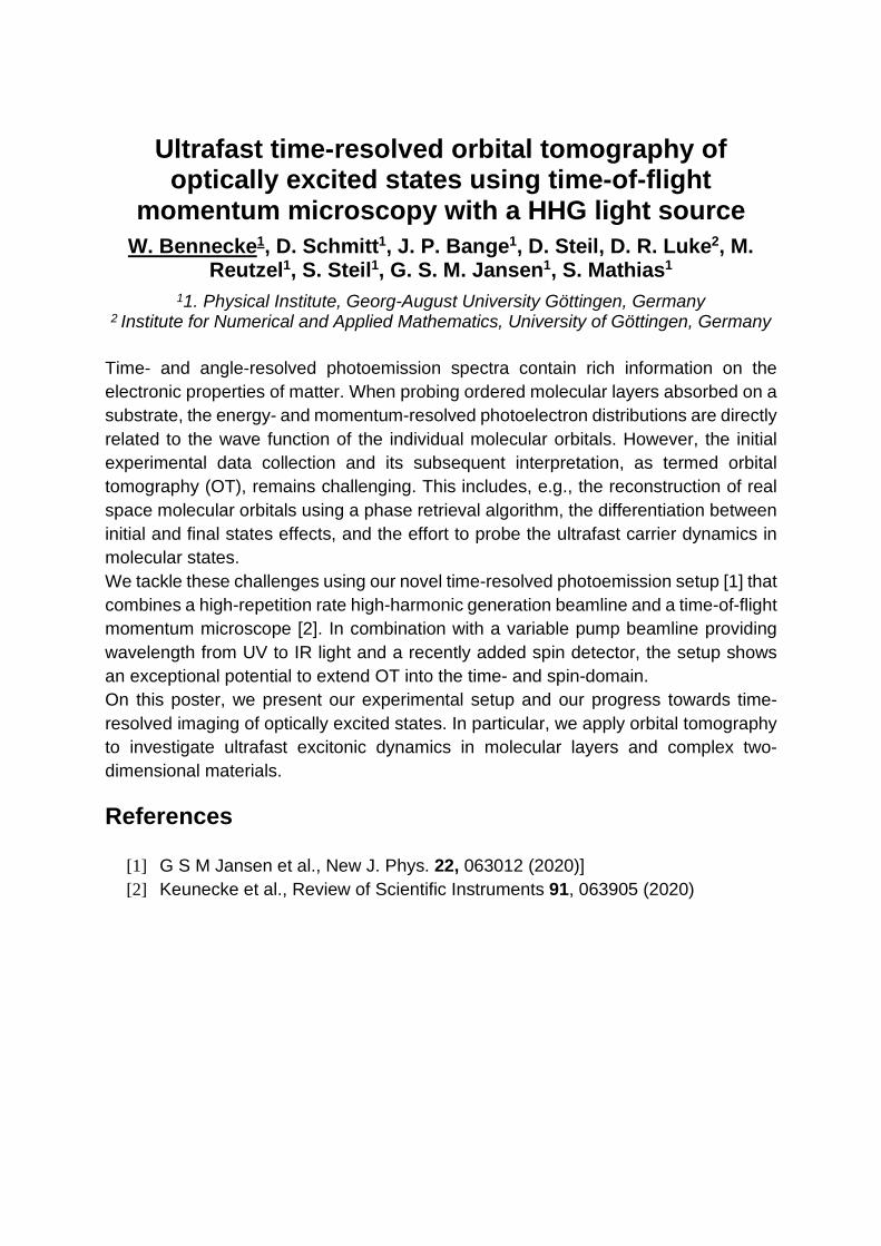
2 Institute for Numerical and Applied Mathematics, University of Göttingen, Germany Time- and angle-resolved photoemission spectra contain rich information on the electronic properties of matter. When probing ordered molecular layers absorbed on a substrate, the energy- and momentum-resolved photoelectron distributions are directly related to the wave function of the individual molecular orbitals. However, the initial experimental data collection and its subsequent interpretation, as termed orbital tomography (OT), remains challenging. This includes, e.g., the reconstruction of real space molecular orbitals using a phase retrieval algorithm, the differentiation between initial and final states effects, and the effort to probe the ultrafast carrier dynamics in molecular states. We tackle these challenges using our novel time-resolved photoemission setup [1] that combines a high-repetition rate high-harmonic generation beamline and a time-of-flight momentum microscope [2]. In combination with a variable pump beamline providing wavelength from UV to IR light and a recently added spin detector, the setup shows an exceptional potential to extend OT into the time- and spin-domain. On this poster, we present our experimental setup and our progress towards time-resolved imaging of optically excited states. In particular, we apply orbital tomography to investigate ultrafast excitonic dynamics in molecular layers and complex two-dimensional materials. References
[1] G S M Jansen et al., New J. Phys. 22, 063012 (2020)] [2] Keunecke et al., Review of Scientific Instruments 91, 063905 (2020)

Interplay of Organo-Metallic Interface Charge Transfer and Adsorbate Orientation
Thomas Georg Boné†*, Andreas Windischbacher†, Marie S. Sättele‡¶, Katharina Greulich‡, Larissa Egger†, Thomas Jauk§, Florian Lackner§, Holger F.
Bettinger¶, Heiko Peisert‡, Thomas Chassé‡, Mike Ramsey†, Martin Sterrer†, Georg Koller† and Peter Puschnig†
†Institute of Physics, University of Graz, Graz, Austria ‡ Institute of Physical and Theoretical Chemistry, University of Tübingen, Germany ¶ Institute of Experimental Physics, Graz University of Technology, Graz, Austria
§ Institute of Organic Chemistry, University of Tübingen, Tübingen, Germany *E-Mail: [email protected]
Thin layers of acenes on metals like copper or silver are discussed to induce complex electronic characteristics, depending on factors like substrate metal workfunction and molecule adsorbate electron affinity [1]. In this study the growth and energy level alignment of the rod-like, long chain acene heptacene (7A) on a copper(110) (Cu(110)) surface is evaluated. Our results show that the orientation of the 7A molecules can be controlled by the preparation conditions. Photoemission tomography is used to investigate molecular geometries and assign specific emissions to molecule orbitals [2]. Thermal cycloreversion of diheptacene isomers during evaporation produces highly oriented and epitaxial monolayers of heptacene on the metal surface. The molecules are oriented either along or perpendicular to the close-packed metal rows of Cu(110). Our combined experimental and computational results show that for heptacene oriented along the Cu rows, the lowest unoccupied molecular orbital (LUMO) and the LUMO+1 are occupied. The LUMO+1 receives no charge for molecules aligned perpendicular to the Cu rows. The possibility to tune the energy level alignment and charge transfer at organic-metal interfaces by means of adjustable molecular alignment is fully corroborated by our experiments and density functional calculations. Acknowledgment Financial support through FWF (FWF project number I 4145) References [1] Hollerer, Michael, et al. “Charge Transfer and Orbital Level Alignment at
Inorganic/Organic Interfaces: The Role of Dielectric Interlayers.” ACS Nano, vol. 11, no. 6, 2017, pp. 6252–6260.
[2] Hurdax, Philipp, et al. “Controlling the Electronic and Physical Coupling on Dielectric Thin Films.” Beilstein Journal of Nanotechnology, vol. 11, no. 1, 2020, pp. 1492–1503.

kMap.py: A Python program for simulation and dataanalysis in photoemission tomography
D. Brandstetter 1, X. Yang1,2,3,4, D. Lüftner1, F. S. Tautz2,3,4, P.Puschnig1
1Karl-Franzens-Universität Graz, Institut für Physik, 8010 Graz, Austria2Peter Grünberg Institut (PGI-3), Forschungszentrum Jülich, 52425 Jülich, Germany
3Jülich Aachen Research Alliance (JARA), Fundamentals of Future InformationTechnology, 52425 Jülich, Germany
4Experimentalphysik IV A, RWTH Aachen University, 52074 Aachen, Germany
kMap.py is a Python program that enables the user, via a PyQt-based graphical userinterface, to simulate photoemission momentum maps of molecular orbitals and toperform a one-to-one comparison between simulation and experiment. Based on theplane wave approximation for the final state, simulated momentum maps arecomputed numerically from a fast Fourier transform (FFT) of real space molecularorbital distributions which are used as program input and which are usually obtainedfrom density functional calculations. The user can vary a number of simulationparameters such as the final state kinetic energy, the molecular orientation or thepolarization state of the incident light field. Moreover, also experimentalphotoemission data can be loaded into the program, enabling a direct visualcomparison as well as an automatic optimization procedure to minimize thedifference between simulated and measured momentum maps. Thereby, structuralparameters of the molecules and the weights of molecular orbitals to experimentallyobserved emission features can be determined.kMap.py provides an easy-to-use Python API for its core features and datastructures, making it possible to employ them for similar custom analysis scriptscircumventing the GUI entirely. As a pure Python package, kMap.py has beenpublished on PyPI and is thus simple to install on all major platforms.
References[1] D. Brandstetter et al., Computer Physics Communications 263, 107905 (2021)

Ordered assemblies of bis-perylene derivatives on metal single crystals
Giovanni Di Santo1, Tanja Miletic2, Mathias Schwendt3, Luca Floreano4, Andrea Goldoni1, Peter Puschnig3, Luca Petaccia1 and
Davide Bonifazi5 1Sincrotrone Trieste S.C.p.A. s.s.14 Km. 163.5, 34149 - Trieste, Italy
2School of Chemistry, Cardiff University, Park Place, CF10 3AT, Cardiff (UK) 3Institute of Physics, University of Graz, NAWI Graz, 8010 Graz, Austria
4CNR-IOM Lab. TASC in Area Science Park, s.s. 14, Km 163.5 - 34149 Trieste, Italy 5Institute of Organic Chemistry, Faculty of Chemistry, University of Vienna ,
Universitätsring 1 - 1010 Vienna, Austria The assembly of molecular building blocks on crystalline surfaces into ordered nano–architectures is an established approach towards the creation of novel materials with outstanding properties. The surface science methodology allows to investigate the morphology of novel molecular structures in the monolayer or sub-monolayer regime (e.g. self-assembled long range ordered 2D networks), and their electronic properties (e.g. density of states, charge transfer, HOMO-LUMO coupling). Eventually it may lead to a clear understanding of the molecule–to–substrate adsorption geometry together with the interaction between the first layer and the hosting surface. Recently reported O-doped polycyclic aromatic hydrocarbons (PAHs) bearing pyranopyranil or furanyl core showed exceptionally high emission yields and tunable optoelectronic properties, resulting as appealing candidates for photoelectronic applications.[1] Molecular derived electronic states of this class of extended molecule can be disclosed and revealed with the ARPES based momentum mapping methodology[2] with the support of complementary techniques such as Scanning Tunneling Microscopy (STM) and Low Energy Electron Diffraction (LEED). That allowed to shed light on most of the unknown of these π-conjugated systems sorting out the adsorbates electronic structure and their geometric arrangement. Molecular patterns of a specific group of bis-Perylen derivatives are self-assembling on Cu(111) forming ordered extended domains and expressing the mutual recognition of molecules with opposite chirality. Similar assemblies on Au(111) have been studied following the doping route with alkali metal. Subsequent Potassium (K) evaporations have filled the empty molecular states. References
[1] T. Miletic, A. Fermi, I. Orfanos, A. Avramopoulos, F. De Leo, N. Demitri, G. Bergamini, P. Ceroni, M.G. Papadopoulos, S. Couris, D. Bonifazi, Chemistry A European Journal 23, 2363–2378 (2017)
[2] P. Puschnig, S. Berkebile, A.J. Fleming, G. Koller, K. Emtsev, T. Seyller, J.D. Riley, C. Ambrosch-Draxl, F.P. Netzer, M.G. Ramsey. Science 326, 702–706 (2009)
Reciprocal-space imaging of σ-orbitals for chemical analysis Anja Haags1,2,3, Xiaosheng Yang1,2,3, Larissa Egger4, Dominik Brandstetter4, Hans Kirschner5, François C. Bocquet1,2, Georg Koller4, Alexander Gottwald5, Mathias Richter5, J. Michael Gottfried6, Michael G. Ramsey4, Peter Puschnig4, Serguei Soubatch1,2 and F. Stefan Tautz1,2,3
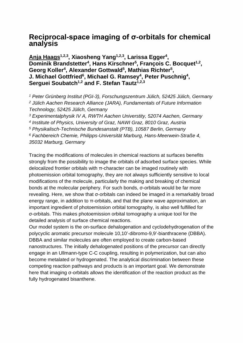
1 Peter Grünberg Institut (PGI-3), Forschungszentrum Jülich, 52425 Jülich, Germany 2 Jülich Aachen Research Alliance (JARA), Fundamentals of Future Information Technology, 52425 Jülich, Germany 3 Experimentalphysik IV A, RWTH Aachen Universtity, 52074 Aachen, Germany 4 Institute of Physics, University of Graz, NAWI Graz, 8010 Graz, Austria 5 Physikalisch-Technische Bundesanstalt (PTB), 10587 Berlin, Germany 6 Fachbereich Chemie, Philipps-Universität Marburg, Hans-Meerwein-Straße 4, 35032 Marburg, Germany Tracing the modifications of molecules in chemical reactions at surfaces benefits strongly from the possibility to image the orbitals of adsorbed surface species. While delocalized frontier orbitals with π-character can be imaged routinely with photoemission orbital tomography, they are not always sufficiently sensitive to local modifications of the molecule, particularly the making and breaking of chemical bonds at the molecular periphery. For such bonds, σ-orbitals would be far more revealing. Here, we show that σ-orbitals can indeed be imaged in a remarkably broad energy range, in addition to π-orbitals, and that the plane wave approximation, an important ingredient of photoemission orbital tomography, is also well fulfilled for σ-orbitals. This makes photoemission orbital tomography a unique tool for the detailed analysis of surface chemical reactions. Our model system is the on-surface dehalogenation and cyclodehydrogenation of the polycyclic aromatic precursor molecule 10,10’-dibromo-9,9’-bianthracene (DBBA). DBBA and similar molecules are often employed to create carbon-based nanostructures. The initially dehalogenated positions of the precursor can directly engage in an Ullmann-type C-C coupling, resulting in polymerization, but can also become metalated or hydrogenated. The analytical discrimination between these competing reaction pathways and products is an important goal. We demonstrate here that imaging σ-orbitals allows the identification of the reaction product as the fully hydrogenated bisanthene.
Photoemission tomography of excitonic states in molecular materials by time-resolved two-photon
momentum microscopy R. Hemm1, M. Mitkov1, F. Haag1, K. M. Yu1, T. Feuerbach1, M.
Aeschlimann1, and B. Stadtmüller2
1University of Kaiserslautern (TUK) and Research Center OPTIMAS, Erwin-Schrödinger-Str. 46, 67663 Kaiserslautern, Germany
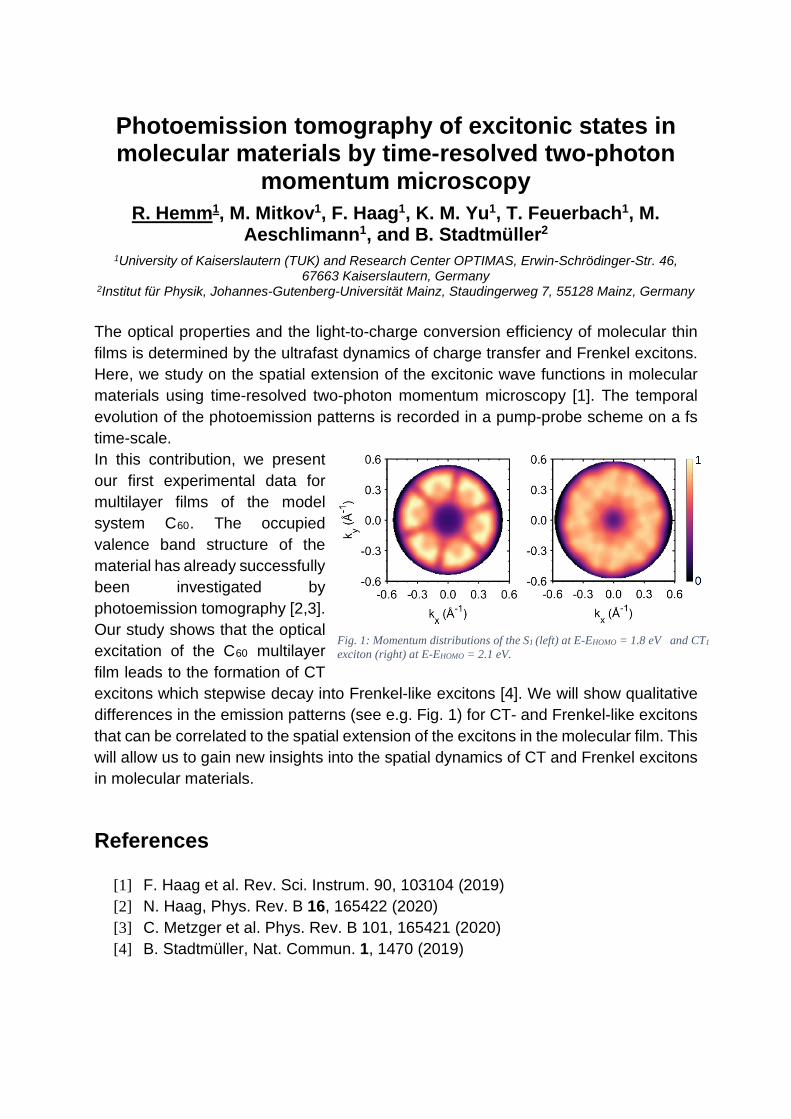
2Institut für Physik, Johannes-Gutenberg-Universität Mainz, Staudingerweg 7, 55128 Mainz, Germany The optical properties and the light-to-charge conversion efficiency of molecular thin films is determined by the ultrafast dynamics of charge transfer and Frenkel excitons. Here, we study on the spatial extension of the excitonic wave functions in molecular materials using time-resolved two-photon momentum microscopy [1]. The temporal evolution of the photoemission patterns is recorded in a pump-probe scheme on a fs time-scale. In this contribution, we present our first experimental data for multilayer films of the model system C60. The occupied valence band structure of the material has already successfully been investigated by photoemission tomography [2,3]. Our study shows that the optical excitation of the C60 multilayer film leads to the formation of CT excitons which stepwise decay into Frenkel-like excitons [4]. We will show qualitative differences in the emission patterns (see e.g. Fig. 1) for CT- and Frenkel-like excitons that can be correlated to the spatial extension of the excitons in the molecular film. This will allow us to gain new insights into the spatial dynamics of CT and Frenkel excitons in molecular materials. References
[1] F. Haag et al. Rev. Sci. Instrum. 90, 103104 (2019) [2] N. Haag, Phys. Rev. B 16, 165422 (2020) [3] C. Metzger et al. Phys. Rev. B 101, 165421 (2020) [4] B. Stadtmüller, Nat. Commun. 1, 1470 (2019)
Fig. 1: Momentum distributions of the S1 (left) at E-EHOMO = 1.8 eV and CT1 exciton (right) at E-EHOMO = 2.1 eV.

REF [1] Y.Kuroda et al 2019 JJAP. 58 SIIB27
Momentum microscopy of highly oriented organic thin films Masato Iwasawa1, Fumihiko Matsui2, Satoshi Kera2, and Yoichi Yamada1 1 Institute of Applied Physics, University of Tsukuba, Tsukuba, Japan
2 Institute for Molecular Science, Okazaki, Japan E-mail: [email protected]
Introduction We investigated the Photoelectron Momentum Maps (PMM) of the organic
semiconductor molecules with extremely high mobility, such as Picene and [1]benzothieno[3,2-b] [1]benzothiophene (BTBT), in order to clarify the mechanisms of the electric conduction in detail. Although the mobility of many organic semiconductors, such as picene, has been understood by the energy and the spatial distribution of HOMO, a recent study has shown that the contribution of HOMO-1 is significant in BTBT derivatives[1]. In this contribution, we compare the PMMs of Picene and DPh-BTBT to discuss the origin of the different conduction mechanism. Experiment
For PMM measurements, we fabricated multilayer films of these molecules with relatively simple structure, utilizing the anisotropic Ag(110) surface. The molecular structure of the films were examined using a scanning tunneling microscope (STM) and low energy electron diffraction (LEED). The PMM measurements were performed using momentum microscope at UVSOR BL6U. Result & Discussion
Fig.1(a, c) shows the STM images of Picene and DPh-BTBT multilayer films on Ag(110). On both films, the molecular long axis is nearly parallel to the [11
_
0] direction of the substrate, suitable for the PMM measurement.
Fig.1(b, d) shows the angle-integrated energy distribution curves (EDCs) and PMMs of Picene and DPh-BTBT film on Ag(110). In the case of Picene, HOMO and HOMO-1 are energetically overlapped, while the energetic overlapping between HOMO and HOMO-1 is not very significant in the case of DPh-BTBT. However, in DPh-BTBT, each orbital is found to be broadened and fitted with two Gaussian functions, suggesting that orbital splitting due to the overlap of neighboring molecules. In PMM, the HOMO and HOMO-1 of picene showed a clearly differen distribution, which corresponds well to the Fourier transform (FT) of isolated picene. On the other hand, the PMM of DPh-BTBT showed similar distribution in HOMO and HOMO-1, and they are not completely reproduced by the FT of isolated DPh-BTBT, suggesting the significant hybridization of the frontier orbitals in the film.

Co porphyrins deposited on the passivated Fe(100)-p(1x1)O surface: a photoemission tomography study
David Janas1, Henning Sturmeit1, Iulia Cojocariu2, Vitaliy Feyer2, Stefano Ponzoni1, Giovanni Zamborlini1, and Mirko Cinchetti1
1Experimentelle Physik VI, TU Dortmund, Otto-Hahn-Straße 2, 44227 Dortmund, Germany
2Peter Grünberg Institut (PGI-6), Forschungszentrum Jülich, Leo-Brandt-Straße, 52425 Jülich, Germany
In order to enhance the performance of molecular spintronic devices, a detailed knowledge of the physical properties at the interface is crucial [1]. In this context photoemission tomography (PT), which combines ab-initio calculations and angle-resolved photoelectron spectroscopy (ARPES), stands out as a powerful and reliable tool to unravel the orbital structure of molecular thin films at metal/organic interfaces [2]. In this work we apply PT to investigate the interaction of paramagnetic molecules, namely CoTPP, on the passivated Fe(100)-p(1x1)O surface. Recently, it was shown that the interactions at this interface lead to an emergent magnetic ordering of the magnetic layer [3]. Here, we use PT to further characterize this intriguing system. In particular, we determine the energy level alignment at the interface, the nature of the frontier orbitals, and the azimuthal orientation of the CoTPP molecules with respect to the substrate. References
[1] Cinchetti, M., Dediu, V. Hueso, L., Activating the molecular spinterface. Nature Mater 16, 507515 (2017)
[2] Puschnig, P. et al., Reconstruction of molecular orbital densities from photoemission data. Science 326, 702706 (2009)
[3] M. S. Jagadeesh et al., Room temperature magnetism of ordered porphyrin layers on Fe. Appl. Phys. Lett. 115, 082404 (2019)
Photoemission Tomography on the Time-Domain: Simulation of Photoelectron Spectroscopy from
Time-Dependent Density Functional Theory C. S. Kern1, X. Yang2, U. De Giovannini3,4, S. Subach2, F. S. Tautz2, A. Rubio3,5, M. Ramsey1, Peter Puschnig1
1 Institute of Physics, NAWI Graz, University of Graz, 8010 Graz, Austria 2 Peter Grünberg Institut (PGI-3), Forschungszentrum Jülich, 52425 Jülich, Germany 3 Max Planck Institute for the Structure and Dynamics of Matter and Center for Free-
Electron Laser Science, 22761 Hamburg, Germany 4IKERBASQUE, Basque Foundation for Science, E-48011 Bilbao, Spain
5Center for Computational Quantum Physics, The Flatiron Institute, New York, USA
Angular-Resolved Photoelectron Spectroscopy (ARPES) can benefit greatly from theoretical simulations as the observed momentum-space signature of the electronic structure is often quite involved. With the recent developments in ultra-fast laser physics, ARPES is now being used to also investigate time-resolved phenomena, e.g. for excited-states (pump-probe experiments). In this contribution, we use real-time Time-Dependent Density Functional Theory to simulate time-resolved ARPES [1]. Accounting for dynamical processes directly is an advance over established methods and reproduces experimental findings such as circular dichroism in organic molecules such as Tetracene and Pentacene. The method also gives results for the photonenergy dependence of ARPES intensities that compare well to experimental findings, which will be shown for a graphene monolayer. Furthermore, we present how this method can be used to directly observe excitations in the electronic structure in time. References
[1] De Giovannini et. al., Journal of Chemical Theory and Computation 13, 265-273 (2017)
Photoemission Tomography on Photoionization Cross Sections from s-States of Noble Gases
H. Kirschner1, A. Gottwald1, M. Richter1 1Physikalisch-Technische Bundesanstalt, Abbestr. 2-12, 190587 Berlin, Germany
Photoemission tomography (PT) gives information about the interaction of the initial and the final state of a photoelectron, according to Fermi’s Golden rule. Assuming a
basic plane wave as the final state, the initial state can be reconstructed by applying a Fourier Transformation (FT) to the measured orbital representation in momentum space. We investigate if both the theoretical and the experimental parts of the PT might be generalized to the photoemission from atoms in gas phase, characterized by photoionization cross section. Hence, the initially introduced FT is exchanged by an integral transformation, which serves the spherical symmetry of atoms better than the plane symmetry of molecule layers. Additionally, the Coulomb field of the remaining ion must be considered. These two properties are merged into a so-called Coulomb wave function, which is used as the final state instead of a plane wave description. A numerical integral transformation is implemented. Thus, Slater type orbitals can be applied for the initial state and are fitted to photoionization cross section data, taken from literature. The measurements for the PT are conducted with a toroidal analyser, which uses the radiation of PTB’s insertion device beamline (IDB) at the Metrology Light Source (MLS). For measuring photoemission from gas phase, the analyser is modified with a gas inlet in form of a capillary tube. The measured k-maps will be matched to the data from literature, in order to show that also the experimental method of PT can be applied to gas phase experiments.

1-1’-Bitetracene – a Precursor Molecule for a Surface-Assisted Cyclodehydrogenation to
Peritetracene Maren Klein1, Marie S. Sättele1, Holger F. Bettinger2, Heiko Peisert1,
Thomas Chassé1 1Institut für Physikalische und Theoretische Chemie, Tübingen, Germany
2Institut für Organische Chemie, Tübingen, Germany
Acenes and peri-acenes can be considered as zigzag shaped nanographene units with noble electronic properties for future electronic devices[1]. Peri-acenes are formed in bottom-up fabrications via on-surface synthesis[2,3]. This organic synthesis based on precursor molecules can achieve band-gap tuning of nanographene’s. We successfully synthesized 1,1’-Bitetracene for the first time and deposited it on a Cu (110) substrate. This unique molecule undergoes a surface-assisted cyclodehydrogenation to peri-tetracene on this surface. The Scholl reaction was investigated by the complementary techniques: Scanning tunneling microscope (STM), X-ray photoelectron spectroscopy (XPS) and density functional calculations (DFT).
Figure 1: a) Scheme for the surface-assisted reaction of 1-1’-bitetracene to peritetracene. b) C1s core level spectra monolayer (bottom), heated monolayer (middle) and multilayer (top) bitetracene on Cu (110). According to DFT calculations all spectra can be essentially described by three components. c) STM microscopic picture of heated bitetracene (peritetracene) on Cu (110). One unit has the diameters indicated in the Figure.
References: [1] Anthony, J. E. Functionalized acenes and heteroacenes for organic electronics.
Chem Rev 106, 5028-5048, doi:10.1021/cr050966z (2006). [2] Talirz, L., Ruffieux, P. & Fasel, R. On-Surface Synthesis of Atomically Precise
Graphene Nanoribbons. Adv Mater 28, 6222-6231, doi:10.1002/adma.201505738 (2016).
[3] Rogers, C. et al. Closing the Nanographene Gap: Surface-Assisted Synthesis of Peripentacene from 6,6'-Bipentacene Precursors. Angew Chem Int Ed Engl 54, 15143-15146, doi:10.1002/anie.201507104 (2015).
C36H1610 : 6 : 2
C36H227 : 7 : 4
Peri-tetracene
Bitetracene
c)Peritetracene
d(H-H) = 1.164 nm
d(H-
H) =
0.9
937
nm
250 °C / 30 min
a)

Implementation of a polychromatic beamline for time-resolved two-photon momentum microscopy
Martin Mitkov1, Ralf Hemm1, Tobias Feuerbach1, Florian Haag1, Ka Man Yu1, Martin Aeschlimann1, Benjamin Stadtmüller1,2
1 Department of Physics and Research Center OPTIMAS, University of Kaiserslautern, 67663 Kaiserslautern, Germany
2 Institut für Physik, Johannes-Gutenberg-Universität Mainz, Mainz 55128, Germany E-mail: [email protected]
Time-resolved two-photon photoelectron spectroscopy is an ideal tool for mapping the ultrafast dynamics of excited states in a large variety of material systems. Thereby, different excited states can be addressed by tuning the photon energy of optically exciting light pulses to the energy of the corresponding resonant optical transition. This is particularly important for molecular materials which exhibit a manifold of localized excited states at well-defined energies. Here, we present our latest progress in the implementation of a polychromatic time-resolved two-photon momentum microscopy experiment [1]. For the optical excitation, we can select either the beam of the second harmonic radiation (2.95 eV) of a Ti:Sa oscillator or of an optical parametric oscillator (OPO) that is seeded by a Ti:Sa oscillator. The photon energy of the OPO beam can be tuned between 1.8 and 2.3 eV. In both cases, the transient population of the excited states can be probed by the fourth harmonic radiation (5.9 eV) of the Ti:Sa oscillator. The large photon energy of the probe pulses together with the exceptionally large acceptance angle of the momentum microscopy detector system allows us to image the momentum space signatures of excited molecular states in a large range of momenta on ultrafast timescales. We show first results of optically excited states in C60 multilayer films on Cu(111) that are a first demonstration of the capabilities and performance of our experimental setup. References
[1] F. Haag et al., Rev. Sci. Instr. 90, 103104 (2019)

Orbital-resolved observation of singlet fission Alexander Neef1 (Presenter), Samuel Beaulieu3, Sebastian Hammer2,
Shuo Dong1, Tommaso Pincelli1, Julian Maklar1, Martin Wolf1, Laurenz Rettig1, Jens Pflaum2 and Ralph Ernstorfer1
1Fritz Haber Institute of the Max Planck Society, Faradayweg 4-6, 14195 Berlin,
Germany
2Experimental Physics VI, Julius- Maximilian University Wuerzburg, 97074
Wuerzburg, Germany
3Université de Bordeaux - CNRS - CEA, CELIA, UMR5107, F33405, Talence, France
Great strides have been made in observing ultrafast molecular processes on their intrinsic length and time scales. Some techniques directly image molecular motion [1], while others access spatial information in momentum space [2]. Ideally, these techniques should allow to follow the dynamics of electrons and atoms in time, space and energy. Experiments so far have shown potential but exploiting them to unravel major mysteries in ultrafast molecular processes has not been achieved. Here we investigate ultrafast singlet fission – the process of a singlet exciton splitting into two triplet excitons [3] – in crystalline pentacene by using time- and angle-resolved photoemission spectroscopy. Using insights from orbital tomography, we were able to match singlet and triplet excitons to their orbital character based on their respective momentum maps. Furthermore, we separated the dynamics of states with different orbital character and thereby gained mechanistic insights about singlet fission.
References
[1] Cocker, Peller et al., Nature 539, 263–267 (2016)[2] Wallauer et al., Science 371, 1056-1059 (2021)[3] Smith & Michl, Chem. Rev. 110, 6891–6936 (2010)

Analysis of pump–probe ultrafast photoemission M. Haniuda, M. Nozaki, and K. Niki
Gradurate School of Science and Engineering, Chiba, Japan
Detailed studies using femtosecond time-resolved X-ray photoemission spectroscopy (TR-XPS) on organic donor-acceptor systems have been reported. The femtosecond TR-XPS technique provides direct, real-time access to important relaxation channels. According to Roth et al., in the CuPc:C60 system, the triplet states of CuPc play an important role in charge separation at donor/acceptor interfaces [1]. We proposed a theory to analyze TR-XPS spectrum based on the Keldysh
Green's function approach [2]. This theory allows us to calculate these time-dependent Dyson orbitals and their natural amplitudes. The time-dependent photoemission intensity Ik detecting photo-electrons with momentum k is given
(1)
Where 𝑓𝑓𝒌𝒌−∗ and ∅C are final and initial core state. A(1) is electron-photon interaction by probe X-ray at time t1. The time-dependent intrinsic amplitude Sm(t1) describes those pump-probe processes
(2)
𝑈𝑈 �(t1,t0) is time evolution operator contains the effect of pump light between times t0 and t1. This term depends on the transition probability from the ground state ( |0>) to excited states (|l>) with the energy 𝐸𝐸l . Photoelectrons are emitted by probe light ( b that annihilates an electron at x). |m*> describes m-th hole state with 𝐸𝐸m
∗ . This term includes the core hall effect. As a result, it is possible to calculate the intensities of C1s photoemission under the excited state. In this presentation, we have estimated the dependence of XPS intensities
after pump excitation on the transition probability and the number of electrons in the valence band. References [1] R. Friedrich et al., NAT. COMMUN. 12, 1196 (2021).[2] T. Fujikawa and K. Niki J. Electron Spectros. Relat. Phenom., 206, 74
(2016).

Molecular level alignment at the NiTPP/O-Cu(100) interface
Jonah Elias Nitschke1, Henning Sturmeit1, Iulia Cojocariu2, Vitaliy Feyer2, Alessandro Sala2, Stefano ponzoni1, Giovanni Zamborlini1,
Mirko Cinchetti1 1 Experimentelle Physik VI, TU Dortmund, Otto-Hahn-Straße 2, 44227 Dortmund,
Germany 2 Peter Grünberg Institut (PGI-6), Forschungszentrum Jülich, Leo-Brandt-Straße,
52425 Jülich, Germany
The last decades have witnessed an increasing interest for molecular-based electronics. Molecular building blocks offer the ability to design specific molecular components with tailored electronic and chemical properties. A key method to get a deeper understanding of the various interactions and effects is photoemission tomography (PT), which provides access to the energy landscape of the interface and allows the identification of single molecular orbitals [1]. I will present recent insights on the coupling/decoupling between Nickel tetraphenylporphyrin molecules (NiTPP) and a transition-metal surface. NiTPPs form long-range ordered structures on a clean Cu(100) surface, whereas the strong molecule metal interaction leads to the population of formerly unoccupied molecular states (LUMOs) [2]. I will show how this interaction can be suppressed by decoupling the NiTPP from the Cu surface with an oxygen interlayer. By using a multi-technique approach, which combines low energy electron diffraction and scanning tunnelling microscopy, we determine the unit cell of the NiTPP molecules on the oxidized Cu(100). The STM images reveal a coverage dependent superstructure with multiple domains that reduces into a single domain for high coverage with two different adsorption geometries of the NiTPP molecules. Additional PT measurements unveil two molecular states (HOMO and HOMO-1), of which the DOS could be extracted by fitting theoretical calculations to the observed images. Both orbitals are split in two components separated by 0.4 eV, which could be caused by the two different adsorption configurations. Furthermore, the absence of charge transfer confirms the oxygen interlayer as a good opportunity to achieve a physical decoupling of the molecules and the substrate. Additionally, the full energy level alignment at the interface can be probed by means of 2 photon photoemission that provide access to the unoccupied molecular states above the Fermi level. References [1] Puschnig et al. Phys.Rev.B 84, 235427, 2011 [2] Zamborlini et al. Nature Communications 8.1, 335, 2017

Restoring the molecular properties: K intercalation of the flexible DBP on Ag(111)
Felix Otto1, Tino Kirchhuebel1, Anu Baby2, Falko Sojka1, Guido Fratesi3, Roman Forker1, Torsten Fritz1
1 Institute of Solid State Physics, Friedrich Schiller University Jena, Germany 2 Department of Materials Science, University of Milano-Bicocca, Italy
3 ETSF and Dipartimento di Fisica “Aldo Pontremoli”, Università degli Studi di Milano, Italy
The polycyclic aromatic hydrocarbon tetraphenyldibenzoperiflanthene (DBP) has advantageous properties for applications in the field of organic electronics. These are a consequence of its chemical structure consisting of an aromatic backbone with four phenyl rings nearly perpendicular to the molecular plane. We are interested in the effect of potassium intercalation on the structural, optical, and electronic properties of DBP adsorbed on Ag(111). Several structurally reordered stable bonding stages were obtained with K intercalation. LEED and STM measurements combined with differential reflectance spectroscopy (DRS) as well as XPS and UPS including photoelectron momentum maps (PMMs) were used to investigate the system and yield a variety of different information. These results can only be interpreted unambiguously with the help of DFT calculations. We find that the added K atoms adsorb below the molecule and thereby obstruct the bonding channels between DBP and Ag. These alterations of the rather complex host molecule - guest atom - substrate system lead to severe changes of the electronic structure. The strong metal-organic hybridization of DBP and K2DBP yields PMMs which cannot be explained by assuming free molecules. In contrast, K6DBP essentially behaves like an electronically decoupled monolayer of DBP dianions. [1] References
[1] F. Otto, J. Phys. Chem. C 124, 4114-4127 (2020).

Tracing orbital images on ultrafast time scales: The PTCDA/Cu(001)-2O-system
M. Raths, S. Soubatch, X. Yang, C. Kumpf, F. C. Bocquet, F. S. Tautz Peter Grünberg Institut (PGI-3), Forschungszentrum Jülich, Germany R. Wallauer, K. Stallberg, L. Münster, J. Güdde, U. Höfer
Department of Physics, Philipps-Universität Marburg, Germany D. Brandstetter, P. Puschnig
Institute of Physics, University of Graz, Austria e-mail:[email protected]
We combined time-resolved photoemission (using high laser harmonics) and time-of-flight momentum microscopy to establish a femtosecond pump-probe photoemission tomography experiment of unoccupied molecular orbitals. The full momentum-space distribution of electrons, transiently excited into the lowest unoccupied molecular orbital (LUMO), was measured [1]. The selection of the sample system was crucial for this experiment: On the one hand, photoemission tomography requires a sufficiently corrugated substrate that promotes a small number of well-defined azimuthal orientations of the adsorbed molecules. On the other hand, the molecules must be electronically decoupled from the substrate since electrons excited into otherwise unoccupied states shall be (photo)emitted. The PTCDA/Cu(001)-2O-system fulfills both conditions: The ultrathin oxide layer provides a �√2 × 2√2�𝑅𝑅45° -2O reconstructed surface imposing only two, clearly defined azimuthal orientations of PTCDA (see Fig. 1), and at the same time suppresses any static charge transfer between the molecules and the metal surface. On this poster, we discuss the structure and the preparation of the PTCDA/Cu(001)-2O-system in detail. Fig. 1 PTCDA on Cu(001)-�√2 × 2√2�𝑅𝑅45°-2O: (A,B) Adsorption geometry (top and side view) of one of the four equivalent domains. In (B) adsorption heights are given according to [2]. (C,D) Measured and simulated LEED patterns. References
[1] R. Wallauer et al., Science 371, 1056 (2021) [2] X. Yang et al., Chem. Commun. 54, 9039 (2018)
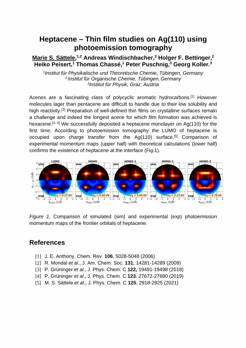
Heptacene – Thin film studies on Ag(110) using photoemission tomography
Marie S. Sättele,1,2 Andreas Windischbacher,3 Holger F. Bettinger,2 Heiko Peisert,1 Thomas Chassé,1 Peter Puschnig,3 Georg Koller.3
1Institut für Physikalische und Theoretische Chemie, Tübingen, Germany 2 Institut für Organische Chemie, Tübingen, Germany
3Institut für Physik, Graz, Austria Acenes are a fascinating class of polycyclic aromatic hydrocarbons.[1] However molecules lager than pentacene are difficult to handle due to their low solubility and high reactivity.[2] Preparation of well-defined thin films on crystalline surfaces remain a challenge and indeed the longest acene for which film formation was achieved is hexacene.[3, 4] We successfully deposited a heptacene monolayer on Ag(110) for the first time. According to photoemission tomography the LUMO of heptacene is occupied upon charge transfer from the Ag(110) surface.[5] Comparison of experimental momentum maps (upper half) with theoretical calculations (lower half) confirms the existence of heptacene at the interface (Fig.1).
Figure 1. Comparison of simulated (sim) and experimental (exp) photoemission momentum maps of the frontier orbitals of heptacene. References
[1] J. E. Anthony, Chem. Rev. 106, 5028-5048 (2006) [2] R. Mondal et al., J. Am. Chem. Soc. 131, 14281-14289 (2009) [3] P. Grüninger et al., J. Phys. Chem. C 122, 19491-19498 (2018) [4] P. Grüninger et al., J. Phys. Chem. C 123, 27672-27680 (2019) [5] M. S. Sättele et al., J. Phys. Chem. C 125, 2918-2925 (2021)
Enhancement of the contrast of photoelectron momentum maps M. Schaal, F. Otto, M. Gruenewald, R. Forker, and T. Fritz
Institute of Solid State Physics, Friedrich Schiller University Jena, Helmholtzweg 5, 07743 Jena, Germany
Photoemission tomography is a powerful surface science technique for identifying spectroscopic features in ultraviolet photoelectron (UP) spectra of organic thin films with corresponding occupied molecular orbitals [1]. When a charge transfer occurs from the substrate to the adsorbate, the former lowest unoccupied molecular orbital (LUMO) can be also investigated [2, 3]. However, the analysis of photoelectron momentum maps (PMMs) is quite challenging when the valence bands of the substrate superimpose the highest occupied states of the adsorbate (including the former LUMO) [4]. To solve this problem, a suitable background could be considered. In this study, we investigated a 2D organic-inorganic heterostructure consisting of PTCDA molecules on 2D hexagonal boron nitride (h-BN) on Ni(111). Using scanning tunneling spectroscopy (STS), we observed that the first monolayer consists of neutral and negatively charged molecules. However, the 3d bands of nickel superimpose the molecular feature of the adsorbate, making the identification of the molecular orbitals in the recorded PMMs very difficult. Inspired by the analysis of X-ray photoelectron diffraction (XPD) patterns, we applied a similar method to obtain a contrast enhancement that allows the identification of the former LUMO and HOMO. Therefore, we propose that this procedure can be used to identify spectroscopic features with molecular orbitals even when the substrate valence bands superimpose the molecular electronic states. References
[1] H. Offenbacher et al., J. Electron Spectrosc. Relat. Phenom. 204, 92-101 (2015)
[2] M. Willenbockel et al., Phys. Chem. Chem. Phys. 17, 1530-1548 (2015) [3] F. Otto et al., J. Phys. Chem. C 124, 4114-4127 (2020) [4] P. Kliuiev et al., Phys. Rev. B 98, 085426 (2018)

Lippmann-Schwinger approach to the Final State in Photoemission
M. Schwendt1 and P.Puschnig 1 1University of Graz, Graz, Austria
The final state in photoemission is obtained from the Lippmann-Schwinger equation with the Kohn-Sham potential of the modeled system. The initial state of the matrix element is also taken from Density functional theory. This approach is applied to both localised (e.g. atoms and molecules) as well as extended systems (e.g. clean and adsorbate covered surfaces). Special attention will be paid to the energy dependence of the photoemission intensity. The results are compared with analytical calculations and the widely used plane wave approximation for the final state.
Comparison between plane-wave approximation (left) and Lippmann-Schwinger equation (right).

Microscopy with Momentum and Imaging Spin-Filter (Au/Ir)
M. Patt, N.B. Weber, M. Escher, T.-J. Kühn and M. Merkel FOCUS GmbH, 65510 Hünstetten, Germany
Momentum Microscopy is a new technique in surface science, in which the momentum (or the real-space) distribution of photoelectrons is projected onto an image plane by using a photoemission electron microscope (PEEM) column. In case of momentum imaging the kx-ky plane can be energy-filtered by a double-hemispherical electrostatic analyzer (IDEA) to achieve a monochromatic momentum distribution. The ability of the method to map the complete angular distribution of photoelectrons is quite successfully used for photoemission orbital tomography (e.g. at the NanoESCA in Trieste [1]). An innovative extension of this technique is to use the monochromatic electron distribution behind the double-hemispherical analyzer for 2D imaging spin-filtering. Early experiments with a NanoESCA were performed with a W(100) single crystal as electron-mirror with spin-polarization dependent reflection [2], but only the move to a Au passivated Ir(100) single crystal with long-term stable scattering properties paved the way to a broadly based scientific useability. We will show results from our two first commercial Au/Ir 2D Imaging Spin Filters. Pre-characterizations of the Au/Ir crystal were done with LEED and a Ferrum-Detector setup [3] to find optimal preparation conditions and scattering energies for a high single-point figure-of-merit (with Sherman function >60% and Reflectivity >1%) [4]. Spin-filtered images of magnetic domains show that along the diameter of the field of view (e.g. 66 µm, Figure 1) more than 100 separate image points can be resolved. This increases the effective 2D figure-of-merit of this spin-filter by nearly four orders of magnitude compared to single-point analyzers. References
[1] M. Wießner et al., Nature Comm. 5, 4156 (2014) [2] C. Tusche et al., Appl. Phys. Lett. 99, 032505 (2011) [3] Escher, et al., e-J. Surf. Sci. Nanotech. Vol. 9, 340-343 (2011) [4] C.Tusche et al., Ultramicroscopy 159, 520–529 (2015)
Figure 1: Spin polarization (vertical axis) of photo electrons, excited with a Hg-lamp from magnetic domains in a polycrystalline iron sample (FoV 66µm).
On the Simulation of Different Spin States of Ni-Containing Molecular Interfaces
A. Windischbacher1 and P. Puschnig1 1Institute of Physics, Universitaetsplatz 5, 8010 Graz, Austria
In recent years, increasing experimental effort was put into the control of the local magnetic moment of metal ions at the nanoscale resulting in hybrid organic-metal interfaces with surprising spin configurations [1], [2]. The interpretations of such experiments, however, often rely on the comparison with theoretical calculations. This poster wants to evaluate the impact of different levels of theory on spin states of molecular interfaces. For this, we have calculated Ni-containing molecules with varying electronic configurations adsorbed on copper surfaces employing GGA, GGA+U and hybrid functionals. Differences in the calculations are presented and compared to experiments. References
[1] W.Auwärter et al., Nature Chemistry 7, 105 (2015) [2] Z.Xu et al., Nanomaterials 10, 2393 (2020)

Momentum-resolved hybridization of molecular and metal states
Xiaosheng Yang1,2, Philipp Hurdax3, Peter Puschnig3, Matteo Jugovac4, Giovanni Zamborlini4, Vitaliy Feyer4, Georg Koller3,
Michael G. Ramsey3, F. Stefan Tautz1,2 and Serguei Soubatch1,2 1Peter Grünberg Institut (PGI-3), Forschungszentrum Jülich, 52425 Jülich, Germany
2Jülich Aachen Research Alliance (JARA), Fundamentals of Future Information Technology, 52425 Jülich, Germany
3Institut für Physik, Karl-Franzens-Universität Graz, NAWI Graz, 8010 Graz, Austria 4Peter Grünberg Institut (PGI-6), Forschungszentrum Jülich, 52425 Jülich, Germany
The hybridization of adsorbate molecules' orbitals at a metal surface is of significance to the understanding of molecule–metal surface electronic properties. The concomitant phenomena, such as energy level alignment and charge transfer, are often regarded as the figure of merit from a device point of view. However, it is demonstrated in a photoemission tomography (PT) study on the bonding of p-sexiphenyl with the Cu(110) surface that for hybridization to occur, not only do states require overlap in energy and space, but also in momentum [1]. In this contribution, six similar oligomer systems, the monolayers of p-sexiphenyl, p-quinquephenyl and pentacene on Cu(110) and on Ag(110), respectively, are investigated by PT. On Cu(110), the Shockley surface state at the Y point of the substrate’s surface Brillouin zone (SBZ) disappears and the electronic states around the bulk gap scatter to a k-position appearing as ellipses, with a scattering vector following the reciprocal unit cell of the molecular overlayer. When the scattered states overlap with the corresponding lowest unoccupied molecular orbital (LUMO) in momentum, the LUMO intensity is evidently curtailed. On Ag(110), the molecular states are altered less as hinted by the unchanged Shockley surface state. References
[1] S. Berkebile et al., Phys. Chem. Chem. Phys. 13, 3604 (2011)