physics 3.43 | physics
TRANSCRIPT
Physics 3, 43 (2010)
Viewpoint
Cellular aggregates under pressure
Gabor Forgacs and Ioan KosztinDepartment of Physics and Astronomy, University of Missouri, Columbia, MO 65211, USA
Published May 24, 2010
Researchers develop a new approach to measuring the viscoelastic properties of multicellular aggregates by usinga micropipette aspiration technique.
Subject Areas: Biological Physics
A Viewpoint on:Aspiration of Biological Viscoelastic DropsKarine Guevorkian, Marie-Josée Colbert, Mélanie Durth, Sylvie Dufour and Françoise Brochard-WyartPhys. Rev. Lett. 104, 218101 (2010) – Published May 24, 2010
It is widely accepted by now that the mechanical char-acteristics of cells and tissues play an important role innumerous biological processes, such as early morpho-genesis [1, 2], signal transduction [3], cell adhesion [4],and even stem cell differentiation [5, 6]. In particu-lar, it was suggested and experimentally demonstratedthat a number of phenomena in embryonic tissues canbe interpreted through their viscoelastic properties [7].Tissue mechanical properties have often been quanti-fied by measurements using spherical aggregates of cells(as models of tissues) and parallel-plate tensiometry [8].In this technique the aggregates are exposed to a fixedstrain by compression between the plates and the vis-coelastic parameters such as surface tension (γ), elas-tic constant (E), and viscosity (η) are determined fromthe relaxation of the compressive force (i.e., stress) toequilibrium. In a work published in Physical ReviewLetters[9], Karine Guevorkian and collaborators fromCNRS-Institut Curie and Ecole Polytechnique, both inFrance, and McMaster University, Canada, use, for thefirst time, micropipette aspiration—a method normallyapplied to individual cells—to test the mechanical be-havior of multicellular spheroids and to determine γ, E,and η.
In this approach, the spheroid is aspirated into apipette of much smaller diameter than that of thespheroid, with a constant suction pressure ∆P. Thusthe approach is complementary to the parallel-platemethod, as now the applied stress is constant and theviscoelastic properties of the tissue are deduced fromthe variation of the strain (i.e., the change in length ofthe cellular material as it flows inside the pipette). Astissues are complex materials with complex constitutiveequations, it is not obvious that the two methods shouldlead to the same set of viscoelastic parameters. Indeed,Guevorkian et al. find that the surface tension of cell ag-gregates composed of a murine sarcome cell line (trans-fected with the cell adhesion molecule E-cadherin) de-
pends on ∆P, whereas in parallel-plate experiments, nodependence of the surface tension on the compressiveforce was observed.
As shown in Ref. [9], cells aspirated with force fa, im-posed by ∆P, reach steady inflow into the pipette withvelocity va = C(∆P − ∆Pc). Here, C = Rp/3πη, Rp isthe radius of the pipette, and ∆Pc ≈ 2γ/Rp is the crit-ical (Laplace) pressure, at which flow stops (Fig. 2 inRef. [9]). The above analytic expression for the aspi-ration velocity va is obtained by balancing the aspira-tion force fa = πR2
p(∆P − ∆Pc), with the resistive vis-cous force fv = 3π2ηRpva. Aspiration is followed byretraction (upon setting ∆P = 0), leading to steady out-flow from the pipette with vr = C∆Pc. The measurablequantities va, vr, and ∆P are used to determine γ and η,through C and ∆Pc. Assuming that the latter parame-ters are the same in the aspiration and retraction phases,∆Pc = ∆Pvr/(vr + va) and C = (va + vr)/∆P. The newfinding in the work is that vr , and therefore ∆Pc and γ,depend significantly on ∆P. (Unfortunately, va + vr vs∆P is not shown in Ref. [9], so one can only assume thatthis relationship was found to be linear, thus implyingthat C ∼ 1/η is indeed independent of ∆P.)
The ∆P dependence of vr means that despite the factthat during retraction no external pressure is imposedon the system, the cells remember what the aspirationpressure was when they were moving into the pipetteunder ∆P 6= 0. The authors interpret this result as anactive cellular response to mechanical force leading tothe remodeling of the cytoskeleton. This is an appealingexplanation and consistent with other findings on theeffect of mechanical forces on cells [10].
The work of Guevorkian et al. represents an impor-tant contribution to the field of tissue mechanics. How-ever, the application of the micropipette aspiration tech-nique to cellular aggregates raises several questions.First, most likely it is not applicable to weakly adhesivecells. (It would be useful to know how strong the ad-
DOI: 10.1103/Physics.3.43URL: http://link.aps.org/doi/10.1103/Physics.3.43
c© 2010 American Physical Society
Physics 3, 43 (2010)
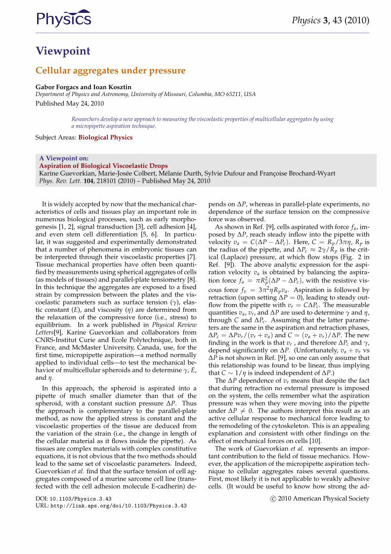
FIG. 1: Scanning electron microscopy images of 300-micron-diameter multicellular aggregates of (A) human umbilicalsmooth muscle cells, and (B) N-cadherin transfected Chinesehamster ovary cells. The aggregate with larger surface tension(A) displays a smooth surface, while the one with smaller sur-face tension (B) has a berrylike surface. (Illustration: From Ref.[11])
hesion between the E-cadherin transfected murine sar-coma cells is, in comparison to other cell types usedto measure surface tension). Figure 1 shows the sur-face of 300-micron-diameter aggregates composed ofhuman umbilical smooth muscle cells (HUSMC) (Fig. 1,A) and N-cadherin transfected Chinese hamster ovary(CHO) cells (Fig. 1, B), with respective surface ten-sions of 279 mN/m and 23 mN/m (as measured withparallel-plate tensiometer [11] and respective cell num-bers of 8, 000 and 12, 000). The aggregate of higher ten-sion displays a smooth and uniform morphology, whilethe aggregate of lower tension shows a berrylike sur-face. The differing morphologies reflect differential ad-hesion between the respective cells, with adhesion be-tween CHO cells being significantly weaker than be-tween HUSMC. As aspiration stretches the adhesivebonds between cells, at sufficiently weak adhesion andstrong aspiration, the method may simply separate cellsfrom one another.
Another problem arises from the finite number ofcells aspirated into the pipette. For a 400 micron aggre-gate and a 2Rp = 70-micron-diameter pipette, there areabout 12 cells within the pipette’s cross sectional area(we assume the linear dimension of a murine sarcomacell to be around 20 micron). Thus the quantity γ, mea-sured by Guevorkian et al. reflects the properties of asmall number of cells rather than those of the entire ag-gregate. Therefore, despite the fact that their measuredsurface tension has the same order of magnitude as thatmeasured by other methods where the entire aggregateis probed (parallel-plate tensiometry [8, 11], centrifu-gation [12]), it is not clear what γ really means. Fur-thermore, surface tension is a strictly equilibrium quan-tity, but here, γ is obtained from kinetic measurements.(Given that both va and vr are small, the system mostprobably can be regarded as being in quasi-equilibrium,
thus γ is well defined.) Finally, it is somewhat puzzlingthat while γ depends on ∆P the viscosity η of the cellu-lar assembly in the pipette appears to be independent of∆P.
If the authors are right and indeed the cellular systemhas memory of the magnitude of the aspiration pressurewhen it retracts (when ∆P = 0) and it is this memorythat causes γ to depend on ∆P, one would expect to seea similar effect in the compression experiments upon thevariation of the compressive pressure. However, tissuesurface tension measured by compressive tensiometrywas found to be independent of the compressive pres-sure [11]. What may cause this discrepancy? It mayresult from the difference between the magnitudes ofthe applied aspiration and compressive pressures. At asufficiently large pressure, it is probably not surprisingthat indeed the cellular system reorganizes. However,we find the aspiration pressures applied by Guevorkianet al. and the compressive pressures to be of the sameorder of magnitude. On the other hand, the aspirationpressure is applied effectively to a much smaller numberof cells than the compressive pressure. If it is indeed thenumber of relevant cells that is responsible for the ob-served variation of γ on ∆P, then the question of whatis the meaning of the measured surface tension remainsto be answered. This could be accomplished by measur-ing the surface tension of aggregates of the murine sar-coma cells with compression tensiometry and to com-pare it with γ(∆P = 0), as extrapolated from the valuesobtained using the micropipette aspiration technique.
References
[1] D. E. Ingber, Int. J. Dev. Biol. 50, 255 (2006).[2] R. Keller, L. A. Davidson, and D. R. Shook, Differentiation 71, 171
(2003).[3] D. J. Tschumperlin, G. D. I. V. Maly, T. Kikuchi, L. H. Laiho, A. K.
McVittie, K. J. Haley, C. M. Lilly, P. T. C. So, D. A. Lauffenburger,R. D. Kamm, and J. M. Drazen, Nature 429, 83 (2004).
[4] J. Schmitz, and K-E. Gottschalk, Soft Matter 4, 1373 (2008).[5] A. J. Engler, S. Sen, H. L. Sweeney, and D. E. Discher, Cell 126, 677
(2006).[6] F. Chowdhury, S. Na, D. Li, Y-C. Poh, T. S. Tanaka, F. Wang, and
N. Wang, Nature Mater. 9, 82 (2009).[7] G. Forgacs and S. Newman, Biological physics of the developing em-
bryo (Cambridge University Press, Cambridge, 2005).[8] R. A. Foty, C. M. Pfleger, G. Forgacs, and M. S. Steinberg, Devel-
opment 122, 1611 (1996).[9] K. Guevorkian, M-J. Colbert, M. Durth, S. Dufour, and F.
Brochard-Wyart, Phys. Rev. Lett. 104, 218101 (2010).[10] B. J. Damon, N. Mezentseva, J. S. Kumaratilake, G. Forgacs, and
S. A. Newman, Dev. Biol. 321, 319 (2008).[11] C. Norotte, F. Marga, A. Neagu, I. Kosztin, and G. Forgacs, Euro.
Phys. Lett. 81, 46003 (2008).[12] A. Kalantarian, H. Ninomiya, S. M. I. Saad, R. David,
R.Winklbauer and A. W. Neumann, Biophys. J. 96, 1606 (2009).
DOI: 10.1103/Physics.3.43URL: http://link.aps.org/doi/10.1103/Physics.3.43
c© 2010 American Physical Society
Physics 3, 43 (2010)
About the Authors
Gabor Forgacs
Gabor Forgacs is George H. Vineyard Professor of Biophysics at the University of Missouri.He received his Ph.D. in theoretical physics from the Roland Eötvös University, Budapest,Hungary. His present research interest is in the physical mechanisms acting in early em-bryonic development. He is applying these mechanisms to engineering and building livingstructures of prescribed shape and functionality by bioprinting.
Ioan Kosztin
Ioan Kosztin is an Associate Professor of Biological Physics at the University of Missouri.He received his Ph.D. in theoretical condensed matter physics from the University of Illi-nois at Urbana-Champaign. His current research interest is to develop and apply theo-retical and computational models for investigating different aspects of the dynamics ofbiomolecular complexes and of multicellular aggregates.
DOI: 10.1103/Physics.3.43URL: http://link.aps.org/doi/10.1103/Physics.3.43
c© 2010 American Physical Society