pincher … · development/plasticity/repair pincher-mediatedmacroendocytosisunderliesretrograde...
TRANSCRIPT

Development/Plasticity/Repair
Pincher-Mediated Macroendocytosis Underlies RetrogradeSignaling by Neurotrophin Receptors
Gregorio Valdez,1 Wendy Akmentin,1 Polyxeni Philippidou,1 Rejji Kuruvilla,2 David D. Ginty,2 and Simon Halegoua1
1Department of Neurobiology and Behavior, Center for Brain and Spinal Cord Research, State University of New York at Stony Brook, Stony Brook, NewYork 11794-5230, and 2Department of Neuroscience, Howard Hughes Medical Institute, The Johns Hopkins University School of Medicine, Baltimore,Maryland 21205
Retrograde signaling by neurotrophins is crucial for regulating neuronal phenotype and survival. The mechanism responsible forretrograde signaling has been elusive, because the molecular entities that propagate Trk receptor tyrosine kinase signals from the nerveterminal to the soma have not been defined. Here, we show that the membrane trafficking protein Pincher defines the primary pathwayresponsible for neurotrophin retrograde signaling in neurons. By both immunofluorescence confocal and immunoelectron microscopy,we find that Pincher mediates the formation of newly identified clathrin-independent macroendosomes for Trk receptors in soma, axons,and dendrites. Trk macroendosomes are derived from plasma membrane ruffles and subsequently processed to multivesicular bodies.Pincher similarly mediates macroendocytosis for NGF (TrkA) and BDNF (TrkB) in both peripheral (sympathetic) and central (hippocam-pal) neurons. A unique feature of Pincher–Trk endosomes is refractoriness to lysosomal degradation, which ensures persistent signalingthrough a critical effector of retrograde survival signaling, Erk5 (extracellular signal-regulated kinase 5). Using sympathetic neuronsgrown in chamber cultures, we find that block of Pincher function, which prevents Trk macroendosome formation, eliminates retro-gradely signaled neuronal survival. Pincher is the first distinguishing molecular component of a novel mechanistic pathway for endoso-mal signaling in neurons.
Key words: apoptosis; axon; neurite; nerve growth factor (NGF); pheochromocytoma (PC12); trophic; trafficking; ERK; signal transduc-tion; neurotrophin; endocytosis; clathrin independent
IntroductionThe neurotrophin (NT) family of target-derived growth factorsregulates neuronal survival and physiology in both the peripheralnervous system and the CNS (Levi-Montalcini, 1987; Lewin andBarde, 1996). NT signaling occurs through the Trk receptor ty-rosine kinases after binding the specific ligands NGF (TrkA),BDNF (TrkB), and NT3 (TrkC). Signal transduction by the re-ceptors is mediated by various effector pathways, which elicitlong-term regulation of cell physiology and survival by alteringpatterns of gene expression (Segal, 2003).
The highly asymmetric neuronal morphology has necessitatedevolution of a specialized NT signaling system that is initiated indistal axons and axonally propagated back to the soma. Mecha-nistic studies in neurons have relied primarily on chamber cul-tures in which axons are separated from somas. These studieshave shown that both NT and Trk are retrogradely transportedup the axon via microtubules (Hendry et al., 1974; Ehlers et al.,
1995), an event that appears to be required for signal propagation(Ginty and Segal, 2002). However, the identification of an ultra-rapid, ligand-independent component of retrograde phosphory-lated Trk (P-Trk) propagation has questioned the necessity forNT/Trk retrograde transport (MacInnis and Campenot, 2002)and has led to proposals of Trk-generated retrograde signalingintermediates (Campenot and MacInnis, 2004; Howe and Mob-ley, 2004). Neuronal survival is in part mediated by Trk retro-grade signals that lead to activation of the transcription factorcAMP response element-binding protein (Riccio et al., 1997).Retrograde signaling at the soma may be selective for specificMAP (mitogen-activated protein) kinase effectors such as Erk5(extracellular signal-regulated kinase 5) (Watson et al., 2001),although putative Trk signaling endosomes associated withErk1/2 have been identified (Howe et al., 2001; Delcroix et al.,2003). An important step toward resolving the controversies isthe unambiguous identification of the specific molecules andstructures that mediate Trk retrograde signaling.
The first vesicular mechanism proposed for retrograde signalingespoused the idea of an endosomal vesicle containing NGF, its re-ceptor, and associated signaling effectors, which was internalized atthe nerve terminal and retrogradely transported to the soma (Hale-goua et al., 1991). The identification of cytoplasmic vesicles contain-ing Trk receptors lent credence to this model (Beattie et al., 1996),and recent studies indicated that endosomal signaling is the ma-jor retrograde mechanism for neuronal survival (Ye et al., 2003;
Received Dec. 14, 2004; revised April 19, 2005; accepted April 21, 2005.This work was supported by National Institutes of Health Grant NS18218 (S.H.) and by the Howard Hughes
Medical Institute (D.D.G.). We thank P. Brehm and G. Mandel for helpful discussions, N. Mendell for the statisticalanalyses, J. Rosenbaum for valuable technical assistance, M. Goldfarb for providing the EGFR/TrkB construct, D.Kaplan for the anti-TrkBin antibody, M. Chao for the TrkA construct, and R. Segal for the TrkB–GFP adenovirus.
Correspondence should be addressed to Simon Halegoua, Department of Neurobiology and Behavior, Center forBrain and Spinal Cord Research, State University of New York at Stony Brook, Stony Brook, NY 11794-5230. E-mail:[email protected].
DOI:10.1523/JNEUROSCI.5104-04.2005Copyright © 2005 Society for Neuroscience 0270-6474/05/255236-12$15.00/0
5236 • The Journal of Neuroscience, May 25, 2005 • 25(21):5236 –5247

Heerssen et al., 2004). To provide a viable retrograde signalingvehicle, the internalized Trk endosome must engage the micro-tubule-based transport machinery, avoid degradation or recy-cling, and selectively recruit specific signaling complexes to reg-ulate gene expression. No endocytic mechanism for Trks inneurons has been identified that would satisfy these require-ments. Although evidence exists for a role for clathrin in Trkendosome formation (Howe et al., 2001), it is unknown whetherit is required for Trk retrograde signaling. Additionally, becauseclathrin-based endocytosis is involved normally in lysosomaldegradation and/or recycling to the plasma membrane, thismechanism is not ideally suited for retrograde signaling in neu-rons. Indeed, several features of Trk signaling suggest alternativemodes of Trk internalization. For example, Trk activation is seenat membrane ruffles where macropinocytosis occurs (Shao et al.,2002; Jullien et al., 2003) and with cholesterol-enriched mem-brane rafts (Bilderback et al., 1999; Huang et al., 1999) in whichcaveolas are internalized. However, a link between these specificmodes of internalization and Trk retrograde axonal signaling hasnot been explored.
We recently identified a membrane trafficking protein,Pincher (pinocytic chaperone), that mediates internalization ofactivated TrkA in pheochromocytoma PC12 cells at plasmamembrane ruffles (Shao et al., 2002). Here, we sought to deter-mine whether Pincher-mediated internalization is responsiblefor Trk-signaling endosomes and retrograde signaling inneurons.
Materials and MethodsCell culture. Superior cervical ganglia (SCGs) and hippocampi were dis-sected from postnatal day 1 (P1) and embryonic day 18 (E18) SpragueDawley rats, respectively. SCG neurons were dissociated, plated ontocollagen-coated tissue culture dishes or glass slides, and maintained asdescribed previously (Mains and Patterson, 1973; Geetha and Wooten,2003; Ye et al., 2003). Hippocampi were dissociated, plated onto poly-D-lysine-coated glass slides, and cultured as described previously (Mat-suoka et al., 1998). PC12 cells (Greene and Tischler, 1976) and TrkA–PC12 (Hempstead et al., 1992) were grown on tissue culture dishes orpolylysine plus laminin-coated dishes in DMEM supplemented with 5%fetal bovine serum (FBS) and 10% horse serum.
Real-time quantitative PCR. Total cellular RNA was isolated from 8 dcultured SCGs or freshly isolated E18 hippocampi using the RNAeasy kit(Qiagen, Hilden, Germany) and treated with RNAase-free DNase I(Roche Products, Welwyn Garden City, UK) for 30 min at 37°C. TheRNA was reversed transcribed using SuperScript (Invitrogen, San Diego,CA) and random primers to generate cDNA. Real-time PCR was per-formed using the ABI Prism sequence detector 7700 (Applied Biosys-tems, Foster City, CA) with SYBR Green PCR core reagents and Pincher3� untranslated region-specific primers (forward, 5�TGCCACTGACTT-GCTGAATGTC3�; reverse, 5�TGCCCTACATATGAGGAAACT-GAG3�) or rat glyceraldehyde-3-phosphate dehydrogenase (GAPDH)primers (Applied Biosystems) for normalization.
Recombinant cDNA constructs and adenoviruses. The cDNA constructs,rat TrkA in pCMV5 (Yano et al., 2001), epidermal growth factor receptor(EGFR)/TrkB, a chimera containing the EGFR extracellular domain andTrkA juxtamembrane and intracellular regions (Wang et al., 1996), hem-agglutinin (HA)–Pincher, and HA–PincherG68E in pCGN-HA (Shao etal., 2002), were used as described in the text. HA–Pincher- and HA–PincherG68E-containing defective recombinant adenoviruses were cre-ated using the Ad-Easy system (Stratagene, La Jolla, CA). Recombinantadenoviruses containing green fluorescent protein (GFP) (Vaillant et al.,1999), �-galactosidase (LacZ) plus dynaminK44A (Ye et al., 2003), andTrkB–GFP (Watson et al., 1999) have been described previously. Specificsmall interfering RNAs (siRNAs) for Pincher (AAGACCACTTTCAT-CAGATAC) and Eps15 homology domain-containing protein (EHD)1-3 (AAGAAAGAGCTGGTGAACAAC) were cloned into the shuttle
vector 1.0-CMV and recombinant adenoviruses created using the pSi-lencer adeno system 1.0-CMV (Ambion, Austin, TX).
Transfections and viral infections. Transient transfection with Lipo-fectamine 2000 (Invitrogen) was used to introduce constructs containingHA–Pincher, HA–PincherG68E, TrkA, and EGFR/TrkB into PC12 cellsand neuronal cultures that were seeded on polylysine plus laminin-coated coverslips. SCG neurons were maintained in C2 medium (Lein etal., 2002) supplemented with 100 ng/ml NGF for 24 h, at which time thecells were washed two times with and transfected in DMEM. After 4 h, thecells were washed two times and incubated in DMEM supplemented with10% FBS, 100 ng/ml NGF for 48 h. Hippocampal neurons grown onpolylysine-coated coverslips were transfected after 5 d of culture as de-scribed above for SCGs. For biochemical analyses by Western blot, theadenoviruses were titered, and cultures were infected in media with lowserum (1% horse serum for PC12 cells and 1% FBS for SCG neurons) toobtain virally encoded gene expression in 70 – 80% of the cells. For sur-vival and immunofluorescence studies, 30 – 40% of the neurons wereinfected.
Western blot analysis. Proteins were extracted from PC12 cells, 8 dcultured SCG neurons, and E18 hippocampi using radioimmunopre-cipitation assay (RIPA) buffer and analyzed by Western blot. The follow-ing antibodies were used to probe Western blots: rabbit anti-Pincher(1:5000) (Shao et al., 2002), rabbit anti-Trk-C14 (1:250; Santa Cruz Bio-technology, Santa Cruz, CA), rabbit anti-TrkA (1:200; Chemicon, Te-mecula, CA), mouse monoclonal antibody (mAb) anti-tubulin(1:10,000; Sigma, St. Louis, MO), mouse mAb anti-HA (1:200; SantaCruz Biotechnology), and mouse mAb anti-Flag-M5 (1:350; Sigma).Films were analyzed using a Super CoolScan9000ED film scanner (Ni-kon, Tokyo, Japan), and quantitation of scanned images was done usingImageJ (NIH).
Trk internalization and degradation assays. Internalization and degra-dation of cell-surface TrkA was assessed using surface TrkA biotinyla-tion. PC12 cells or sympathetic neurons, grown as described above, werecooled to 4°C and biotinylated on ice (1.5 mg/ml EZ-Link NHS-LC-biotin in PBS; Pierce, Rockford, IL) for 20 min. Unreacted biotin wasremoved in the cold using PBS containing 50 mg/ml glycine. For TrkAinternalization measurements, cells were infected with GFP control virusor with siRNA viruses specific for EHD1–3 or Pincher. To measure sur-face TrkA remaining after NGF-induced internalization, 72 h after infec-tion, the cells were NGF treated for 30 min before cell-surface biotinyla-tion, and the cell-surface biotin–TrkA was measured as described below.Internalized TrkA was directly assessed after biotinylation followed bydebiotinylation after NGF treatment, as described previously (Kuruvillaet al., 2004). For TrkA degradation measurements, cells infected withviruses containing GFP or Pincher were starved for both serum and NGFfor 24 h. After surface biotinylation, the cells were shifted to 37°C for 3 hin the presence of NGF (100 ng/ml) to allow endocytosis and degrada-tion. Proteins were extracted using RIPA buffer, and detection of biotin-ylated TrkA was performed as described previously (Kuruvilla et al.,2004). For PC12 cell extracts, equal amounts of protein were used inimmunoprecipitations from different samples, and for SCG extracts,Western blot analysis for tubulin in whole-cell extracts was used fornormalization.
EGF–Alexa555 internalization. EGF–Alexa555 (Molecular Probes, Eu-gene, OR) internalization was examined as described previously (Sato etal., 2001), with modification. Forty-eight hours after transfection ofEGFR/TrkB chimera and infection with HA–Pincher adenovirus as de-scribed above, hippocampal neurons were cooled in DMEM plus 1%BSA (M-BSA) and treated with ice-cold M-BSA containing 1 �g/mlEGF–Alexa555. After internalization, the cells were incubated with coldM-BSA to terminate internalization, and surface-bound EGF–Alexa555was removed by washing the cells three times with ice-cold acid-saltsolution (0.2 M acetic acid, pH 2.8, 0.5 M sodium chloride). The cells werethen fixed and processed for immunofluorescence microscopy as de-scribed below.
Immunofluorescence staining and confocal microscopy. To monitorevents in the cell bodies of sympathetic neurons and in cell bodies, axons,and dentrites of hippocampal neurons, the cells were cultured as de-scribed above. Before treatments, neurons were neurotrophin and serum
Valdez et al. • Trk Macroendocytosis and Retrograde Signaling J. Neurosci., May 25, 2005 • 25(21):5236 –5247 • 5237
starved for 2 h. For assessing SCG axons, dissociated neurons were platedon polylysine plus laminin-coated glass slides and 24 h later infected withadenoviruses as indicated. Thirty-six hours after infection, neurons werereplated onto polylysine plus laminin-coated glass slides for another 8 hin growth media. Neurons were then starved for serum and neurotro-phin for 2 h before treatments. In all cases, after treatments as indicated inthe text, SCG or hippocampal neurons were washed with cold PBS andfixed with 4% paraformaldehyde in PBS for 15 min at room temperature(RT). After fixation, the cells were washed two times with cold PBS andincubated in blocking solution (5.5% goat serum, 0.1% Triton/PBS) for1 h at RT. After exposing the cells to primary antibodies diluted in block-ing solution for 2 h at RT or overnight at 4°C, the cells were washed threetimes with PBS and then probed with fluorescently labeled secondaryantibodies, washed three times, and mounted on slides. Primary anti-bodies used were the following: rabbit anti-P-Erk5 (1:500; Biosource,Camarillo, CA), anti-HA (1:200; Santa Cruz Biotechnology), rabbit anti-P-Trk (1:100; Cell Signaling Technology, Beverly, MA), rabbit anti-TrkB(1:200) (Fryer et al., 1996), rabbit anti-Pincher (1:1000) (Shao et al.,2002), and mouse mAb anti-dynamin (1:250; Transduction Laborato-ries, Lexington, KY). The fluorophore-conjugated secondary antibodiesused in this study were the following: goat anti-mouse Alexa 488/Alexa546 or goat anti-rabbit-Alexa 488/Alexa 546 (Molecular Probes), goatanti-rabbit-cyanine 5 (Cy5), or goat anti-mouse-Cy5 (Jackson Immuno-Research, West Grove, PA). All images were obtained using an LSM 510laser scanning confocal microscope (Zeiss, Oberkochen, Germany). Toavoid bleed-through, fluorophores were activated and signal capturedsequentially using the following filters: 505–530/GFP/Alexa488, 560 –610/Alexa546, and LP 650/Cy5.
Neuronal survival. Sympathetic neurons were cultured in compart-mentalized chambers as described previously (Tsui-Pierchala and Ginty,1999). Eight to 10 d after plating, the cells were infected with LacZ, GFP,HA–Pincher, HA–G68Epincher, and dynaminK44A viruses in growthmedium consisting of 1% FBS for 8 h. After washing the cells three timeswith DMEM, the cell bodies were kept in 1% FBS medium, and the distalaxons were maintained in 10% FBS. The cell bodies and distal axoncompartments were treated with 10 ng of NGF or anti-NGF (1:1000;Sigma), as indicated in the figure legends. To score projecting cell bodiesin the survival assay, 40 nm fluorescent microspheres (1:500; MolecularProbes) were added to distal axon compartments. Seventy-two hoursafter infections, the cells were fixed in 4% paraformaldehyde at roomtemperature for 15 min. Detection of cells expressing HA–Pincher, HA-G68Epincher, and dynaminK44A was performed as described above.The nucleus was stained with 4�,6-diamidino-2-phenylindole (1:1000;Vector Laboratories, Burlingame, CA) at room temperature for 5 min. Acell with a condensed, fragmented, or with no nucleus was scored asapoptotic.
Electron microscopy and immunogold labeling. SCG and hippocampalneurons were plated on collagen and poly-D-lysine-coated ACLAR film(Ted Pella, Redding, CA), respectively, and cultured as described above.Immunogold electron microscopy (EM) was performed as described forPC12 cells (Shao et al., 2002), except that cells were microwave fixed for30 s at 650 W using a Pelco3451 microwave (Ted Pella) in a cold mixtureof 2% paraformaldehyde and 0.05% glutaraldehyde in 0.1 M phosphatebuffer, pH 7.4. To detect TrkB–GFP and Pincher, we used rabbit anti-GFP (1:20,000; Molecular Probes or 1:6000; Eusera, Edmonton, Alberta,Canada) and rabbit anti-Pincher (1:50,000), respectively, on ultrathinsections of Durcapan-embedded cells.
Image processing and quantitative analysis. All fluorescence imageswere digitally analyzed using Aim 3.2 software (Zeiss) and processedusing Photoshop (Adobe Systems, San Jose, CA). The weighted colocal-ization coefficient, defined as the sum of intensities of colocalizing pixelsfor Pincher or P-TrkA compared with the overall sum of pixel intensities(above threshold) for Pincher or P-TrkA, was determined. This was cal-culated using the Aim 3.2 colocalization macro (Zeiss) according toManders et al. (1993), which gives a higher value to brighter pixels.
Unless otherwise indicated, statistical analyses on quantified experi-ments were performed using the Mantel–Haenszal (M–H) test for equalrates (odds ratio of 1.0).
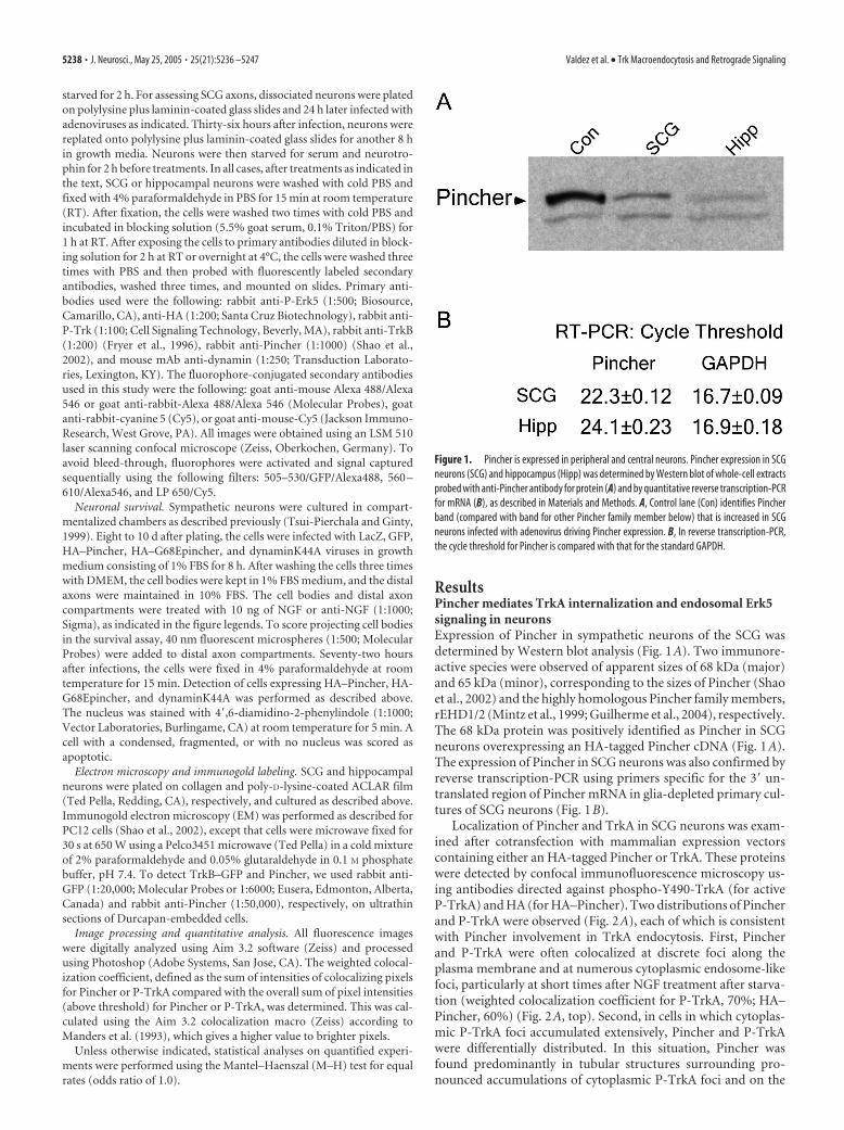
ResultsPincher mediates TrkA internalization and endosomal Erk5signaling in neuronsExpression of Pincher in sympathetic neurons of the SCG wasdetermined by Western blot analysis (Fig. 1A). Two immunore-active species were observed of apparent sizes of 68 kDa (major)and 65 kDa (minor), corresponding to the sizes of Pincher (Shaoet al., 2002) and the highly homologous Pincher family members,rEHD1/2 (Mintz et al., 1999; Guilherme et al., 2004), respectively.The 68 kDa protein was positively identified as Pincher in SCGneurons overexpressing an HA-tagged Pincher cDNA (Fig. 1A).The expression of Pincher in SCG neurons was also confirmed byreverse transcription-PCR using primers specific for the 3� un-translated region of Pincher mRNA in glia-depleted primary cul-tures of SCG neurons (Fig. 1B).
Localization of Pincher and TrkA in SCG neurons was exam-ined after cotransfection with mammalian expression vectorscontaining either an HA-tagged Pincher or TrkA. These proteinswere detected by confocal immunofluorescence microscopy us-ing antibodies directed against phospho-Y490-TrkA (for activeP-TrkA) and HA (for HA–Pincher). Two distributions of Pincherand P-TrkA were observed (Fig. 2A), each of which is consistentwith Pincher involvement in TrkA endocytosis. First, Pincherand P-TrkA were often colocalized at discrete foci along theplasma membrane and at numerous cytoplasmic endosome-likefoci, particularly at short times after NGF treatment after starva-tion (weighted colocalization coefficient for P-TrkA, 70%; HA–Pincher, 60%) (Fig. 2A, top). Second, in cells in which cytoplas-mic P-TrkA foci accumulated extensively, Pincher and P-TrkAwere differentially distributed. In this situation, Pincher wasfound predominantly in tubular structures surrounding pro-nounced accumulations of cytoplasmic P-TrkA foci and on the
Figure 1. Pincher is expressed in peripheral and central neurons. Pincher expression in SCGneurons (SCG) and hippocampus (Hipp) was determined by Western blot of whole-cell extractsprobed with anti-Pincher antibody for protein (A) and by quantitative reverse transcription-PCRfor mRNA (B), as described in Materials and Methods. A, Control lane (Con) identifies Pincherband (compared with band for other Pincher family member below) that is increased in SCGneurons infected with adenovirus driving Pincher expression. B, In reverse transcription-PCR,the cycle threshold for Pincher is compared with that for the standard GAPDH.
5238 • J. Neurosci., May 25, 2005 • 25(21):5236 –5247 Valdez et al. • Trk Macroendocytosis and Retrograde Signaling

plasma membrane (Fig. 2A, bottom). These distributions areconsistent with Pincher sorting from P-TrkA endosomes andrecycling back to the plasma membrane (Shao et al., 2002).
The activated Erk5 kinase (P-Erk5) mediates downstreamevents of TrkA and is a critical component of endosomal Trksignaling (Watson et al., 2001; Shao et al., 2002; Geetha and Woo-ten, 2003). To confirm that P-Erk5 is recruited by Pincher–Trkendosomes, its distribution was examined after Pincher overex-pression. Large punctate accumulations of P-Erk5 were distrib-uted in the cytoplasm, surrounded by Pincher (Fig. 2C) in a man-ner similar to that seen above for P-TrkA. In three independentexperiments, cytoplasmic accumulations of P-Erk5 foci (Fig. 2C)were observed in 66% of cells (n � 264) overexpressing Pinchercompared with only 5% in control cells (n � 401; p � 0.0001).Extensive colocalization of cytoplasmic yellow fluorescent pro-tein–TrkA and P-Erk5 was observed (data not shown), substan-tiating the conclusion that P-Erk5 is recruited to the Trkendosomes.
If endogenous Pincher function is re-quired for TrkA internalization in SCGneurons, a dominant-negative mutantform of Pincher (G68E), which blocksTrkA internalization in PC12 cells (Shao etal., 2002), should result in a reduction ofcytoplasmic P-TrkA staining. Indeed, cy-toplasmic accumulations of active P-TrkAwere found in only 11% (from three inde-pendent experiments) of cells (n � 95) ex-pressing PincherG68E compared with67% in control uninfected cells (n � 176;p � 0.0001). Moreover, a punctate distri-bution of P-TrkA was observed that wasprimarily limited to the plasma membraneand colocalized with the mutantPincherG68E (Fig. 2B). In addition, inter-nalization of endogenous TrkA was pre-vented by expression of Pincher-specificRNA interference (RNAi), which resultedin elimination of HA–Pincher expression(Fig. 3A) and downregulation of endoge-nous Pincher protein (by 61 and 65% intwo independent experiments) (Fig. 3A).Adenovirus-driven expression of PincherRNAi in PC12 cells resulted in increasedaccumulation of endogenous TrkA at thecell surface by 1.5 � 0.06-fold (from threeindependent experiments). NGF treat-ment caused a 50% reduction in surfaceTrkA in control cells as a result of internal-ization, which was significantly inhibited(by 60%) as a result of Pincher RNAi ex-pression (only 20% reduction of surfaceTrkA). Thus, the amount of surface TrkAin NGF-treated cells was 2.6 � 0.76-fold(from three independent experiments)higher in Pincher RNAi-expressing cells(Fig. 3B), with a corresponding reductionin the percentage of the biotinylated cell-surface TrkA that was found in the inter-nalized compartment after NGF treatment(2.8- and 1.9-fold reduction, respectively,from two independent experiments) (Fig.3C). A similarly enhanced accumulation
(1.8 � 0.33-fold, from three independent experiments) of cell-surface TrkA was seen in NGF-treated SCG neurons infected withPincher RNAi virus (Fig. 3D). Pincher RNAi expression causedno change in total cellular Trk levels in PC12 cells or sympatheticneurons (Fig. 3B–D). These effects were specific for PincherRNAi, because no effects on either Pincher expression or TrkAinternalization were seen with expression of RNAi that was in-stead specific for other Pincher family members (data notshown).
Pincher effects on Trk endocytosis are not restricted to NGF,TrkA, or peripheral neuronsPincher would be expected to mediate a similar process of endo-cytosis of other NT/Trk family members. Thus, the endocyticprofiles of TrkB and Pincher were examined in primary culturesof SCG neurons double infected with TrkB–GFP and HA–Pincher adenoviruses. Similar to P-TrkA, two intracellular distri-butions of activated P-TrkB and Pincher were observed after
Figure 2. Pincher mediates formation of TrkA-signaling endosomes that associate with active Erk5 kinase in SCG neurons.Cultured SCG neurons were transfected with a TrkA construct alone (A, B, left) or infected with a HA–Pincher-containing adeno-virus alone (C) or together with TrkA transfection (A, right) or were infected with dominant-negative mutant HA–PincherG68E-containing adenovirus together with TrkA transfection (B, right) or uninfected (C, left). Neurons were then treated with NGF for 15min (A, top row, C) or continuously (A, bottom row, B), fixed, stained, and confocal microscopy was performed as described inMaterials and Methods. Active, P-TrkA (green; A, B), HA–Pincher (red; A, C), HA–PincherG68E (HA-PinG68E; B), and P-Erk5(green; C) were visualized using anti-P-TrkA, anti-HA, and anti-P-Erk5 antibodies, respectively. Scale bar: (in A) 2 �m.
Valdez et al. • Trk Macroendocytosis and Retrograde Signaling J. Neurosci., May 25, 2005 • 25(21):5236 –5247 • 5239

BDNF treatment. First, Pincher and P-TrkB colocalized at cyto-plasmic foci (Fig. 4A, top). Second, Pincher was found in tubularstructures that surrounded cytoplasmic accumulations ofP-TrkB foci (Fig. 4A, bottom). When TrkB–GFP was expressedtogether with dominant-negative HA–PincherG68E, a nearlycomplete inhibition of P-TrkB internalization was observed.P-TrkB and PincherG68E were instead colocalized in large punctialong the plasma membrane (Fig. 4B).
TrkB is expressed in both peripheral and central neurons, inwhich it mediates events leading to neuronal survival and plastic-ity. Both Pincher mRNA and protein are expressed in the hip-pocampus of E18 rats (Fig. 1A), which is nearly devoid of glia(Maher et al., 1999). Although they normally express TrkB, cul-tured hippocampal neurons do not require neurotrophins fortheir survival. Thus, permitting growth in media lacking BDNFprovided an opportunity to examine the roles of the Trk ligand aswell as Pincher in mediating endocytosis and Trk endosomalsignaling. In neurons expressing TrkB–GFP, significant levels ofP-TrkB were seen in cytoplasmic foci resembling endosomalstructures (data not shown). In the absence of BDNF ligand, 36%(from three independent experiments) of infected cells (n � 90)contained cytoplasmic accumulations of P-TrkB, indicating thatin cells overexpressing TrkB, significant activation and internal-ization of TrkB occurs. As expected, when TrkB–GFP-expressingcells were treated with BDNF for 30 min, increased staining ofboth plasma membrane and endocytosed P-TrkB was observed,
and the number of cells (n � 94) with cytoplasmic accumulationswas greatly increased to 68% ( p � 0.0001). The ability of Pincherto mediate internalization of P-TrkB was examined in hippocam-pal neurons coinfected with adenoviruses harboring TrkB–GFPor HA–Pincher and treated with BDNF. The distribution ofPincher and P-TrkB was similar to that seen in sympathetic neu-rons, with colocalization at both plasma membrane and cytoplas-mic foci (Fig. 4C, top) or Pincher surrounding cytoplasmic accu-mulations of P-TrkB endosomes (Fig. 4C, bottom). Even in theabsence of BDNF, the extent of cytoplasmic accumulation of ac-tive P-TrkB (62%; n � 213 from three independent experiments)was greatly increased by coexpression of Pincher ( p � 0.0001).Importantly, as in sympathetic neurons, endocytosis of activatedTrkB in hippocampal neurons depended on Pincher function.Coexpression of dominant-negative mutant PincherG68E re-sulted in the codistribution of P-TrkB with PincherG68E at largepuncti, restricted to the plasma membrane (Fig. 4D).
In neurons, neurotrophins are internalized after binding toboth p75 and Trk receptors. To determine whether signalingthrough TrkB (but not p75) was sufficient for Pincher-mediatedinternalization, a chimeric protein was introduced that containsthe extracellular domain of EGF receptor fused to the transmem-brane and cytoplasmic domains of TrkB (EGFR/TrkB). This chi-meric receptor binds to EGF, but the signaling pathways are me-diated by TrkB (Wang et al., 1996). Cultured hippocampalneurons were transfected with the EGFR/TrkB construct and in-fected with adenovirus harboring HA–Pincher. The neuronswere then exposed to fluorescently tagged EGF (EGF–Alexa555)to monitor internalized EGF, followed by staining with anti-HAand anti-P-TrkB antibodies. As shown in Figure 4E, EGF wasfound together with P-TrkB in cytoplasmic accumulations sur-rounded by tubulo-vesicular arrays of HA–Pincher, thus demon-strating that Pincher-mediated endocytosis of ligand–Trk com-plexes is not dependent on ligand binding to p75.
Pincher mediates a novel process of Trk macroendocytosisTo gain insight into both the nature and the mode of formation ofTrkB and Pincher-containing structures, they were ultrastructur-ally characterized by immunogold electron microscopy. To cir-cumvent an incompatability of available anti-TrkB antibodiesand their visualization with high-quality ultrastructure, we usedneurons expressing TrkB–GFP and HA–Pincher and analyzedthem using anti-GFP (for TrkB–GFP) and anti-Pincher (for HA–Pincher) antibodies. As expected, when TrkB–GFP was ex-pressed, it could be seen associated with cytoplasmic endoplas-mic reticulum (ER) (data not shown). In neurons, TrkB–GFPand HA–Pincher were associated with four endocytic structuralarrangements as exemplified in Figure 5. In the first arrangement,both TrkB–GFP (Fig. 5A, top) and HA–Pincher (data not shown)were concentrated at typical membrane ruffles that appeared asplasma membrane loops in ultra-thin sections. Although goldlabeling for both TrkB–GFP and HA–Pincher could also be seensporadically all along the plasma membrane, neither was seenassociated with clathrin-coated pits (data not shown). TrkB–GFPand HA–Pincher were primarily coconcentrated at complex cell-surface ruffles that encompassed accumulated pinocytic vesiclesof varied sizes, as seen by gold labeling for both proteins in serialthin sections (Fig. 5A, second panel). Varying the relative expres-sion levels of TrkB–GFP and HA–Pincher indicated that withmore Pincher, the vesicular structures within ruffles were moreevenly sized and in a more ordered arrangement (data notshown). TrkB–GFP and HA–Pincher were additionally cocon-centrated in a distinctive structural arrangement in the cytoplasm
Figure 3. RNAi-mediated repression of Pincher expression retards TrkA internalization. A,The ability of Pincher-specific RNAi to block Pincher protein expression was determined in cellextracts taken from PC12 cells transfected with a HA–Pincher (Con) construct alone or togetherwith a Pincher–RNAi (RNAi) construct using Western blots probed with anti-HA antibody (top).Levels of endogenous Pincher probed using anti-Pincher antibody was decreased after infectionwith adenovirus driving Pincher–RNAi (bottom). The band corresponding to Pincher was iden-tified by its increased intensity (compared with other Pincher family members), seen afterinfection of cells with adenovirus driving HA–Pincher expression, as in Figure 1 A (data notshown). B, Internalization of TrkA was compared between control and Pincher–RNAi-expressing PC12 cells. After NGF treatments as indicated, cell-surface TrkA was biotinylated,immunoprecipitated, and identified by Western blot using anti-TrkA antibody, as described inMaterials and Methods. Equal amounts of PC12 extract proteins were loaded in each lane. C, Theeffects of Pincher–RNAi expression on internalized surface TrkA were directly assessed in PC12cells. Cells were surface biotinylated, treated with NGF, then debiotinylated as described inMaterials and Methods. The remaining biotinylated (internalized) TrkA was detected as in B. D,The effects of Pincher–RNAi expression on internalization of surface TrkA was determined inSCG neurons. NGF-treated neurons were infected, surface biotinylated, and TrkA was detectedas in B. �-Tubulin, probed for in the same blot as the SCG extracts (data not shown), was usedto control for protein loading. Total TrkA protein was determined from the same PC12 cell andSCG extracts used to measure surface TrkA internalization. Con, Control.
5240 • J. Neurosci., May 25, 2005 • 25(21):5236 –5247 Valdez et al. • Trk Macroendocytosis and Retrograde Signaling

at what appeared to be internalized complex pinocytic mem-brane ruffles, which we termed macroendosomes. The same mac-roendosomal structures were gold labeled for both TrkB–GFPand HA–Pincher as seen in serial thin sections (Fig. 5A, thirdpanel), suggesting that TrkB and Pincher are coendocytosedwithin these structures. In the fourth structural arrangement,TrkB–GFP and HA—Pincher were seen in accumulations ofmultivesicular bodies (MVBs). Gold-labeled TrkB–GFP wasfound preferentially in internal vesicles of the MVBs but was alsopresent at the outer membrane (Fig. 5A, bottom). The sameMVBs often contained gold-labeled HA–Pincher along the outerendosomal membrane. Compared with the distribution of la-beled TrkB–GFP, gold-labeled HA–Pincher was rarely found inthe internal vesicles, as shown in labeled serial sections (Fig. 5A,fourth panel).
Trk–Pincher MVBs appeared to be formed directly from themacroendosomes, because intermediates in this process were ob-served. In TrkB–GFP- (Fig. 5B, right) and/or HA–Pincher- (Fig.5B, left) expressing neurons, gold-labeled structures were ob-served in the cytoplasm that appeared to contain macroendo-somes that were contiguous with multivesicular body-like struc-tures, suggesting intermediates in endosome formation andprocessing. As expected, gold-labeled TrkB–GFP was associatedwith both macroendosomal and multivesicular body-like struc-tures, whereas gold-labeled HA–Pincher was primarily associatedwith the macroendosomal areas of the intermediates. As reportedpreviously (Shao et al., 2002), HA–Pincher-expressing cells alsocontained putative recycling structures consisting of largePincher-labeled tubules throughout the cytoplasm (data notshown), suggesting that after processing of macroendosomes toMVBs, excess Pincher is recycled to the plasma membrane.
Increased expression of TrkB–GFP and/or Pincher modulatedendosomal processing. In neurons expressing TrkB–GFP, mac-roendosomes as well as MVBs (and the ER) were more numer-ous, and the MVBs were more often vesicle filled than those seenin control-infected neurons (Fig. 6, Table 1), suggesting that in-creased expression of Trk drove more endosome formation andmaturation. Many more of the gold-labeled MVBs were also elec-tron dense in TrkB–GFP-overexpressing neurons (Fig. 6, Table1), indicative of increased lysosomal processing with Trk overex-pression. Overexpression of HA–Pincher alone also caused anenhanced accumulation (approximately threefold) of MVBs, butthese were much larger and for the most part only partially filledand thus less mature (Fig. 6, Table 1). When HA–Pincher wascoexpressed with TrkB–GFP, the number of MVBs was dramat-ically increased (over ninefold), but they were again only partiallyfilled with vesicles (Fig. 6, Table 1). This result indicates thatTrkB–GFP and HA–Pincher synergistically enhanced productionof MVBs while keeping them unfilled. Remarkably, the endoso-mal MVBs seen in both the Pincher-overexpressing and thePincher and TrkB–GFP-overexpressing neurons were almostnever electron dense (Fig. 6, Table 1), regardless of the extent towhich they were vesicle filled, indicating that they were refractoryto lysosomal processing. These results suggested that Pincher re-tarded the maturation of Trk-containing MVBs, making themresistant to degradation. To more directly assess effects of Pincheroverexpression on endosome-mediated Trk degradation, thedegradation of endogenous cell-surface TrkA was assessed insympathetic neurons. For this, cell-surface TrkA was biotinylatedin control and HA–Pincher-overexpressing cultured SCG neu-rons starved for 2 h of NGF. After treatment with NGF for 3 h, weobserved 2.4- and 2.8-fold (from two independent experiments)more biotinylated TrkA remaining in whole-cell extracts of neu-
Figure 4. Pincher mediates endocytosis of ligand-bound TrkB in SCG and hippocampal neu-rons. Cultured SCG (A, B) or hippocampal (C–E) neurons were double infected with adenovi-ruses containing TrkB–GFP or HA–Pincher (A, C), TrkB–GFP or HA–PincherG68E (B, D), anEGFR/TrkB chimera (with extracellular domain of EGFR and transmembrane with intracellulardomains of TrkB or HA–Pincher; E). Neurons were treated with BDNF for 15 min (A, C, top) or 1 h(A–C, bottom, D) or treated for 15 min with EGF coupled to Alexa555 (E). Neurons were fixedand stained as in Figure 2 for P-TrkB (green; A–E), HA–Pincher (red; A, C, E), HA–PincherG68E(HA-PinG68E; red; B, D), and EGF–Alexa555 (blue; EGF-A555; E), which were visualized directly.Scale bar: (in C) 2 �m.
Valdez et al. • Trk Macroendocytosis and Retrograde Signaling J. Neurosci., May 25, 2005 • 25(21):5236 –5247 • 5241
rons expressing HA–Pincher comparedwith control cells (data not shown). Thus,increased Pincher expression conferredprotection from degradation on cell-surface TrkA.
Pincher-mediated Trk endocytosis isglobally mediated in allneuronal compartmentsFor Pincher to mediate internalization ofTrk in axons and axon terminals, it mustbe expressed there. To visualize Pincherdistribution along axons, primary SCGneurons were infected with an adenovirusindependently driving expression ofPincher and GFP. GFP-labeled neuronswere fixed and stained with anti-Pincherantibody. As seen in Figure 7A, Pincherwas present in axon terminals and washighly localized to membrane ruffles. En-dogenous Pincher was also present in ax-ons as determined by Western blot analy-sis of the distal axon compartment ofneurons grown in chamber cultures (datanot shown). To determine whetherPincher mediated the internalization ofTrk in axons, SCG neurons were doubleinfected with adenoviruses driving TrkB–GFP or HA–Pincher expression. As shownin Figure 7B, after infected neurons weretreated with BDNF for 15 min, large accu-mulations of colocalized TrkB and Pincherwere observed predominantly at the ter-minal (weighted colocalization coefficientfor TrkB, 0.56 � 0.20; Pincher, 0.54 �0.19; n � 24), and costained puncti werealso seen along the axon (Fig. 7B, left). TheTrkB appearing at these foci was activated,because similar results were seen with anti-P-Trk staining. After 1 h of BDNF treat-ment, large foci of colocalized P-Trk andHA–Pincher were seen along the axon(weighted colocalization coefficient forP-TrkB, 0.70 � 0.08; Pincher, 0.59 � 0.10;n � 22) (Fig. 7B, right). Serial optical sec-tions of axons and terminals indicated thatmany foci were axoplasmically internal-ized (data not shown). To determinewhether Pincher is required for Trk inter-nalization at the axon terminal, TrkB–GFPwas coexpressed in SCG neurons alongwith the dominant-negative mutantPincherG68E. Even after BDNF treatmentof these neurons, P-Trk was found to behighly colocalized with PincherG68E atdiscrete plasma membrane locations (Fig.7C) and, as in the cell bodies, was notinternalized.
Pincher distribution was also examinedin cultured hippocampal neurons, inwhich axons and dendrites are distin-guished by enhanced tau and MAP2 label-ing, respectively (Craig and Banker, 1994).
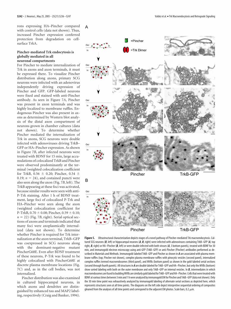
Figure 5. Ultrastructural characterization depicts stages of a novel pathway of Pincher-mediated Trk macroendocytosis. Cul-tured SCG neurons (B, left) or hippocampal neurons (A, B, right) were infected with adenoviruses containing TrkB–GFP (A, topright, B, right) or HA–Pincher (B, left) or were double infected with both viruses (A, 3 bottom panels), treated with BDNF for 30min, and immunogold electron microscopy using anti-GFP (TrkB–GFP) or anti-Pincher (Pincher) antibodies performed as de-scribed in Materials and Methods. Immunogold-labeled TrkB–GFP and Pincher as shown in A are associated with plasma mem-brane ruffles (top; Pincher not shown), complex plasma membrane ruffles with pinocytic vesicles (second panel), internalizedcomplex ruffles termed macroendosomes (third panel), and MVBs (bottom panel) as shown in the gold-labeled serial sections(second through fourth panels). All structures in A are double labeled for TrkB–GFP and HA–Pincher, but only the MVBs (bottom)show sorted labeling with both on the outer membrane and only TrkB–GFP on internal vesicles. In B, intermediates in whichmacroendosomes are fused to budding MVBs are similarly gold labeled for TrkB–GFP and HA–Pincher. Cells that were treated withBDNF at various times between 3 min and 1 h were analyzed by immunogold EM for Pincher and TrkB–GFP (data not shown). Onlythe 30 min time point was exhaustively analyzed by immunogold labeling of alternate serial sections as depicted here, whichrepresents structures seen at all time points. The diagrams on the left side depict interpretive sequential ordering of compositesgleaned from the analyses of all time points and correspond to the adjacent EM photos. Scale bars, 0.2 �m.
5242 • J. Neurosci., May 25, 2005 • 25(21):5236 –5247 Valdez et al. • Trk Macroendocytosis and Retrograde Signaling

When these neurons were infected with adenovirus driving HA–Pincher expression, Pincher was found in the cell body and in allaxonal and dendritic processes labeled or not for either MAP2 ortau in a similarly even punctate pattern (data not shown). Inneurons expressing TrkB–GFP and HA–Pincher, colocalizationof these proteins in large foci was observed regularly distributedalong all processes of these neurons (weighted colocalization co-efficient for TrkB–GFP, 0.81 � 0.07; HA–Pincher, 0.71 � 0.11;n � 10) (Fig. 7D). The internalized Pincher and TrkB–GFP struc-tures seen in virus-infected sympathetic and hippocampal axonsand/or dendrites were further analyzed by immunogold electronmicroscopy using anti-Pincher and anti-GFP antibodies, respec-tively. TrkB–GFP and Pincher were seen at surface complex ruf-fling structures, macroendosomes, and MVBs, similar to thoseseen in the soma (data not shown). The existence of putativePincher recycling tubules, abundant in the cell soma, appeared tobe restricted to the proximal segment of the axonal or dendriticprocess, adjacent to the soma. However, ultrastructurally definedstructures in axons and dendrites were scarce, probably becauseof the much lower levels of Trk and Pincher expression in theneuronal processes after viral infection. We were thus unable toreliably determine quantitatively the relative abundance of eachtype of structure.
To verify that TrkB–GFP and Pincher colabeled structuresrepresent ligand-induced internalized receptors, endocytosis ofthe EGF/TrkB chimeric protein was examined. When EGFR/TrkB expressing neurons were treated with a fluorescently taggedEGF, tagged EGF was found in puncta along with TrkB andPincher all along the processes (Fig. 7E), as was seen for BDNF-internalized TrkB–GFP and Pincher (Fig. 7D). Low pH treatmentof the neurons removed labeled EGF from surface receptors aftercold temperature (4°C) incubation (data not shown) but not
after the chase at 37°C (Fig. 7E), indicatingthat the labeled EGF seen after warmingwas internalized.
Pincher mediates NGF retrogradeaxonal signaling in neuronsThe above results suggest that Pincher isinvolved in both internalization and retro-grade transport of Trk endosomes in ax-ons. To determine whether Pincher is re-quired for retrograde signaling, we askedwhether survival, a property that dependson this event, is blocked by expression ofthe dominant-negative PincherG68E mu-tant. These studies were performed inchamber cultures in which axons extend
into a biochemically separate chamber from the cell bodies, al-lowing differential treatments of cell bodies and distal axons(Campenot, 1982). We used a retrograde survival assay in whichNGF treatment selective to the distal axons mediates neuronalsurvival by preventing apoptosis (Ye et al., 2003). Neurons in thechambers were infected with adenoviruses driving expression ofGFP, Pincher, dominant-negative PincherG68E, or dominant-negative dynaminK44A (Zhang et al., 2000; Watson et al., 2001;Ye et al., 2003). Treatment of the distal axon chamber with NGFprevented apoptotic death of neurons expressing GFP or Pincher(Fig. 8A). However, in neurons expressing PincherG68E, NGFdid not prevent cell death (Fig. 8A), similar to the effects ofdynaminK44A (Fig. 8A) (Ye et al., 2003). The resulting cell deathwas predominantly apoptotic as evidenced by prevention withthe caspase inhibitor Boc-aspartyl fluoromethyl ketone (BAF)(Fig. 8A). When NGF was added to the cell bodies of infectedneurons, thus bypassing retrograde axonal signaling, apoptoticdeath caused by PincherG68E or dynaminK44A was, as expected,significantly reduced (Fig. 8A). However, in the case ofPincherG68E, the reduction was only partial (40% reduction forPincherG68E vs 75% reduction for dynaminK44A), perhaps theresult of a contribution by Pincher to signaling events in thesoma. Because retrograde P-Erk5 signaling to the nucleus occursvia an endosomal pathway to prevent apoptosis (Watson et al.,2001), we examined the distribution of P-Erk5 in the cells bodiesafter expression of PincherG68E or dynaminK44A. Expression ofPincherG68E resulted in accumulation of P-Erk5 that was lim-ited to the plasma membrane in all cells examined (from threeindependent experiments with a total of 108 cells) (Fig. 8B, top).In contrast, expression of dynaminK44A did not cause plasmamembrane accumulation of P-Erk5 and did not prevent translo-
Table 1. Expression of Trk and Pincher affects endosomal processing of multivesicular bodies
Viral infection(s) MVB/cell* Diameter (�m) Partial (%) Filled (%) e� dense (%) Int. (% gold) Ext. (% gold)
GFP (control) 1.42 � 0.96(31)a 0.24 � 0.07(44)b 93 7 0TrkB–GFP 3.00 � 1.18(14)a 0.27 � 0.10(42)b 3 52 45 88 (256/40)c 12 (34/40)c
TrkB–GFP plus Pincher 13.25 � 11.09(4)a 0.27 � 0.08(53)b 92 6 2 ND NDPincher 4.29 � 2.69(7)a 0.49 � 0.16(30)b,** 57 43 0 23 (19/30)c 77 (62/30)c
The table presents data obtained from EM photomicrographs of soma from cultured SCG neurons that were single or double infected with the indicated adenoviruses as described in Materials and Methods. MVBs were scored for thecharacteristics indicated, as to mean number per cell (MVB/cell), mean diameter, partially filled (Partial), or completely filled (Filled) with vesicles, the proportion that was lysosomally dark or electron dense (e� dense), and the portion ofgold particles found on the internal vesicles (Int.) or on the external MVB membrane (Ext.). Comparisons were statistically evaluated using the Welch–Satterthwaite t test. ND, Not determined.an value for cells.bn value for endosomes.cNumber of gold particles/number of endosomes.
*Comparisons of values to GFP control were significant to p � 0.0001.
**Comparison to GFP control, p � 0.0001.
Figure 6. Pincher is associated with Trk endosomes that resist lysosomal processing. Cultured neurons were infected withadenoviruses driving expression of and immunogold EM labeled for GFP (Control), HA–Pincher (Pincher), TrkB–GFP (TrkB), ordouble infected with the latter two viruses (TrkB � Pincher; only TrkB–GFP labeling shown), as in Figures 4 and 5. Photomicro-graphs depict typical examples of MVBs in each case. Forty-five percent of the MVBs in cells expressing TrkB–GFP alone waselectron dense, indicating lysosomal processing. Scale bars, 0.2 �m.
Valdez et al. • Trk Macroendocytosis and Retrograde Signaling J. Neurosci., May 25, 2005 • 25(21):5236 –5247 • 5243

cation of P-Erk5 from the cytoplasm to thenucleus in any cells (from three indepen-dent experiments with a total of 150 cells)(Fig. 8B, bottom).
DiscussionPincher-mediated endocytosis represents aunique mechanistic pathway for Trk endo-somal signaling. Intermediate stages of thispathway were identified by both confocalimmunofluorescence microscopy and im-munogold electron microscopic analysis.Exposure of neurons to neurotrophins re-sulted in formation of extensive plasmamembrane ruffles. Pincher and Trk wereboth concentrated within complex plasmamembrane ruffles and cointernalized withinthe complex ruffles into structures termedmacroendosomes. Pincher- and Trk-con-taining membranes were subsequentlysorted from the macroendosomes to formMVBs containing Trk vesicles. This series ofevents is intermediate between macropino-cytosis and receptor-mediated endocytosis.It is macropinocytic-like in that internaliza-tion occurs at plasma membrane ruffles thatunusually encompass multiple pinocyticvesicles of varied sizes. It is endocytic-likewith respect to the high concentration of Trkreceptors at the site of internalization and itsdependence on dynamin (G. Valdez, W. Ak-mentin, and S. Halegoua, unpublished ob-servations). To reflect the shared features, wehave named the Pincher-mediated processmacroendocytosis.
Although Trk internalization has beensuggested to occur via clathrin-mediatedendocytosis (Howe et al., 2001; Heerssenet al., 2004), we have not observed Trk orPincher associated with clathrin-coatedpits or vesicles. Although it is possiblethat both clathrin-dependent and-independent processes each contribute tothe formation of Trk endosomes, the mac-roendocytic process driven by Pincherrepresents the primary mode of activatedTrk endocytosis in neurons. This was evi-dent by the prevalence of Trk macroendo-somal structures in neurons overexpress-ing Trk and the ability of dominant-
Figure 7. Pincher mediates endocytosis and trafficking of ligand-bound TrkB in axons and dendrites. Cultured SCG neurons(A–C) or hippocampal neurons (D, E) were infected with adenovirus either independently driving expression of both GFP andHA–Pincher(A)oronlyHA–Pincher(E)orweredoubleinfectedwithvirusescontainingTrkB–GFPtogetherwithHA–Pincher(B,D)orwith
4
HA–PincherG68E (HA-PinG68E; C). EGFR/TrkB construct wastransfected into HA–Pincher-expressing neurons (E). SCG ax-onal processes with growth cones (A–C) and hippocampalprocesses (D, E) were visualized (P-TrkB, green; HA–Pincher,red) after fixation and staining as in Figure 4, except thatsurface-bound EGF–Alexa555 (blue) was removed by acid-salt wash before fixation, TrkB (green) was seen using anti-TrkB in antibody, and GFP (green) was visualized directly, allas described in Materials and Methods. In B, the axon andgrowth cone were outlined in the Merge to depict the loca-tion of the limiting plasma membrane. Scale bars, 2 �m.
5244 • J. Neurosci., May 25, 2005 • 25(21):5236 –5247 Valdez et al. • Trk Macroendocytosis and Retrograde Signaling

negative mutant PincherG68E or Pincher RNAi to selectively andeffectively block internalization of endogenous surface Trk. Theformation of clathrin-independent Trk macroendosomes wasnot aberrantly caused by overexpression of Trk and/or Pincher,because a similar overexpression of the EGF receptor in neuronsresulted in increased formation of EGF receptor-labeled clathrin-coated pits and vesicles, as well as the usual variety of early andlate endosomal structures (Valdez, Akmentin, and Halegoua, un-published observations). However, depending on the expressionlevels of both Trk and Pincher, the complex ruffles and macroendo-somes would be expected to normally vary in both size and mem-brane complexity. Although dynamin is also required for Trk endo-
cytosis, it participates more generally in vesicle formation underlyinga variety of trafficking and signaling processes (McNiven et al.,2000). Defining the interplay between Pincher and dynamin and theidentity of Pincher partners involved in the macroendocytic processwill provide additional insights into this unique mode ofendocytosis.
The intermediates in the Trk endocytic pathway identified byultrastructural analysis suggested an atypical pathway for Trk endo-somal processing. In neurons overexpressing Trk and/or Pincher,Trk-containing vesicles and MVBs appeared to be processed directlyfrom the macroendosomes. This process was Pincher mediated, be-cause Pincher overexpression enhanced both the number and thesize of macroendosomal intermediates and MVBs containing bothTrk and Pincher. Pincher was primarily sorted from MVBs intomembrane tubules that recycled to the plasma membrane [as inPC12 cells (Shao et al., 2002)], but some also remained associatedwith the outer membrane of Trk MVBs. Pincher expression had apronounced inhibitory effect on lysosomal processing and degrada-tion within Trk endosomes, suggesting that the degree of Pincherassociation with endosomal structures might govern their half-lifeand thus the lifetime of endosomal signaling.
Pincher mediates the internalization of Trk along axons anddendrites in a similar manner to the cell body. Trk internalizationin all neuronal compartments was blocked by expression of thedominant-negative PincherG68E, indicating that Pincher-mediated endocytosis was the primary mode of Trk internaliza-tion. In contrast to the cell body, Pincher-mediated Trk endocy-tosis in axonal and dendritic processes appears to involveincomplete endosomal processing, with little evidence of Pincherrecycling, suggesting that Trk endosomal structures are associ-ated with Pincher along its route of retrograde transport. Al-though the mechanics of Pincher sorting and recycling are not yetknown, it seems that in axons and dendrites the factors requiredfor these events are either limiting or blocked. The predominantendosomal structure, macroendosome or multivesicular body,that is retrogradely transported may be determined by the relativeamounts of Trk and Pincher. Previous studies have shown thepresence, by electron microscopy, of NGF-containing vesiclesand MVBs in axons in vivo (Delcroix et al., 2003; Weible andHendry, 2004). The idea that MVBs participate in retrogradetransport of the signaling endosome has also been proposed(Weible and Hendry, 2004), but how these may be integrated intothe transport process and whether they are long lived are notknown. Our finding that Pincher inhibits lysosomal processing inthe cell body suggests that the persistence of Pincher specificallyin axonal and dendritic endosomes may likewise prevent degra-dation of Trk endosomes in the axons and dendrites, thus pro-viding for long-range and long-term retrograde transport andfunctioning of the signaling endosome.
The activated form of the critical downstream effector, Erk5,which mediates retrograde axonal survival signaling by Trk(Watson et al., 2001), was associated with the Pincher-internalized Trk endosomal structures. This indicates that Erk5has an affinity for and is activated by signaling complexes associ-ated with Trk endosomes. Indeed, in neurons expressing thedominant-negative mutant PincherG68E, activated Erk5 wasstrongly associated with plasma membrane complexes contain-ing both PincherG68E and accumulated activated Trk that wasnot internalized. However, when Trk internalization was blockedby a dominant-negative dynaminK44A mutant, Erk5 was notdrawn to the Trk that was restricted to the plasma membrane,suggesting that Pincher specifically nucleates a specializedendosomal-signaling complex that carries the retrograde signal.
Figure 8. Pincher and dynamin mediate NGF retrograde axonal signaling, but Pincher selec-tively recruits P-Erk5. A, SCG neurons, cultured in compartmentalized chambers (CB, cell body;DA, distal axon) for 7–9 d, were infected with viruses driving expression of LacZ, HA–Pincher,HA–PincherG68E, or dynaminK44A. The neurons were then subjected to treatment with NGFand/or BAF (caspase inhibitor) for 72 h, as indicated. Infected neurons were visualized by stain-ing for LacZ, HA (Pincher) antibody, and dynamin (K44A) antibody, and apoptosis was assessedas described in Materials and Methods. Values are derived from the average of three indepen-dent experiments (error bars indicate SDs). Comparative values were statistically assessed usingthe M–H test. Comparisons of values between wild-type Pincher (wt-Pincher) and LacZ controlswere not statistically significant ( p � 0.2). Asterisks indicate comparisons to LacZ with a p �0.0001. Double asterisks indicate differences among PincherG68E values with p � 0.0001. Inthe total absence of added NGF, all virally infected neurons (expressing LacZ, wt-Pincher,PincherG68E, or dynaminK44A) died (data not shown). B, SCG neurons cultured in the contin-uous presence of NGF were infected for 2 d with viruses containing HA–PincherG68E anddynaminK44A. Active P-Erk5 (green), HA–PincherG68E (red; top), and dynaminK44A (red; bot-tom) were visualized using anti-P-Erk5, anti-HA, and anti-dynamin antibodies, respectively.
Valdez et al. • Trk Macroendocytosis and Retrograde Signaling J. Neurosci., May 25, 2005 • 25(21):5236 –5247 • 5245
Endosome formation and signaling is mediated specificallythrough the Trk receptor tyrosine kinase. Pincher mediated the en-docytosis of the ligand–receptor complex of an EGF/TrkB chimerathat contained activated Trk, indicating that Trk endocytosis doesnot require ligand binding to the p75 NT receptor. This result isconsistent with previous studies showing that EGFR/TrkB chimerascan mediate long-term signaling for neuronal differentiation ofPC12 cells (Wang et al., 1996) and that internalization of p75 canfollow a route of internalization distinct from TrkA (Bronfman et al.,2003). For both TrkA and TrkB, Pincher mediates the internaliza-tion of activated and autophosphorylated receptors, whether or notthe activation is caused by NT binding. The independence of NTbinding is consistent with previous studies reporting density-dependent Trk activation (Hempstead et al., 1992; Saragovi et al.,1998) and Trk retrograde signaling in the absence of ligand (MacIn-nis and Campenot, 2002; Heerssen et al., 2004).
Pincher plays a general role in mediating endosomal signaling forthe neurotrophins and their Trk receptors in neurons of the CNSand peripheral nervous system. Retrograde axonal signaling via Trkreceptors has been best demonstrated to be crucial for survival ofperipheral neurons. We show that Pincher-mediated Trk macroen-docytosis is responsible for this event. Interestingly, Trk retrogradesignaling in hippocampal dendrites has been linked to long-termpotentiation (LTP) (Patterson et al., 2001); the finding that Pincher-mediated Trk endocytosis occurs in hippocampal dendrites suggeststhat LTP may involve retrograde signaling mechanisms similar tothose in axons. Throughout the nervous system, families of growthfactor/cytokine receptors are recruited for long-term and retrogradesignaling. It will be of interest to determine whether Pincher-mediated macroendocytosis is a more universal mechanism for me-diating this class of signaling events.
ReferencesBeattie EC, Zhou J, Grimes ML, Bunnett NW, Howe CL, Mobley WC (1996)
A signaling endosome hypothesis to explain NGF actions: potential im-plications for neurodegeneration. Cold Spring Harb Symp Quant Biol61:389 – 406.
Bilderback TR, Gazula VR, Lisanti MP, Dobrowsky RT (1999) Caveolin in-teracts with Trk A and p75(NTR) and regulates neurotrophin signalingpathways. J Biol Chem 274:257–263.
Bronfman FC, Tcherpakov M, Jovin TM, Fainzilber M (2003) Ligand-induced internalization of the p75 neurotrophin receptor: a slow route tothe signaling endosome. J Neurosci 23:3209 –3220.
Campenot RB (1982) Development of sympathetic neurons in compart-mentalized cultures. II. Local control of neurite survival by nerve growthfactor. Dev Biol 93:13–21.
Campenot RB, MacInnis BL (2004) Retrograde transport of neurotrophins:fact and function. J Neurobiol 58:217–229.
Craig AM, Banker G (1994) Neuronal polarity. Annu Rev Neurosci17:267–310.
Delcroix JD, Valletta JS, Wu C, Hunt SJ, Kowal AS, Mobley WC (2003) NGFsignaling in sensory neurons: evidence that early endosomes carry NGFretrograde signals. Neuron 39:69 – 84.
Ehlers MD, Kaplan DR, Price DL, Koliatsos VE (1995) NGF-stimulated ret-rograde transport of trkA in the mammalian nervous system. J Cell Biol130:149 –156.
Fryer RH, Kaplan DR, Feinstein SC, Radeke MJ, Grayson DR, Kromer LF(1996) Developmental and mature expression of full-length and trun-cated TrkB receptors in the rat forebrain. J Comp Neurol 374:21– 40.
Geetha T, Wooten MW (2003) Association of the atypical protein kinaseC-interacting protein p62/ZIP with nerve growth factor receptor TrkAregulates receptor trafficking and Erk5 signaling. J Biol Chem278:4730 – 4739.
Ginty DD, Segal RA (2002) Retrograde neurotrophin signaling: Trk-ingalong the axon. Curr Opin Neurobiol 12:268 –274.
Greene LA, Tischler AS (1976) Establishment of a noradrenergic clonal lineof rat adrenal pheochromocytoma cells which respond to nerve growthfactor. Proc Natl Acad Sci USA 73:2424 –2428.
Guilherme A, Soriano NA, Bose S, Holik J, Bose A, Pomerleau DP, FurcinittiP, Leszyk J, Corvera S, Czech MP (2004) EHD2 and the novel EH do-main binding protein EHBP1 couple endocytosis to the actin cytoskele-ton. J Biol Chem 279:10593–10605.
Halegoua S, Armstrong RC, Kremer NE (1991) Dissecting the mode of ac-tion of a neuronal growth factor. Curr Top Microbiol Immunol165:119 –170.
Heerssen HM, Pazyra MF, Segal RA (2004) Dynein motors transport acti-vated Trks to promote survival of target-dependent neurons. Nat Neuro-sci 7:596 – 604.
Hempstead BL, Rabin SJ, Kaplan L, Reid S, Parada LF, Kaplan DR (1992)Overexpression of the trk tyrosine kinase rapidly accelerates nerve growthfactor-induced differentiation. Neuron 9:883– 896.
Hendry IA, Stockel K, Thoenen H, Iversen LL (1974) The retrograde axonaltransport of nerve growth factor. Brain Res 68:103–121.
Howe CL, Mobley WC (2004) Signaling endosome hypothesis: a cellularmechanism for long distance communication. J Neurobiol 58:207–216.
Howe CL, Valletta JS, Rusnak AS, Mobley WC (2001) NGF signaling fromclathrin-coated vesicles: evidence that signaling endosomes serve as aplatform for the Ras-MAPK pathway. Neuron 32:801– 814.
Huang CS, Zhou J, Feng AK, Lynch CC, Klumperman J, DeArmond SJ,Mobley WC (1999) Nerve growth factor signaling in caveolae-like do-mains at the plasma membrane. J Biol Chem 274:36707–36714.
Jullien J, Guili V, Derrington EA, Darlix JL, Reichardt LF, Rudkin BB (2003)Trafficking of TrkA-green fluorescent protein chimerae during nervegrowth factor-induced differentiation. J Biol Chem 278:8706 – 8716.
Kuruvilla R, Zweifel LS, Glebova NO, Lonze BE, Valdez G, Ye H, Ginty DD(2004) A neurotrophin signaling cascade coordinates sympathetic neu-ron development through differential control of TrkA trafficking andretrograde signaling. Cell 118:243–255.
Lein PJ, Beck HN, Chandrasekaran V, Gallagher PJ, Chen HL, Lin Y, Guo X,Kaplan PL, Tiedge H, Higgins D (2002) Glia induce dendritic growth incultured sympathetic neurons by modulating the balance between bonemorphogenetic proteins (BMPs) and BMP antagonists. J Neurosci22:10377–10387.
Levi-Montalcini R (1987) The nerve growth factor 35 years later. Science237:1154 –1162.
Lewin GR, Barde YA (1996) Physiology of the neurotrophins. Annu RevNeurosci 19:289 –317.
MacInnis BL, Campenot RB (2002) Retrograde support of neuronal sur-vival without retrograde transport of nerve growth factor. Science295:1536 –1539.
Maher MP, Pine J, Wright J, Tai YC (1999) The neurochip: a new multielec-trode device for stimulating and recording from cultured neurons. J Neu-rosci Methods 87:45–56.
Mains RE, Patterson PH (1973) Primary cultures of dissociated sympatheticneurons. I. Establishment of long-term growth in culture and studies ofdifferentiated properties. J Cell Biol 59:329 –345.
Manders E, Verbeek F, Aten J (1993) Measurement of colocalization of ob-jects in dual-color confocal images. J Microsc 169:375–382.
Matsuoka Y, Li X, Bennett V (1998) Adducin is an in vivo substrate forprotein kinase C: phosphorylation in the MARCKS-related domain in-hibits activity in promoting spectrin-actin complexes and occurs in manycells, including dendritic spines of neurons. J Cell Biol 142:485– 497.
McNiven MA, Cao H, Pitts KR, Yoon Y (2000) The dynamin family ofmechanoenzymes: pinching in new places. Trends Biochem Sci25:115–120.
Mintz L, Galperin E, Pasmanik-Chor M, Tulzinsky S, Bromberg Y, Kozak CA,Joyner A, Fein A, Horowitz M (1999) EHD1–an EH-domain-containingprotein with a specific expression pattern. Genomics 59:66–76.
Patterson SL, Pittenger C, Morozov A, Martin KC, Scanlin H, Drake C, Kan-del ER (2001) Some forms of cAMP-mediated long-lasting potentiationare associated with release of BDNF and nuclear translocation ofphospho-MAP kinase. Neuron 32:123–140.
Riccio A, Pierchala BA, Ciarallo CL, Ginty DD (1997) An NGF-TrkA-mediated retrograde signal to transcription factor CREB in sympatheticneurons. Science 277:1097–1100.
Saragovi HU, Zheng W, Maliartchouk S, DiGugliemo GM, Mawal YR, Ka-men A, Woo SB, Cuello AC, Debeir T, Neet KE (1998) A TrkA-selective,fast internalizing nerve growth factor-antibody complex induces trophicbut not neuritogenic signals. J Biol Chem 273:34933–34940.
Sato Y, Takahashi M, Shibukawa Y, Jain SK, Hamaoka R, Miyagawa J, Yagi-
5246 • J. Neurosci., May 25, 2005 • 25(21):5236 –5247 Valdez et al. • Trk Macroendocytosis and Retrograde Signaling

numa Y, Honke K, Ishikawa M, Taniguchi N (2001) Overexpression ofN-acetylglucosaminyltransferase III enhances the epidermal growthfactor-induced phosphorylation of ERK in HeLaS3 cells by up-regulationof the internalization rate of the receptors. J Biol Chem 276:11956 –11962.
Segal RA (2003) Selectivity in neurotrophin signaling: theme and variations.Annu Rev Neurosci 26:299 –330.
Shao Y, Akmentin W, Toledo-Aral JJ, Rosenbaum J, Valdez G, Cabot JB,Hilbush BS, Halegoua S (2002) Pincher, a pinocytic chaperone for nervegrowth factor/TrkA signaling endosomes. J Cell Biol 157:679 – 691.
Tsui-Pierchala BA, Ginty DD (1999) Characterization of an NGF-P-TrkAretrograde-signaling complex and age-dependent regulation of TrkAphosphorylation in sympathetic neurons. J Neurosci 19:8207– 8218.
Vaillant AR, Mazzoni I, Tudan C, Boudreau M, Kaplan DR, Miller FD (1999)Depolarization and neurotrophins converge on the phosphatidylinositol3-kinase-Akt pathway to synergistically regulate neuronal survival. J CellBiol 146:955–966.
Wang JK, Xu H, Li HC, Goldfarb M (1996) Broadly expressed SNT-likeproteins link FGF receptor stimulation to activators of Ras. Oncogene13:721–729.
Watson FL, Heerssen HM, Moheban DB, Lin MZ, Sauvageot CM, Bhatta-charyya A, Pomeroy SL, Segal RA (1999) Rapid nuclear responses totarget-derived neurotrophins require retrograde transport of ligand-receptor complex. J Neurosci 19:7889 –7900.
Watson FL, Heerssen HM, Bhattacharyya A, Klesse L, Lin MZ, Segal RA(2001) Neurotrophins use the Erk5 pathway to mediate a retrograde sur-vival response. Nat Neurosci 4:981–988.
Weible II MW, Hendry IA (2004) What is the importance of multivesicularbodies in retrograde axonal transport in vivo? J Neurobiol 58:230 –243.
Yano H, Lee FS, Kong H, Chuang J, Arevalo J, Perez P, Sung C, Chao MV(2001) Association of Trk neurotrophin receptors with components ofthe cytoplasmic dynein motor. J Neurosci 21:RC125(1–7).
Ye H, Kuruvilla R, Zweifel LS, Ginty DD (2003) Evidence in support ofsignaling endosome-based retrograde survival of sympathetic neurons.Neuron 39:57– 68.
Zhang Y, Moheban DB, Conway BR, Bhattacharyya A, Segal RA (2000) Cellsurface Trk receptors mediate NGF-induced survival while internalizedreceptors regulate NGF-induced differentiation. J Neurosci20:5671–5678.
Valdez et al. • Trk Macroendocytosis and Retrograde Signaling J. Neurosci., May 25, 2005 • 25(21):5236 –5247 • 5247