pka-i holoenzyme structure reveals a mechanism for camp-dependent
TRANSCRIPT

PKA-I Holoenzyme StructureReveals a Mechanismfor cAMP-Dependent ActivationChoel Kim,1,4 Cecilia Y. Cheng,1,4 S. Adrian Saldanha,1 and Susan S. Taylor1,2,3,*1Department of Chemistry and Biochemistry2Department of Pharmacology3Howard Hughes Medical Institute
University of California, San Diego, La Jolla, CA 92093-0654, USA4These authors contributed equally to this work.
*Correspondence: [email protected]
DOI 10.1016/j.cell.2007.07.018
SUMMARY
Protein kinase A (PKA) holoenzyme is one of themajor receptors for cyclic adenosine mono-phosphate (cAMP), where an extracellularstimulus is translated into a signaling response.We report here the structure of a complex be-tween the PKA catalytic subunit and a mutantRI regulatory subunit, RIa(91–379:R333K), con-taining both cAMP-binding domains. Uponbinding to the catalytic subunit, RI undergoesa dramatic conformational change in whichthe two cAMP-binding domains uncouple andwrap around the large lobe of the catalytic sub-unit. This large conformational reorganizationreveals the concerted mechanism required tobind and inhibit the catalytic subunit. The struc-ture also reveals a holoenzyme-specific saltbridge between two conserved residues,Glu261 and Arg366, that tethers the two ade-nine capping residues far from their cAMP-binding sites. Mutagenesis of these residuesdemonstrates their importance for PKA activa-tion. Our structural insights, combined withthe mutagenesis results, provide a molecularmechanism for the ordered and cooperativeactivation of PKA by cAMP.
INTRODUCTION
Cyclic adenosine monophosphate (cAMP) signaling
through cAMP-dependent protein kinase A (PKA) is a ubiq-
uitous mammalian signaling pathway conserved in all
eukaryotes, with the exception of the plant phyla. While
the catalytic (C) subunit has served as a prototype for
the protein kinase superfamily, the regulatory (R) subunit
defines the mechanism whereby the second messenger,
cAMP, translates an extracellular signal into an intracellu-
1032 Cell 130, 1032–1043, September 21, 2007 ª2007 Elsevie
lar biological response. This mechanism of cAMP regula-
tion is conserved from bacteria to man, and the domain
that recognizes cAMP is likewise universal.
The crystal structure of the catalytic subunit defined for
the first time the conserved structural features of the pro-
tein kinase superfamily (Knighton et al., 1991b). The cata-
lytic subunit is a globular bilobal protein with a highly
dynamic small lobe that serves as the binding site for
ATP, burying the adenine ring in a deep hydrophobic
pocket and positioning the g-phosphate for transfer to
a protein substrate. The stable large lobe serves as a
framework for the catalytic machinery and also as a dock-
ing scaffold for binding to protein partners that act as sub-
strates or inhibitors (Cheng et al., 2001; Johnson et al.,
2001; Knighton et al., 1991a, 1991b). The activation loop
is a characteristic motif of the protein kinase family that
upon phosphorylation optimizes the catalytic machinery
for phosphoryl transfer (Adams et al., 1995; Nolen et al.,
2004; Steinberg et al., 1993). In PKA, this loop (residues
191–197, VKGRTWT) also functions as a major binding
surface for the R subunit (Kim et al., 2005).
In contrast, the R subunit is a highly dynamic and mod-
ular protein that serves as one of the major receptors for
cAMP in eukaryotic cells. At the N terminus is a helical
dimerization/docking (D/D) domain that interacts with
scaffold proteins, referred to collectively as A-kinase
anchoring proteins (AKAPs) (Kinderman et al., 2006; New-
lon et al., 2001). Following this domain is a variable and
flexible linker region containing an inhibitor site that docks
to the active-site cleft of the C subunit. Two tandem
cAMP-binding domains (domain A and domain B) lie at
the C terminus. Each cAMP-binding domain consists of
a b sandwich and a noncontiguous helical subdomain.
Among the four known protein families that bind cyclic
nucleotides (PKA/PKG, catabolite activator protein [CAP],
hyperpolarization-activated cyclic nucleotide-modulated
channel [HCN], and exchange protein directly activated
by cAMP [EPAC]), the most conserved feature of each
domain is the phosphate-binding cassette (PBC), a he-
lix-loop region where cAMP binds. The C subunit is locked
in a dormant state in the absence of cAMP through
r Inc.

formation of a holoenzyme inhibitory complex, where the
R subunit dimer binds to two C subunits. Binding of cAMP
to the R subunit unleashes the catalytic subunit, thereby
allowing phosphorylation of PKA substrates. There are two
major classes of R subunits, RI and RII, which are func-
tionally nonredundant; within these classes are a and b
subtypes (RIa, RIb, RIIa, and RIIb) (Brandon et al., 1997).
While crystal structures for separate catalytic and regu-
latory subunits of PKA are known, understanding the
molecular features of this universal signaling pathway
requires a structure of the holoenzyme complex. The re-
cent structure of the C subunit bound to a deletion mutant
of RIa containing only domain A provides clues to the
dramatic conformational switch that the R subunit must
undergo to release the C subunit and bind cAMP (Kim
et al., 2005). This complex, however, lacked the second
cAMP-binding domain, which is crucial for allosteric acti-
vation of PKA by cAMP. Activation of the type I holoenzyme
by cAMP is a highly ordered process. In holoenzyme com-
plexes with R subunits containing both cAMP-binding
domains, domain A is inaccessible until cAMP occupies
domain B (Herberg et al., 1996; Ogreid and Doskeland,
1981a, 1981b). This obligatory activation pathway led
to the designation of domain B as a ‘‘gatekeeper’’ for
domain A.
Here we report a holoenzyme structure that contains
both cAMP-binding domains of RIa (domain A, residues
123–259, and domain B, residues 260–379). The complex
reveals an extended R/C interface that protects sites in the
C subunit essential for catalysis and substrate binding. It
shows an extended interface surrounding the activation
loop and defines a novel interaction site between domain
B in RIa and an S-shaped loop (residues 276–286) on the
large lobe of the catalytic subunit (subsequently referred
to as the aH-aI loop). The structure also shows local con-
formational changes within the helical regions as well as
dramatic global rearrangement of the two cAMP-binding
domains as the R subunit wraps around the large lobe of
the catalytic subunit. Finally, this new structure reveals
a highly conserved holoenzyme-specific salt bridge
formed in domain B involving two residues (Glu261 and
Arg366) that are solvent exposed in the cAMP-bound con-
formation. In the holoenzyme, this salt bridge tethers the
two adenine capping residues (Trp260 and Tyr371). The
importance of this salt bridge and the two capping resi-
dues in facilitating PKA activation is confirmed by muta-
genesis. Taken together, the molecular features revealed
by this structure allow understanding of the communica-
tion pathway between the two cAMP-binding domains
and provide a mechanism for the ordered and cooperative
activation of PKA by cAMP.
RESULTS AND DISCUSSION
Structural Overview of the RIa(91–379):C ComplexThe holoenzyme crystal structure of a mutant RIa(91–
379:R333K) in complex with the C subunit, AMP-PNP,
and two Mn2+ ions was solved to 2.3 A resolution using
Cell
the RIa(91–244):C complex as a molecular replacement
probe (Kim et al., 2005). We attempted to crystallize three
deletion mutants of RIa (RIa(91–379), RIa(91–379:R209K),
and RIa(91–379:R333K)) in complex with the C subunit (for
rationale, see the Supplemental Results and Discussion in
the Supplemental Data available with this article online),
but only the holoenzyme formed with RIa(91–379:R333K)
(subsequently referred to as RIa*) produced crystals that
diffracted. The RIa*:C structure was crystallized in
a P3221 space group with 73% solvent and shows mini-
mum contact between symmetrically related molecules
(Table S1). The surface area on the catalytic subunit that
is masked by binding of RIa is approximately 3800 A2 (Fig-
ure 1). As with the previous RIa(91–244) holoenzyme
structure, the C subunit adopts a closed conformation
with its active site bound to AMP-PNP, two Mn2+ions,
and the inhibitor site of the R subunit. The previous
RIa(91–244):C structure showed major reorganization of
domain A upon binding to the C subunit (Kim et al.,
2005). Our new holoenzyme structure defines the full
extent of the conformational change in the R subunit
that must occur to accommodate the C subunit. As RIa
adopts an extended dumbbell shape that complements
the large lobe of the C subunit, the two cAMP-binding
domains become uncoupled. The largest binding
interface lies between the C subunit and domain A of the
R subunit, while domain B extends the interaction surface
and makes a contact to the aH-aI loop in the C subunit.
Domain B of the Regulatory Subunit Providesan Additional Docking Surface for the ActivationLoop and Presents a Novel Interaction Siteon the Catalytic SubunitAs defined previously (Kim et al., 2005), an extended
surface on the C subunit is utilized for binding to the R sub-
unit: (1) site 1, the predominantly acidic active site
(Figure S1); (2) site 2, the substrate binding loop (P+1
loop, residues 198–205) and the hydrophobic aG helix
(Figure S2); and (3) site 3, the activation loop (Figure 2B).
The new RIa*:C structure reveals expanded interaction
surfaces at sites 1 and 3 and defines a fourth site, the
aH-aI loop, that interacts uniquely with domain B in the
R subunit (Figure 2C), supporting previous hydrogen/deu-
terium exchange data (Anand et al., 2003). We first discuss
new features of site 3 that are revealed by the structure
and then describe site 4. Additional information regarding
sites 1 and 2 can be found in the Supplemental Data.
At site 3, separation of the RIa cAMP-binding domains
provides an additional docking surface that fully encloses
the activation loop of the C subunit within the R/C interface
(Figures 2A and 2B). The previous RIa(91–244):C complex
lacking domain B showed a dramatic extension of the aB/
C helix in domain A, which docks against the activation
loop and masks the region extending from the P+1 loop
to the activation loop (Kim et al., 2005). In the RIa*:C
structure, the activation loop is now completely enclosed
within the R/C interface and is sandwiched between
the two cAMP-binding domains. The additional docking
130, 1032–1043, September 21, 2007 ª2007 Elsevier Inc. 1033

Figure 1. Overview of the PKA RIa(91–379):C Holoenzyme Complex
Top: domain organization of the catalytic and regulatory subunits. The two red spheres indicate the phosphorylation sites Thr197C and Ser338C in the
catalytic subunit.
(A and C) (A) shows a view of the inhibitor sequence of the regulatory subunit bound to the active-site cleft of the catalytic subunit. Boxed regions
indicate interaction sites between the R and C subunits at the active site (site 1, left) and the aG helix (site 2, right). (C) shows a 180� rotation of
the view in (A). Boxed regions indicate the interaction site at the activation loop (site 3, top) and aH-aI loop (site 4, bottom). The regulatory subunit
is shown as a cartoon representation with domain A in dark teal, domain B in cyan, the phosphate-binding cassette (PBC) in yellow, and the aB/C
helix and inhibitor site in dark red.
(B and D) Surface representation of both subunits in the same view as in (A) and (C), respectively. The catalytic subunit is bound to AMP-PNP (black
sticks) and Mn2+ (blue spheres) with the small lobe (light tan) and the large lobe (dark tan) in surface rendering.
surface arises from complete extension of the aB/C helix
from residues 226 to 250 through to the aA helix in domain
B. The entire extension was not observed in the previous
structure since the RIa construct terminated at residue
244. Strikingly, the residue used to cap the cAMP-binding
site in domain A, Trp260R (hereafter, residues in the regu-
latory subunit are followed by a superscript R, while resi-
1034 Cell 130, 1032–1043, September 21, 2007 ª2007 Elsevie
dues in the catalytic subunit are followed by a superscript
C), packs against the N-terminal tip of the activation loop
(Figure 2B, right). As described later, binding of the C sub-
unit causes Trp260R to move nearly 30 A away from the
PBC in domain A.
In addition to the first three sites that surround the C
subunit active site, the RIa*:C structure reveals a fourth
r Inc.
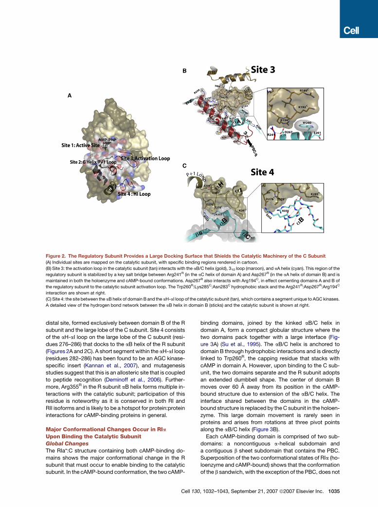
Figure 2. The Regulatory Subunit Provides a Large Docking Surface that Shields the Catalytic Machinery of the C Subunit
(A) Individual sites are mapped on the catalytic subunit, with specific binding regions rendered in cartoon.
(B) Site 3: the activation loop in the catalytic subunit (tan) interacts with the aB/C helix (gold), 310 loop (maroon), and aA helix (cyan). This region of the
regulatory subunit is stabilized by a key salt bridge between Arg241R (in the aC helix of domain A) and Asp267R (in the aA helix of domain B) and is
maintained in both the holoenzyme and cAMP-bound conformations. Asp267R also interacts with Arg194C, in effect cementing domains A and B of
the regulatory subunit to the catalytic subunit activation loop. The Trp260R:Lys285C:Asn283C hydrophobic stack and the Arg241R:Asp267R:Arg194C
interaction are shown at right.
(C) Site 4: the site between the aB helix of domain B and the aH-aI loop of the catalytic subunit (tan), which contains a segment unique to AGC kinases.
A detailed view of the hydrogen bond network between the aB helix in domain B (sticks) and the catalytic subunit is shown at right.
distal site, formed exclusively between domain B of the R
subunit and the large lobe of the C subunit. Site 4 consists
of the aH-aI loop on the large lobe of the C subunit (resi-
dues 276–286) that docks to the aB helix of the R subunit
(Figures 2A and 2C). A short segment within the aH-aI loop
(residues 282–286) has been found to be an AGC kinase-
specific insert (Kannan et al., 2007), and mutagenesis
studies suggest that this is an allosteric site that is coupled
to peptide recognition (Deminoff et al., 2006). Further-
more, Arg355R in the R subunit aB helix forms multiple in-
teractions with the catalytic subunit; participation of this
residue is noteworthy as it is conserved in both RI and
RII isoforms and is likely to be a hotspot for protein:protein
interactions for cAMP-binding proteins in general.
Major Conformational Changes Occur in RIaUpon Binding the Catalytic SubunitGlobal Changes
The RIa*:C structure containing both cAMP-binding do-
mains shows the major conformational change in the R
subunit that must occur to enable binding to the catalytic
subunit. In the cAMP-bound conformation, the two cAMP-
Cell 1
binding domains, joined by the kinked aB/C helix in
domain A, form a compact globular structure where the
two domains pack together with a large interface (Fig-
ure 3A) (Su et al., 1995). The aB/C helix is anchored to
domain B through hydrophobic interactions and is directly
linked to Trp260R, the capping residue that stacks with
cAMP in domain A. However, upon binding to the C sub-
unit, the two domains separate and the R subunit adopts
an extended dumbbell shape. The center of domain B
moves over 60 A away from its position in the cAMP-
bound structure due to extension of the aB/C helix. The
interface shared between the domains in the cAMP-
bound structure is replaced by the C subunit in the holoen-
zyme. This large domain movement is rarely seen in
proteins and arises from rotations at three pivot points
along the aB/C helix (Figure 3B).
Each cAMP-binding domain is comprised of two sub-
domains: a noncontiguous a-helical subdomain and
a contiguous b sheet subdomain that contains the PBC.
Superposition of the two conformational states of RIa (ho-
loenzyme and cAMP-bound) shows that the conformation
of the b sandwich, with the exception of the PBC, does not
30, 1032–1043, September 21, 2007 ª2007 Elsevier Inc. 1035
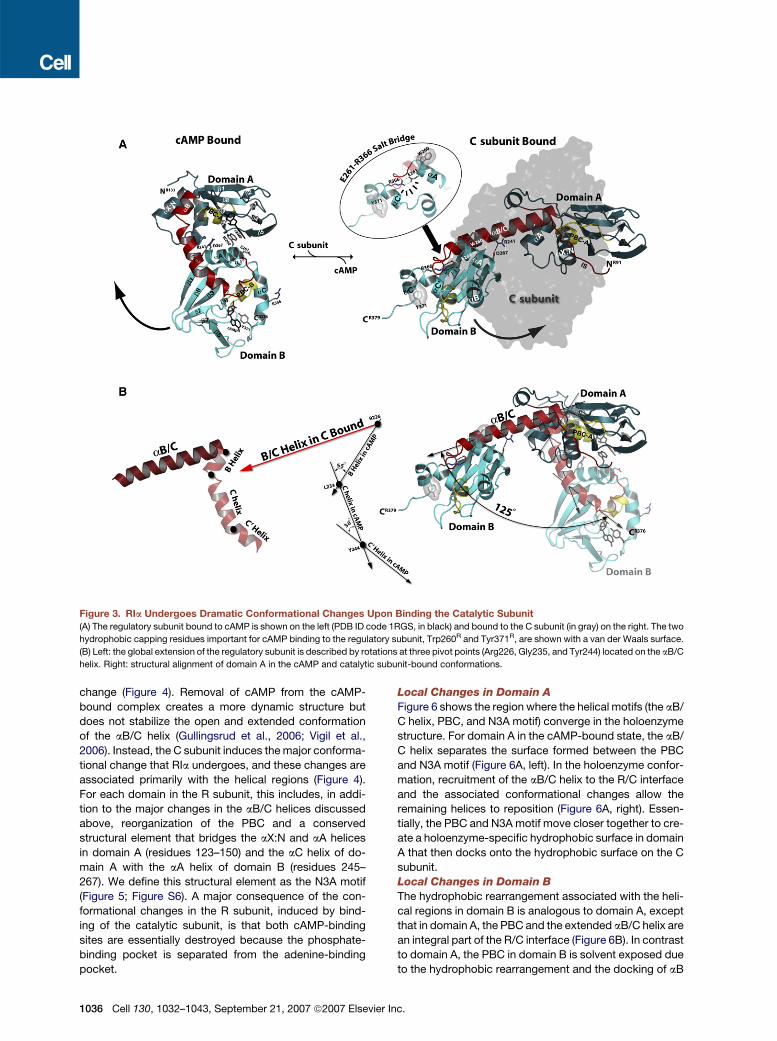
Figure 3. RIa Undergoes Dramatic Conformational Changes Upon Binding the Catalytic Subunit
(A) The regulatory subunit bound to cAMP is shown on the left (PDB ID code 1RGS, in black) and bound to the C subunit (in gray) on the right. The two
hydrophobic capping residues important for cAMP binding to the regulatory subunit, Trp260R and Tyr371R, are shown with a van der Waals surface.
(B) Left: the global extension of the regulatory subunit is described by rotations at three pivot points (Arg226, Gly235, and Tyr244) located on the aB/C
helix. Right: structural alignment of domain A in the cAMP and catalytic subunit-bound conformations.
change (Figure 4). Removal of cAMP from the cAMP-
bound complex creates a more dynamic structure but
does not stabilize the open and extended conformation
of the aB/C helix (Gullingsrud et al., 2006; Vigil et al.,
2006). Instead, the C subunit induces the major conforma-
tional change that RIa undergoes, and these changes are
associated primarily with the helical regions (Figure 4).
For each domain in the R subunit, this includes, in addi-
tion to the major changes in the aB/C helices discussed
above, reorganization of the PBC and a conserved
structural element that bridges the aX:N and aA helices
in domain A (residues 123–150) and the aC helix of do-
main A with the aA helix of domain B (residues 245–
267). We define this structural element as the N3A motif
(Figure 5; Figure S6). A major consequence of the con-
formational changes in the R subunit, induced by bind-
ing of the catalytic subunit, is that both cAMP-binding
sites are essentially destroyed because the phosphate-
binding pocket is separated from the adenine-binding
pocket.
1036 Cell 130, 1032–1043, September 21, 2007 ª2007 Elsevie
Local Changes in Domain A
Figure 6 shows the region where the helical motifs (the aB/
C helix, PBC, and N3A motif) converge in the holoenzyme
structure. For domain A in the cAMP-bound state, the aB/
C helix separates the surface formed between the PBC
and N3A motif (Figure 6A, left). In the holoenzyme confor-
mation, recruitment of the aB/C helix to the R/C interface
and the associated conformational changes allow the
remaining helices to reposition (Figure 6A, right). Essen-
tially, the PBC and N3A motif move closer together to cre-
ate a holoenzyme-specific hydrophobic surface in domain
A that then docks onto the hydrophobic surface on the C
subunit.
Local Changes in Domain B
The hydrophobic rearrangement associated with the heli-
cal regions in domain B is analogous to domain A, except
that in domain A, the PBC and the extended aB/C helix are
an integral part of the R/C interface (Figure 6B). In contrast
to domain A, the PBC in domain B is solvent exposed due
to the hydrophobic rearrangement and the docking of aB
r Inc.

Figure 4. Conformational Changes in the Regulatory Subunit Are a Result of Structural Rearrangements in the Helical Regions
(A) Structural alignment of the regulatory subunit cAMP-binding domains in the holoenzyme conformation. Domains A and B are shown in red and
black, respectively. Ninety-two equivalent Ca atoms from the b barrel region overlap with a root-mean-square deviation of 1.1 A, excluding a short
insert between b4-b5.
(B and C) Comparisons between the two cAMP-binding domains in the cAMP and catalytic subunit-bound conformations. The cAMP-bound
conformation is shown on the left, and the holoenzyme conformation is shown on the right. The two conformations are superimposed in the center.
The aB/C helix is shown in red, and the PBC in yellow. In the two center panels, the cAMP-bound conformation is shown in gray.
helix in domain B to the C subunit (Figure 1). The highly
accessible cAMP-binding site in domain B observed in
our structure explains kinetic studies showing domain B
as the fast association site for cAMP in holoenzyme
(Ogreid and Doskeland, 1981a).
Comparison of domains A and B shows that the aB and
aC helices do not extend in domain B as they do in domain
A. Instead, they remain as distinct helices and form a helix-
turn-helix motif that covers the hydrophobic surface
(Figure 6C). This helix-turn-helix motif provides the hydro-
phobic lid (from Tyr371R) for the PBC in the cAMP-bound
state.
Residues Required for Stabilizing cAMPin the Regulatory Subunit Are Trapped at a RemoteSite in the Holoenzyme StructureThe extended conformation of RIa in the holoenzyme not
only partitions the two cAMP-binding domains but also
separates many of the key residues that anchor cAMP in
the PBC, effectively destroying both cAMP-binding sites.
A common feature for cAMP-binding proteins is hydro-
Cell
phobic capping of the cAMP adenine ring (Berman et al.,
2005). For CAP, HCN, and domain B of RIa, the hydropho-
bic capping residue is located in the aC helix of the cAMP-
binding domain. For domain A of RIa, the capping residue
is Trp260R, located at the beginning of the aA helix of
domain B. Thus, for RIa, both capping residues (Trp260R
for domain A and Tyr371R for domain B) are in domain B
(Figure 3A).
In the holoenzyme structure, the capping residues are
far removed from their respective PBCs. Trp260R moves
over 30 A and docks onto the C subunit activation loop
(Figure 2B). Trp260R, the only residue from domain B
that binds directly to cAMP in domain A, is important for
communication between the two cAMP-binding domains
(Canaves et al., 2000). As illustrated in Figure 6B, Tyr371R
in the cAMP-bound state has a dual role—aromatic stack-
ing with the adenine base and hydrogen bonding to the
conserved Glu324R in the PBC, which binds to the 20OH
of the ribose ring of cAMP. Mutational studies confirm
the importance of this residue for cAMP binding (Bubis
et al., 1988a, 1988b; Kapphahn and Shabb, 1997). In
130, 1032–1043, September 21, 2007 ª2007 Elsevier Inc. 1037

Figure 5. Schematic Diagram of the Structural Motifs in the Regulatory Subunit
(A) Sequence alignment of domains A and B. Residues in the gray boxes belong to domain A but are also aligned as part of the N3A motif of domain B.
(B) Cartoon schematic of the major structural elements of the regulatory subunit in the holoenzyme conformation.
contrast, in the holoenzyme conformation, Tyr371R is 13 A
away from the PBC. Thus, binding of the catalytic subunit
to RIa prohibits many interactions that are needed to sta-
bilize the cAMP-bound structure (see Supplemental Data
for additional sites).
The Glu261-Arg366 Salt Bridge Functions to Trapthe Two cAMP Capping ResiduesThe holoenzyme structure reveals a salt bridge formed
between Glu261R and Arg366R. These two residues not
only position the RIa C-terminal tail but also sequester
the two adenine capping residues (Trp260R and Tyr371R)
away from their cAMP-binding sites. In the cAMP-bound
conformation, both of these highly conserved salt bridge
residues are 15 A apart where Arg366R is exposed to sol-
vent and Glu261R is near the domain interface. It is only in
the holoenzyme conformation that their true function can
be appreciated. In effect, the salt bridge traps both hydro-
phobic residues far away from the cAMP-binding sites
and forms a communication path that links the two R sub-
unit cAMP-binding domains. Our holoenzyme structure
provides a molecular model explaining the ordered and
highly cooperative pathway for the activation of the type
I holoenzyme. The biochemical details for this model
1038 Cell 130, 1032–1043, September 21, 2007 ª2007 Elsevie
were first proposed based on kinetic arguments (Ogreid
and Doskeland, 1981a, 1981b) and were then confirmed
with mutants of the essential arginine residues in the
cAMP-binding pocket (Arg209R for domain A and
Arg333R for domain B) (Herberg et al., 1996).
To test this model and the contribution of the electro-
static trapping of the capping residues, we engineered
four RIa(91–379) mutants (W260A, Y371A, E261A, and
R366A) and measured the effect of these mutations on
PKA activation. For each mutant, the inhibition of the C
subunit was not affected (Figure S4). In contrast, there
are differences in cAMP-mediated activation of PKA, as
measured by a catalytic coupled assay (Cook et al.,
1982) and a fluorescence polarization binding assay (Sal-
danha et al., 2006). Holoenzyme complexes formed with
RIa mutants that contain a substitution of either Trp260R
or Tyr371R with alanine were less sensitive to cAMP
compared to the RIa(91–379) holoenzyme. RIa(91–
379:W260A) requires 4.6-fold more cAMP, while RIa(91–
379:Y371A) requires 9-fold more cAMP (Figure 7A;
Figure S5). The difference for the W260A mutation can
be attributed to the missing hydrophobic capping abilities
of the aromatic side chain. The larger difference observed
for the Y371A mutation is most likely due to the absence of
r Inc.

Figure 6. Binding of the Catalytic Subunit Reorganizes the N3A Motif and the Phosphate-Binding Cassette in the Regulatory
Subunit to Create a Contiguous Hydrophobic Interface
(A) Comparison of the helical regions in domain A between the cAMP (left) and catalytic subunit-bound (right) conformations. Movement of the helical
regions is mediated by hydrophobic rearrangement of the hinge residues in the PBC (Ile203R and Leu204R), aB helix (Tyr229R), and 310 loop (Leu135R).
(B) Comparison of domain B in the cAMP and catalytic subunit-bound conformations, highlighting the C-terminal tail (red). In domain B, the helical
rearrangements are similar to domain A where residues in the PBC (Leu327R and Leu328R), aB helix (Phe353R), and 310 loop (Ile253R and Leu254R)
come together.
(C) Comparison between domains A and B in the holoenzyme conformation. In domain A, the N3A motif (residues 123–150) and PBC come together
and serve as a docking surface for the P+1 loop (black) and the aG helix (dark tan) of the catalytic subunit. In domain B, a similar hydrophobic interface
is formed between the N3A motif (residues 245–367) and PBC; however, the C-terminal tail (aB, aC0, and aC0 0 helices) lies on top of the hydrophobic
interface.
both the aromatic cap and hydrogen bond, which together
help stabilize cAMP in domain B.
In contrast, holoenzyme complexes formed with RIa
mutants that contain a substitution of either Glu261R or
Arg366R with alanine were more sensitive to cAMP activa-
tion. The EC50 decreased from 13.5 nM for RIa(91–379)
to 4.7 and 6.6 nM for RIa(91–379:E261A) and RIa(91–
379:R366A) mutants, respectively. The differences in
Cell 1
EC50 values for these salt bridge-deficient mutants are
likely to be greater than 3-fold since 10 nM C subunit
was used in our assays. Nevertheless, these results con-
clusively show that disrupting the salt bridge makes the
holoenzyme more sensitive to cAMP and shifts the equilib-
rium toward a more ‘‘activation-prone’’ state. Not only are
Glu261R and Arg366R conserved in all regulatory subunit
isoforms, their homologous counterparts in domain A
30, 1032–1043, September 21, 2007 ª2007 Elsevier Inc. 1039

Figure 7. A RIa Electrostatic Interaction in the Holoenzyme Conformation Functions as a ‘‘Capping Residue Trap’’ Important forPKA Activation
(A) Left: the salt bridge between Glu261R and Arg366R structurally couples the two hydrophobic capping residues for domain A and domain B,
Trp260R and Tyr371R, respectively. Center: the effect of RIa(91–379) (black squares), RIa(91–379:W260A) (upward-pointing triangles), RIa(91–
379:Y371A) (downward-pointing triangles), RIa(91–379:E261A) (red circles), and RIa(91–379:R366A) (blue diamonds) on PKA activation by cAMP
measured by the fluorescence polarization assay. Right: fold changes are given relative to RIa(91–379) data. Binding curves were fit using GraphPad
Prism 4 software; error bars indicate the standard error of the mean.
(B) Stepwise model of PKA activation by cAMP.
(Glu143R and Arg241R, respectively) provide equally
important contributions to the molecular architecture of
the holoenzyme (Figure S6).
The highly cooperative interaction between the two tan-
dem cAMP-binding domains of RIa allows the enzyme to
respond rapidly to the second messenger cAMP. Data
from our study and others suggest that several factors con-
tribute to this cooperative process. Comparison of the Hill
coefficients in the cAMP activation data for both capping
residue mutants relative to wild-type RIa suggests that
these residues play an important role in the cooperative
cAMP activation process. The Hill coefficient was reduced
significantly from 1.5 for wild-type to 0.9 and 1 for W260A
and Y371A, respectively (Figure 7A). Mutations that re-
move the salt bridge between Glu261 and Arg366 also
show reductions in the Hill coefficient (1.2 for both E261A
1040 Cell 130, 1032–1043, September 21, 2007 ª2007 Elsevie
and R366A), but the protein concentrations used in our
assays may have limited our ability to determine true Hill
coefficients since titration effects will also influence these
values. Furthermore, previous studies show that Arg241R,
which mediates a salt bridge between domain A
(Arg241R) and domain B (Asp267R) (Figure 3A; Figure S6),
not only disrupts high-affinity cAMP binding but also plays
an important role in the cooperative coupling between the
two domains (Symcox et al., 1994). In light of both our
data and others, it is apparent that cooperativity involves
not just one residue, but a number of residues that all con-
tribute to the activation process in a synergistic way.
Model of PKA Activation by cAMPPrevious biochemical data proposed an ordered and
sequential pathway of cAMP binding to the type Ia
r Inc.

holoenzyme in which cAMP must first bind to domain B
and then to domain A (Herberg et al., 1996). Our structural
and mutagenesis data together provide corroboration for
this mechanism and allow us to propose a molecular ex-
planation for the highly ordered pathway for activation
by cAMP in which domain B serves as a ‘‘gatekeeper’’
for cAMP access to domain A (Figure 7B).
� Step 1: cAMP first binds to the PBC in domain B. The
PBC in domain B is more accessible than in domain
A. The cAMP-binding site in domain A is masked by
the R/C interface so that Trp260R and Arg241R, key
residues that stabilize cAMP binding, are not acces-
sible. In fact, Trp260R, the hydrophobic capping res-
idue for domain A, not only is 30 A from the PBC in
domain A but also is docked to the activation loop
of the C subunit (Figure 2B). Furthermore, the PBC
in domain A is partially occluded by the C subunit
at the site 2 interface (Figure 1; Figure S2). Specifi-
cally, Tyr247C (in the aG helix of the C subunit)
hydrogen bonds to Tyr205R, and the two subunits
are docked through a hydrophobic interface at this
site. These structural details are consistent with
studies that find domain B to be the fast association
site for cAMP in holoenzyme (Ogreid and Doskeland,
1981a).
� Step 2: We predict that recruitment of the C-terminal
tail to stabilize cAMP in the PBC of domain B will dis-
rupt the Glu261R-Arg366R salt bridge. Both our mu-
tational studies and others (Kapphahn and Shabb,
1997) show Tyr371R to be a critical element that
influences PKA activation by cAMP. Mutation of
Tyr371R to alanine results in a 9-fold increase in the
level of cAMP needed to activate PKA compared
to wild-type RIa, presumably due to removal of the
hydrophobic and hydrogen-bonding capabilities of
this residue. In addition, single point mutations of ei-
ther Glu261R or Arg366R that disrupt the salt bridge
require 3-fold less cAMP to activate PKA, suggesting
that positioning of the C-terminal tail is destabilized
in the absence of the salt bridge.
� Step 3: The R subunit undergoes a large conforma-
tional change in response to uncoupling the
Glu261R-Arg366R salt bridge. Breaking the salt
bridge also releases Trp260R, the capping residue
for cAMP binding in domain A. Several observations
support this idea. First, as seen in Figure 2C, the aB
helix in domain B interacts with the aH-aI loop of C.
The movement of the C-terminal tail toward the PBC
in domain B weakens the interaction between the C
subunit and domain B, thereby facilitating the con-
formational change. Second, in the holoenzyme
complex, Trp260R is buried in the R/C interface.
Since the Glu261R-Arg366R interaction structurally
couples the two hydrophobic capping residues,
Trp260R and Tyr371R, docking of cAMP to domain
B breaks the salt bridge and pulls Trp260R away
from the C subunit activation loop. These motions
Cell
collectively destabilize the extended aB/C helix,
and the concerted motions of domain B bring
Trp260R toward the PBC in domain A.
� Step 4: Binding of a second molecule of cAMP to the
PBC in domain A is stabilized by Trp260R. Mutation
of Trp260R to alanine showed a 4.6-fold decrease
in cAMP sensitivity for PKA activation. It is apparent
in our holoenzyme structure that a second cAMP
molecule can only bind to the PBC in domain A if
this domain is dislodged from the C subunit. It
remains to be established whether the C subunit dis-
sociates from the pseudosubstrate site in the R sub-
unit before or after trapping cAMP in domain A, or
whether these steps are coordinated.
� Step 5: In the final step, release of the C subunit from
the inhibitor site of the R subunit leads to activation
of PKA.
ConclusionIn this report, we describe the structure of the PKA cata-
lytic subunit bound to a deletion mutant of RIa containing
both cAMP-binding domains. The structure demonstrates
the exceptional mobility of the cAMP-binding domains in
RIa and confirms that there is a large movement of domain
B relative to domain A as the R subunit shuttles between
its binding partners, namely the catalytic subunit and
cAMP. The conversion of the globular conformation of
the cAMP-bound structure into a dumbbell-shaped holo-
enzyme complex, in which the two cAMP-binding
domains are separated, is mediated by extension of the
aB/C helix of domain A. The RIa*:C structure also shows
that the aB and aC helices in domain B are equally
dynamic, but their conformations are very different from
the aB/C helices in domain A. When bound to the C sub-
unit, RIa utilizes a unique set of residues that stabilize the
C subunit-bound conformation without directly participat-
ing in the R/C interaction. We show through mutagenesis
that a conserved salt bridge plays a significant role in
cAMP activation of PKA, most likely by trapping the two
adenine capping residues in RIa away from their cAMP-
binding sites. These hydrophobic capping residues also
contribute to the cooperative activation of the enzyme
through cAMP. These data provide for the first time a mo-
lecular explanation for the highly ordered pathway
whereby binding of cAMP to domain B leads to the even-
tual activation of kinase activity.
EXPERIMENTAL PROCEDURES
Protein Preparation
The catalytic subunit was expressed and purified in E. coli as described
previously (Gangal et al., 1998). For crystallography, three RIa mutants
(RIa(91–379), RIa(91–379:R209K), and RIa(91–379:R333K)) were gen-
erated by QuikChange site-directed mutagenesis according to the
Stratagene protocol. These mutants lacked the N-terminal dimeriza-
tion/docking domain (residues 1–90). The essential arginine in the PBC
of each cAMP-binding domain was also mutated, Arg209 in domain A
and Arg333 in domain B. Four additional mutants (RIa(91–379:E261A),
RIa(91–379:R366A), RIa(91–379:W260A), and RIa(91–379:Y371A))
130, 1032–1043, September 21, 2007 ª2007 Elsevier Inc. 1041

were generated by QuikChange mutagenesis for biochemical analysis.
All RIa mutants were expressed in E. coli BL21 (DE3) cells (Novagen)
and purified as described previously (Su et al., 1995; Wu et al., 2004).
Holoenzyme Formation for Crystallography
Three RIa mutants (RIa(91–379), RIa(91–379:R209K), and RIa(91–
379:R333K)) were mixed with wild-type C subunit in a 1:1.2 molar ratio
and dialyzed by concentration at 4�C in 10 mM MOPS (pH 7.0), 2 mM
MnCl2, 50 mM NaCl, 2 mM EDTA and EGTA, 1 mM TCEP-HCl, 0.2 mM
AMP-PNP, and 10% glycerol. Holoenzyme was separated from
excess C subunit by gel filtration chromatography as described previ-
ously (Wu et al., 2004).
Crystallization and Data Collection
The RIa(91–379:R333K):C complex was crystallized at 25�C in hanging
drops using the vapor diffusion method in 2.0 M (NH4)2SO4, 0.1 M
citrate (pH 5.5). The crystals were transferred to a cryoprotectant solu-
tion (mother liquor containing 20% glycerol) and flash cooled in liquid
nitrogen. X-ray diffraction data were collected at the SER-CAT inser-
tion device beamline 19ID (Advanced Photon Source, Argonne
National Laboratory, Argonne, IL, USA) on SBC2 3k 3 3k CCD
(ANL). Diffraction data were processed and scaled using HKL2000
(Otwinowski and Minor, 1997). Initial indexing clearly indicated a prim-
itive hexagonal lattice without any ambiguity (distortion index 0.12%).
The final data were integrated and scaled in P3221 (a = b = 125.9 A, c =
141.0 A) with satisfactory statistics. Data processing statistics are
presented in Table S1.
Structure Determination and Refinement
Initial phases of the RIa(91–379:R333K):C complex were generated by
molecular replacement using the RIa(91–244):C complex (Protein Data
Bank ID code 1U7E) (Kim et al., 2005) as a search model in Phaser
(Storoni et al., 2004). Although our initial solvent content analysis pre-
dicted that there would be two molecules per asymmetric unit (VM = 2.2
A3/dalton), a Phaser run in single-model mode unambiguously found
only one molecule (Z score 24-60) in the asymmetric unit, correspond-
ing to a solvent content of 72.3% (VM = 4.5 A3/dalton). The phases
obtained from the Phaser run were improved by solvent flattening
using DM (Cowtan, 1994). The resulting Fo map calculated from the
improved phases showed a well-defined electron density for RIa do-
main B. Secondary structure of RIa domain B was built manually using
XtalView, followed by iterative cycles of structure refinement using RE-
FMAC (CCP4, 1994). The final refinement implementing TLS refine-
ment (Winn et al., 2001) for each chain converged to R and Rfree values
of 0.192 and 0.212, respectively, with excellent geometry (Table S1).
The final model contained residues 13–350 for the C subunit and res-
idues 90–379 for the R subunit and was evaluated using PROCHECK
(Table S1) (Laskowski et al., 1993). Water molecules were
incorporated using wARP (Murshudov et al., 1997) and manually
verified. All figures were made using PyMOL (DeLano Scientific).
cAMP Activation of PKA by Fluorescence Polarization
A new fluorescence polarization assay developed for measuring the
apparent activation constant of PKA for cAMP (EC50) was performed
in parallel with the standard Cook assay. FAM-IP20 used in this study
was synthesized as described previously (Saldanha et al., 2006). Holo-
enzyme was formed in situ by incubating 7 nM C subunit and 8.4 nM R
subunit mutants for 20 min in 25 mM HEPES (pH 7.0), 75 mM KCl, 10
mM MgCl2, 2 mM ATP, 2 mM DTT, 0.005% Triton X-100. FAM-IP20
(1.5 nM) was then added and incubated for an additional 10 min.
Seventy-five microliters of this holoenzyme solution was aliquoted
into each well of a 384-well solid black Fluotrac 200 plate (Greiner
Bio-One, part no. 781076). In all cases, 2-fold dilutions of cAMP rang-
ing from 4 to 4096 nM were added to each well and incubated for
60 min at 25�C. The assay was performed using a GENios Pro micro-
plate reader (Tecan) in which fluorescence polarization was measured
with 485 nm excitation (20 nm band-pass) and 535 nm emission (20 nm
1042 Cell 130, 1032–1043, September 21, 2007 ª2007 Elsevie
band-pass) filters. Data were analyzed using Prism 4 software (Graph-
Pad). Each protein was tested in quadruplicate.
Supplemental Data
Supplemental Data include Supplemental Results and Discussion,
Supplemental Experimental Procedures, Supplemental References,
one table, and six figures and can be found with this article online at
http://www.cell.com/cgi/content/full/130/6/1032/DC1/.
ACKNOWLEDGMENTS
We thank T. Huxford for the data collection. Without him, it would not
have been possible to assess the quality of crystals quickly and to
obtain a full data set on the fragile crystals. We thank J. Gullingsrud
for insights and helpful discussions on the salt bridge; M.S. Deal for
preparation of the catalytic subunit; N.H. Cheung and E.V. Smith-
Nguyen for preparation of the regulatory subunit; N. Nguyen at the
UCSD X-ray facility for assistance; and A. Joachimiak, R. Zhang, Y.
Kim, and the staff of the beamline 19ID at Advanced Photon Source,
Argonne National Laboratory. We specially thank T. Koller for analyzing
the holoenzyme samples by mass spectrometry and verifying muta-
tions. Use of the Advanced Photon Source was supported by the US
Department of Energy, Office of Science, Office of Basic Energy Sci-
ences. This work was funded in part by NIH grant DK 54441 to
S.S.T. C.K. is supported by the American Cancer Fellowship grant
PF-05-238-01-GMC. C.Y.C. is supported by NIH grant GM08326.
Received: December 19, 2006
Revised: March 23, 2007
Accepted: July 13, 2007
Published: September 20, 2007
REFERENCES
Adams, J.A., McGlone, M.L., Gibson, R., and Taylor, S.S. (1995). Phos-
phorylation modulates catalytic function and regulation in the cAMP-
dependent protein kinase. Biochemistry 34, 2447–2454.
Anand, G.S., Law, D., Mandell, J.G., Snead, A.N., Tsigelny, I., Taylor,
S.S., Ten Eyck, L.F., and Komives, E.A. (2003). Identification of the pro-
tein kinase A regulatory RIalpha-catalytic subunit interface by amide H/
2H exchange and protein docking. Proc. Natl. Acad. Sci. USA 100,
13264–13269.
Berman, H.M., Ten Eyck, L.F., Goodsell, D.S., Haste, N.M., Kornev, A.,
and Taylor, S.S. (2005). The cAMP binding domain: an ancient signal-
ing module. Proc. Natl. Acad. Sci. USA 102, 45–50.
Brandon, E.P., Idzerda, R.L., and McKnight, G.S. (1997). PKA iso-
forms, neural pathways, and behaviour: making the connection.
Curr. Opin. Neurobiol. 7, 397–403.
Bubis, J., Neitzel, J.J., Saraswat, L.D., and Taylor, S.S. (1988a). A point
mutation abolishes binding of cAMP to site A in the regulatory subunit
of cAMP-dependent protein kinase. J. Biol. Chem. 263, 9668–9673.
Bubis, J., Saraswat, L.D., and Taylor, S.S. (1988b). Tyrosine-371 con-
tributes to the positive cooperativity between the two cAMP binding
sites in the regulatory subunit of cAMP-dependent protein kinase I.
Biochemistry 27, 1570–1576.
Canaves, J.M., Leon, D.A., and Taylor, S.S. (2000). Consequences of
cAMP-binding site mutations on the structural stability of the type I
regulatory subunit of cAMP-dependent protein kinase. Biochemistry
39, 15022–15031.
Cheng, X., Phelps, C., and Taylor, S.S. (2001). Differential binding of
cAMP-dependent protein kinase regulatory subunit isoforms Ialpha
and IIbeta to the catalytic subunit. J. Biol. Chem. 276, 4102–4108.
CCP4 (Collaborative Computational Project, Number 4) (1994). The
CCP4 suite: programs for protein crystallography. Acta Crystallogr.
D Biol. Crystallogr. 50, 760–763.
r Inc.

Cook, P.F., Neville, M.E., Jr., Vrana, K.E., Hartl, F.T., and Roskoski, R.,
Jr. (1982). Adenosine cyclic 30,50-monophosphate dependent protein
kinase: kinetic mechanism for the bovine skeletal muscle catalytic sub-
unit. Biochemistry 21, 5794–5799.
Cowtan, K. (1994). ‘dm’: An automated procedure for phase improve-
ment by density modification. Joint CCP4 and ESF-EACBM Newslett.
Protein Crystallogr. 31, 34–38.
Deminoff, S.J., Howard, S.C., Hester, A., Warner, S., and Herman, P.K.
(2006). Using substrate-binding variants of the cAMP-dependent pro-
tein kinase to identify novel targets and a kinase domain important for
substrate interactions in Saccharomyces cerevisiae. Genetics 173,
1909–1917.
Gangal, M., Cox, S., Lew, J., Clifford, T., Garrod, S.M., Aschbaher, M.,
Taylor, S.S., and Johnson, D.A. (1998). Backbone flexibility of five sites
on the catalytic subunit of cAMP-dependent protein kinase in the open
and closed conformations. Biochemistry 37, 13728–13735.
Gullingsrud, J., Kim, C., Taylor, S.S., and McCammon, J.A. (2006).
Dynamic binding of PKA regulatory subunit RI alpha. Structure 14,
141–149.
Herberg, F.W., Taylor, S.S., and Dostmann, W.R. (1996). Active site
mutations define the pathway for the cooperative activation of
cAMP-dependent protein kinase. Biochemistry 35, 2934–2942.
Johnson, D.A., Akamine, P., Radzio-Andzelm, E., Madhusudan, M.,
and Taylor, S.S. (2001). Dynamics of cAMP-dependent protein kinase.
Chem. Rev. 101, 2243–2270.
Kannan, N., Haste, N., Taylor, S.S., and Neuwald, A.F. (2007). The hall-
mark of AGC kinase functional divergence is its C-terminal tail, a cis-
acting regulatory module. Proc. Natl. Acad. Sci. USA 104, 1272–1277.
Kapphahn, M.A., and Shabb, J.B. (1997). Contribution of the carboxyl-
terminal regional of the cAMP-dependent protein kinase type I alpha
regulatory subunit to cyclic nucleotide interactions. Arch. Biochem.
Biophys. 348, 347–356.
Kim, C., Xuong, N.H., and Taylor, S.S. (2005). Crystal structure of
a complex between the catalytic and regulatory (RIalpha) subunits of
PKA. Science 307, 690–696.
Kinderman, F.S., Kim, C., von Daake, S., Ma, Y., Pham, B.Q., Sprag-
gon, G., Xuong, N.H., Jennings, P.A., and Taylor, S.S. (2006). A
dynamic mechanism for AKAP binding to RII isoforms of cAMP-depen-
dent protein kinase. Mol. Cell 24, 397–408.
Knighton, D.R., Xuong, N.H., Taylor, S.S., and Sowadski, J.M. (1991a).
Crystallization studies of cAMP-dependent protein kinase. Cocrystals
of the catalytic subunit with a 20 amino acid residue peptide inhibitor
and MgATP diffract to 3.0 A resolution. J. Mol. Biol. 220, 217–220.
Knighton, D.R., Zheng, J.H., Ten Eyck, L.F., Ashford, V.A., Xuong,
N.H., Taylor, S.S., and Sowadski, J.M. (1991b). Crystal structure of
the catalytic subunit of cyclic adenosine monophosphate-dependent
protein kinase. Science 253, 407–414.
Laskowski, R.A., MacArthur, M.W., Moss, D.S., and Thornton, J.M.
(1993). PROCHECK: a program to check the stereochemical quality
of protein structures. J. Appl. Cryst. 26, 283–291.
Murshudov, G.N., Vagin, A.A., and Dodson, E.J. (1997). Refinement of
macromolecular structures by the maximum-likelihood method. Acta
Crystallogr. D Biol. Crystallogr. 53, 240–255.
Cell 1
Newlon, M.G., Roy, M., Morikis, D., Carr, D.W., Westphal, R., Scott,
J.D., and Jennings, P.A. (2001). A novel mechanism of PKA anchoring
revealed by solution structures of anchoring complexes. EMBO J. 20,
1651–1662.
Nolen, B., Taylor, S., and Ghosh, G. (2004). Regulation of protein
kinases; controlling activity through activation segment conformation.
Mol. Cell 15, 661–675.
Ogreid, D., and Doskeland, S.O. (1981a). The kinetics of association of
cyclic AMP to the two types of binding sites associated with protein
kinase II from bovine myocardium. FEBS Lett. 129, 287–292.
Ogreid, D., and Doskeland, S.O. (1981b). The kinetics of the interaction
between cyclic AMP and the regulatory moiety of protein kinase II.
Evidence for interaction between the binding sites for cyclic AMP.
FEBS Lett. 129, 282–286.
Otwinowski, Z., and Minor, W. (1997). Processing of X-ray diffraction
data collected in oscillation mode. Methods Enzymol. 276, 307–326.
Saldanha, S.A., Kaler, G., Cottam, H.B., Abagyan, R., and Taylor, S.S.
(2006). Assay principle for modulators of protein-protein interactions
and its application to non-ATP-competitive ligands targeting protein
kinase A. Anal. Chem. 78, 8265–8272.
Steinberg, R.A., Cauthron, R.D., Symcox, M.M., and Shuntoh, H.
(1993). Autoactivation of catalytic (C alpha) subunit of cyclic AMP-de-
pendent protein kinase by phosphorylation of threonine 197. Mol. Cell.
Biol. 13, 2332–2341.
Storoni, L.C., McCoy, A.J., and Read, R.J. (2004). Likelihood-en-
hanced fast rotation functions. Acta Crystallogr. D Biol. Crystallogr.
60, 432–438.
Su, Y., Dostmann, W.R., Herberg, F.W., Durick, K., Xuong, N.H., Ten
Eyck, L., Taylor, S.S., and Varughese, K.I. (1995). Regulatory subunit
of protein kinase A: structure of deletion mutant with cAMP binding
domains. Science 269, 807–813.
Symcox, M.M., Cauthron, R.D., Ogreid, D., and Steinberg, R.A. (1994).
Arg-242 is necessary for allosteric coupling of cyclic AMP-binding
sites A and B of RI subunit of cyclic AMP-dependent protein kinase.
J. Biol. Chem. 269, 23025–23031.
Vigil, D., Lin, J.H., Sotriffer, C.A., Pennypacker, J.K., McCammon, J.A.,
and Taylor, S.S. (2006). A simple electrostatic switch important in the
activation of type I protein kinase A by cyclic AMP. Protein Sci. 15,
113–121.
Winn, M.D., Isupov, M.N., and Murshudov, G.N. (2001). Use of TLS
parameters to model anisotropic displacements in macromolecular
refinement. Acta Crystallogr. D Biol. Crystallogr. 57, 122–133.
Wu, J., Jones, J.M., Nguyen-Huu, X., Ten Eyck, L.F., and Taylor, S.S.
(2004). Crystal structures of RIalpha subunit of cyclic adenosine 50-
monophosphate (cAMP)-dependent protein kinase complexed with
(Rp)-adenosine 30,50-cyclic monophosphothioate and (Sp)-adenosine
30,50-cyclic monophosphothioate, the phosphothioate analogues of
cAMP. Biochemistry 43, 6620–6629.
Accession Numbers
The coordinates for the structure described herein have been depos-
ited in the Protein Data Bank under the ID code 2QCS.
30, 1032–1043, September 21, 2007 ª2007 Elsevier Inc. 1043