plasma exchange for thrombotic microangiopathy secondary
TRANSCRIPT
CASE REPORT Open Access
Plasma exchange for thromboticmicroangiopathy secondary todermatomyositis associated with acutekidney injury and complement activation: acase report with literature reviewNorifumi Hayashi1*, Keiichirou Okada1, Yuko Tsuruyama1, Yu Kagaya1, Sho Kumano1, Yuki Ishikura2,Kiminobu Takeda2, Masayuki Nanbu2, Keiji Fujimoto1, Hiroki Adachi1 and Hitoshi Yokoyama1
Abstract
Background: Thrombotic microangiopathy (TMA) in patients with connective tissue disease is rare but life-threatening. In particular, the survival rate of patients with dermatomyositis (DM) that develop TMA is low. Theeffectiveness of plasma exchange (PEX) therapy is unclear for the treatment of TMA secondary to DM.
Case presentation: We describe a case of a 28-year-old woman who developed severe DM complicated byaspiration pneumonia from dysphagia and acute kidney injury. The patient was unresponsive to corticosteroids andintravenous immunoglobulin (IVIG) therapy and developed TMA. In this case, immunofluorescence of skin biopsyrevealed that complement activation was involved in the pathogenesis of DM. After 6 PEX therapies,thrombocytopenia improved. She was successfully treated by intensive care and PEX therapy.
Conclusions: PEX therapy was effective to treat TMA secondary to DM associated with complement activation.
Keywords: Dermatomyositis, Thrombotic microangiopathy, Plasma exchange, Complement activation, ADAMTS13
BackgroundDermatomyositis (DM) is an idiopathic inflammatorymuscle disease with characteristic cutaneous manifes-tations such as a periorbital heliotrope rash withedema and violaceous eruption on the knuckles (Got-tron’s papules) [1]. Skin manifestations often accom-pany or precede muscle weakness, which is typicallydistributed symmetrically and proximally [2]. Throm-botic microangiopathy (TMA) in patients with con-nective tissue disease (CTD) is rare but life-threatening. In particular, the survival rate of patientswith DM that develop TMA is low, only 18.8% in asmall case series study [3].
For the treatment of corticosteroid-resistant DM,plasma exchange (PEX) therapy is not effective for musclestrength and functional capacity [4]. Furthermore, becausethe disease is rare, the effectiveness of PEX is unclear forthe treatment of TMA secondary to DM.Here, we describe a patient with DM accompanied by
DM-associated TMA who survived by intensive care andPEX therapy.
Case presentationA 28-year-old woman with no significant medical historywas admitted to the dermatology department for thetreatment of a rash and muscle pain. Eighteen days be-fore admission, erythema appeared over the trunk, face,and extremities. Three days later, pain and weakness ofthe bilateral thigh muscles developed. The muscle weak-ness progressed including difficulty climbing stairs. Onadmission, physical examination revealed Gottron’s
© The Author(s). 2019 Open Access This article is distributed under the terms of the Creative Commons Attribution 4.0International License (http://creativecommons.org/licenses/by/4.0/), which permits unrestricted use, distribution, andreproduction in any medium, provided you give appropriate credit to the original author(s) and the source, provide a link tothe Creative Commons license, and indicate if changes were made. The Creative Commons Public Domain Dedication waiver(http://creativecommons.org/publicdomain/zero/1.0/) applies to the data made available in this article, unless otherwise stated.
* Correspondence: [email protected] of Nephrology, Kanazawa Medical University, 1-1 Daigaku,Uchinada, Ishikawa 920-0293, JapanFull list of author information is available at the end of the article
Hayashi et al. Renal Replacement Therapy (2019) 5:48 https://doi.org/10.1186/s41100-019-0244-5

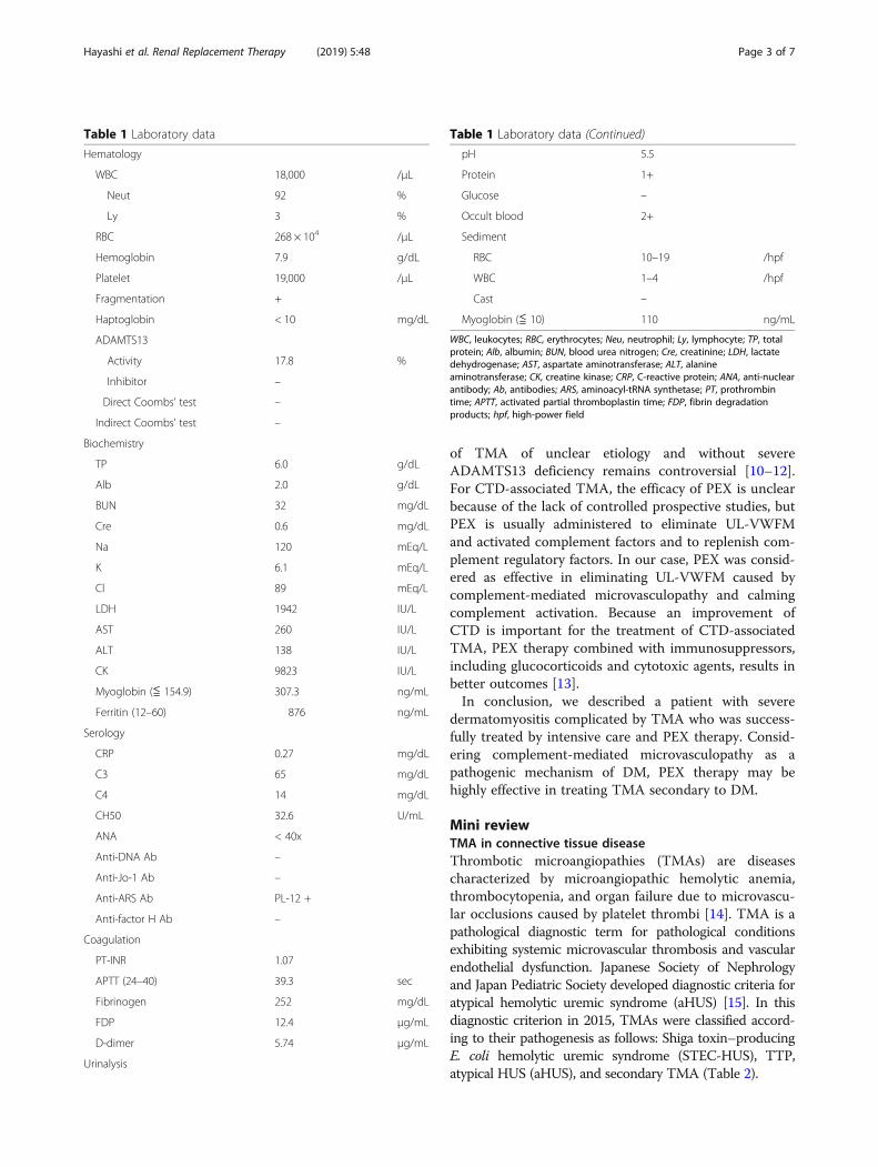
papules on the metacarpophalangeal joints (Fig. 1),heliotrope rash, and muscular pain and weakness in thethighs and upper arms. Laboratory evaluation revealedelevated levels of creatine kinase (CK, 9823 U/L) andmyoglobin (307.3 ng/mL). Serum anti-PL-12 antibody,an anti-aminoacyl tRNA synthetase antibody, was weaklypositive and anti-factor H antibodies were negative(Table 1). Magnetic resonance imaging (MRI) revealedhigh intensity in the lesions of the bilateral thigh mus-cles in T2-weighted and STIR (short tau inversion recov-ery) images (Fig. 2a, b). The pathological findings from askin biopsy showed a perivascular infiltration of inflam-matory cells in the superficial dermis (Fig. 2c), whereasin muscle biopsy, muscle fiber necrosis and inflamma-tory infiltrates were not observed. Immunofluorescenceof the skin biopsy revealed deposition of IgM, fibrinogen,C3d, C4d, and C5b-9 on dermal vessels (Fig. 2d).According to these findings, she was diagnosed with
dermatomyositis (DM), and therapy was initiated withmethylprednisolone pulse (1 g/day for 3 days). However,the response to this therapy was poor, her muscle weak-ness progressed, and she developed severe dysphagia.Therefore, intravenous immunoglobulin (IVIG) therapy(20 g/day for 3 days) was performed. She developed acutekidney injury (AKI) and was referred to our nephrologydepartment. On the 18th day, continuous hemodiafiltra-tion (CHDF) was initiated because of AKI with anuria,hyperkalemia, and hypermyoglobinemia. Furthermore, re-spiratory failure developed because of complications withaspiration pneumonia and exhausted respiratory muscles,and mechanical ventilation was started. Laboratory evalu-ation revealed marked thrombocytopenia and hemolyticanemia with fragmented erythrocytes. Her kidney injuryand the instability of her consciousness suggested a diag-nosis of thrombotic microangiopathy (TMA), and we initi-ated PEX therapy using fresh frozen plasma at the dose of
1.5 times the estimated plasma volume (EPV). EPV wascalculated from the equation given below [5],
EPV Lð Þ ¼ body weight kgð Þ � 0:065� 1−hematocritð Þ
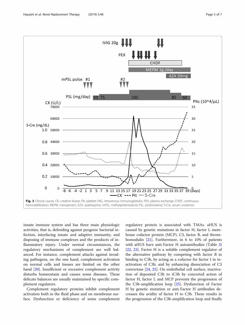
In PEX therapy, we used Plasmacure PE-05 (KurarayMedical, Tokyo, Japan) as a plasma separator. In ourcase, body weight was 49.0 kg and hematocrit was 0.23,respectively, as the initiation of PEX therapy.Her clinical course is shown in Fig. 3. After 6 PEX
therapies, thrombocytopenia improved. After pneumoniawas resolved, azathioprine was added to oral prednisol-one to control DM. On the 36th day, she was withdrawnfrom mechanical ventilation. Her urine volume recov-ered gradually, and hemodialysis therapy was discontin-ued on the 37th day. After long-term rehabilitation, hermuscle strength gradually recovered, and she was dis-charged on the 200th day.
Discussion and conclusionsOur patient was diagnosed with DM by characteristiccutaneous manifestations, progressive muscle weakness,muscle enzyme elevation, and MRI findings. Althoughmuscle biopsy revealed no characteristic features of DM,we considered this result was caused by sampling error.The patient was unresponsive to corticosteroids andIVIG in the first line therapy and developed TMA. How-ever, immunosuppressive therapy was not increased be-cause of aspiration pneumonia from dysphagia, which isfrequently observed in severe DM [6].The pathogenic mechanism underlying the relation-
ship between DM and TMA is unclear. In the pathogen-esis of DM, however, complement C5b-9 is activatedand deposited on the endothelial cell wall of endomysialcapillaries, which leads to necrosis, ischemia, and musclefiber destruction [2]. Low-serum complement level andC5b-9 deposition on dermal vessels suggest thatcomplement-mediated microvasculopathy was involvedin the pathogenesis of DM and TMA in our patient.Our patient did not exhibit a severe deficiency of
ADAMTS13 activity (i.e., < 5% of normal). For CTD-associated TMA, there are fewer patients who have a se-vere deficiency of ADAMTS13 activity (21%) than pa-tients who have a mild-to-moderate deficiency ofADMATS13 activity (79%) [7]. In contrast to thromboticthrombocytopenic purpura (TTP), most patients withCTD-associated TMA have high plasma levels of vonWillebrand factor (VWF) and a mild-to-moderate defi-ciency of ADMATS13 activity [8].For TTP treatment, survival rate improves markedly
by PEX therapy [9]. PEX works by replenishingADMATS13 and by removing ADAMTS13 inhibitorsand UL-VWFM. The efficacy of PEX for the treatment
Fig. 1 A pathognomonic manifestation of dermatomyositis,Gottron’s papule is seen on the metacarpophalangeal joints
Hayashi et al. Renal Replacement Therapy (2019) 5:48 Page 2 of 7
of TMA of unclear etiology and without severeADAMTS13 deficiency remains controversial [10–12].For CTD-associated TMA, the efficacy of PEX is unclearbecause of the lack of controlled prospective studies, butPEX is usually administered to eliminate UL-VWFMand activated complement factors and to replenish com-plement regulatory factors. In our case, PEX was consid-ered as effective in eliminating UL-VWFM caused bycomplement-mediated microvasculopathy and calmingcomplement activation. Because an improvement ofCTD is important for the treatment of CTD-associatedTMA, PEX therapy combined with immunosuppressors,including glucocorticoids and cytotoxic agents, results inbetter outcomes [13].In conclusion, we described a patient with severe
dermatomyositis complicated by TMA who was success-fully treated by intensive care and PEX therapy. Consid-ering complement-mediated microvasculopathy as apathogenic mechanism of DM, PEX therapy may behighly effective in treating TMA secondary to DM.
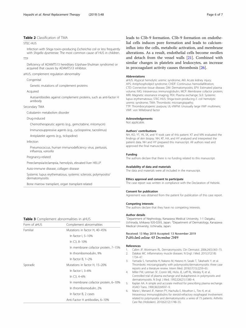
Mini reviewTMA in connective tissue diseaseThrombotic microangiopathies (TMAs) are diseasescharacterized by microangiopathic hemolytic anemia,thrombocytopenia, and organ failure due to microvascu-lar occlusions caused by platelet thrombi [14]. TMA is apathological diagnostic term for pathological conditionsexhibiting systemic microvascular thrombosis and vascularendothelial dysfunction. Japanese Society of Nephrologyand Japan Pediatric Society developed diagnostic criteria foratypical hemolytic uremic syndrome (aHUS) [15]. In thisdiagnostic criterion in 2015, TMAs were classified accord-ing to their pathogenesis as follows: Shiga toxin–producingE. coli hemolytic uremic syndrome (STEC-HUS), TTP,atypical HUS (aHUS), and secondary TMA (Table 2).
Table 1 Laboratory data
Hematology
WBC 18,000 /μL
Neut 92 %
Ly 3 %
RBC 268 × 104 /μL
Hemoglobin 7.9 g/dL
Platelet 19,000 /μL
Fragmentation +
Haptoglobin < 10 mg/dL
ADAMTS13
Activity 17.8 %
Inhibitor –
Direct Coombs’ test –
Indirect Coombs’ test –
Biochemistry
TP 6.0 g/dL
Alb 2.0 g/dL
BUN 32 mg/dL
Cre 0.6 mg/dL
Na 120 mEq/L
K 6.1 mEq/L
Cl 89 mEq/L
LDH 1942 IU/L
AST 260 IU/L
ALT 138 IU/L
CK 9823 IU/L
Myoglobin (≦ 154.9) 307.3 ng/mL
Ferritin (12–60) 876 ng/mL
Serology
CRP 0.27 mg/dL
C3 65 mg/dL
C4 14 mg/dL
CH50 32.6 U/mL
ANA < 40x
Anti-DNA Ab –
Anti-Jo-1 Ab –
Anti-ARS Ab PL-12 +
Anti-factor H Ab –
Coagulation
PT-INR 1.07
APTT (24–40) 39.3 sec
Fibrinogen 252 mg/dL
FDP 12.4 μg/mL
D-dimer 5.74 μg/mL
Urinalysis
Table 1 Laboratory data (Continued)
pH 5.5
Protein 1+
Glucose –
Occult blood 2+
Sediment
RBC 10–19 /hpf
WBC 1–4 /hpf
Cast –
Myoglobin (≦ 10) 110 ng/mL
WBC, leukocytes; RBC, erythrocytes; Neu, neutrophil; Ly, lymphocyte; TP, totalprotein; Alb, albumin; BUN, blood urea nitrogen; Cre, creatinine; LDH, lactatedehydrogenase; AST, aspartate aminotransferase; ALT, alanineaminotransferase; CK, creatine kinase; CRP, C-reactive protein; ANA, anti-nuclearantibody; Ab, antibodies; ARS, aminoacyl-tRNA synthetase; PT, prothrombintime; APTT, activated partial thromboplastin time; FDP, fibrin degradationproducts; hpf, high-power field
Hayashi et al. Renal Replacement Therapy (2019) 5:48 Page 3 of 7

In a Japanese registry of 919 TMA patients [7], CTD-associated TMA is the most common cause of secondaryTMA (221 out of 382 patients, 57.8%). In CTD-associated TMA, TMA complicated by systemic lupuserythematosus (SLE) is the most frequent (41.6%)followed by systemic sclerosis (23.0%) and polymyositis/dermatomyositis (6.3%).TMA occurs in 3 to 9% cases of SLE patients
[16]. The differential diagnosis of TMA with SLEpatients includes malignant hypertension, antipho-spholipid syndrome (APS)/catastrophic APS, TTP,and drug-induced TMA. Careful evaluation of per-ipheral blood smear is useful for the diagnosis ofthese clinical syndromes. The patients with SLE ac-companied by malignant hypertension and TTPshow fragmented red blood cells in peripheral bloodsmear [17].
In the management of CTD-associated TMA, treatingunderlying autoimmune disorders is important. Further-more, in suspected cases, anticoagulation, corticoste-roids, PEX therapy, and IVIG have been used. In somecases showing resistance for these conventional therap-ies, the efficacy of anti-C5 monoclonal antibody eculizu-mab has been reported [16, 18]. Eculizumab binds to theC5 complement with high affinity and blocks the assem-bly of C5b-9. Eculizumab has shown the effectivenessand been approved for the treatment of patients withaHUS [19]. However, the effectiveness of eculizumab forthe treatment of TMA secondary to SLE is stillcontroversial.
The roles of complements in TMADysregulation of complement cascade plays a major rolein the pathogenesis of TMAs. Complement is part of the
Fig. 2 MRI and histological findings of the dermal biopsy. MRI showed high intensity in the lesions of the bilateral thigh muscles in T2-weighted(a) and short tau inversion recovery (b) imaging. In a skin biopsy, a perivascular infiltration of inflammatory cells in the superficial dermis, slightbasal liquefaction degeneration (arrow), and some apoptotic cells (asterisk) in the epidermis were observed (c). C5b-9 deposits on the endothelialcell wall of the dermal vasculature (d)
Hayashi et al. Renal Replacement Therapy (2019) 5:48 Page 4 of 7
innate immune system and has three main physiologicactivities, that is, defending against pyogenic bacterial in-fection, interfacing innate and adaptive immunity, anddisposing of immune complexes and the products of in-flammatory injury. Under normal circumstances, theregulatory mechanisms of complement are well bal-anced. For instance, complement attacks against invad-ing pathogens, on the one hand, complement activationon normal cells and tissues are limited on the otherhand [20]. Insufficient or excessive complement activitydisturbs homeostasis and causes some diseases. Thesedelicate balances are usually maintained by specific com-plement regulators.Complement regulatory proteins inhibit complement
activation both in the fluid phase and on membrane sur-face. Dysfunction or deficiency of some complement
regulatory protein is associated with TMAs. aHUS iscaused by genetic mutations in factor H, factor I, mem-brane cofactor protein (MCP), C3, factor B, and throm-bomodulin [21]. Furthermore, in 6 to 10% of patientswith aHUS have anti-Factor H autoantibodies (Table 3)[22, 23]. Factor H is a soluble complement regulator ofthe alternative pathway by competing with factor B inbinding to C3b, by acting as a cofactor for factor I in in-activation of C3b, and by enhancing dissociation of C3convertase [24, 25]. On endothelial cell surface, inactiva-tion of deposited C3b to iC3b by concerted action offactor H, factor I, and MCP prevents the progression ofthe C3b-amplification loop [25]. Dysfunction of FactorH by genetic mutation or anti-Factor H antibodies de-creases the avidity of factor H to C3b. These results inthe progression of the C3b-amplification loop and finally
Fig. 3 Clinical course. CK, creatine kinase; Plt, platelet; IVIG, intravenous immunoglobulin; PEX, plasma exchange; CHDF, continuoushemodiafiltration; MEPM, meropenem; AZA, azathioprine; mPSL, methylprednisolone; PSL, prednisolone; S-Cre, serum creatinine
Hayashi et al. Renal Replacement Therapy (2019) 5:48 Page 5 of 7
leads to C5b-9 formation. C5b-9 formation on endothe-lial cells induces pore formation and leads to calcium-influx into the cells, metabolic activation, and membranealterations. As a result, endothelial cells become swollenand detach from the vessel walls [21]. Combined withsimilar changes in platelets and leukocytes, an increasein procoagulant activity causes thrombosis [26].
AbbreviationsaHUS: Atypical hemolytic uremic syndrome; AKI: Acute kidney injury;APS: Antiphospholipid syndrome; CHDF: Continuous hemodiafiltration;CTD: Connective tissue disease; DM: Dermatomyositis; EPV: Estimated plasmavolume; IVIG: Intravenous immunoglobulin; MCP: Membrane cofactor protein;MRI: Magnetic resonance imaging; PEX: Plasma exchange; SLE: Systemiclupus erythematosus; STEC-HUS: Shiga-toxin-producing E. coli hemolyticuremic syndrome; TMA: Thrombotic microangiopathy;TTP: Thrombocytopenic purpura; UL-VWFM: Unusually large VWF multimers;VWF: von Willebrand factor
AcknowledgementsNot applicable.
Authors’ contributionsNH, KO, YT, YK, SK, and YI took care of this patient. KT and MN evaluated thefindings of skin biopsy. NH, KF, HA, and HY analyzed and interpreted thepatient data. NH and HY prepared this manuscript. All authors read andapproved the final manuscript.
FundingThe authors declare that there is no funding related to this manuscript.
Availability of data and materialsThe data and materials were all included in the manuscript.
Ethics approval and consent to participateThe case report was written in compliance with the Declaration of Helsinki.
Consent for publicationAgreement was obtained from the patient for publication of this case report.
Competing interestsThe authors declare that they have no competing interests.
Author details1Department of Nephrology, Kanazawa Medical University, 1-1 Daigaku,Uchinada, Ishikawa 920-0293, Japan. 2Department of Dermatology, KanazawaMedical University, Uchinada, Japan.
Received: 13 May 2019 Accepted: 13 November 2019
References1. Callen JP, Wortmann RL. Dermatomyositis. Clin Dermatol. 2006;24(5):363–73.2. Dalakas MC. Inflammatory muscle diseases. N Engl J Med. 2015;372(18):
1734–47.3. Yamada S, Yamashita H, Nakano M, Hatano H, Sasaki T, Takahashi Y, et al.
Thrombotic microangiopathy with polymyositis/dermatomyositis: three casereports and a literature review. Intern Med. 2018;57(15):2259–65.
4. Miller FW, Leitman SF, Cronin ME, Hicks JE, Leff RL, Wesley R, et al.Controlled trial of plasma exchange and leukapheresis in polymyositis anddermatomyositis. N Engl J Med. 1992;326(21):1380–4.
5. Kaplan AA. A simple and accurate method for prescribing plasma exchange.ASAIO Trans. 1990;36(3):M597–9.
6. Marie I, Menard JF, Hatron PY, Hachulla E, Mouthon L, Tiev K, et al.Intravenous immunoglobulins for steroid-refractory esophageal involvementrelated to polymyositis and dermatomyositis: a series of 73 patients. ArthritisCare Res (Hoboken). 2010;62(12):1748–55.
Table 3 Complement abnormalities in aHUS
Form of aHUS Complement abnormalities
Familial Mutations in factor H, 40–45%
In factor I, 5–10%
In C3, 8–10%
In membrane cofactor protein, 7–15%
In thrombomodulin, 9%
In factor B, 1–2%
Sporadic Mutations in factor H, 15–20%
In factor I, 3–6%
In C3, 4–6%
In membrane cofactor protein, 6–10%
In thrombomodulin, 2%
In factor B, 2 cases
Anti-Factor H antibodies, 6–10%
Table 2 Classification of TMA
STEC-HUS
Infection with Shiga toxin–producing Escherichia coli or less frequentlywith Shigella dysenteriae. The most common cause of HUS in children.
TTP
Deficiency of ADAMTS13 hereditary (Upshaw-Shulman syndrome) oracquired that causes by ADAMTS13 inhibitor.
aHUS, complement regulation abnormality
Congenital
Genetic mutations of complement proteins
Acquired
Autoantibodies against complement proteins, such as anti-factor Hantibody
Secondary TMA
Cobalamin metabolism disorder
Drug-induced
Chemotherapeutic agents (e.g., gemcitabine, mitomycin)
Immunosuppressive agents (e.g., cyclosporine, tacrolimus)
Antiplatelet agents (e.g., ticlopidine)
Infection
Pneumococcus, human immunodeficiency virus, pertussis,influenza, varicella
Pregnancy-related
Preeclampsia/eclampsia, hemolysis, elevated liver HELLP
Auto-immune disease, collagen disease
Systemic lupus erythematosus, systemic sclerosis, polymyositis/dermatomyositis
Bone marrow transplant, organ transplant-related
Hayashi et al. Renal Replacement Therapy (2019) 5:48 Page 6 of 7

7. Fujimura Y, Matsumoto M. Registry of 919 patients with thromboticmicroangiopathies across Japan: database of Nara Medical University during1998-2008. Intern Med. 2010;49(1):7–15.
8. Matsuyama T, Kuwana M, Matsumoto M, Isonishi A, Inokuma S, Fujimura Y.Heterogeneous pathogenic processes of thrombotic microangiopathies inpatients with connective tissue diseases. Thromb Haemost. 2009;102(2):371–8.
9. Rock GA, Shumak KH, Buskard NA, Blanchette VS, Kelton JG, Nair RC, et al.Comparison of plasma exchange with plasma infusion in the treatment ofthrombotic thrombocytopenic purpura. Canadian Apheresis Study Group. NEngl J Med 1991;325(6):393–397.
10. Li A, Makar RS, Hurwitz S, Uhl L, Kaufman RM, Stowell CP, et al. Treatmentwith or without plasma exchange for patients with acquired thromboticmicroangiopathy not associated with severe ADAMTS13 deficiency: apropensity score-matched study. Transfusion. 2016;56(8):2069–77.
11. Vesely SK, George JN, Lammle B, Studt JD, Alberio L, El-Harake MA, et al.ADAMTS13 activity in thrombotic thrombocytopenic purpura-hemolyticuremic syndrome: relation to presenting features and clinical outcomes in aprospective cohort of 142 patients. Blood. 2003;102(1):60–8.
12. George JN. Measuring ADAMTS13 activity in patients with suspectedthrombotic thrombocytopenic purpura: when, how, and why? Transfusion.2015;55(1):11–3.
13. Jiang H, An X, Li Y, Sun Y, Shen G, Tu Y, et al. Clinical features andprognostic factors of thrombotic thrombocytopenic purpura associatedwith systemic lupus erythematosus: a literature review of 105 cases from1999 to 2011. Clin Rheumatol. 2014;33(3):419–27.
14. Moake JL. Thrombotic microangiopathies. N Engl J Med. 2002;347(8):589–600.
15. Sawai T, Nangaku M, Ashida A, Fujimaru R, Hataya H, Hidaka Y, et al.Diagnostic criteria for atypical hemolytic uremic syndrome proposed by theJoint Committee of the Japanese Society of Nephrology and the JapanPediatric Society. Clin Exp Nephrol. 2014;18(1):4–9.
16. Kello N, Khoury LE, Marder G, Furie R, Zapantis E, Horowitz DL. Secondarythrombotic microangiopathy in systemic lupus erythematosus andantiphospholipid syndrome, the role of complement and use ofeculizumab: case series and review of literature. Semin Arthritis Rheum.2019;49(1):74–83.
17. Shah AA, Higgins JP, Chakravarty EF. Thrombotic microangiopathichemolytic anemia in a patient with SLE: diagnostic difficulties. Nat Clin PractRheumatol. 2007;3(6):357–62.
18. de Holanda MI, Porto LC, Wagner T, Christiani LF, Palma LMP. Use ofeculizumab in a systemic lupus erythemathosus patient presentingthrombotic microangiopathy and heterozygous deletion in CFHR1-CFHR3. Acase report and systematic review. Clin Rheumatol. 2017;36(12):2859–67.
19. Verhave JC, Wetzels JF, van de Kar NC. Novel aspects of atypical haemolyticuraemic syndrome and the role of eculizumab. Nephrol Dial Transplant.2014;29(Suppl 4):iv131–41.
20. Walport MJ. Complement. First of two parts. N Engl J Med. 2001;344(14):1058–66.
21. Meri S. Complement activation in diseases presenting with thromboticmicroangiopathy. Eur J Intern Med. 2013;24(6):496–502.
22. Jozsi M, Licht C, Strobel S, Zipfel SL, Richter H, Heinen S, et al. Factor Hautoantibodies in atypical hemolytic uremic syndrome correlate withCFHR1/CFHR3 deficiency. Blood. 2008;111(3):1512–4.
23. Dragon-Durey MA, Loirat C, Cloarec S, Macher MA, Blouin J, Nivet H, et al.Anti-factor H autoantibodies associated with atypical hemolytic uremicsyndrome. J Am Soc Nephrol. 2005;16(2):555–63.
24. Noris M, Remuzzi G. Atypical hemolytic-uremic syndrome. N Engl J Med.2009;361(17):1676–87.
25. Jokiranta TS, Jaakola VP, Lehtinen MJ, Parepalo M, Meri S, Goldman A.Structure of complement factor H carboxyl-terminus reveals molecular basisof atypical haemolytic uremic syndrome. EMBO J. 2006;25(8):1784–94.
26. Peerschke EI, Yin W, Ghebrehiwet B. Complement activation on platelets:implications for vascular inflammation and thrombosis. Mol Immunol. 2010;47(13):2170–5.
Publisher’s NoteSpringer Nature remains neutral with regard to jurisdictional claims inpublished maps and institutional affiliations.
Hayashi et al. Renal Replacement Therapy (2019) 5:48 Page 7 of 7