plastid encoded rna polymerase activity and expression of ... · plastid encoded rna polymerase...
TRANSCRIPT

ORIGINAL RESEARCH ARTICLEpublished: 12 August 2014
doi: 10.3389/fpls.2014.00385
Plastid encoded RNA polymerase activity and expression ofphotosynthesis genes required for embryo and seeddevelopment in ArabidopsisDmitry Kremnev and Åsa Strand*
Umeå Plant Science Centre, Department of Plant Physiology, Umeå University, Umeå, Sweden
Edited by:
Thomas Pfannschmidt, UniversityJoseph Fourier Grenoble, France
Reviewed by:
Tatjana Kleine,Ludwig-Maximilians-UniversitätMünchen, GermanySilva Mache, Laboratoire dePhysiologie Cellulaire Végétale,France
*Correspondence:
Åsa Strand, Umeå Plant ScienceCentre, Department of PlantPhysiology, Umeå University,S-901 87 Umeå, Swedene-mail: [email protected]
Chloroplast biogenesis and function is essential for proper plant embryo and seed develop-ment but the molecular mechanisms underlying the role of plastids during embryogenesisare poorly understood. Expression of plastid encoded genes is dependent on two differenttranscription machineries; a plastid-encoded bacterial-type RNA polymerase (PEP) anda nuclear-encoded phage-type RNA polymerase (NEP), which recognize distinct typesof promoters. However, the division of labor between PEP and NEP during plastiddevelopment and in mature chloroplasts is unclear. We show here that PLASTID REDOXINSENSITIVE 2 (PRIN2) and CHLOROPLAST STEM-LOOP BINDING PROTEIN 41 kDa(CSP41b), two proteins identified in plastid nucleoid preparations, are essential for properplant embryo development. Using Co-IP assays and native PAGE we have shown adirect physical interaction between PRIN2 and CSP41b. Moreover, PRIN2 and CSP41bform a distinct protein complex in vitro that binds DNA. The prin2.2 and csp41b-2 singlemutants displayed pale phenotypes, abnormal chloroplasts with reduced transcript levels ofphotosynthesis genes and defects in embryo development.The respective csp41b-2prin2.2homo/heterozygote double mutants produced abnormal white colored ovules and shrunkenseeds. Thus, the csp41b-2prin2.2 double mutant is embryo lethal. In silico analysis ofavailable array data showed that a large number of genes traditionally classified as PEPdependent genes are transcribed during early embryo development from the pre-globularstage to the mature-green-stage.Taken together, our results suggest that PEP activity andconsequently the switch from NEP to PEP activity, is essential during embryo developmentand that the PRIN2-CSP41b DNA binding protein complex possibly is important for full PEPactivity during this process.
Keywords: chloroplast, PEP, NEP, embryo development, photosynthesis, PRIN2, CSP41b
INTRODUCTIONThe chloroplasts house the photosynthetic light reactions wheresunlight is converted into chemical energy. Plastids are alsothe location of a number of vital metabolic pathways, includ-ing primary carbon metabolism and the biosynthesis of fattyacids, amino acids, and tetrapyrroles. Chloroplast function isrequired throughout the life cycle of the plant and compromisedactivity can result in embryo lethality. Abortion of developingembryos is known to occur when amino acid, nucleotide orfatty acid biosynthesis is impaired, or when import of chloro-plast proteins and translation are disrupted (McElver et al., 2001;Tzafrir et al., 2003; Hsu et al., 2010; Bryant et al., 2011). In con-trast, disrupting components of the photosynthetic apparatusleads to reduced pigmentation and changed physiology ratherthan embryo lethality (Tzafrir et al., 2003; Bryant et al., 2011).Chloroplasts are detected as early as at the globular stage ofthe embryo (Tejos et al., 2010) and transcript profiling duringembryo development showed a significant increase in the expres-sion of nuclear genes encoding components involved in energyproduction, carbon fixation and photosynthesis already fromthe globular embryogenic stage (Spencer et al., 2007; Le et al.,
2010; Belmonte et al., 2013). Furthermore, it has been indicatedthat chloroplasts can provide the embryo with energy and O2
required for biosynthesis and respiration (Rolletschek et al., 2003).Additionally, it was shown that Brassica embryos were able tofix CO2 and contributing to embryo growth rate and biomass(Goffman et al., 2005).
Chloroplasts, like mitochondria, evolved from free-livingprokaryotic organisms that entered the eukaryotic cell throughendosymbiosis. The gradual conversion from endosymbiontto organelle during the course of evolution has clearly beenaccompanied by a dramatic reduction in genome size as thechloroplasts lost most of their genes to the nucleus. The genesremaining in the chloroplast genome are related to photosyn-thesis or encode components of the plastid gene expressionmachinery (Wakasugi et al., 2001; Martin et al., 2002). The chloro-plast genes of higher plants are transcribed by at least twotypes of RNA polymerases; the nuclear encoded plastid RNApolymerase (NEP), a T3-T7 bacteriophage type that predomi-nantly mediates the transcription of the house keeping genes(Hedtke et al., 1997, 2000; Puthiyaveetil et al., 2010). The othertype, plastid encoded RNA polymerase (PEP) is a bacterial-type
www.frontiersin.org August 2014 | Volume 5 | Article 385 | 1

Kremnev and Strand PEP activity and embryo development
multi-subunit enzyme that predominantly mediates the tran-scription of photosynthesis-related genes (Allison et al., 1996;De Santis-MacIossek et al., 1999). Most chloroplast genes can betranscribed by both polymerases but they utilize different pro-moter elements (Hajdukiewicz et al., 1997; Pfannschmidt andLiere, 2005). The PEP enzyme recognizes the −10 and −35 cis-elements, similar to those found in bacterial promoters whereasthe NEP enzyme recognizes the YRTA-motif, which can also befound upstream of several genes with PEP promoters indicat-ing that these genes can be transcribed by both polymerases(Pfannschmidt and Liere, 2005). Transcription of the plastidencoded photosynthesis genes during chloroplast developmentand the activation of the photosynthetic reactions are accom-panied by a switch from NEP to PEP activity (Hanaoka et al.,2005). However, the mechanisms underlying this change in majorRNA polymerase activity and the division of labor between NEPand PEP in the chloroplast are unknown (Zhelyazkova et al.,2012).
A large number of proteins have been shown to be asso-ciated with the PEP complex and the components associatedwith PEP changes in response to developmental signals andchanges in the environment (Pfalz and Pfannschmidt, 2013).The variation in the composition of the PEP complex suggeststhat regulation of plastid gene expression is both complex andsophisticated. PLASTID REDOX INSENSITIVE 2 (PRIN2) is anovel plant protein localized to the plastid nucleoids and plas-tid transcriptome analyses demonstrated that PRIN2 is requiredfor full expression of genes transcribed by PEP (Kindgren et al.,2012). The role of the PEP associated proteins is unclear andwhether these proteins contribute to the regulation of plastid geneexpression by environmental and developmental cues remains tobe determined. In order to shed light on the complex regula-tion of PEP activity and to understand the function of PRIN2we pursued an assay to identify interacting protein partnersof PRIN2. We report here a direct interaction between PRIN2and CHLOROPLAST STEM-LOOP BINDING PROTEIN 41 kDa(CSP41b) using three different biochemical methods. CSP41bwas described as an RNA binding protein identified in nucleoidpreparation and was attributed numerous plastid functions, forexample CSP41b was suggested to stimulate both transcriptionand translation in the chloroplast (Pfannschmidt et al., 2000;Yamaguchi et al., 2003; Suzuki et al., 2004; Hassidim et al., 2007;Bollenbach et al., 2009). We also demonstrate that PRIN2 andCSP41b form a distinct DNA binding protein complex in vitroand that the csp41b-2prin2.2 double mutant is embryo lethal.Taken together, our results suggest that PEP activity and con-sequently the switch from NEP to PEP activity, is essential alsoduring embryo development and that the PRIN2-CSP41b proteincomplex potentially is important for full PEP activity during thisprocess.
MATERIALS AND METHODSPLANT MATERIAL AND GROWTH CONDITIONSSeedlings of Arabidopsis thaliana were grown on phytoagarplates containing 1 × Murashige and Skoog salt mixture supple-mented with vitamins (Duchefa) and 2% sucrose. The T-DNAinsertion lines: prin2.2 (GK-772D07-024643) and csp41b-2
(SALK_021748), were obtained from the European ArabidopsisStock Centre. All genotypes are in Colombia ecotype. Seedlingsand rosette plants were grown on soil at 23◦C (16 h light 150 μmolphotons m−2 s−1) and 18◦C (8 h dark).
MORPHOLOGICAL ANALYSISEmbryo isolation was done according to (Perry and Wang,2003). Briefly, The siliques at the desired stage of DAP weredissected under a stereo microscope and the seeds were col-lected. Embryos were gently extruded from the seed coat byapplying pressure on a glass plate covering them. Embryo/seedcoat mixture was loaded a 25% Percoll gradient in isolationbuffer (10 mM potassium phosphate, pH 7.0, 50 mM NaCl,0.1 M sucrose) and centrifuged for 10 min at 800 g. The pel-let was subjected to another round of purification using the25% Percoll gradient. The pellet was re-suspended in isolationbuffer and used for subsequent analysis. For transmission elec-tron microscopy (TEM) pictures, 3-weeks-old plants grown onsoil and the isolated seed embryos were prepared according to(Barajas-Lopez Jde et al., 2013).
RNA ISOLATION, cDNA SYNTHESIS AND REAL-TIME PCRTotal RNA was isolated using Plant RNA Mini Kit (EZNA)and genomic DNA contamination was removed by DNase treat-ment (Fermentas). cDNA was synthesized using the iScriptcDNA kit (Bio-Rad) according to the manufacturer’s instruc-tions and 10 × diluted. Real-Time PCR was performedusing iQSYBR Green Supermix (Bio-Rad), with a final vol-ume of 10 μL. The PCR amplification was done using two-step protocol using the CFX96 Real-Time system (C1000Thermal Cycler; Bio-Rad). All experiments were performedwith three biological and three technical replicates, the rel-ative gene expression was normalized to the expression ofRCE1 (At4g36800) and PP2AA3 (At1g13320). Data analy-sis was done by CFX manager (Bio-Rad) and LinRegPCRsoftware.
EXPRESSION AND PURIFICATION OF RECOMBINANT PROTEINSThe coding sequences of PRIN2 and CSP41b were amplified withPCR. The PCR products were cloned using the NcoI–AccI sites intopET_His1a vector. BL21 Escherichia coli cells were transformedwith the expression constructs and induced for 6 h with 1 mMIPTG. Overexpressed proteins were affinity purified on Ni2+-NTA agarose resin (Qiagen). The pET-His1a expression vectorwas kindly provided by Günter Stier, Umeå University, Sweden.
EMSAThe 197 bp probe containing −196 to +1 PsaA promoter regionwas PCR amplified and labeled at the 3′-end with biotin-14-dCTP using biotin labeling kit (Invitrogen) according to themanufacturer’s instructions. DNA–protein interactions were per-formed in 25 mcL reactions containing following reagents: 2.5 mcLof × 10 binding buffer (100 mM Tris HCl, 250 mM KCl, and10 mM DTT), 1 mcg poly dIdC (Sigma–Aldrich), 2,5% glyc-erol, 0.05% Triton X-100, 5 mM MgCl2, 10 mM EDTA. Thereaction mixture was incubated with DNA and protein at roomtemperature for 30min and was run on 6% native TBE-PAGE in
Frontiers in Plant Science | Plant Physiology August 2014 | Volume 5 | Article 385 | 2

Kremnev and Strand PEP activity and embryo development
x0,5 TBE buffer at 100 V. DNA was transferred to nylon + mem-brane (Amersham) and was UV cross-linked to the membrane,incubated with Streptavidin-HRP and detected by Chemolumi-nescence Nucleic Acid Detection Module (Pierce) according tomanufacturer’s instructions (Shaikhali et al., 2012a,b).
Co-IPTo identify PRIN2 interacting partners, a 35S promoter linkedto the full length PRIN2 coding sequence was cloned into thepGWB16_myc expression construct. Col-0 plants were thentransformed with 35S_pGWB16myc_PRIN2 using the floral-dip method (Clough and Bent, 1998). Two weeks old stabletransformants overexpressing PRIN2 were used for chloro-plast isolation in a two-step 50–25% Percoll gradient asdescribed previously (Aronsson and Jarvis, 2002). Chloroplastproteins were extracted and incubated with 3 mg of anti-cMYC monoclonal antibody (Bio-Site) bound to the proteinG-coated magnetic beads (Dynabeads Protein G Immuno-precipitation, Invitrogen) for 1 h at 4◦C. All the wash-ing steps were performed at 4◦C according to the manufac-turer’s instructions. Immunoprecipitated PRIN2 protein com-plexes were eluted in SDS loading buffer containing 100 mMβ-mercaptoethanol and proteins were separated on 6–12% SDSPAGE. Non-transformed Col-0 plants were used as negative con-trol. For direct protein Co-IP assay, full length PRIN2 andCSP41b coding sequences were cloned into NcoI–NcoI sites ofpRT104_3myc and into SacI–NotI sites of pRT104_3HA vec-tor, respectively. Protoplasts obtained from Arabidopsis Ler-0cell culture were transformed with pRT104_PRIN2_myc andpRT104_CSP41b_HA constructs as described previously by(Doelling and Pikaard, 1993).
ISOLATION OF THYLAKOID COMPLEXES AND BLUE NATIVE PAGEChloroplasts were isolated on a two-step 50–25% Percoll gradientfrom 4 weeks old rosette plants grown in short day as described pre-viously (Aronsson and Jarvis, 2002). Thylakoid membranes werethen purified (Hall et al., 2011) and protein complexes were solubi-lized in BN-solubilization buffer (30 mM HEPES, pH 7.4; 150 mMpotassium acetate; 10% glycerole, 4% digitonin (SIGMA); 1% b-Dodecylmaltoside (SIGMA) for 40 min, 4◦C. DM and digitoninin a mixture was shown to be suitable for better preservation ofmegacomplexes and at the same time good for solubilization (Järviet al., 2011). 35 micro gram protein was loaded onto the 4–12%Bis-Tris Gel (NuPAGEH Novex 1.0 mm, Invitrogen) from eachgenotype.
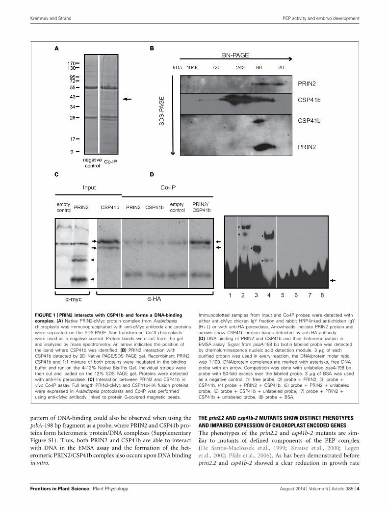
RESULTSPRIN2 AND CSP41B FORM A DISTINCT PROTEIN COMPLEX THAT BINDSDNATo understand the function of PRIN2 we wanted to identifyproteins interacting with PRIN2 in vivo. To achieve this a co-IP approach was used. Full length PRIN2 protein fused to acMyc-tag was expressed in Arabidopsis plants, intact chloroplastswere isolated and PRIN2-containing protein complexes were pre-cipitated with anti-cMyc antibody, proteins were separated onSDS PAGE and distinct bands identified using mass spectrometry(Figure 1A). The Co-IP experiment was performed twice and as
many as 17 bands absent from the negative controls were in totalcut from the gels. Most of the identified proteins were only foundin one experiment and most likely represented unspecific inter-actions. However, one protein, CSP41b was identified in bothexperiments and several peptides with significant scores corre-sponding to CSP41b were identified in each sample (Table S1in Supplementary Material). CSP41b is a conserved chloroplastprotein and the csb41b-2 mutant displayed impaired chloroplasttranscription and plant development (Pfannschmidt et al., 2000;Suzuki et al., 2004; Bollenbach et al., 2009; Qi et al., 2012). Thephenotype of the csb41b-2 mutant was very similar to the phe-notype of the prin2 mutant and it is possible that CSP41b andPRIN2 are involved in the same process. Thus, the identifiedinteraction between PRIN2 and CSP41b was chosen for furtheranalysis.
First, to really confirm the interaction between CSP41b andPRIN2 purified recombinant proteins were separated on 2D elec-trophoresis (Figure 1B; Table S2). PRIN2 forms protein complexesranging from 20–66 kDa while CSP41b is present in high molec-ular weight complexes ranging from 40–700 kDa. When PRIN2and CSP41 are incubated together, PRIN2 seems to break thehigh molecular weight complexes of CSP41b and form a distinctprotein complex with CSP41b suggesting heteromerization. Tofurther confirm in vivo that PRIN2 and CSP41b directly interactwith each other we used a third method by transiently express-ing the full length proteins PRIN2 and CSP41b fused to cMyc-and HA-tags, respectively, in Arabidopsis protoplasts. Two bandsof approximately 50 kDa in size correspond to CSP41b in theCSP41b-HA transformed protoplasts and these could be cyto-plasmic/transite peptide-processed forms of the protein and/orproteolytic fragment of the protein. The PRIN2 protein exists inmonomeric, dimeric and oligomeric forms (unpublished data)and migrates under these conditions as two bands of approxi-mately 20 and 40 kDa, respectively, most likely corresponding tomonomer and dimer. CSP41b was detected in the Co-IP fractionconfirming interaction with PRIN2 (Figure 1C). Thus, using threeindependent methods CSP41b and PRIN2 were shown to directlyinteract.
Both PRIN2 and CSP41b were suggested to regulate transcrip-tion of PEP dependent chloroplast genes (Bollenbach et al., 2009;Kindgren et al., 2012). To investigate if PRIN2 and CSP41b inter-act upon DNA binding in vitro, the proteins were incubated witha 197 bp DNA probe containing −196 to +1 region of the psaApromoter from Arabidopsis. In the electrophoretic mobility shiftassay (EMSA) both PRIN2 and CSP41b bound the labeled probe,PRIN2 formed at least two distinct complexes with DNA, whileCSP41b formed only one DNA-protein complex (Figure 1D).PRIN2 and CSP41b are about 15 and 40 kDa, respectively, andthe difference in the migration of the protein/DNA complexes ofPRIN2 and CSP41b suggests that PRIN2 forms higher molecu-lar weight oligomeric complexes and/or binds to several regionsof the DNA probe. When PRIN2 and CSP41b were incubatedtogether with DNA, a new band, intermediate in size to what wasobserved for the individual proteins, was detected that most likelycorresponded to a psaA197-PRIN2/CSP41b heteromeric complex.The competition reactions with unlabeled psaA-198 bp confirmthe DNA binding capacity of PRIN2 and CSP41b. A similar
www.frontiersin.org August 2014 | Volume 5 | Article 385 | 3
Kremnev and Strand PEP activity and embryo development
FIGURE 1 | PRIN2 interacts with CSP41b and forms a DNA-binding
complex. (A) Native PRIN2-cMyc protein complex from Arabidopsischloroplasts was immunoprecipitated with anti-cMyc antibody and proteinswere separated on the SDS-PAGE. Non-transformed Col-0 chloroplastswere used as a negative control. Protein bands were cut from the geland analyzed by mass spectrometry. An arrow indicates the position ofthe band where CSP41b was identified. (B) PRIN2 interaction withCSP41b detected by 2D Native PAGE/SDS PAGE gel. Recombinant PRIN2,CSP41b and 1:1 mixture of both proteins were incubated in the bindingbuffer and run on the 4–12% Native Bis-Tris Gel. Individual stripes werethen cut and loaded on the 12% SDS PAGE gel. Proteins were detectedwith anti-His peroxidase. (C) Interaction between PRIN2 and CSP41b invivo Co-IP assay. Full length PRIN2-cMyc and CSP41b-HA fusion proteinswere expressed in Arabidopsis protoplasts and Co-IP was performedusing anti-cMyc antibody linked to protein G-covered magnetic beads.
Immunoblotted samples from input and Co-IP probes were detected witheither anti-cMyc chicken IgY fraction and rabbit HRP-linked anti-chicken IgY(H+L) or with anti-HA peroxidase. Arrowheads indicate PRIN2 protein andarrows show CSP41b protein bands detected by anti-HA antibody.(D) DNA binding of PRIN2 and CSP41b and their heteromerization inEMSA assay. Signal from psaA-198 bp biotin labeled probe was detectedby chemoluminescence nucleic acid detection module. 3 μg of eachpurified protein was used in every reaction, the DNA/protein molar ratiowas 1:100. DNA/protein complexes are marked with asterisks, free DNAprobe with an arrow. Competition was done with unlabeled psaA-198 bpprobe with 50-fold excess over the labeled probe. 3 μg of BSA was usedas a negative control. (1) free probe, (2) probe + PRIN2, (3) probe +CSP41b, (4) probe + PRIN2 + CSP41b, (5) probe + PRIN2 + unlabeledprobe, (6) probe + CSP41b + unlabeled probe, (7) probe + PRIN2 +CSP41b + unlabeled probe, (8) probe + BSA.
pattern of DNA-binding could also be observed when using thepsbA-198 bp fragment as a probe, where PRIN2 and CSP41b pro-teins form heteromeric protein/DNA complexes (SupplementaryFigure S1). Thus, both PRIN2 and CSP41b are able to interactwith DNA in the EMSA assay and the formation of the het-eromeric PRIN2/CSP41b complex also occurs upon DNA bindingin vitro.
THE prin2.2 AND csp41b-2 MUTANTS SHOW DISTINCT PHENOTYPESAND IMPAIRED EXPRESSION OF CHLOROPLAST ENCODED GENESThe phenotypes of the prin2.2 and csp41b-2 mutants are sim-ilar to mutants of defined components of the PEP complex(De Santis-MacIossek et al., 1999; Krause et al., 2000; Legenet al., 2002; Pfalz et al., 2006). As has been demonstrated beforeprin2.2 and csp41b-2 showed a clear reduction in growth rate
Frontiers in Plant Science | Plant Physiology August 2014 | Volume 5 | Article 385 | 4
Kremnev and Strand PEP activity and embryo development
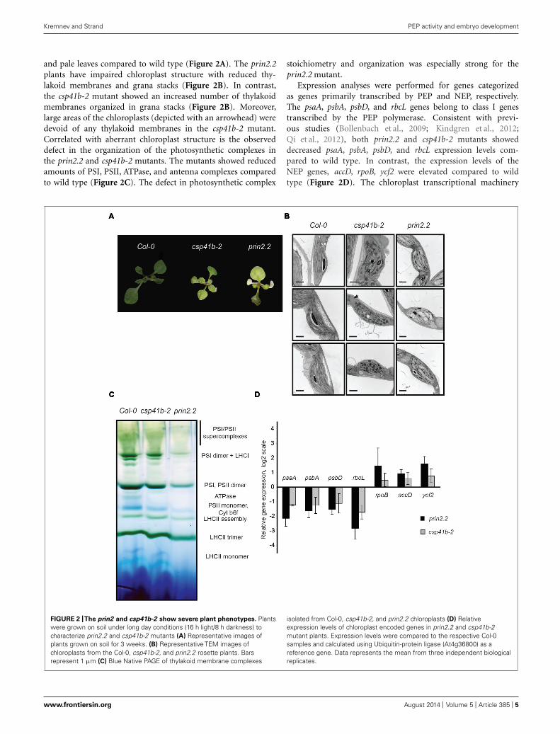
and pale leaves compared to wild type (Figure 2A). The prin2.2plants have impaired chloroplast structure with reduced thy-lakoid membranes and grana stacks (Figure 2B). In contrast,the csp41b-2 mutant showed an increased number of thylakoidmembranes organized in grana stacks (Figure 2B). Moreover,large areas of the chloroplasts (depicted with an arrowhead) weredevoid of any thylakoid membranes in the csp41b-2 mutant.Correlated with aberrant chloroplast structure is the observeddefect in the organization of the photosynthetic complexes inthe prin2.2 and csp41b-2 mutants. The mutants showed reducedamounts of PSI, PSII, ATPase, and antenna complexes comparedto wild type (Figure 2C). The defect in photosynthetic complex
stoichiometry and organization was especially strong for theprin2.2 mutant.
Expression analyses were performed for genes categorizedas genes primarily transcribed by PEP and NEP, respectively.The psaA, psbA, psbD, and rbcL genes belong to class I genestranscribed by the PEP polymerase. Consistent with previ-ous studies (Bollenbach et al., 2009; Kindgren et al., 2012;Qi et al., 2012), both prin2.2 and csp41b-2 mutants showeddecreased psaA, psbA, psbD, and rbcL expression levels com-pared to wild type. In contrast, the expression levels of theNEP genes, accD, rpoB, ycf2 were elevated compared to wildtype (Figure 2D). The chloroplast transcriptional machinery
FIGURE 2 |The prin2 and csp41b-2 show severe plant phenotypes. Plantswere grown on soil under long day conditions (16 h light/8 h darkness) tocharacterize prin2.2 and csp41b-2 mutants (A) Representative images ofplants grown on soil for 3 weeks. (B) Representative TEM images ofchloroplasts from the Col-0, csp41b-2, and prin2.2 rosette plants. Barsrepresent 1 μm (C) Blue Native PAGE of thylakoid membrane complexes
isolated from Col-0, csp41b-2, and prin2.2 chloroplasts (D) Relativeexpression levels of chloroplast encoded genes in prin2.2 and csp41b-2mutant plants. Expression levels were compared to the respective Col-0samples and calculated using Ubiquitin-protein ligase (At4g36800) as areference gene. Data represents the mean from three independent biologicalreplicates.
www.frontiersin.org August 2014 | Volume 5 | Article 385 | 5

Kremnev and Strand PEP activity and embryo development
that primarily utilizes PEP polymerase is clearly impaired in theprin2.2 and csp41b-2 mutants and when the two mutants werecompared side by side the effect was stronger in the prin2.2mutant.
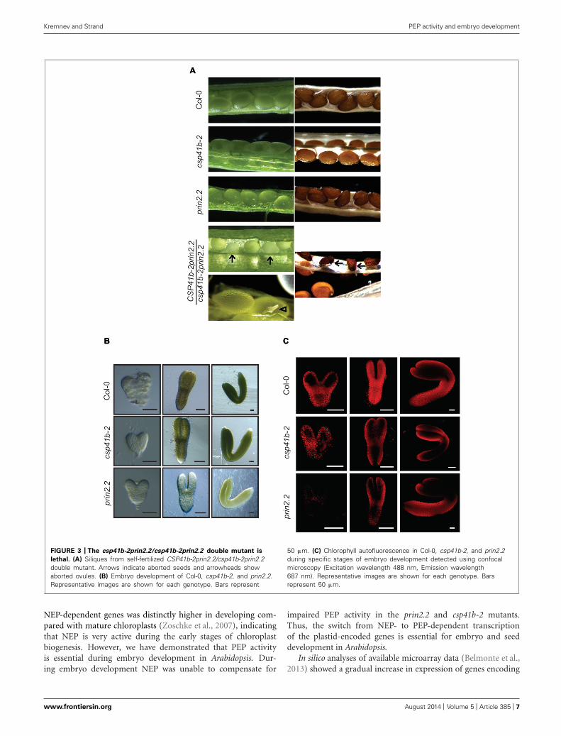
THE prin2.2 AND csp41b-2 MUTANTS SHOW DEFECTS IN EMBRYODEVELOPMENT AND THE csp41b-2 prin2.2 DOUBLE MUTANT ISEMBRYO LETHALIn order to investigate the genetic interaction between PRIN2 andCSP41b we attempted to generate a csp41b-2prin2.2 double mutant(Supplementary Figure S2). However, the csp41b-2prin2.2 dou-ble mutant was embryo lethal and the CSP41b-2prin2.2/csp41b-2prin2.2 mutant produced siliques where 18% (green:albinoovule = 240:51, Chi-square 8,67 for p < 0,05) of all ovulesappeared opaque (Figure 3A, arrows). Those impaired ovulesfinally turned into shrunken, dark colored seeds unable to ger-minate on MS media. A few ovules were also aborted at veryearly developmental stages (Figure 3A, arrowheads). The factthat theoretical 3:1 segregation green/albino is not supported byour statistics could be explained by the observed range in thestage at which the embryo development is arrested. Given theembryo lethality of the double mutant we investigated if therewere any effects during embryo development in the csp41b-2and prin2.2 single mutants. The csp41b-2 embryos were undis-tinguishable from the wild type at the heart stage (Figure 3B).However, at the linear cotyledon and mature green (MG) stagesthe csp41b-2 embryos were not as uniformly green as the wildtype embryos. As has been shown before wild type embryos dis-played a specific pattern of chlorophyll autofluorescence duringembryogenesis (Tejos et al., 2010). This pattern was significantlyaltered at the linear cotyledon stage (LC), where the chloroplastcontaining tissue was mostly localized to the epidermal layers,in the csp41b-2 embryos (Figure 3C). Distribution of chloro-plast containing tissue was even more altered in the prin2.2embryos (Figure 3C). Consistent with these findings are thelight microscopy pictures demonstrating that the prin2.2 embryoswere paler than wild type embryos at all developmental stages(Figure 3B).
To investigate the potential role of PRIN2 and CSP41b duringembryo development, we used TEM to examine morphologi-cal differences in the embryos at the MG stage. Plastids of wildtype embryos develop normally and have numerous thylakoidsorganized in grana stacks indicating that the plastids can be pho-tosynthetically functional at this stage of embryo development(Figure 4). However, csp41b-2 chloroplasts developed less thy-lakoid membranes and fewer grana stacks and exhibit quite oftenchloroplasts with large areas completely devoid of membranes(Figure 4). This specific defect in chloroplast structure of csp41b-2 was also observed for the chloroplasts from 3-week-old plants(Figure 2B). The prin2.2 showed an even stronger impairment inchloroplast development, the prin2-2 plastids showed numerousvesicles and few thylakoid membranes and grana stacks. Thylakoidmembranes were also often mis-oriented (Figure 4). Similarly towhat was observed for the csp41b-2 mutant the chloroplasts fromthe embryos of prin2.2 showed similar defects to what was alsoseen in the adult plants (Figure 2B). Taken together our resultsindicate that chloroplast development in the embryo is impaired
in both prin2.2 and csp41b-2 single mutants and that the ovulesare arrested at early developmental stages in the csp41b-2prin2.2double mutant.
EXPRESSION OF PHOTOSYNTHESIS GENES ESSENTIAL DURINGEMBRYO DEVELOPMENTWe performed in silico analysis of available array data wheregene activity was profiled genome-wide in every organ, tissue,and cell type of Arabidopsis seeds from fertilization throughmaturity (Belmonte et al., 2013). We specifically investigatedthe expression of plastid encoded genes from preglobular (PG)to MG embryo stage. Overall transcription of chloroplast-encoded photosynthesis associated genes was activated fromglobular to MG embryo stage (Figure 5A). The highest foldchange in expression level was observed for the genes encod-ing PSI and PSII core subunits, ATPase, and the ribosomalsubunits (Figure 5A). Thus, from the analysis of the plastidtranscriptome it is clear that photosynthesis associated com-ponents are highly expressed during embryo development andthat therefore PEP mediated transcription most likely is acti-vated during this process. Expression of the nuclear encodedcomponents associated with the PEP complex was demonstratedto increase from PG to LC (Figure 5B). Especially the genesencoding the PTAC proteins showed a very strong upregula-tion during embryo development. In addition, transcript levelsof TRXZ were significantly up-regulated during the transitionfrom PG to LC stage. Also transcription of PRIN2 and CSP41bwere observed during the LC stage of embryo development(Figure 5B).
The observed embryo phenotype in the prin2.2 and csp41b-2mutants encouraged us to study expression of psaA, psbA, andpsbD at the MG embryo stage. Expression of psaA, psbA, and psbDwas strongly down regulated both in prin2.2 and csp41b-2 mutantscompared to wild type (Figure 5B; Supplementary Figure S3).Thus, these results suggest that also under embryo developmentPRIN2 and CSP41b are important for proper transcription of PEPgenes. Taken together, these results suggest that the PEP compo-nent of the chloroplast transcription machinery is active duringembryo development and that its activity is essential for properembryo and seed development.
DISCUSSIONIn leaf tissue, the initiation of chloroplast development in thelight and the activation of the photosynthetic reactions areaccompanied by repression of NEP activity and an increase ofPEP-mediated plastid transcription (Hanaoka et al., 2005). How-ever, the mechanisms underlying this change in major RNApolymerase activity and the division of labor between NEPand PEP in the chloroplast are unknown (Zhelyazkova et al.,2012). Expression of plastid-encoded photosynthetic compo-nents, thought to be mediated exclusively by PEP, was recentlyshown both in tobacco and barley to be driven by NEP in theabsence of functional PEP, suggesting a less strict division oftarget genes between NEP and PEP (Legen et al., 2002; Lyubet-sky et al., 2011; Zhelyazkova et al., 2012). NEP is particularlyactive in non-green tissues and in very young leaves (Emanuelet al., 2006). Similarly, the expression of the previously described
Frontiers in Plant Science | Plant Physiology August 2014 | Volume 5 | Article 385 | 6
Kremnev and Strand PEP activity and embryo development
FIGURE 3 | The csp41b-2prin2.2/csp41b-2prin2.2 double mutant is
lethal. (A) Siliques from self-fertilized CSP41b-2prin2.2/csp41b-2prin2.2double mutant. Arrows indicate aborted seeds and arrowheads showaborted ovules. (B) Embryo development of Col-0, csp41b-2, and prin2.2.Representative images are shown for each genotype. Bars represent
50 μm. (C) Chlorophyll autofluorescence in Col-0, csp41b-2, and prin2.2during specific stages of embryo development detected using confocalmicroscopy (Excitation wavelength 488 nm, Emission wavelength687 nm). Representative images are shown for each genotype. Barsrepresent 50 μm.
NEP-dependent genes was distinctly higher in developing com-pared with mature chloroplasts (Zoschke et al., 2007), indicatingthat NEP is very active during the early stages of chloroplastbiogenesis. However, we have demonstrated that PEP activityis essential during embryo development in Arabidopsis. Dur-ing embryo development NEP was unable to compensate for
impaired PEP activity in the prin2.2 and csp41b-2 mutants.Thus, the switch from NEP- to PEP-dependent transcriptionof the plastid-encoded genes is essential for embryo and seeddevelopment in Arabidopsis.
In silico analyses of available microarray data (Belmonte et al.,2013) showed a gradual increase in expression of genes encoding
www.frontiersin.org August 2014 | Volume 5 | Article 385 | 7

Kremnev and Strand PEP activity and embryo development
FIGURE 4 |The csp41b-2 and 2prin2.2 mutants display impaired chloroplast during embryo development. Representative TEM images of chloroplastsfrom the Col-0, csp41b-2, and prin2.2 mature green (MG) embryos. Bars represent 1 μm.
components involved in photosynthesis and energy productionfrom the PG to the MG stage of embryo development (Figure 5A).During embryogenesis, expression of RPOT encoding the plas-tid NEP polymerase was induced and the maximum expressionlevel was reached as early as at the PG and globular stages (G;Figure 5B). The RPOT expression then diminished already atthe heart stage (H; Figure 5B). The NEP promoter is the onlypromoter in the rpoBC operon, and expression of rpoA, rpoB,and rpoC1/C2 followed the induction of RPOT. The core com-ponents of PEP were transcribed from the globular stage butin contrast to RPOT, expression was maintained all throughthe different embryo developmental stages (Figure 5A). Theexpression profile of the nuclear encoded sigma factors, espe-cially SIGB (SIG2) was similar to the profiles of rpoA, rpoB,and rpoC1/C2 (Figure 5B). Correlated with the induction ofthe genes encoding the core components of PEP was a verystrong induction of the plastid encoded photosynthesis genes.Expression of psbA,B,C,D,E, and psaA,B,J, for example, increasedapproximately 10-fold when the MG embryo was compared tothe PG suggesting that PEP transcription is activated during thisdevelopmental transition. The strong induction of photosynthe-sis related mRNAs during the PG to MG transition was alsoshown in a study where three periods of seed formation wasinvestigated (Allorent et al., 2013). The embryo lethality of the
csp41b-2prin2.2 double mutant further indicates that PEP driventranscription is required for effective transcription of photosyn-thesis genes to sustain the embryo with energy. It was also shownpreviously that genes associated with photosynthesis and carbonmetabolism, are active in all embryo and endosperm sub-regionsduring early seed development (Belmonte et al., 2013) stronglysuggesting there is a need for photosynthetic activity to contributeto embryo growth rate and biomass. Interestingly, at the MGembryo stage transcription of the nuclear encoded componentsof PEP was halted, suggesting that there is no need for furtherexpression of chloroplast genes when the seeds enter the post-MGstage. In contrast, the chloroplast encoded photosynthesis genesshowed a delayed response and maintained high expression alsoat the MG embryo stage. Possibly chloroplast transcription is sub-ject to later anterograde regulation from the nucleus to repressexpression.
True to its cyanobacterial origin, the core subunits of PEP arehomologous to the cyanobacterial RNAP components (Martinet al., 2002). However, PEP also requires additional nuclear-encoded factors for its function (Pfannschmidt et al., 2000; Pfalzet al., 2006). As many as 40–60 proteins appear to be presentin the TAC from chloroplasts. From Arabidopsis and mustardTACs 35 components were identified and 18 of those compo-nents, called pTACs, were novel proteins (Pfalz et al., 2006).
Frontiers in Plant Science | Plant Physiology August 2014 | Volume 5 | Article 385 | 8

Kremnev and Strand PEP activity and embryo development
FIGURE 5 | Induction of photosynthetic gene expression during embryo
development. (A) Heatmap of plastid gene expression in Col-0 embryos atdifferent developmental stages. Scale from 0 (yellow) to 4.8.e3 (red)represents the relative mean signal intensity for each probe (B) Heatmap ofselected nuclear genes in wild type embryos at different developmentalstages. Scale from 0 (yellow) to 3.4.e2 (red) represents the relative mean
signal intensity for each probe. (A) and (B): PG, preglobular stage; G, globularstage; H, heart stage; LC, linear cotyledon stage; MG, mature green embryostage. (C) Expression levels of psaA, psbA, and psbD in prin2.2 and csp41b-2MG embryos compared to the respective embryos of Col-0. Relativeexpression was calculated using PP2AA3 (At1g13320) as a reference gene.Data represent means from three independent biological replicates.
In addition, TAC and sRNAP preparations from pro-plastids,chloroplasts and etioplasts have different protein compositions,suggesting a multifaceted regulation of PEP activity (Reiss andLink, 1985; Pfannschmidt and Link, 1994; Suck et al., 1996). Veryhigh expression levels were observed for genes encoding the pTACcomponents PTAC3, PTAC10, PTAC12, FLN1, PTAC6, and TRXZ
during the globular (G) to LC stages. Expression of PRIN2 andCSP41B was also detected during the LC stage. The expressionof all these additional PEP components suggests that PEP activityrequires many different components already during embryo devel-opment and that regulation of plastid transcription is complex andsophisticated from the very early stages of plant development.
www.frontiersin.org August 2014 | Volume 5 | Article 385 | 9

Kremnev and Strand PEP activity and embryo development
PRIN2 and CSP41b were both identified in nucleoid prepa-rations (Majeran et al., 2012) and our results suggest that thesetwo components and possibly the PRIN2-CSP41b complex areessential for PEP dependent transcription during early embryodevelopment. Using three independent methods CSP41b andPRIN2 were shown to directly interact (Figure 1). PRIN2 havetwo conserved Cys residues that possibly are responsible formonomer/dimer/oligomer formation upon oxidation. Consistentwith this hypothesis is the observation of two bands of 20 and40 kDa that might correspond to PRIN2 monomer and dimer(Figure 1C). Interestingly, the CSP41b protein migrated as con-tinuous multimeric complex ranging from 40 to ∼700 kDa onthe BN-PAGE. The presence of high molecular weight complexeswas previously described for the native CSP41b protein in chloro-plasts (Qi et al., 2012). Moreover, the CSP41b protein containsredox active Cys residues suggested to be putative targets of TRX(Stroher and Dietz, 2008) and the formation of high molecularweight complexes were also shown to be enhanced by oxidized con-ditions in the chloroplast stroma (Qi et al., 2012). When PRIN2and CSP41b were incubated together they migrated on the gelas a distinct band (∼66 kDa; Figure 1B) suggesting the forma-tion of a defined heteromeric protein complex containing bothproteins. PRIN2 and CSP41b were shown to independently bindto promoter fragments of psaA and psbA in EMSA assays. How-ever, when PRIN2 and CSP41b were incubated together, a newband of intermediate size was observed (Figure 1D, Supplemen-tary data Figure S1), suggesting an interaction between PRIN2and CSP41b also upon DNA binding in vitro. CSP41b was previ-ously shown in RIP-chip analysis to be an RNA binding proteinwith specificity toward photosynthesis-related transcripts mainlyexpressed by PEP (Qi et al., 2012). However, many RNA bindingproteins have also been described to have DNA binding proper-ties, including TFIIIA, p53, STAT1, β/β′ subunits of RNAP andσ70, factors well known to regulate transcription (Sakonju et al.,1980; Clemens et al., 1993; Cassiday and Maher, 2002; Suswamet al., 2005). Another plant specific protein that binds both DNAand RNA is GUN1, a key component in retrograde commu-nication between chloroplasts and nucleus (Koussevitzky et al.,2007). Possibly, the observed DNA binding of the PRIN2-CSP41bprotein complex is required for full PEP activity as indicatedby the embryo lethality of the csp41b-2prin2.2 double mutant(Figure 3A).
Correlated with the embryo phenotype observed in prin2.2 andcsp41b-2 single mutants was the impaired expression of chloro-plast photosynthesis genes. The expression levels of psaA, psbA,and psbD were significantly lower in both mutants comparedto wild type in MG embryos (Figure 5C), suggesting that thepreviously described defect in PEP-mediated gene expression inrosette plants is maintained in the mutants also during embryodevelopment. Thus, our results establish a link between PEPactivity and embryo development. Previously compromised trans-lational and post-translational activities in the chloroplasts, suchas mutations in elongation factor G and PPR proteins, have beenshown to lead to embryo lethality (Ruppel and Hangarter, 2007;Khrouchtchova et al., 2012; Sosso et al., 2012). Interestingly, intobacco and barley neither of the knockouts completely lack-ing PEP activity exhibits an embryo lethal phenotype (Allison
et al., 1996; De Santis-MacIossek et al., 1999; Zhelyazkova et al.,2012). However, it should be emphasized that an essential roleof plastid activity and embryo greening during embryogenesishas so far only been reported for oil-seed plants such as Ara-bidopsis and Brassica (He and Wu, 2009; Hsu et al., 2010). InArabidopsis, PEP activity appears essential during embryo devel-opment and during this process NEP is unable to compensatefor the impaired PEP activity. Recently maize mutants lack-ing several PEP-associated proteins such as PTACs and PRIN2demonstrated deficiency of numerous plastid tRNAs. Thus, a rolefor PEP, and for the PEP associated proteins, was demonstratedfor the expression of plastid transfer RNA (Williams-Carrieret al., 2014). These results emphasize the complex division oflabor between NEP and PEP during the initiation of chloro-plast development and that PEP, and its associated proteins, arealso essential for the translation of the photosynthetic compo-nents.
AUTHOR CONTRIBUTIONSDmitry Kremnev carried out the experiments and analyzed thedata. Dmitry Kremnev and Åsa Strand planned the study andwrote the manuscript. Both authors read and approved the finalmanuscript.
ACKNOWLEDGMENTThis work was supported by grants from the Swedish researchfoundation, VR (ÅS).
SUPPLEMENTARY MATERIALThe Supplementary Material for this article can be found onlineat: http://www.frontiersin.org/journal/10.3389/fpls.2014.00385/abstract
REFERENCESAllison, L. A., Simon, L. D., and Maliga, P. (1996). Deletion of rpoB reveals a second
distinct transcription system in plastids of higher plants. EMBO J. 15, 2802–2809.Allorent, G., Florence, C., Chevalier, F., and Lerbs-Mache, S. (2013). Plastid gene
expression during chloroplast differentiation and dedifferentiation inot non-photosynthetic plastids during seed formation. Plant Mol. Biol. 82, 59–70. doi:10.1007/s11103-0037-0
Aronsson, H., and Jarvis, P. (2002). A simple method for isolating import-competent Arabidopsis chloroplasts. FEBS Lett. 529, 215–220. doi: 10.1016/S0014-5793(02)03342-2
Barajas-Lopez Jde, D., Kremnev, D., Shaikhali, J., Pinas-Fernandez, A., andStrand, Å. (2013). PAPP5 is involved in the tetrapyrrole mediated plas-tid signalling during chloroplast development. PLoS ONE 8:e60305. doi:10.1371/journal.pone.0060305
Belmonte, M. F., Kirkbride, R. C., Stone, S. L., Pelletier, J. M., Bui, A. Q., Yeung,E. C., et al. (2013). Comprehensive developmental profiles of gene activity inregions and subregions of the Arabidopsis seed. Proc. Natl. Acad. Sci. U.S.A. 110,E435–E444. doi: 10.1073/pnas.1222061110
Bollenbach, T. J., Sharwood, R. E., Gutierrez, R., Lerbs-Mache, S., and Stern, D. B.(2009). The RNA-binding proteins CSP41a and CSP41b may regulate transcrip-tion and translation of chloroplast-encoded RNAs in Arabidopsis. Plant Mol. Biol.69, 541–552. doi: 10.1007/s11103-008-9436-z
Bryant, N., Lloyd, J., Sweeney, C., Myouga, F., and Meinke, D. (2011). Iden-tification of nuclear genes encoding chloroplast-localized proteins requiredfor embryo development in Arabidopsis. Plant Physiol. 155, 1678–1689. doi:10.1104/pp.110.168120
Cassiday, L. A., and Maher, L. J. III. (2002). Having it both ways: transcrip-tion factors that bind DNA and RNA. Nucleic Acids Res. 30, 4118–4126. doi:10.1093/nar/gkf512
Frontiers in Plant Science | Plant Physiology August 2014 | Volume 5 | Article 385 | 10

Kremnev and Strand PEP activity and embryo development
Clemens, K. R., Wolf, V., Mcbryant, S. J., Zhang, P., Liao, X., Wright, P.E., et al. (1993). Molecular basis for specific recognition of both RNA andDNA by a zinc finger protein. Science 260, 530–533. doi: 10.1126/science.8475383
Clough, S. J., and Bent, A. F. (1998). Floral dip: a simplified method forAgrobacterium-mediated transformation of Arabidopsis thaliana. Plant J. 16,735–743. doi: 10.1046/j.1365-313x.1998.00343.x
De Santis-MacIossek, G., Kofer, W., Bock, A., Schoch, S., Maier, R. M., Wanner, G.,et al. (1999). Targeted disruption of the plastid RNA polymerase genes rpoA, Band C1: molecular biology, biochemistry and ultrastructure. Plant J. 18, 477–489.doi: 10.1046/j.1365-313X.1999.00473.x
Doelling, J. H., and Pikaard, C. S. (1993). Transient expression in Arabidopsisthaliana protoplasts derived from rapidly established cell suspension cultures.Plant Cell Rep. 12, 241–244. doi: 10.1007/BF00237127
Emanuel, C., Von Groll, U., Muller, M., Borner, T., and Weihe, A. (2006).Development- and tissue-specific expression of the RpoT gene family of Arabidop-sis encoding mitochondrial and plastid RNA polymerases. Planta 223, 998–1009.doi: 10.1007/s00425-005-0159-y
Goffman, F. D., Alonso, A. P., Schwender, J., Shachar-Hill, Y., and Ohlrogge, J.B. (2005). Light enables a very high efficiency of carbon storage in develop-ing embryos of rapeseed. Plant Physiol. 138, 2269–2279. doi: 10.1104/pp.105.063628
Hajdukiewicz, P. T., Allison, L. A., and Maliga, P. (1997). The two RNA poly-merases encoded by the nuclear and the plastid compartments transcribedistinct groups of genes in tobacco plastids. EMBO J. 16, 4041–4048. doi:10.1093/emboj/16.13.4041
Hall, M., Mishra, Y., and Schroder, W. P. (2011). Preparation of stroma, thy-lakoid membrane, and lumen fractions from Arabidopsis thaliana chloroplastsfor proteomic analysis. Methods Mol. Biol. 775, 207–222. doi: 10.1007/978-1-61779-237-3_11
Hanaoka, M., Kanamaru, K., Fujiwara, M., Takahashi, H., and Tanaka, K.(2005). Glutamyl-tRNA mediates a switch in RNA polymerase use dur-ing chloroplast biogenesis. EMBO Rep. 6, 545–550. doi: 10.1038/sj.embor.7400411
Hassidim, M., Yakir, E., Fradkin, D., Hilman, D., Kron, I., Keren, N., et al. (2007).Mutations in CHLOROPLAST RNA BINDING provide evidence for the involve-ment of the chloroplast in the regulation of the circadian clock in Arabidopsis.Plant J. 51, 551–562. doi: 10.1111/j.1365-313X.2007.03160.x
He, Y. Q., and Wu, Y. (2009). Oil body biogenesis during Brassica napus embryo-genesis. J. Integr. Plant Biol. 51, 792–799. doi: 10.1111/j.1744-7909.2009.00851.x
Hedtke, B., Borner, T., and Weihe, A. (1997). Mitochondrial and chloro-plast phage-type RNA polymerases in Arabidopsis. Science 277, 809–811. doi:10.1126/science.277.5327.809
Hedtke, B., Borner, T., and Weihe, A. (2000). One RNA polymerase serving twogenomes. EMBO Rep. 1, 435–440. doi: 10.1093/embo-reports/kvd086
Hsu, S. C., Belmonte, M. F., Harada, J. J., and Inoue, K. (2010). Indispensable rolesof plastids in Arabidopsis thaliana embryogenesis. Curr. Genomics 11, 338–349.doi: 10.2174/138920210791616716
Järvi, S., Suorsa, M., Paakkarinen, V., and Aro, E.-M. (2011). Optimized native gelsystems for separation of thylakoid protein complexes: novel super- and mega-complexes. Biochem. J. 439, 207–214. doi: 10.1042/BJ20102155
Khrouchtchova, A., Monde, R. A., and Barkan, A. (2012). A short PPR proteinrequired for the splicing of specific group II introns in angiosperm chloroplasts.RNA 18, 1197–1209. doi: 10.1261/rna.032623.112
Kindgren, P., Kremnev, D., Blanco, N. E., De Dios Barajas Lopez, J., Fernan-dez, A. P., Tellgren-Roth, C., et al. (2012). The plastid redox insensitive 2mutant of Arabidopsis is impaired in PEP activity and high light-dependentplastid redox signalling to the nucleus. Plant J. 70, 279–291. doi: 10.1111/j.1365-313X.2011.04865.x
Koussevitzky, S., Nott, A., Mockler, T. C., Hong, F., Sachetto-Martins,G., Surpin, M., et al. (2007). Signals from chloroplasts converge to regu-late nuclear gene expression. Science 316, 715–719. doi: 10.1126/science.1140516
Krause, K., Maier, R. M., Kofer, W., Krupinska, K., and Herrmann, R. G. (2000).Disruption of plastid-encoded RNA polymerase genes in tobacco: expression ofonly a distinct set of genes is not based on selective transcription of the plastidchromosome. Mol. Gen. Genet. 263, 1022–1030. doi: 10.1007/PL00008690
Le, B. H., Cheng, C., Bui, A. Q., Wagmaister, J. A., Henry, K. F., Pelletier, J., et al.(2010). Global analysis of gene activity during Arabidopsis seed development andidentification of seed-specific transcription factors. Proc. Natl. Acad. Sci. U.S.A.107, 8063–8070. doi: 10.1073/pnas.1003530107
Legen, J., Kemp, S., Krause, K., Profanter, B., Herrmann, R. G., and Maier,R. M. (2002). Comparative analysis of plastid transcription profiles of entireplastid chromosomes from tobacco attributed to wild-type and PEP-deficienttranscription machineries. Plant J. 31, 171–188. doi: 10.1046/j.1365-313X.2002.01349.x
Lyubetsky, V. A., Zverkov, O. A., Rubanov, L. I., and Seliverstov, A. V. (2011).Modeling RNA polymerase competition: the effect of sigma-subunit knockoutand heat shock on gene transcription level. Biol. Direct 6, 3. doi: 10.1186/1745-6150-6-3
Majeran, W., Friso, G., Asakura, Y., Qu, X., Huang, M., Ponnala, L., et al. (2012).Nucleoid-enriched proteomes in developing plastids and chloroplasts from maizeleaves: a new conceptual framework for nucleoid functions. Plant Physiol. 158,156–189. doi: 10.1104/pp.111.188474
Martin, W., Rujan, T., Richly, E., Hansen, A., Cornelsen, S., Lins, T., et al.(2002). Evolutionary analysis of Arabidopsis, cyanobacterial, and chloroplastgenomes reveals plastid phylogeny and thousands of cyanobacterial genes inthe nucleus. Proc. Natl. Acad. Sci. U.S.A. 99, 12246–12251. doi: 10.1073/pnas.182432999
McElver, J., Tzafrir, I., Aux, G., Rogers, R., Ashby, C., Smith, K., et al. (2001).Insertional mutagenesis of genes required for seed development in Arabidopsisthaliana. Genetics 159, 1751–1763.
Perry, S. E., and Wang, H. (2003). Rapid isolation of Arabidopsis thaliana developingembryos. BioTechniques 35, 278–280, 282.
Pfalz, J., Liere, K., Kandlbinder, A., Dietz, K. J., and Oelmuller, R. (2006). pTAC2,-6, and -12 are components of the transcriptionally active plastid chromo-some that are required for plastid gene expression. Plant Cell 18, 176–197. doi:10.1105/tpc.105.036392
Pfalz, J., and Pfannschmidt, T. (2013). Essential nucleoid proteins in early chloroplastdevelopment. Trends Plant Sci. 18, 186–194. doi: 10.1016/j.tplants.2012.11.003
Pfannschmidt, T., and Liere, K. (2005). Redox regulation and modification of pro-teins controlling chloroplast gene expression. Antioxid. Redox Signal. 7, 607–618.doi: 10.1089/ars.2005.7.607
Pfannschmidt, T., and Link, G. (1994). Separation of two classes of plastid DNA-dependent RNA polymerases that are differentially expressed in mustard (Sinapisalba L.) seedlings. Plant Mol. Biol. 25, 69–81. doi: 10.1007/BF00024199
Pfannschmidt, T., Ogrzewalla, K., Baginsky, S., Sickmann, A., Meyer, H. E., andLink, G. (2000). The multisubunit chloroplast RNA polymerase A from mustard(Sinapis alba L.). Integration of a prokaryotic core into a larger complex withorganelle-specific functions. Eur. J. Biochem. 267, 253–261. doi: 10.1046/j.1432-1327.2000.00991.x
Puthiyaveetil, S., Ibrahim, I. M., Jelicic, B., Tomasic, A., Fulgosi, H., and Allen,J. F. (2010). Transcriptional control of photosynthesis genes: the evolutionarilyconserved regulatory mechanism in plastid genome function. Genome Biol. Evol.2, 888–896. doi: 10.1093/gbe/evq073
Qi, Y., Armbruster, U., Schmitz-Linneweber, C., Delannoy, E., De Longevialle, A. F.,Ruhle, T., et al. (2012). Arabidopsis CSP41 proteins form multimeric complexesthat bind and stabilize distinct plastid transcripts. J. Exp. Bot. 63, 1251–1270. doi:10.1093/jxb/err347
Reiss, T., and Link, G. (1985). Characterization of transcriptionally activeDNA-protein complexes from chloroplasts and etioplasts of mustard (Sinapisalba L.). Eur. J. Biochem. 148, 207–212. doi: 10.1111/j.1432-1033.1985.tb08826.x
Rolletschek, H., Weber, H., and Borisjuk, L. (2003). Energy status and its controlon embryogenesis of legumes. Embryo photosynthesis contributes to oxygensupply and is coupled to biosynthetic fluxes. Plant Physiol. 132, 1196–1206. doi:10.1104/pp.102.017376
Ruppel, N. J., and Hangarter, R. P. (2007). Mutations in a plastid-localized elongationfactor G alter early stages of plastid development in Arabidopsis thaliana. BMCPlant Biol. 7:37. doi: 10.1186/1471-2229-7-37
Sakonju, S., Bogenhagen, D. F., and Brown, D. D. (1980). A control region in thecenter of the 5S RNA gene directs specific initiation of transcription: I. The 5’border of the region. Cell 19, 13–25. doi: 10.1016/0092-8674(80)90384-0
Shaikhali, J., Barajas-Lopéz, J., Östvös, K., Kremnev, D., Sánchez Garcia, A.,Srivastava, V., et al. (2012a). The CRYPTOCHROME1-dependent response
www.frontiersin.org August 2014 | Volume 5 | Article 385 | 11
Kremnev and Strand PEP activity and embryo development
to excess light is mediated through the transcriptional activators ZINC FIN-GER PROTEIN EXPRESSED IN INFLORESCENCE MERISTEM LIKE1 and2 in Arabidopsis thaliana. Plant Cell 24, 3009–3025. doi: 10.1105/tpc.112.100099
Shaikhali, J., Norén, L., Barajas-López, J., Srivastava, V., König, J., Sauer, U. H.,et al. (2012b). Redox-mediated mechanisms regulate DNA-binding activity ofthe G-group of bZIP transcription factors in Arabidopsis. J. Biol. Chem. 287,27510–27525. doi: 10.1074/jbc.M112.361394
Sosso, D., Canut, M., Gendrot, G., Dedieu, A., Chambrier, P., Barkan, A., et al.(2012). PPR8522 encodes a chloroplast-targeted pentatricopeptide repeat proteinnecessary for maize embryogenesis and vegetative development. J. Exp. Bot. 63,5843–5857. doi: 10.1093/jxb/ers232
Spencer, M. W., Casson, S. A., and Lindsey, K. (2007). Transcriptional profilingof the Arabidopsis embryo. Plant Physiol. 143, 924–940. doi: 10.1104/pp.106.087668
Stroher, E., and Dietz, K. J. (2008). The dynamic thiol-disulphide redox pro-teome of the Arabidopsis thaliana chloroplast as revealed by differential elec-trophoretic mobility. Physiol. Plant. 133, 566–583. doi: 10.1111/j.1399-3054.2008.01103.x
Suck, R., Zeltz, P., Falk, J., Acker, A., Kossel, H., and Krupinska, K. (1996).Transcriptionally active chromosomes (TACs) of barley chloroplasts contain thealpha-subunit of plastome-encoded RNA polymerase. Curr. Genet. 30, 515–521.doi: 10.1007/s002940050164
Suswam, E. A., Li, Y. Y., Mahtani, H., and King, P. H. (2005). Novel DNA-bindingproperties of the RNA-binding protein TIAR. Nucleic Acids Res. 33, 4507–4518.doi: 10.1093/nar/gki763
Suzuki, J. Y., Ytterberg, A. J., Beardslee, T. A., Allison, L. A., Wijk, K. J., and Maliga,P. (2004). Affinity purification of the tobacco plastid RNA polymerase and invitro reconstitution of the holoenzyme. Plant J. 40, 164–172. doi: 10.1111/j.1365-313X.2004.02195.x
Tejos, R. I., Mercado, A. V., and Meisel, L. A. (2010). Analysis of chlorophyll fluores-cence reveals stage specific patterns of chloroplast-containing cells during Ara-bidopsis embryogenesis. Biol. Res. 43, 99–111. doi: 10.4067/S0716-97602010000100012
Tzafrir, I., Dickerman, A., Brazhnik, O., Nguyen, Q., Mcelver, J., Frye, C., et al.(2003). The Arabidopsis SeedGenes Project. Nucleic Acids Res. 31, 90–93. doi:10.1093/nar/gkg028
Wakasugi, T., Tsudzuki, T., and Sugiura, M. (2001). The genomics of land plantchloroplasts: gene content and alteration of genomic information by RNA editing.Photosynth. Res. 70, 107–118. doi: 10.1023/A:1013892009589
Williams-Carrier, R., Zoschke, R., Belcher, S., Pfalz, J., and Barkan, A. (2014).A major role for the plastid encoded RNA polymerase complex in the expres-sion of plastid transfer RNAs. Plant Physiol. 164, 239–248. doi/10.1104/pp.113.228726
Yamaguchi, K., Beligni, M. V., Prieto, S., Haynes, P. A., Mcdonald, W. H.,Yates, J. R. III, et al. (2003). Proteomic characterization of the Chlamy-domonas reinhardtii chloroplast ribosome. Identification of proteins uniqueto the 70 S ribosome. J. Biol. Chem. 278, 33774–33785. doi: 10.1074/jbc.M301934200
Zhelyazkova, P., Sharma, C. M., Forstner, K. U., Liere, K., Vogel, J., and Borner, T.(2012). The primary transcriptome of barley chloroplasts: numerous noncodingRNAs and the dominating role of the plastid-encoded RNA polymerase. PlantCell 24, 123–136. doi: 10.1105/tpc.111.089441
Zoschke, R., Liere, K., and Borner, T. (2007). From seedling to mature plant: Ara-bidopsis plastidial genome copy number, RNA accumulation and transcriptionare differentially regulated during leaf development. Plant J. 50, 710–722. doi:10.1111/j.1365-313X.2007.03084.x
Conflict of Interest Statement: The authors declare that the research was conductedin the absence of any commercial or financial relationships that could be construedas a potential conflict of interest.
Received: 14 March 2014; accepted: 19 July 2014; published online: 12 August 2014.Citation: Kremnev D and Strand Å (2014) Plastid encoded RNA polymerase activityand expression of photosynthesis genes required for embryo and seed development inArabidopsis. Front. Plant Sci. 5:385. doi: 10.3389/fpls.2014.00385This article was submitted to Plant Physiology, a section of the journal Frontiers inPlant Science.Copyright © 2014 Kremnev and Strand. This is an open-access article distributed underthe terms of the Creative Commons Attribution License (CC BY). The use, distributionor reproduction in other forums is permitted, provided the original author(s) or licensorare credited and that the original publication in this journal is cited, in accordance withaccepted academic practice. No use, distribution or reproduction is permitted whichdoes not comply with these terms.
Frontiers in Plant Science | Plant Physiology August 2014 | Volume 5 | Article 385 | 12