pneumothorax
TRANSCRIPT

Copyright © 2006 by Mosby, Inc.Slide 1
PneumothoraxPneumothorax
GA
DD
CL
Dr.Njia Brg Ellil

Copyright © 2006 by Mosby, Inc.Slide 2
Anatomic Alterations of the LungsAnatomic Alterations of the Lungs
Lung collapseLung collapse
AtelectasisAtelectasis
Chest wall expansionChest wall expansion
Compression of the great veins and Compression of the great veins and decreased cardiac venous returndecreased cardiac venous return

Copyright © 2006 by Mosby, Inc.Slide 3
Etiology—3 WaysEtiology—3 Ways
From the lungs through a perforation of the From the lungs through a perforation of the visceral pleuravisceral pleura
From the surrounding atmosphere through a From the surrounding atmosphere through a perforation of the chest wall and parietal perforation of the chest wall and parietal pleura or, rarely, through an esophageal pleura or, rarely, through an esophageal fistula or a perforated abdominal viscusfistula or a perforated abdominal viscus
From gas-forming microorganisms in an From gas-forming microorganisms in an empyema in the pleural space (rare)empyema in the pleural space (rare)

Copyright © 2006 by Mosby, Inc.Slide 4
Pneumothorax ClassificationsPneumothorax ClassificationsGeneral TermsGeneral Terms
Closed pneumothoraxClosed pneumothorax
Open pneumothoraxOpen pneumothorax
Tension pneumothoraxTension pneumothorax

Copyright © 2006 by Mosby, Inc.Slide 5
Pneumothorax ClassificationsPneumothorax ClassificationsBased on OriginBased on Origin
Traumatic pneumothoraxTraumatic pneumothorax
Spontaneous pneumothoraxSpontaneous pneumothorax
Iatrogenic pneumothoraxIatrogenic pneumothorax

Copyright © 2006 by Mosby, Inc.Slide 6

Copyright © 2006 by Mosby, Inc.Slide 7

Copyright © 2006 by Mosby, Inc.Slide 8
Spontaneous PneumothoraxSpontaneous Pneumothorax

Copyright © 2006 by Mosby, Inc.Slide 9
Iatrogenic PneumothoraxIatrogenic Pneumothorax

Copyright © 2006 by Mosby, Inc.Slide 10
Overview of the Cardiopulmonary Overview of the Cardiopulmonary Clinical Manifestations Associated Clinical Manifestations Associated
with PNEUMOTHORAXwith PNEUMOTHORAX
The following clinical manifestations result from The following clinical manifestations result from the pathophysiologic mechanisms caused (or the pathophysiologic mechanisms caused (or activated) by activated) by AtelectasisAtelectasis

Copyright © 2006 by Mosby, Inc.Slide 11

Copyright © 2006 by Mosby, Inc.Slide 12
Clinical Data Obtained at the Clinical Data Obtained at the Patient’s BedsidePatient’s Bedside
Vital signsVital signs
Increased respiratory rateIncreased respiratory rate Stimulation of peripheral chemoreceptors Stimulation of peripheral chemoreceptors
Other possible mechanismsOther possible mechanisms
• Decreased lung complianceDecreased lung compliance
• Activation of the deflation receptorsActivation of the deflation receptors
• Activation of the irritant receptorsActivation of the irritant receptors
• Stimulation of the J receptorsStimulation of the J receptors
• Pain/anxietyPain/anxiety
Increased heart rate, cardiac output, blood pressureIncreased heart rate, cardiac output, blood pressure

Copyright © 2006 by Mosby, Inc.Slide 13

Copyright © 2006 by Mosby, Inc.Slide 14
Clinical Data Obtained at the Clinical Data Obtained at the Patient’s BedsidePatient’s Bedside
CyanosisCyanosis
Chest assessment findingsChest assessment findings Hyperresonant percussion note over the Hyperresonant percussion note over the
pneumothoraxpneumothorax
Diminished breath sounds over the pneumothoraxDiminished breath sounds over the pneumothorax
Tracheal shiftTracheal shift
Displaced heart soundsDisplaced heart sounds
Increased thoracic volume on the affected sideIncreased thoracic volume on the affected side
• Particularly in tension pneumothoraxParticularly in tension pneumothorax

Copyright © 2006 by Mosby, Inc.Slide 15

Copyright © 2006 by Mosby, Inc.Slide 16

Copyright © 2006 by Mosby, Inc.Slide 17

Copyright © 2006 by Mosby, Inc.Slide 18
Clinical Data Obtained from Clinical Data Obtained from Laboratory Tests and Special Laboratory Tests and Special
ProceduresProcedures

Copyright © 2006 by Mosby, Inc.Slide 19
Pulmonary Function Study: Pulmonary Function Study: Lung Volume and Capacity Findings Lung Volume and Capacity Findings
VT RV FRC TLC
N or
VC IC ERV RV/TLC%
N
VT RV FRC TLC
N or
VC IC ERV RV/TLC%
N
Copyright © 2006 by Mosby, Inc.Slide 20
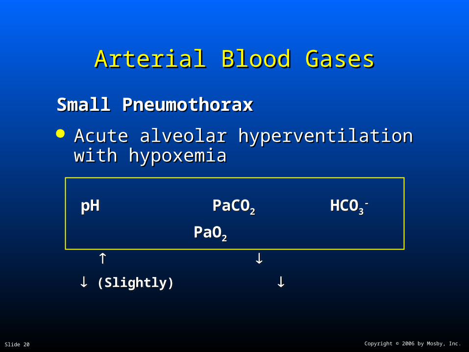
Arterial Blood GasesArterial Blood Gases
Small PneumothoraxSmall Pneumothorax
Acute alveolar hyperventilation with Acute alveolar hyperventilation with hypoxemiahypoxemia
pH PaCO2 HCO3- PaO2
(Slightly)
pH PaCO2 HCO3- PaO2
(Slightly)

Copyright © 2006 by Mosby, Inc.Slide 21
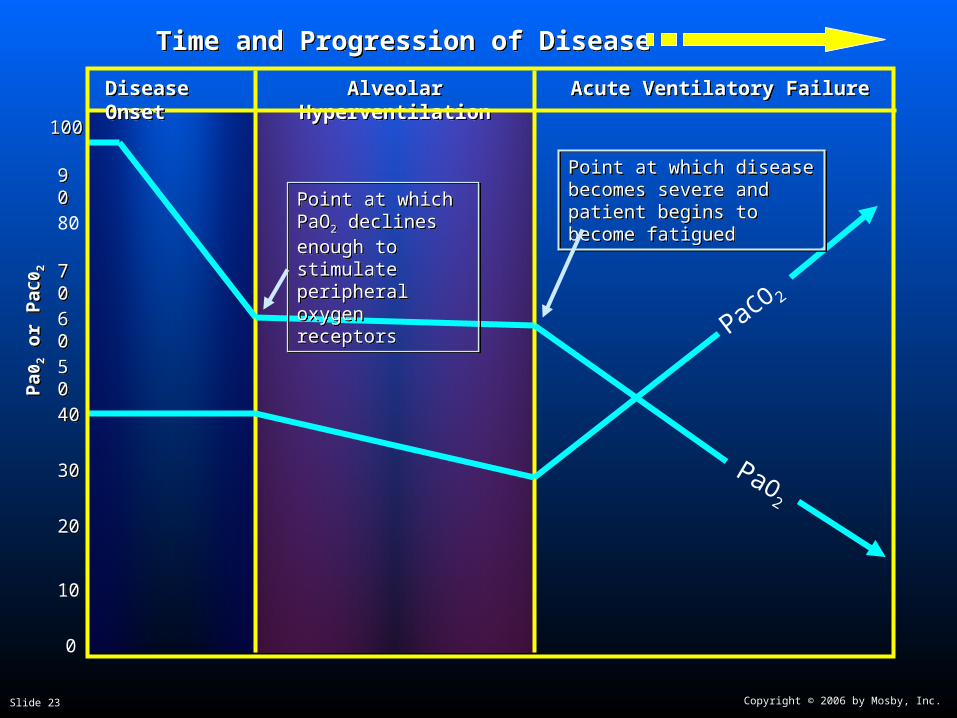
Time and Progression of Disease Time and Progression of Disease
100100
5050
3030
8080
00
PaCO2
1010
2020
4040
Alveolar HyperventilationAlveolar Hyperventilation
6060
7070
9090 Point at which PaO2 declines enough to stimulate peripheral oxygen receptors
Point at which PaO2 declines enough to stimulate peripheral oxygen receptors
PaO2
Disease OnsetDisease OnsetP
aO2
or
PaC
O2
PaO
2 o
r P
aCO
2
Copyright © 2006 by Mosby, Inc.Slide 22
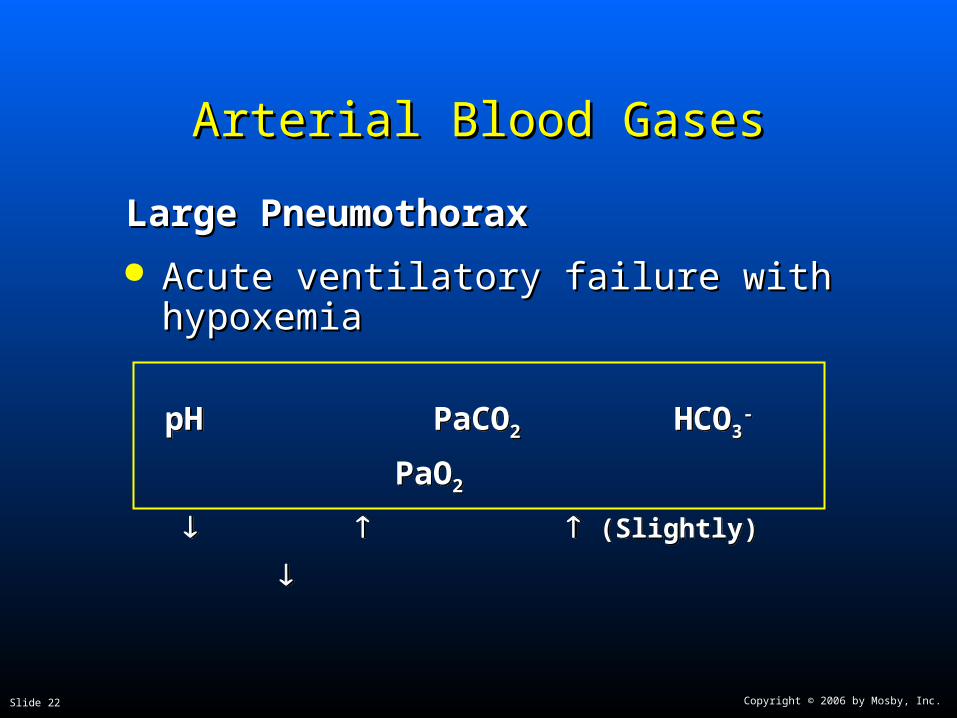
Arterial Blood GasesArterial Blood Gases
Large PneumothoraxLarge Pneumothorax
Acute ventilatory failure with hypoxemiaAcute ventilatory failure with hypoxemia
pH PaCO2 HCO3- PaO2
(Slightly)
pH PaCO2 HCO3- PaO2
(Slightly)
Copyright © 2006 by Mosby, Inc.Slide 23
Time and Progression of DiseaseTime and Progression of Disease
100100
5050
3030
80
0
PaO2
1010
2020
4040
Alveolar HyperventilationAlveolar Hyperventilation
6060
7070
9090Point at which PaO2 declines enough to stimulate peripheral oxygen receptors
Point at which PaO2 declines enough to stimulate peripheral oxygen receptors
PaCO 2
Acute Ventilatory Failure Acute Ventilatory FailureDisease OnsetDisease Onset
Point at which disease becomes severe and patient begins to become fatigued
Point at which disease becomes severe and patient begins to become fatigued
Pa0
2 o
r P
aC0 2
Pa0
2 o
r P
aC0 2

Copyright © 2006 by Mosby, Inc.Slide 24
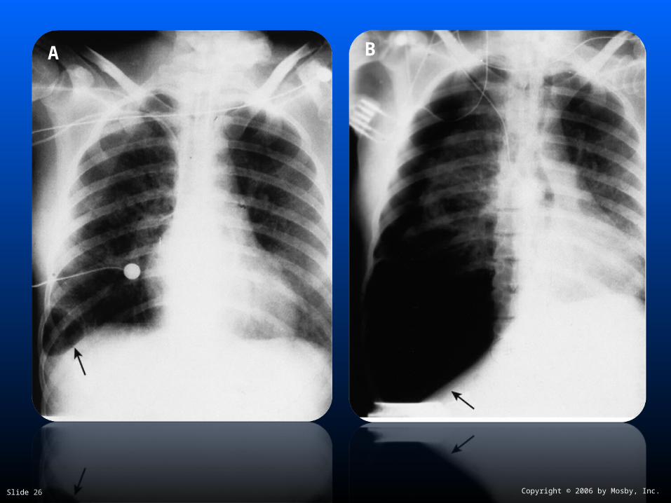
Radiologic FindingsRadiologic Findings
Chest radiographChest radiograph
Increased translucencyIncreased translucency
Mediastinal shift to unaffected side Mediastinal shift to unaffected side in tension pneumothoraxin tension pneumothorax
Depressed diaphragmDepressed diaphragm
Lung collapseLung collapse
AtelectasisAtelectasis

Copyright © 2006 by Mosby, Inc.Slide 25
Copyright © 2006 by Mosby, Inc.Slide 26
A B
Copyright © 2006 by Mosby, Inc.Slide 27
General Management of General Management of PneumothoraxPneumothorax
>20%—gas should be evacuated >20%—gas should be evacuated
Negative pressure—5 to 12 cm HNegative pressure—5 to 12 cm H22O O
Should not exceed negative 12 cm HShould not exceed negative 12 cm H22OO

Copyright © 2006 by Mosby, Inc.Slide 28
General Management of General Management of PneumothoraxPneumothorax
Respiratory care treatment protocolsRespiratory care treatment protocols
Oxygen therapy protocolOxygen therapy protocol
Hyperinflation therapy protocolHyperinflation therapy protocol
Mechanical ventilation protocolMechanical ventilation protocol

Copyright © 2006 by Mosby, Inc.Slide 29
General Management of General Management of PneumothoraxPneumothorax
PLEURODESISPLEURODESIS
Chemical or medication injected into the Chemical or medication injected into the chest cavitychest cavity TalcTalc
TetracyclineTetracycline
Bleomycin sulfateBleomycin sulfate
Produces inflammatory reaction between Produces inflammatory reaction between lungs and inner chest cavitylungs and inner chest cavity Causes lung to stick to chest cavityCauses lung to stick to chest cavity

Copyright © 2006 by Mosby, Inc.Slide 30
Conditions requiring chest Conditions requiring chest drainagedrainage
Air between the pleurae Air between the pleurae is a is a pneumothoraxpneumothorax
Copyright © 2006 by Mosby, Inc.Slide 31
Conditions requiring chest Conditions requiring chest drainagedrainage
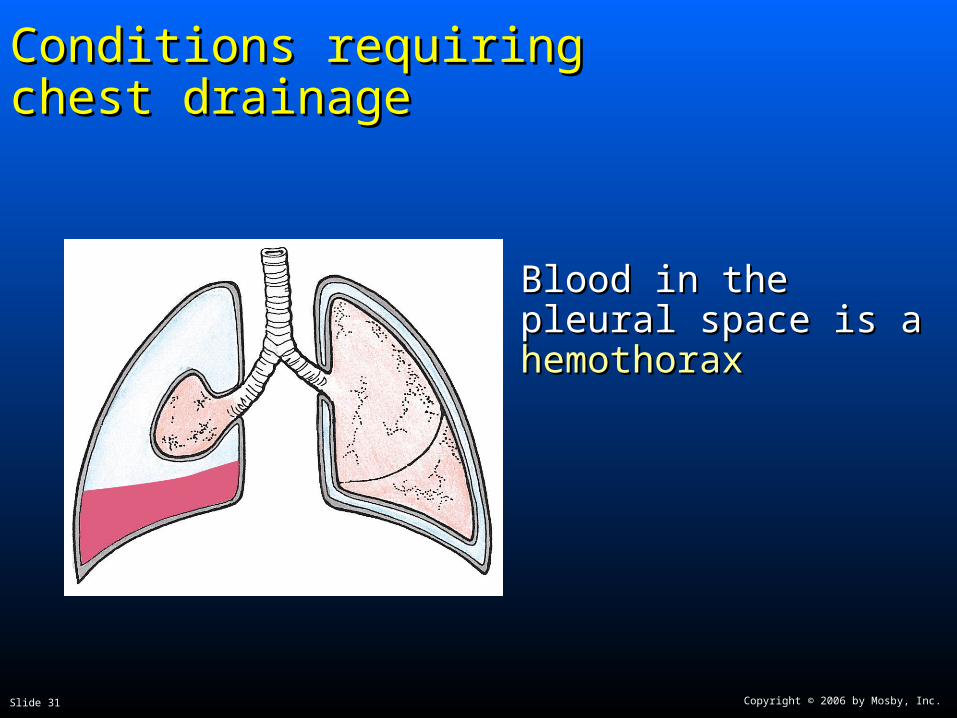
Blood in the pleural Blood in the pleural space is a space is a hemothoraxhemothorax

Copyright © 2006 by Mosby, Inc.Slide 32
Conditions requiring chest drainageConditions requiring chest drainage
Transudate or exudate Transudate or exudate in the pleural space is a in the pleural space is a pleural effusionpleural effusion

Copyright © 2006 by Mosby, Inc.Slide 33
Conditions requiring chest drainage: Conditions requiring chest drainage: pneumothoraxpneumothorax
PneumothoraxPneumothorax Occurs when there is an opening on the surface of Occurs when there is an opening on the surface of
the lung or in the airways, in the chest wall — or the lung or in the airways, in the chest wall — or bothboth
The opening allows air to enter the pleural space The opening allows air to enter the pleural space between the pleurae, creating an actual spacebetween the pleurae, creating an actual space

Copyright © 2006 by Mosby, Inc.Slide 34
Conditions requiring chest drainage: Conditions requiring chest drainage: open pneumothoraxopen pneumothorax
Open pneumothoraxOpen pneumothorax Opening in the chest Opening in the chest
wall (with or without wall (with or without lung puncture)lung puncture)
Allows atmospheric air Allows atmospheric air to enter the pleural to enter the pleural spacespace
Penetrating trauma: Penetrating trauma: stab, gunshot, stab, gunshot, impalementimpalement
SurgerySurgery

Copyright © 2006 by Mosby, Inc.Slide 35
Conditions requiring chest drainage: closed Conditions requiring chest drainage: closed pneumothoraxpneumothorax
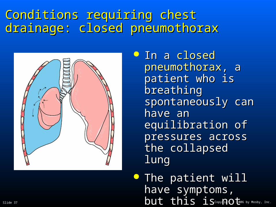
Closed pneumothoraxClosed pneumothorax Chest wall is intactChest wall is intact
Rupture of the lung Rupture of the lung and visceral pleura (or and visceral pleura (or airway) allows air into airway) allows air into the pleural spacethe pleural space

Copyright © 2006 by Mosby, Inc.Slide 36
Conditions requiring chest drainage: open Conditions requiring chest drainage: open pneumothoraxpneumothorax
An An open pneumothoraxopen pneumothorax is also called a “sucking is also called a “sucking chest wound”chest wound”
With the pressure changes in the chest that With the pressure changes in the chest that normally occur with breathing, air moves in and normally occur with breathing, air moves in and out of the chest through the opening in the chest out of the chest through the opening in the chest wallwall
Looks bad and sounds worse, but the opening Looks bad and sounds worse, but the opening acts as a vent so pressure from trapped air acts as a vent so pressure from trapped air cannot build up in the chestcannot build up in the chest
Copyright © 2006 by Mosby, Inc.Slide 37
Conditions requiring chest drainage: closed Conditions requiring chest drainage: closed pneumothoraxpneumothorax
In a In a closed closed pneumothoraxpneumothorax, a , a patient who is breathing patient who is breathing spontaneously can spontaneously can have an equilibration of have an equilibration of pressures across the pressures across the collapsed lungcollapsed lung
The patient will have The patient will have symptoms, but this is symptoms, but this is not life-threateningnot life-threatening

Copyright © 2006 by Mosby, Inc.Slide 38
Conditions requiring chest drainage: Conditions requiring chest drainage: tension pneumothoraxtension pneumothorax
A A tension pneumothoraxtension pneumothorax can kill can kill
Chest wall is intactChest wall is intact
Air enters the pleural space from the lung or Air enters the pleural space from the lung or airway, and it has no way to leaveairway, and it has no way to leave
There is no vent to the atmosphere as there is in There is no vent to the atmosphere as there is in an open pneumothoraxan open pneumothorax
Most dangerous when patient is receiving Most dangerous when patient is receiving positive pressure ventilation in which air is positive pressure ventilation in which air is forced into the chest under pressureforced into the chest under pressure

Copyright © 2006 by Mosby, Inc.Slide 39
Conditions requiring chest drainage: Conditions requiring chest drainage: tension pneumothoraxtension pneumothorax
Tension pneumothoraxTension pneumothorax occurs when a occurs when a closed closed pneumothoraxpneumothorax creates creates positive pressure in the positive pressure in the pleural space that pleural space that continues to buildcontinues to build
That pressure is then That pressure is then transmitted to the transmitted to the mediastinum (heart and mediastinum (heart and great vessels)great vessels)
Copyright © 2006 by Mosby, Inc.Slide 40
Conditions requiring chest drainage: Conditions requiring chest drainage: mediastinal shiftmediastinal shift
Mediastinal shift
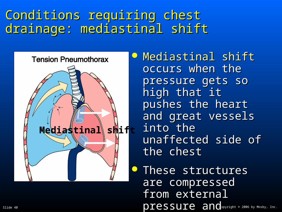
Mediastinal shiftMediastinal shift occurs occurs when the pressure gets when the pressure gets so high that it pushes the so high that it pushes the heart and great vessels heart and great vessels into the unaffected side into the unaffected side of the chestof the chest
These structures are These structures are compressed from compressed from external pressure and external pressure and cannot expand to accept cannot expand to accept blood flowblood flow

Copyright © 2006 by Mosby, Inc.Slide 41
Conditions requiring chest drainage: Conditions requiring chest drainage: mediastinal shiftmediastinal shift
Mediastinal shiftMediastinal shift can quickly lead to can quickly lead to cardiovascular collapsecardiovascular collapse
The vena cava and the right side of the heart The vena cava and the right side of the heart cannot accept venous returncannot accept venous return
With no venous return, there is no cardiac outputWith no venous return, there is no cardiac output
No cardiac output = not able to sustain lifeNo cardiac output = not able to sustain life
Copyright © 2006 by Mosby, Inc.Slide 42
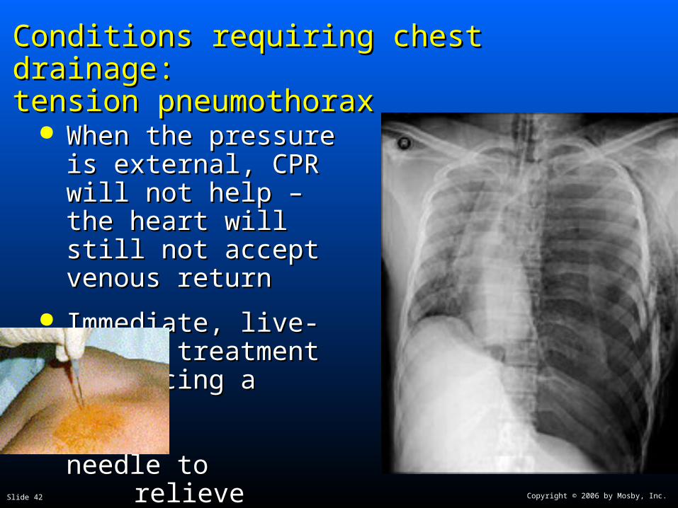
Conditions requiring chest drainage:Conditions requiring chest drainage:tension pneumothoraxtension pneumothorax
When the pressure is When the pressure is external, CPR will not external, CPR will not help – the heart will still help – the heart will still not accept venous returnnot accept venous return
Immediate, live-saving Immediate, live-saving treatment is placing a treatment is placing a
needle to needle to relieve pressure relieve pressure followed by followed by chest tubechest tube

Copyright © 2006 by Mosby, Inc.Slide 43
Conditions requiring chest drainage: Conditions requiring chest drainage: hemothoraxhemothorax
HemothoraxHemothorax occurs after thoracic surgery and occurs after thoracic surgery and many traumatic injuriesmany traumatic injuries
As with pneumothorax, the negative pressure As with pneumothorax, the negative pressure between the pleurae is disrupted, and the lung will between the pleurae is disrupted, and the lung will collapse to some degree, depending on the collapse to some degree, depending on the amount of bloodamount of blood
The risk of mediastinal shift is insignificant, as the The risk of mediastinal shift is insignificant, as the amount of blood needed to cause the shift would amount of blood needed to cause the shift would result in a life-threatening intravascular lossresult in a life-threatening intravascular loss

Copyright © 2006 by Mosby, Inc.Slide 44
Remove fluid & airRemove fluid & air
ThoracostomyThoracostomy creates an opening in the chest wall creates an opening in the chest wall through which a chest tube (also called thoracic through which a chest tube (also called thoracic catheter) is placed, which allows air and fluid to catheter) is placed, which allows air and fluid to flow out of the chestflow out of the chest

Copyright © 2006 by Mosby, Inc.Slide 45
Remove fluid and airRemove fluid and air

Copyright © 2006 by Mosby, Inc.Slide 46
Remove fluid & airRemove fluid & air
Choose site
Explore with finger
Place tube with clamp
Suture tube to chest

Copyright © 2006 by Mosby, Inc.Slide 47
Remove fluid & air through chest tubeRemove fluid & air through chest tube

Copyright © 2006 by Mosby, Inc.Slide 48
Remove fluid and air after thoracic Remove fluid and air after thoracic surgerysurgery
At the end of the At the end of the procedure, the procedure, the surgeon makes a surgeon makes a stab wound in the stab wound in the chest wall through chest wall through which the chest which the chest tube is placed into tube is placed into the pleural spacethe pleural space
Copyright © 2006 by Mosby, Inc.Slide 49
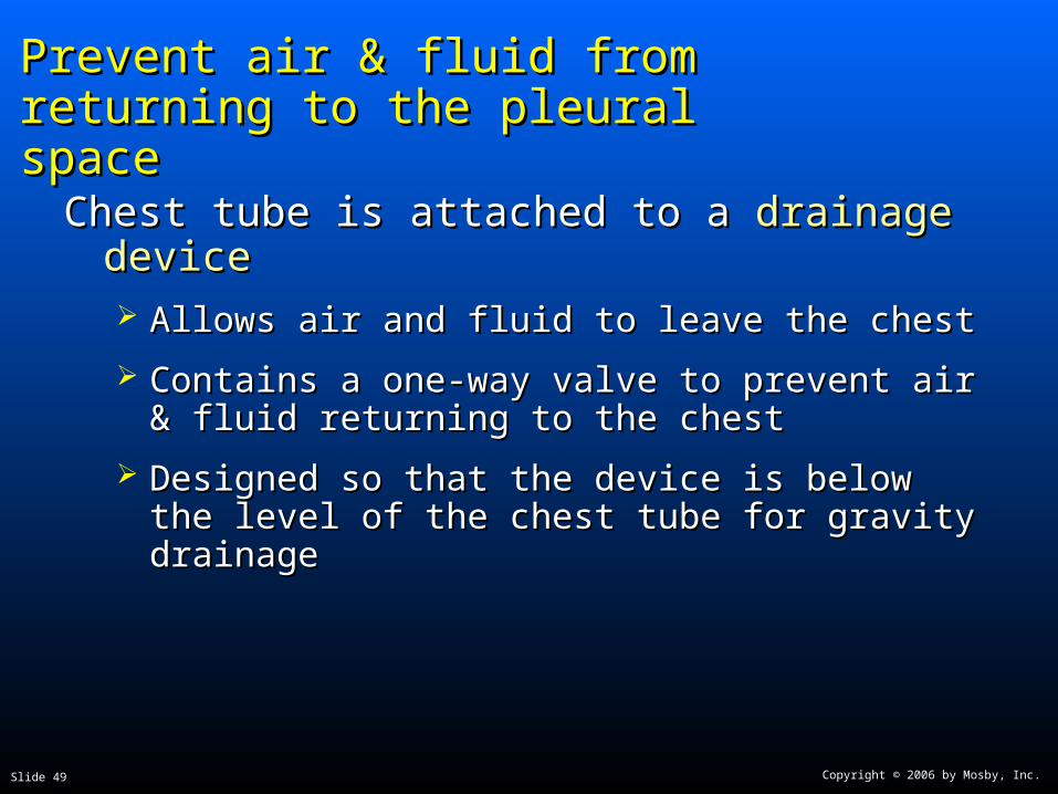
Prevent air & fluid from returning to Prevent air & fluid from returning to the pleural spacethe pleural space
Chest tube is attached to a Chest tube is attached to a drainage devicedrainage device Allows air and fluid to leave the chestAllows air and fluid to leave the chest
Contains a one-way valve to prevent air & fluid Contains a one-way valve to prevent air & fluid returning to the chestreturning to the chest
Designed so that the device is below the level of the Designed so that the device is below the level of the chest tube for gravity drainagechest tube for gravity drainage

Copyright © 2006 by Mosby, Inc.Slide 50
Prevent air & fluid from returning to Prevent air & fluid from returning to the pleural spacethe pleural space
How does a chest drainage system work?How does a chest drainage system work?
It’s all about It’s all about bottles and bottles and
strawsstraws

Copyright © 2006 by Mosby, Inc.Slide 51
Prevent air & fluid from returning to Prevent air & fluid from returning to the pleural spacethe pleural space
Tube from patient
Tube open to atmosphere vents air
Most basic conceptMost basic concept
Straw attached to Straw attached to chest tube from patient chest tube from patient is placed under 2cm of is placed under 2cm of fluid (water seal)fluid (water seal)
Just like a straw in a Just like a straw in a drink, air can push drink, air can push through the straw, but through the straw, but air can’t be drawn back air can’t be drawn back up the strawup the straw

Copyright © 2006 by Mosby, Inc.Slide 52
Prevent air & fluid from returning to Prevent air & fluid from returning to the pleural spacethe pleural space
This system works if only air is leaving the chestThis system works if only air is leaving the chest
If fluid is draining, it will add to the fluid in the If fluid is draining, it will add to the fluid in the water seal, and increase the depthwater seal, and increase the depth
As the depth increases, it becomes harder for As the depth increases, it becomes harder for the air to push through a higher level of water, the air to push through a higher level of water, and could result in air staying in the chestand could result in air staying in the chest
Copyright © 2006 by Mosby, Inc.Slide 53
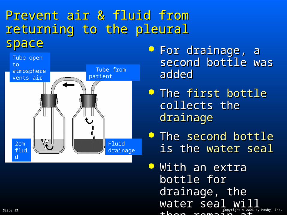
Prevent air & fluid from returning to Prevent air & fluid from returning to the pleural spacethe pleural space
Tube open to atmosphere vents air Tube from patient
Fluid drainage
2cm fluid
For drainage, a second For drainage, a second bottle was addedbottle was added
The The first bottlefirst bottle collects collects the the drainagedrainage
The The second bottlesecond bottle is is the the water sealwater seal
With an extra bottle for With an extra bottle for drainage, the water drainage, the water seal will then remain at seal will then remain at 2cm 2cm

Copyright © 2006 by Mosby, Inc.Slide 54
Prevent air & fluid from returning to Prevent air & fluid from returning to the pleural spacethe pleural space
The two-bottle system is the key for chest The two-bottle system is the key for chest drainagedrainage A place for drainage to collectA place for drainage to collect
A one-way valve that prevents air or fluid from A one-way valve that prevents air or fluid from returning to the chestreturning to the chest

Copyright © 2006 by Mosby, Inc.Slide 55
Restore negative pressure in the pleural Restore negative pressure in the pleural spacespace
Many years ago, it was believed that suction was Many years ago, it was believed that suction was always required to pull air and fluid out of the always required to pull air and fluid out of the pleural space and pull the lung up against the pleural space and pull the lung up against the parietal pleuraparietal pleura
However, recent research has shown that suction However, recent research has shown that suction may actually prolong air leaks from the lung by may actually prolong air leaks from the lung by pulling air through the opening that would pulling air through the opening that would otherwise close on its ownotherwise close on its own
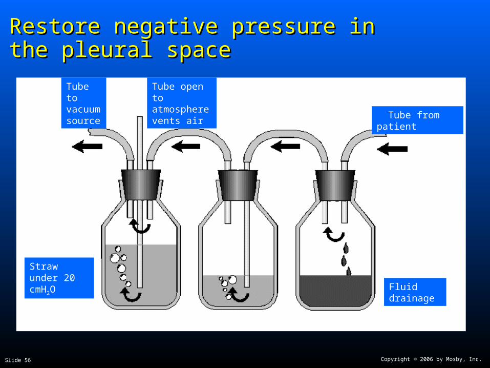
If If suctionsuction is required, a is required, a third bottlethird bottle is added is added
Copyright © 2006 by Mosby, Inc.Slide 56
Restore negative pressure in the Restore negative pressure in the pleural spacepleural space
Tube to vacuum source
Tube open to atmosphere vents air
Tube from patient
Fluid drainage
Straw under 20 cmH2O

Copyright © 2006 by Mosby, Inc.Slide 57
Restore negative pressure in the pleural Restore negative pressure in the pleural spacespace
The straw submerged in the suction control bottle The straw submerged in the suction control bottle (typically to 20cmH(typically to 20cmH22O) limits the amount of O) limits the amount of negative pressure that can be applied to the negative pressure that can be applied to the pleural space – in this case -20cmHpleural space – in this case -20cmH22OO
The submerged straw is open at the topThe submerged straw is open at the top
As the vacuum source is increased, once bubbling As the vacuum source is increased, once bubbling begins in this bottle, it means atmospheric begins in this bottle, it means atmospheric pressure is being drawn in to limit the suction levelpressure is being drawn in to limit the suction level
Copyright © 2006 by Mosby, Inc.Slide 58
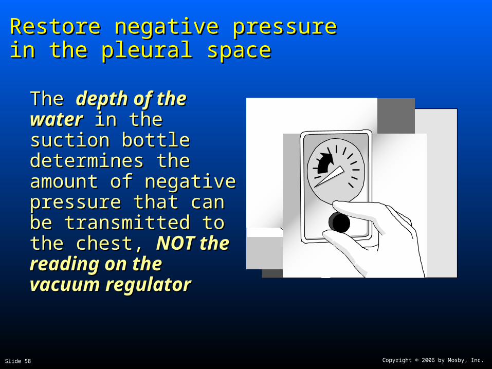
Restore negative pressure in the Restore negative pressure in the pleural spacepleural space
The The depth of the waterdepth of the water in the suction bottle in the suction bottle determines the amount determines the amount of negative pressure of negative pressure that can be transmitted that can be transmitted to the chest, to the chest, NOT the NOT the reading on the reading on the vacuum regulatorvacuum regulator

Copyright © 2006 by Mosby, Inc.Slide 59
Restore negative pressure in the pleural Restore negative pressure in the pleural spacespace
There is no research to support this number of -There is no research to support this number of -20cmH20cmH22O, just conventionO, just convention
Higher negative pressure can increase the flow Higher negative pressure can increase the flow rate out of the chest, but it can also damage rate out of the chest, but it can also damage tissuetissue

Copyright © 2006 by Mosby, Inc.Slide 60
How a chest drainage system How a chest drainage system worksworks
Expiratory positive pressureExpiratory positive pressure from the patient from the patient helps push air and fluid out of the chest (cough, helps push air and fluid out of the chest (cough, Valsalva)Valsalva)
GravityGravity helps fluid drainage as long as the chest helps fluid drainage as long as the chest drainage system is below the level of the chestdrainage system is below the level of the chest
SuctionSuction can improve the speed at which air and can improve the speed at which air and fluid are pulled from the chestfluid are pulled from the chest

Copyright © 2006 by Mosby, Inc.Slide 61
From bottles to a boxFrom bottles to a box
The bottle system worked, but it was bulky at the The bottle system worked, but it was bulky at the bedside and with 16 pieces and 17 connections, bedside and with 16 pieces and 17 connections, it was difficult to set up correctly while it was difficult to set up correctly while maintaining sterility of all of the partsmaintaining sterility of all of the parts
In 1967, a one-piece, disposable plastic box was In 1967, a one-piece, disposable plastic box was introducedintroduced
The box did everything the bottles did – and The box did everything the bottles did – and moremore
Copyright © 2006 by Mosby, Inc.Slide 62
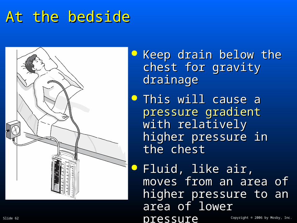
At the bedsideAt the bedside
Keep drain below the chest Keep drain below the chest for gravity drainagefor gravity drainage
This will cause a This will cause a pressure pressure gradientgradient with relatively with relatively higher pressure in the chesthigher pressure in the chest
Fluid, like air, moves from Fluid, like air, moves from an area of higher pressure an area of higher pressure to an area of lower pressureto an area of lower pressure
Same principle as raising an Same principle as raising an IV bottle to increase flow IV bottle to increase flow raterate

Copyright © 2006 by Mosby, Inc.Slide 63
Monitoring intrathoracic pressureMonitoring intrathoracic pressure
The water seal chamber and suction control The water seal chamber and suction control chamber provide intrathoracic pressure monitoringchamber provide intrathoracic pressure monitoring
Gravity drainage without suctionGravity drainage without suction: Level of water in : Level of water in the the water seal chamber = intrathoracic pressurewater seal chamber = intrathoracic pressure (chamber is calibrated manometer)(chamber is calibrated manometer)
Slow, gradual rise in water level over time means more Slow, gradual rise in water level over time means more negative pressure in pleural space and signals healingnegative pressure in pleural space and signals healing
Goal is to return to -8cmHGoal is to return to -8cmH2200
With suctionWith suction: Level of water in : Level of water in suction control +suction control + level of water in level of water in water sealwater seal chamber chamber = = intrathoracic pressureintrathoracic pressure
Copyright © 2006 by Mosby, Inc.Slide 64
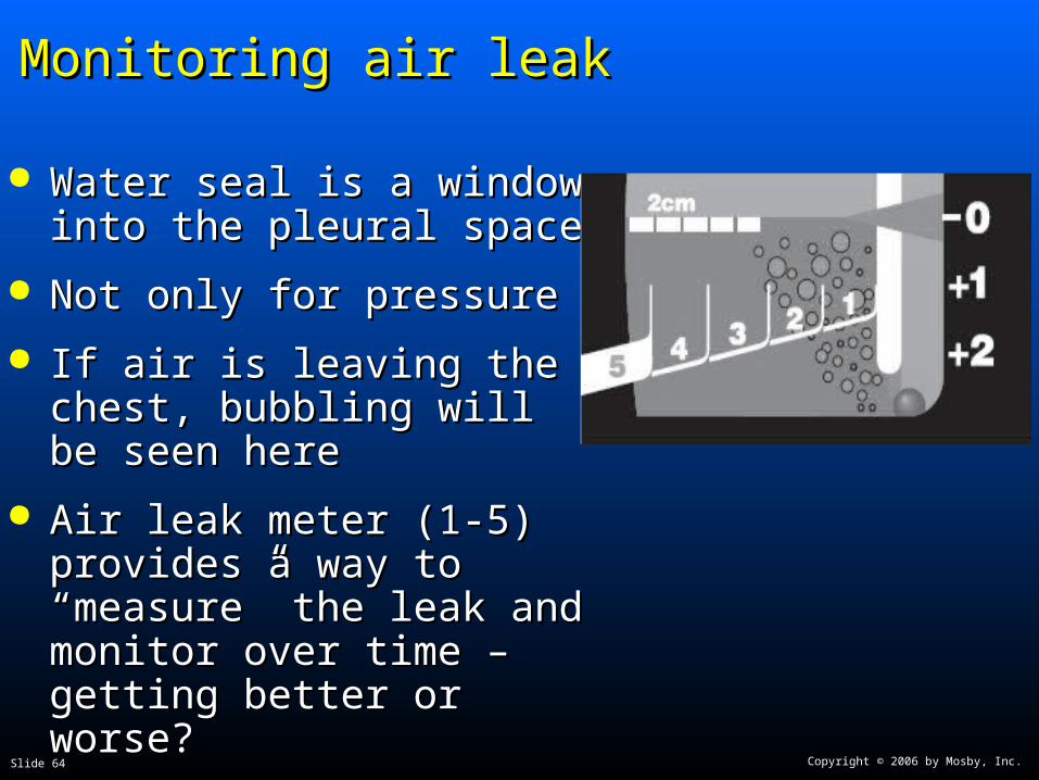
Monitoring air leakMonitoring air leak
Water seal is a window into Water seal is a window into the pleural spacethe pleural space
Not only for pressureNot only for pressure
If air is leaving the chest, If air is leaving the chest, bubbling will be seen herebubbling will be seen here
Air leak meter (1-5) provides a Air leak meter (1-5) provides a way to “measure” the leak and way to “measure” the leak and monitor over time – getting monitor over time – getting better or worse?better or worse?

Copyright © 2006 by Mosby, Inc.Slide 65
Setting up the drainSetting up the drain
Follow the manufacturer’s instructions for adding Follow the manufacturer’s instructions for adding water to the 2cm level in the water seal chamber, water to the 2cm level in the water seal chamber, and to the 20cm level in the suction control and to the 20cm level in the suction control chamber (unless a different level is ordered)chamber (unless a different level is ordered)
Connect 6' patient tube to thoracic catheterConnect 6' patient tube to thoracic catheter
Connect the drain to vacuum, and slowly increase Connect the drain to vacuum, and slowly increase vacuum until vacuum until gentle bubblinggentle bubbling appears in the appears in the suction control chambersuction control chamber

Copyright © 2006 by Mosby, Inc.Slide 66
Setting up suctionSetting up suction
You don’t need to boil spaghetti!You don’t need to boil spaghetti!
Vigorous bubbling is Vigorous bubbling is loudloud and and disturbing to most patientsdisturbing to most patients
Will also cause Will also cause rapid evaporationrapid evaporation in the in the chamber, which will chamber, which will lower suction levellower suction level
Too much bubbling is Too much bubbling is not needed clinicallynot needed clinically in in 98% of patients – more is not better98% of patients – more is not better
If too much, turn down vacuum source until If too much, turn down vacuum source until bubbles go away, then slowly increase until they bubbles go away, then slowly increase until they reappear, then stopreappear, then stop
Copyright © 2006 by Mosby, Inc.Slide 67
Disposable chest drainsDisposable chest drains
Collection chamberCollection chamber Fluids drain directly into chamber, calibrated in mL fluid, Fluids drain directly into chamber, calibrated in mL fluid,
write-on surface to note level and timewrite-on surface to note level and time
Water sealWater seal One way valve, U-tube design, can monitor air leaks & One way valve, U-tube design, can monitor air leaks &
changes in intrathoracic pressurechanges in intrathoracic pressure
Suction control chamberSuction control chamber U-tube, narrow arm is the atmospheric vent, large arm is U-tube, narrow arm is the atmospheric vent, large arm is
the fluid reservoir, system is regulated, easy to control the fluid reservoir, system is regulated, easy to control negative pressurenegative pressure