poli: a virtual screening pipeline based on template...
TRANSCRIPT

PoLi: A Virtual Screening Pipeline Based on Template Pocket andLigand SimilarityAmbrish Roy, Bharath Srinivasan, and Jeffrey Skolnick*
Center for the Study of Systems Biology, Georgia Institute of Technology, 250 14th Street NW, Atlanta, Georgia 30318, United States
*S Supporting Information
ABSTRACT: Often in pharmaceutical research the goal is to identify smallmolecules that can interact with and appropriately modify the biologicalbehavior of a new protein target. Unfortunately, most proteins lack bothknown structures and small molecule binders, prerequisites of many virtualscreening, VS, approaches. For such proteins, ligand homology modeling,LHM, that copies ligands from homologous and perhaps evolutionarily distanttemplate proteins, has been shown to be a powerful VS approach to identifypossible binding ligands. However, if we want to target a specific pocket forwhich there is no homologous holo template protein structure, then LHM willnot work. To address this issue, in a new pocket-based approach, PoLi, wegeneralize LHM by exploiting the fact that the number of distinct smallmolecule ligand-binding pockets in proteins is small. PoLi identifies similarligand-binding pockets in a holo template protein library, selectively copiesrelevant parts of template ligands, and uses them for VS. In practice, PoLi is ahybrid structure and ligand-based VS algorithm that integrates 2D fingerprint-based and 3D shape-based similarity metrics forimproved virtual screening performance. On standard DUD and DUD-E benchmark databases, using modeled receptorstructures, PoLi achieves an average enrichment factor of 13.4 and 9.6, respectively, in the top 1% of the screened library. Incontrast, traditional docking-based VS using AutoDock Vina and homology-based VS using FINDSITEfilt have an averageenrichment of 1.6 (3.0) and 9.0 (7.9) on the DUD (DUD-E) sets, respectively. Experimental validation of PoLi predictions ondihydrofolate reductase, DHFR, using differential scanning fluorimetry, DSF, identifies multiple ligands with diverse molecularscaffolds, thus demonstrating the advantage of PoLi over current state-of-the-art VS methods.
■ INTRODUCTION
Identifying lead molecules that bind to a given target protein isa fundamental challenge in pharmaceutical research. This issuehas been addressed using both experimental high-throughputscreening (HTS) and computational in silico (commonlyreferred as virtual screening, VS) approaches.1 Although HTSis currently the best method for lead identification, thedependence of the results on experimental factors, “chemicalspace” coverage, and applicability for all targets, along with thecost and time required to perform such screens limit theirapplicability.2 For this reason, new computational approachesthat can efficiently screen large databases are needed, as theynot only complement HTS but also have much higherthroughput and greatly reduced cost and increased speed.3
On the basis of the availability of target protein structures,either structure- or ligand-based VS calculations are performedto identify potential lead molecules. The most commonly usedstructure-based VS approach is molecular docking, which doesnot require a priori knowledge of known binders4 and cantarget a specific binding pocket of interest. Molecular dockinginvolves screening database molecules based on their calculatedinteraction energy with the receptor binding site.5 As such, itsperformance relies heavily on the receptor structure quality andflexibility.6 For example, about 90% of docking accuracy is lost
if models of trypsin and HIV-1 protease with a root-mean-square deviation, RMSD, greater than 1.5 Å from native areused.7 It also depends on the presence of water molecules, theconformations of database molecules, and the sensitivity of thescoring function used for evaluating protein−ligand inter-actions.8 Another structure-based variant docks small moleculefragments to screen for promising leads.9 However, distinguish-ing binding and nonbinding fragments is a challenge in thesemethods, as fragments bind with very low weak binding affinity,which cannot be captured using the inaccurate scoringfunctions that we currently have.10 Moreover, like other smallmolecule docking approaches, fragment-based approaches alsorequire a high-resolution structure, which is not alwaysavailable. To address this problem, homology models that arevery closely related to the template proteins in the PDB havebeen used; moreover, the models frequently require a lot ofside-chain refinement.11
In the absence of a target receptor structure, ligand-based VSapproaches are generally used. Ligand-based VS is robust butrequires at least one known bioactive molecule that is used as aseed to fish out database molecules with similar chemotypes.
Received: April 23, 2015
Article
pubs.acs.org/jcim
© XXXX American Chemical Society A DOI: 10.1021/acs.jcim.5b00232J. Chem. Inf. Model. XXXX, XXX, XXX−XXX
Most common ligand-based VS approaches evaluate a 2Dfingerprint,12 pharmacophore,13 or 3D shape-based similarity14
between known bioactive and database molecules. Thus, moststructure and ligand-based VS methods require either anexperimentally solved receptor structure or an experimentallydetermined bioactive molecule. As such, they cannot be readilyapplied to many proteins of therapeutic interest.To address these significant limitations, we recently
described two new virtual screening approaches.15,16 The first,FINDSITEfilt,15 can use either experimental or predicted low-resolution target protein structures to screen databasemolecules based on 2D fingerprint similarity with templateligands in the PDB holo template library. FINDSITEcomb
includes FINDSITEfilt for proteins having holo templates, butfor those proteins lacking holo templates, it also uses anartificially generated template library of predicted tertiarystructures whose binding ligands are found in the ChEMBL17
and DrugBank18 ligand-binding databases. Template ligands arecopied from globally related protein structures based onstructural similarity to the target, without considering where theligand actually binds in the template protein. These methodshave the inherent advantages of speed, lack of requirement ofhigh-resolution protein structures, and do not need knownbinders. Although both approaches achieve good enrichment inidentifying out active molecules, FINDSITEfilt, in particular,depends on the availability of proteins with a similar fold in theholo template library for effective virtual screening. Moreimportantly, both FINDSITEfilt and FINDSITEcomb were notdeveloped with the goal of targeting a specific binding pocket.To begin to generalize our approach, we developed a shape-
based virtual screening algorithm, LIGSIFT,16 that screensdatabase molecules based on their 3D shape and chemicalfeature similarity to a target seed ligand. LIGSIFT wasbenchmarked using the 3D similarity of database moleculesto a known binding ligand to the target protein, as provided bythe DUD database,19 and its performance without knownbinding ligands was not established. Thus, a new pocket centricapproach that can target a specific binding pocket of interest,overcome the requirement of global fold similarity between
template and target structures, and combines both 2D- and 3D-based ligand similarity metrics for virtual screening usingligands identified from holo templates is needed.On the basis of these ideas and the fact that the space of
protein−ligand-binding pockets is small and close tocomplete,20 we developed a new virtual screening pipeline,PoLi, that first predicts the ligand-binding pocket in the targetprotein, selectively copies parts of template ligands based onbinding-pocket alignment, and then performs virtual screeningof database molecules based on combined 2D and 3D ligandsimilarity metrics to the selected template small molecules.Large-scale in silico benchmarking followed by in vitro high-throughput experimental validation of predictions on E. coliDHFR establishes PoLi as an effective virtual screeningapproach.
■ RESULTS
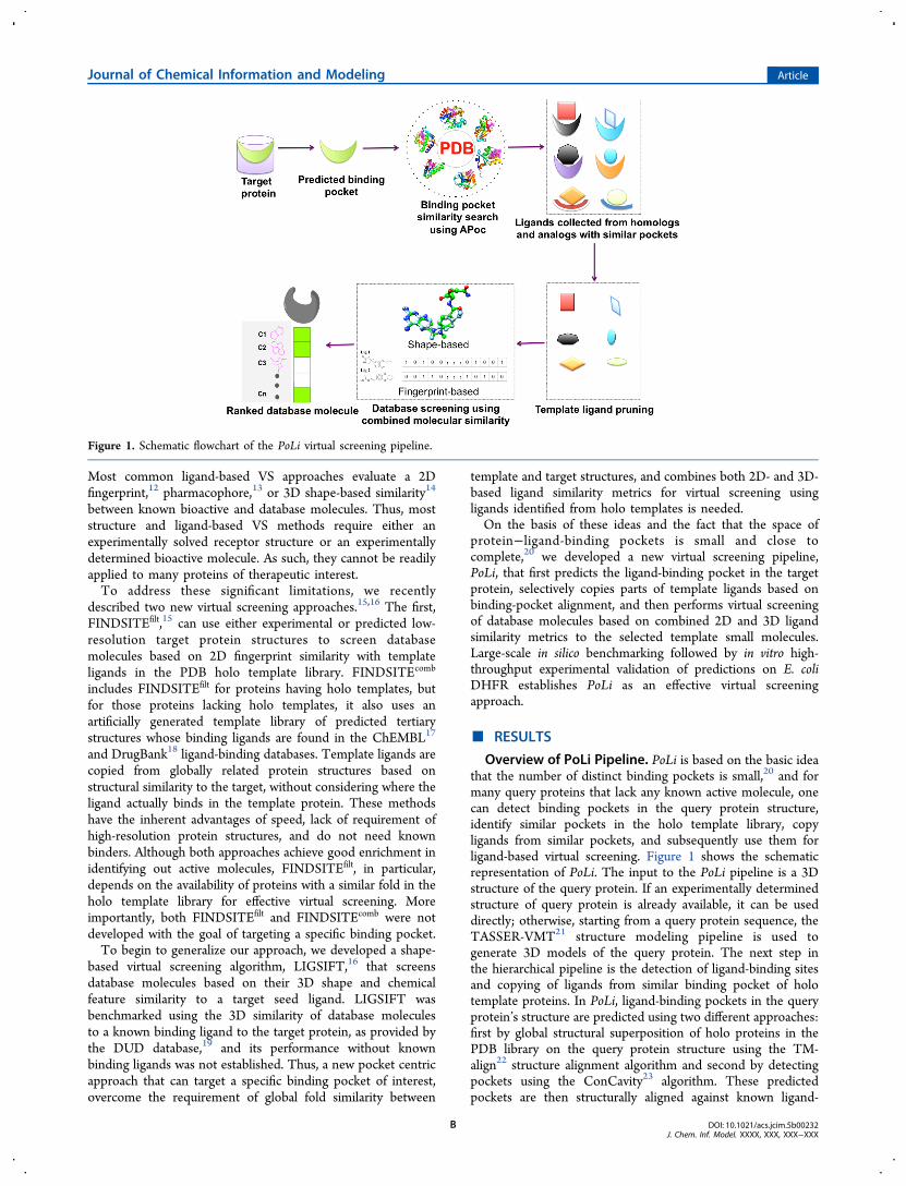
Overview of PoLi Pipeline. PoLi is based on the basic ideathat the number of distinct binding pockets is small,20 and formany query proteins that lack any known active molecule, onecan detect binding pockets in the query protein structure,identify similar pockets in the holo template library, copyligands from similar pockets, and subsequently use them forligand-based virtual screening. Figure 1 shows the schematicrepresentation of PoLi. The input to the PoLi pipeline is a 3Dstructure of the query protein. If an experimentally determinedstructure of query protein is already available, it can be useddirectly; otherwise, starting from a query protein sequence, theTASSER-VMT21 structure modeling pipeline is used togenerate 3D models of the query protein. The next step inthe hierarchical pipeline is the detection of ligand-binding sitesand copying of ligands from similar binding pocket of holotemplate proteins. In PoLi, ligand-binding pockets in the queryprotein’s structure are predicted using two different approaches:first by global structural superposition of holo proteins in thePDB library on the query protein structure using the TM-align22 structure alignment algorithm and second by detectingpockets using the ConCavity23 algorithm. These predictedpockets are then structurally aligned against known ligand-
Figure 1. Schematic flowchart of the PoLi virtual screening pipeline.
Journal of Chemical Information and Modeling Article
DOI: 10.1021/acs.jcim.5b00232J. Chem. Inf. Model. XXXX, XXX, XXX−XXX
B

binding pockets in the PDB holo template library, using thesequence-order independent binding-site comparison algorithmAPoc,24 and template ligands from similar binding pockets arecopied in the query ligand-binding pocket. These copiedtemplate ligands are later pruned to remove parts of thetemplate ligand that interact with the unaligned region oftemplate binding pocket and then used in ligand-based VS.Virtual screening in PoLi is performed using a combination of
2D fingerprint-based and 3D-shape-based similarity metrics,where the 2D path-based fingerprint is generated usingOpenBabel,25 and 3D similarity is calculated using a variantof the LIGSIFT16 algorithm. LIGSIFT is a small moleculestructural alignment algorithm that uses an atom-centeredsmooth Gaussian function to describe the ligand structure andperform rapid overlay to measure 3D shape and chemicalsimilarity. The ligand 3D similarity between molecules inLIGSIFT is evaluated using a size-independent scoring function(a scaled TC). The statistical significance of the similarity score(p-value) is estimated based on millions of comparisons ofrandomly selected ligands.16 A detailed description of thepipeline modules is provided in the Materials and Methodssection.Detection of Template Seed Ligands for Virtual
Screening. We first validate our approach to detect chemicallysimilar ligands using a pocket-based search. These detectedligands will be used as seed ligands for ligand-based virtualscreening. The objective of this exercise is to show that theligands copied from template proteins have a statisticallysignificant chemical similarity to the native ligand when theycome from structurally similar pockets as assessed using theligand-binding pocket structural comparison algorithm APoc.24
In practice, we selected a nonredundant set of 30,000 ligand-pairs with statistically significant chemical similarity (LIGSIFT3D chemical similarity p-value < 0.001) and 35,000 ligand pairsthat lack significant chemical similarity score from the PDBholo template library, such that the corresponding receptorpairs share less than 30% sequence identity. Figure 2 shows theperformance of APoc’s pocket similarity24 to detect templatesthat have chemically similar ligands bound to them, incomparison to TM-align22 (global structural similarity) andHHalign26 (threading). Predictions are labeled as correct if thep-value of the 3D chemical similarity score between copied
template ligand and the query ligand is less than 0.001. Asshown in Figure 2, the pocket similarity-based approach(APoc) outperforms both TM-align and HHalign in detectingtrue positives. For instance, at 95% specificity, APoc identifies34% of chemically similar ligand pairs; TM-align globalstructural template matching recovers 18.5% true positives,while HHalign only identifies 14.5% true positives. Thisestablishes that pocket similarity is the best of the threeapproaches to identify templates that have ligands withoverlapping chemical features.
Benchmarking Performance on DUD and DUD-EDatabases. Benchmarking of virtual screening algorithmswas done on 40 DUD database proteins19 and 65 proteinsincluded in the DUD-E database (Table S1).27 Both areroutinely used for testing scoring functions and virtualscreening methods. Our objective here is to analyze the overallperformance of the PoLi pipeline, which includes structuremodeling of the receptor, binding site prediction, and virtualscreening of database molecules (Materials and Methods). Wealso ran the same pipeline using experimentally solved proteinstructures to assess the effect of model quality on virtualscreening performance. For structure modeling of targetproteins and binding site predictions using both modeled andexperimental structure, closely related homologous proteinswere excluded from template libraries using a sequence identitythreshold of 30%.
Model Quality of Target Proteins. Since model quality andaccuracy of binding site predictions are expected determinantsof structure-based methods for virtual screening, including PoLi,we first examine the quality of predicted protein structures.Figure 3A and B present the global and local structure qualityof predicted TASSER21 models. The global structure quality ofmodels is measured as TM-score,28 with values rangingbetween [0,1] with a higher score indicating a better structuralmatch between the model and native structure. Statistically, aTM-score less than 0.3 means random structural similarity andTM-score greater than 0.4 indicates that the protein pairs havea similar fold. The average TM-scores of DUD and DUD-E setproteins are 0.76 ± 0.18 and 0.73 ± 0.12, respectively (Figure3A), clearly highlighting that the predicted structure of mostproteins share high structural similarity with the experimentallydetermined structure. Two proteins in both sets, namely, hmgr(hydroxymethylglutaryl-CoA reductase) and sahh (S-adenosyl-homocysteine hydrolase) in the DUD set, and nos1 (nitricoxide synthase) and pa2ga (phospholipase A2 group IIA) in theDUD-E set, have incorrectly predicted structures, i.e., the TM-score between the model and experimental structure is less than0.4. For these proteins, the structural confidence C-score ofmodel29 is also less than −3, i.e. they can be easily recognized ashaving poorly predicted structures even in the absence ofexperimentally determined structures (Table S2). Figure 3Bshows the structure quality of the predicted models near theknown ligand-binding site of the co-crystallized ligand. Themean Cα RMSD of binding pocket residues (residues within<4.5 Å from co-crystallized ligand in experimental structure) inthe DUD proteins is 4.3 ± 5.7 Å, and in the DUD-E proteins, itis 3.3 ± 3.2 Å. In most cases, the structure near the knownligand-binding pocket is also reasonably well predicted (TableS2), with some local structural variations (typical of anyhomology based structure modeling algorithm). This is notsurprising, as functionally important regions are generally moreconserved than other parts of the protein and are more likely tobe correctly predicted. Nevertheless, for some proteins, the
Figure 2. Comparison of a pocket-based method (APoc) with globalstructure alignment and homology-based approaches to detect similarligands. The benchmark shows the ability of different approaches torecognize 30,000 pairs of similar ligands from 35,000 pairs ofchemically dissimilar ligands.
Journal of Chemical Information and Modeling Article
DOI: 10.1021/acs.jcim.5b00232J. Chem. Inf. Model. XXXX, XXX, XXX−XXX
C

structural variations of the binding pocket residues can be large(Cα RMSD > 5 Å) because of two reasons: (a) The globalstructure itself is incorrectly predicted and so is the bindingpocket (e.g., in hydroxymethylglutaryl-CoA reductase). (b)While the global fold is basically correct, the structure of theligand-binding site is only partially correct. For example, itcould be an interdomain binding pocket with one incorrectlypredicted domain (e.g., in glycogen phosphorylase beta). Suchstructural variations affect both binding pocket predictions andhave a seriously adverse effect on the performance of moleculardocking methods that use these models.Analysis of Binding Site Predictions. Figure 3C shows the
performance of the PoLi pipeline in recapitulating knownligand-binding sites as provided in the DUD and DUD-Edatabases19,27 using both modeled and experimental structures.Using modeled structures, ligand-binding pockets are correctlyidentified (within 5 Å from the geometric center of theexperimentally solved ligand−protein) in 32 of the 40 DUD set
proteins and in 52 of the 65 DUD-E set proteins. Whenexperimental structures are used, binding pockets can becorrectly predicted for 36 proteins in DUD set and 60 proteinsin DUD-E set, within the same distance cutoff. Among themodeled protein structures with incorrectly predicted bindingpockets (pocket distance >5 Å), 5 of the 8 proteins in DUD setand 7 of the 13 proteins in DUD-E set have binding pocketresidues with a Cα RMSD > 5 Å. For the remaining predictedand experimental structures even though binding pocketcavities could be detected, they lacked a significant match (p-value < 0.001 and PS-score > 0.35) to known ligand-bindingpockets in the PDB holo template library. This is one of themain limitations of LHM-based binding site predictions. Thus,these VS predictions are of poor quality. Also in some targets(e.g., in HIV reverse transcriptase), the known ligand-bindingsite is interfacial (formed by contacts of protein chains in acomplex) and cannot be always predicted using monomericstructures alone (especially in those models having structural
Figure 3. Structure quality and binding site prediction accuracy for DUD and DUD-E proteins. Box and whiskers plot of (A) TM-score and (B)ligand-binding pocket Cα RMSD of TASSER models to the experimentally determined structures. (C) Distance between the geometric center of theligand in the co-crystallized complex and the center of the best predicted ligand-binding pocket in the 40 DUD and 65 DUD-E protein targets.
Table 1. Virtual Screening Performance of PoLi on 40 DUD Targets and 65 DUD-E Targets Using Experimental and ModeledStructuresa
target receptor AUC av | sd EF1% av | sd EF5% av | sd EF10% av | sd HR1% av | sd HR5% av | sd HR10% av | sd
DUD-E (LIGSIFT) 0.73 ± 0.20 18.7 ± 18.1 6.6 ± 4.4 4.2 ± 2.3 29.2 ± 25.7 20.0 ± 14.1 25.3 ± 14.6DUD-E (exp.) 0.72 ± 0.16 9.6 ± 13.5 4.8 ± 4.4 3.4 ± 2.3 14.6 ± 19.1 14.0 ± 13.9 20.1 ± 15.1DUD-E (model TM > 0.5) 0.74 ± 0.16 9.9 ± 13.5 4.9 ± 4.3 3.6 ± 2.3 14.9 ± 18.8 14.5 ± 13.4 21.4 ± 15.3DUD-E (model pdist < 5 Å) 0.76 ± 0.16 11.0 ± 14.3 5.4 ± 4.4 3.9 ± 2.3 16.6 ± 19.9 15.9 ± 13.9 23.4 ± 15.6DUD-E (model) 0.73 ± 0.16 9.6 ± 12.7 4.7 ± 4.2 3.6 ± 2.3 14.3 ± 17.0 13.9 ± 13.2 21.1 ± 15.3DUD (LIGSIFT) 0.77 ± 0.20 17.4 ± 11.1 7.8 ± 5.4 4.7 ± 2.7 49.4 ± 31.5 39.2 ± 27.1 47.2 ± 27.5DUD (exp.) 0.78 ± 0.19 15.2 ± 11.4 7.2 ± 5.2 4.7 ± 2.7 43.3 ± 32.4 31.9 ± 24.4 41.3 ± 26.0DUD (model TM > 0.5) 0.78 ± 0.18 14.1 ± 10.1 7.2 ± 4.7 4.6 ± 2.7 40.1 ± 28.8 31.8 ± 22.1 40.7 ± 25.5DUD (model pdist < 5 Å) 0.80 ± 0.19 15.9 ± 9.9 8.1 ± 4.7 5.1 ± 2.7 45.2 ± 28.3 34.9 ± 21.8 44.1 ± 25.4DUD (model) 0.78 ± 0.18 13.4 ± 10.3 7.0 ± 4.8 4.6 ± 2.9 38.0 ± 29.3 30.7 ± 22.3 39.5 ± 26.1
aav, average; sd, standard deviation; exp, experimentally determined structure; TM, TM-score of model to experimental structure; pdist, distancebetween predicted pocket and center of mass of ligand in crystal structure.
Journal of Chemical Information and Modeling Article
DOI: 10.1021/acs.jcim.5b00232J. Chem. Inf. Model. XXXX, XXX, XXX−XXX
D

variations near the pocket), which is a limitation of thisapproach.Virtual Screening Performance on DUD and DUD-E
targets. The above analyses have shown the following: (a) Apocket-based approach is better than both global similarity- andhomology-based approaches in detecting templates whoseligands have similar chemical features. (b) For most proteins,computationally generated models have a correctly predictedfold, whose ligand-binding pockets can also be correctlyidentified in about 80% of the cases. In this section, weexamine the next module of the PoLi pipeline: the ability toidentify active molecules in the DUD19 and DUD-E27
databases. Performance is evaluated using standard evaluationmetrics: (a) the Enrichment Factor (EF) of the screenedcompound library, (b) the Hit Rate (HR) of active molecules,and (c) the Receiver Operating Characteristic (ROC) curve.Descriptions of these metrics are given in Materials andMethods.Table 1 shows the virtual screening performance of PoLi
using both computationally generated models and experimen-tally determined receptor structures. The average enrichment inthe top 1% of the screened library is 13.4 and 9.6 for DUD andDUD-E set modeled receptor structures, respectively, and theaverage hit rates are 38.0 and 14.3, respectively. Whenexperimental structures are used, the enrichment rates in thetop 1% increase to 15.2 and 9.6, respectively, and the hit rateincreases to 43.3 and 14.6, respectively, for the DUD andDUD-E sets. A paired Wilcoxon signed rank t-test betweenEF1% achieved using model and experimental structures has ap-value of 0.44 on the DUD set and 0.30 on the DUD-E set,suggesting that the difference in VS performance using modeland experimental structure is not statistically significant.Moreover, using a known binder of each target protein(taken from the experimental structure in PDB), the bestaverage EF1% obtained using LIGSIFT shape-based screeningis 17.4 and 18.7 for the DUD and DUD-E sets, respectively;this is notable since PoLi predictions were generated by usingligands copied from templates with <30% sequence identity.Since model quality and accuracy of binding pocket
predictions directly affect the performance of the PoLi pipeline,we further analyzed the VS results only for proteins withreasonable quality model (TM-score > 0.5) and for thoseproteins in which one of the predicted pockets overlap with theknown ligand-binding site in the experimental structure (pocketdistance < 5 Å). Since most proteins are reasonably wellpredicted, using correctly modeled structures, the EF1% onDUD and DUD-E set, marginally improved to 14.1 and 9.9,respectively (Table 1), an increase of approximately 3−5%compared to EF1% obtained for all the proteins. A moresignificant improvement is observed when proteins in which theknown ligand-binding site was recapitulated as one of thebinding site predictions. The EF1% values for DUD and DUD-
E are 15.9 and 11.0, respectively, an improvement ofapproximately 14−18%.It is interesting to observe that using both modeled and
experimental structures the performance of PoLi is consistentlylower on the DUD-E set compared to the DUD set, whileperformance remains similar when known binders are used asinput for LIGSIFT-based VS. This decrease in performancecannot be attributed to either structure quality, as the averageTM-score for both sets about 0.7, or to the accuracy of bindingpocket predictions, as just 20% of modeled proteins and about10% of experimental structures in both the sets have predictedpockets at a distance greater than 5 Å from the geometriccenter of the experimentally solved ligand location in theprotein’s structure.We sought to analyze this further by examining the highest
3D and 2D molecular similarity between database moleculesand collected template ligands. Table 2 clearly shows that themain reason for the decrease in performance on the DUD-E set(using both experimental and modeled structures) is because ofincreased overlap between the active and decoy molecularsimilarity distributions. More specifically, there is an overalldecrease of 3D similarity scores in the DUD-E compared to theDUD database. A detailed statistical analysis performed bytaking random samples from the DUD and DUD-E databasesand analyzing the difference between 3D similarity scores ofactives and decoy molecules reveals that the mean of thedifference distribution is 0.08 in the DUD set and 0.02 in theDUD-E set. Also a Welch’s t-test performed on the differencedistributions has a p-value less than 2.2 × 10⁻¹⁶, suggesting thatdifference between the highest similarity scores of actives anddecoys in the DUD set was significantly greater than in theDUD-E set.
Comparison with Control Methods for Virtual Screening.Without known binders, molecular docking is the most widelyused virtual screening approach and has been benchmarked onnumerous occasions using experimental structures.30,31 Anothervirtual screening approach that is becoming increasinglypopular copies ligands from homologous/structurally analogoustemplate proteins and uses them as seeds for ligand-basedvirtual screening.15,32 Here, template ligands are copied, andeither a single or combination of different 2D molecularsimilarity metrics is used for ranking the database molecules.As our experimental control, we employed the widely used
molecular docking tool AutoDock Vina,33 our in-housedeveloped VS algorithm FINDSITEfilt (as it also uses a PDBholo template library), and shape-based VS using LIGSIFT.Docking runs of AutoDock Vina were performed with defaultoptions, and the entire receptor structure was enclosed within abox during the docking simulations (as if the binding pocketwere unknown). Furthermore, to avoid any bias arising due todifferences in holo template library, both FINDSITEfilt andLIGSIFT used the same set of templates as PoLi for virtual
Table 2. Analysis of Molecular Similarity Scores between Database Molecules and Template Ligands To Understand theDecrease in Performance of PoLi on DUD-E Databasea
3D similarity 2D similarity
database receptor structure actives av | sd decoys av | sd actives av | sd decoys av | sd
DUD-E experimental 0.52 ± 0.06 0.50 ± 0.04 0.61 ± 0.09 0.57 ± 0.06model 0.52 ± 0.06 0.50 ± 0.04 0.61 ± 0.09 0.56 ± 0.06
DUD experimental 0.58 ± 0.09 0.53 ± 0.05 0.62 ± 0.09 0.58 ± 0.06model 0.57 ± 0.06 0.53 ± 0.04 0.62 ± 0.09 0.57 ± 0.06
aav, average; sd, standard deviation.
Journal of Chemical Information and Modeling Article
DOI: 10.1021/acs.jcim.5b00232J. Chem. Inf. Model. XXXX, XXX, XXX−XXX
E

screening. FINDSITEfilt uses a 2D fingerprint similarity metric(eq 6) between these selected templates and databasemolecules, while LIGSIFT uses these template ligands asseeds (without any pruning) for shape-based structuralalignment with database molecules (eq 4). Thus, FINDSITEfilt
is the VS performance achieved using a 2D approach, whileLIGSIFT is representative of a 3D VS algorithm.Table 3 reports the AUC, EF, and HR obtained on the DUD
and DUD-E sets using modeled protein structures. The averageenrichment factors of PoLi, LIGSIFT, FINDSITEfilt, andAutoDock Vina in the top 1% of the screened library(EF1%) are 13.4, 11.8, 9.0, and 1.6, respectively, on theDUD set. A similar trend is also observed on the DUD-E set,where PoLi, LIGSIFT, FINDSITEfilt, and AutoDock Vinaachieve EF1% values of 9.6, 5.9, 7.9, and 3.0, respectively.Figure S1 shows the distribution of AUC and EF1% for theDUD and DUD-E set proteins using a boxplot. A pairedWilcoxon signed rank t-test between EF1% of PoLi and controlmethods (LIGSIFT, FINDSITEfilt, and AutoDock Vina), afterBoneferroni correction for multiple comparison, have p-valuesof 8.45 × 10−02, 4.18 × 10−02, and 1.51 × 10−06, respectively, onthe DUD set and 0.0004, 0.0099, and 0.0011, respectively, onthe DUD-E set of proteins. It is clear from these results thatestablishing which molecular similarity metrics (3D shape-based or 2D fingerprint-based) is better is difficult, as their
performance can vary with the protein target. Nevertheless,fusion of 2D and 3D similarity metrics based on their Z-score(eq 7) shows the best performance in virtual screening on thetested databases. The observed improvement of PoLi is alsopartially due to the pruning of template ligands. Biasedstructural overlap of ligands near the hot spot regions alsocontributed to the enrichment of actives in the DUD set, whereEF1% increased from 12.3 for unbiased structural overlap to13.4 for biased overlap. For the DUD-E set, the performancewas similar, where EF1% was 9.7 for unbiased structural overlapand 9.6 for biased overlap. Molecular docking using AutoDockVina has the worst performance in identifying active molecules.One might expect that without explicitly providing the exactlocation of target binding site, molecular docking will certainlyresult in poor enrichment of active molecule. However, asimilar analysis done by Feinstein and Brylinski32 have shownthat even when the predicted binding site in modeled receptorstructures of the DUD-E set were specified, the resulting EF1%was 2.45 and 2.86 on high and medium quality models. Theseresults suggest that traditional docking-based approachescannot correctly evaluate protein−ligand interactions onpredicted protein structures, as they frequently have incorrectside-chain orientations.
Predictions Using Globally Unrelated Template Proteins.An important advantage of PoLi over existing template-based
Table 3. Performance of PoLi, LIGSIFT, FINDSITEfilt, and AutoDock Vina on DUD and 65 DUD-E Targets Using ModeledStructuresa,b
method AUC av | sd EF1% av | sd EF5% av | sd EF10% av | sd HR1% av | sd HR5% av | sd HR10% av | sd
DUD-E databaseAutoDockVina 0.60 ± 0.13 3.0 ± 2.8 2.1 ± 1.5 1.8 ± 1.0 4.9 ± 4.7 6.3 ± 4.8 11.1 ± 6.9FINDSITEfilt 0.69 ± 0.16 7.9 ± 11.3 3.9 ± 3.8 2.9 ± 2.2 12.1 ± 17.0 11.2 ± 11.6 16.8 ± 13.1LIGSIFT 0.67 ± 0.14 5.9 ± 8.7 3.5 ± 3.4 2.7 ± 1.9 8.9 ± 11.1 10.3 ± 10.1 15.8 ± 12.3PoLiunbiased 0.72 ± 0.17 9.7 ± 13.5 4.8 ± 4.2 3.5 ± 2.3 14.4 ± 17.4 14.0 ± 13.0 21.0 ± 15.3
PoLi 0.73 ± 0.16 9.6 ± 12.7 4.7 ± 4.2 3.6 ± 2.3 14.3 ± 17.0 13.9 ± 13.2 21.1 ± 15.3DUD database
AutoDockVina 0.50 ± 0.16 1.6 ± 2.2 1.5 ± 1.3 1.3 ± 1.0 4.6 ± 6.1 6.7 ± 5.9 11.6 ± 9.3FINDSITEfilt 0.70 ± 0.20 9.0 ± 10.3 4.4 ± 4.5 3.1 ± 2.5 25.8 ± 29.4 20.8 ± 22.5 28.8 ± 25.4LIGSIFT 0.71 ± 0.20 11.8 ± 11.5 5.4 ± 4.6 3.7 ± 2.6 33.3 ± 32.9 23.0 ± 19.9 31.3 ± 21.9PoLiunbiased 0.77 ± 0.19 12.3 ± 10.4 6.6 ± 4.8 4.5 ± 2.9 35.0 ± 29.8 29.0 ± 22.2 39.3 ± 26.9
PoLi 0.78 ± 0.18 13.4 ± 10.3 7.0 ± 4.8 4.6 ± 2.9 38.0 ± 29.3 30.7 ± 22.3 39.5 ± 26.1aPoLiunbiased performance is obtained without performing biased structural overlap in hot spot regions; bav, average; sd, standard deviation.
Table 4. Performance of PoLi on DUD-E and DUD Database Using Templates with Similar Fold and Those with RandomStructure Similarity (TM-score < 0.4)a,b,c
templates AUC av | sd EF1% av | sd EF5% av | sd EF10% av | sd HR1% av | sd HR5% av | sd HR10% av | sd
DUD-E databasesame fold 0.71 ± 0.17 8.7 ± 13.3 4.3 ± 4.2 3.2 ± 2.5 12.9 ± 17.4 12.7 ± 12.5 19.0 ± 15.6
unrelated fold 0.62 ± 0.15 2.7 ± 4.1 2.1 ± 2.2 1.9 ± 1.6 4.3 ± 6.6 6.2 ± 6.5 11.4 ± 10.3same foldb 0.75 ± 0.16 10.5 ± 14.0 5.2 ± 4.2 3.8 ± 2.3 15.6 ± 18.0 15.3 ± 12.2 22.9 ± 14.4combinedb 0.74 ± 0.16 10.0 ± 13.1 4.8 ± 4.0 3.7 ± 2.3 14.7 ± 16.5 14.1 ± 11.4 21.7 ± 14.2
failed (combined) 0.69 ± 0.25 7.3 ± 10.4 4.1 ± 5.3 3.0 ± 2.8 12.5 ± 19.4 12.7 ± 19.9 18.0 ± 20.5DUD database
same fold 0.74 ± 0.21 12.0 ± 11.0 6.3 ± 5.3 4.2 ± 3.3 33.9 ± 31.0 27.5 ± 23.5 35.9 ± 29.5unrelated fold 0.68 ± 0.20 7.1 ± 8.7 4.3 ± 4.3 3.1 ± 2.5 20.0 ± 25.0 19.2 ± 20.6 27.9 ± 24.1same foldc 0.80 ± 0.20 15.0 ± 10.4 7.9 ± 4.8 5.2 ± 2.9 42.3 ± 29.4 34.4 ± 21.6 44.9 ± 26.7combinedc 0.80 ± 0.16 15.9 ± 10.0 7.7 ± 4.7 5.0 ± 2.6 44.9 ± 28.5 33.5 ± 22.2 43.3 ± 25.3
failed (combined) 0.70 ± 0.27 4.0 ± 4.6 3.8 ± 3.9 3.1 ± 2.4 11.1 ± 12.7 18.2 ± 19.0 29.3 ± 23.7aav, average; sd, standard deviation. bAverage over 54 DUD-E targets with predictions generated using similar fold template. cAverage over 32 DUDtargets with predictions generated using similar fold template. Combined: Predictions generated using both similar and unrelated fold templates.Failed: Proteins targets where no predictions could be generated due to lack of similar pockets in similar fold templates.
Journal of Chemical Information and Modeling Article
DOI: 10.1021/acs.jcim.5b00232J. Chem. Inf. Model. XXXX, XXX, XXX−XXX
F

methods15,32 for virtual screening is that it can copy ligandsfrom proteins with different folds but similar pockets and usethem for ligand-based virtual screening. To examine this ingreater detail, we performed an experiment in which bindingsite predictions and ligand copying were done using templateswith unrelated fold (TM-score < 0.4) and templates withsimilar fold. Table 4 shows the result of this analysis on theDUD and DUD-E databases. It is encouraging to observe thatusing ligands copied from globally unrelated template proteins,PoLi can achieve an EF1% of 7.1 on the DUD set and 2.7 onDUD-E set proteins. These EF1% values are significantly higheron the DUD set and are similar for DUD-E targets whencompared to the EF1% obtained using molecular docking(Table 3), which is currently the best approach for screeningdatabase molecules in the absence of any homologous/
structurally analogous holo template protein. Similarly, whenwe restrict PoLi to only use template ligands from related folds(TM-score > 0.4), the EF1% on DUD and DUD-E targetsincreases to 12.0 and 8.7, respectively, which is still lower thanthat achieved using default PoLi pipeline (Table 1) that uses alltemplates ligands irrespective of the fold they were collectedfrom. It needs to be mentioned that when we restricted PoLi touse only template proteins with similar global fold, then 8proteins in the DUD set and 11 proteins in the DUD-E setfailed to generate any predictions because of lack of similartemplate pockets. For the subset of proteins where predictionscould be made using globally related template proteins, theEF1% is 15.0 and 10.5 on DUD and DUD-E sets, respectively.On the same set, a combination of both globally related andunrelated template ligands yield EF1% values of 15.9 and 10.0,
Table 5. Pocket Specific Predictions by PoLi on DUD-E and DUD Databases
pocket (# protein) AUC av | sd EF1% av | sd EF5% av | sd EF10% av | sd HR1% av | sd HR5% av | sd HR10% av | sd
DUD-E databasepocket 1 (65) 0.74 ± 0.13 9.4 ± 13.1 4.7 ± 4.1 3.5 ± 2.3 14.2 ± 17.0 14.0 ± 4.8 20.9 ± 15.3pocket 2 (45) 0.64 ± 0.13 2.5 ± 3.1 2.1 ± 2.1 1.9 ± 1.4 4.1 ± 5.5 6.4 ± 7.9 11.6 ± 10.2pocket 3 (27) 0.60 ± 0.16 2.5 ± 3.5 2.2 ± 1.9 1.9 ± 1.6 3.8 ± 5.2 6.2 ± 5.6 11.1 ± 9.4pocket 4 (12) 0.66 ± 0.13 2.4 ± 2.9 2.5 ± 1.8 2.4 ± 1.5 3.9 ± 5.0 7.5 ± 6.4 14.1 ± 10.1pocket 5 (6) 0.60 ± 0.18 3.5 ± 5.3 2.6 ± 4.3 2.0 ± 2.5 5.0 ± 7.1 7.7 ± 13.2 12.0 ± 15.5
DUD databasepocket 1 (40) 0.77 ± 0.15 13.3 ± 9.1 6.8 ± 4.4 4.5 ± 2.7 37.7 ± 26.1 30.0 ± 21.8 38.8 ± 25.5pocket 2 (36) 0.64 ± 0.18 2.3 ± 4.0 2.4 ± 3.0 2.2 ± 2.2 6.5 ± 11.6 11.8 ± 15.2 21.3 ± 22.5pocket 3 (23) 0.63 ± 0.21 4.1 ± 5.7 2.8 ± 3.3 2.3 ± 2.3 11.6 ± 16.0 13.2 ± 16.0 22.0 ± 22.9pocket 4 (14) 0.65 ± 0.22 3.3 ± 9.7 2.8 ± 3.6 2.5 ± 2.1 9.1 ± 26.9 13.3 ± 17.9 24.2 ± 21.8pocket 5 (8) 0.74 ± 0.13 4.9 ± 5.9 3.9 ± 3.1 3.3 ± 2.1 14.3 ± 17.4 18.7 ± 16.0 31.5 ± 21.4
Figure 4. Thermal unfolding curves of E. coli DHFR. (A) Primary unfolding curves for hits belonging to the 1,3,5 triazine-2, 4-diamine group. (B)Primary unfolding curves for hits belonging to the quinazoline-1,3-diamine group. (C) Primary unfolding curves for hits belonging to thepyrimidinediamine and aminopteridine group. (D) Primary unfolding curves for hits belonging to chemical classes distinct from any reported DHFRinhibitors. (E) Gaussian fit of first-derivative for curves in (A). (F) Gaussian fit of first-derivative for curves in (B). (G) Gaussian fit of first-derivativefor curves in (C). (H) Gaussian fit of first-derivative for curves in (D). On plots A−D, the y-axis represents the normalized fluorescence and the x-axis represents the temperature in degrees Celsius. The experimental data points were fit to the respective equations using the nonlinear curve-fittingalgorithm of GraphPad Prism v 6.0e.
Journal of Chemical Information and Modeling Article
DOI: 10.1021/acs.jcim.5b00232J. Chem. Inf. Model. XXXX, XXX, XXX−XXX
G

respectively. These results highlight that even though templateligands copied from globally related proteins on average yieldbetter enrichment during virtual screening, ligands copied fromunrelated folds improve prediction coverage. For example,EF1% for targets that could only be predicted after copyingligands from globally unrelated template structures (shown asFailed in Table 4) are 4.0 and 7.3 on the DUD and DUD-E setproteins, respectively. Also, unrelated fold template ligandscomplement the ligands templates copied from globally relatedtemplate proteins to improve the overall virtual screeningperformance, as observed for DUD database proteins (shownas combined in Table 4).Pocket Specific Virtual Screening Performance. Another
important advantage of PoLi compared to other LHMmethods15,32 is its ability to generate pocket specificpredictions, similar to docking approaches. To analyze ifpocket specific predictions can yield better virtual screeningperformance, we analyzed the EF1% and AUC of rankeddatabase molecules for the top 5 predicted pockets treatedindividually (Table 5). As shown in the table, in both the DUDand DUD-E databases, the best virtual screening performance(both EF1% and AUC) is achieved using the top predictedpocket, which has the maximum number of superposed
template ligands (pocket 1). Using modeled receptorstructures, pocket 1 results in an average AUC and EF1% of0.77 and 13.3 on the DUD set, respectively, and 0.74 and 9.4on the DUD-E set, respectively. Interestingly, virtual screeningon other predicted pockets (pockets 2−5) also resulted innonrandom ranking of database molecules (AUC > 0.5 andEF1% > 0). Moreover, the combined ranking procedure used inPoLi, which combines predictions from all the pockets, resultsin slightly improved predictions compared to individual pocket-based predictions (compare Tables 1 and 5). This suggests thatsome of the experimentally known active molecules in theDUD and DUD-E databases could bind in pockets differentfrom pocket 1. For example, both experimentally verifiedcanonical and alternate binding sites in PPAR34 were predictedby PoLi and resulted in nonrandom predictions (AUC > 0.5and EF1% > 0) for both sites.
Experimental Validation of PoLi VS. To demonstrate theutility of PoLi as a better VS option in identifying smallmolecule binders, experimental validation was carried out usinga high-throughput DSF approach. The method relies on theincrease in fluorescence quantum-yield of the extrinsicfluorophore reporter dye Sypro orange upon its interactionwith an unfolded protein. In the presence of the ligand that
Figure 5. Structures of small molecules showing binding to E. coli DHFR as assessed by the thermal shift assay methodology. (A) 1,3,5-triazine-2,4-diamine derivatives. (B) Quinazoline-1,3-diamine derivatives. (C) Pyrimidinediamine and diaminopetridine derivatives. (D) 2,4-dihydroxyphenylderivatives. The SDF files for the small molecules were downloaded from Pubchem (http://pubchem.ncbi.nlm.nih.gov), and the figure wasgenerated using ChemBioDraw 14.0.
Journal of Chemical Information and Modeling Article
DOI: 10.1021/acs.jcim.5b00232J. Chem. Inf. Model. XXXX, XXX, XXX−XXX
H

binds to and stabilizes the protein of interest, the transitionmidpoint of unfolding shifts to higher temperatures, themagnitude of which is proportional to the strength of binding.Escherichia coli DHFR, an enzyme that is the sole source of
cellular tetrahydrofolate and thus pivotal for nucleic acidsynthesis, was chosen for its immense medical importance.35
The top 90 predictions from PoLi (approximately the top 3% ofthe ligand library) were tested. Out of 76 interpretable curves(i.e., those showing a single sigmoidal transition and reasonablygood Q values; see Methods), 14 curves showed a substantialshift in their thermal unfolding transition midpoint indicative ofligand binding (Figures 4 and 5). This indicates a success rateof 18.4%. Table 6 shows the thermal shift assay parameters forall hits. Seven out of the 14 hits obtained were within the top10 ranks assigned by the PoLi VS algorithm with a distinctpositive skew to the distribution of top ranking hits whenplotted against the rank. Moreover, 13 of the 14 hits haveconsistently low μM affinities in spite of the high Tm of 51.9 °Cfor the protein alone. This is a clear indication of the strengthof the methodology in identifying experimentally verifiedbinders as top ranking predictions.Figure 4 shows the thermal melting curves, their first
derivatives, and the nonlinear fits used to estimate thermal meltparameters for the various classes of molecules that showedunambiguous binding to prokaryotic DHFR. Figure 5 providesthe chemical structures for these hits.The algorithm was capable of picking up derivatives of 1,3,5-
triazine-2,4-diamine; this represents the most populated groupof identified ligands (Figure 5A). Among molecules belongingto this class, NSC133071 shows the highest shift with a ΔTm of14.3 °C followed by NSC168184 and CHEMBL597262 withabout 10.6 °C each, CHEMBL333873 with 9.6 °C,NSC117268 with 8.5 °C, and NSC104129 with 5.4 °C(Table 6 and Figure 4A and E, and Figure 5A). An approximateestimate of the dissociation constant for NSC133071 showsthat it binds tightly to E. coli DHFR, with a 6.4 ± 1.5 μM KD(Table 6). The tighter binding of this molecule compared toothers from this class can be ascribed to possible favorablecontacts made by the [3-chloro-4-(3-phenoxypropoxy)phenyl]
substituent at the first position of the triazine ring. It should benoted here that cycloguanil, a molecule belonging to the 1,3,5-triazine-2,4-diamine class, is a known inhibitor of Plasmodiumfalciparum DHFR.36 However, to the best of our knowledge, noreport exists on either binding or inhibition of E. coli DHFR bymolecules predicted by PoLi VS and experimentally validated inthe current study. Thus, all hits are novel binders. Moreover, inspite of the presence of 1,3,5-triazine-2,4-diamine ring, it wouldbe difficult to predict the binding of NSC117268 to E. coliDHFR solely relying on 2-D ligand comparison methodologiesor SAR intuition (Table 6). The presence of two bulky orthoring substituents at the first and sixth position on the core ringprecludes intuitive assumptions about binding. We posit thatthe 3D method of comparison facilitated the prediction ofNSC117268 as a potential binder.The second class of molecules predicted to bind to E. coli
DHFR, and subsequently validated experimentally, arederivatives of quinazoline-1,3-diamine (Figure 4B and F andFigure 5B). In previous studies from our lab,37−39 we havedemonstrated the binding and potent inhibition of E. coliDHFR by two of these molecules (NSC339578 andNSC309401) both contain a pyrroloquinazoline core ring.The prediction of these molecules by PoLi as potential bindersvalidates the VS approach and demonstrates its predictivepower. Furthermore, a novel molecule NSC305782 showedbinding to the enzyme with a ΔTm of 14.4 °C, indicative ofstrong binding.The third class of predicted molecules contains either a
diaminopteridine ring (NSC740) or a diaminopyrimidine ring(NSC7364 and NSC71669) (Figures 4C and G and Figure5C). NSC740, commonly known as methotrexate, is a well-known DHFR inhibitor acting on both prokaryotic andeukaryotic homologues.35,40 Likewise, NSC7364 is commonlyknown as metoprine and is also a known inhibitor of DHFRfrom various sources.41 Prediction of the above two moleculesserves as an internal quality control of the VS algorithm’spredictive ability and reinforces our confidence in the novelligands that are predicted. The sole novel hit from this class,NSC71669, with two trifluoromethyl phenyl substituents on
Table 6. Summary of Virtual Ligand Screening, Thermal Shift Assay and Binding Parameters for the Hits Obtained on E. coliDHFR
identity rank rank2D Q# Tm (° C) ΔTm (° C) KD (μM)c
protein NA NA 1.00 51.9 NA NANSC339578a 6 777 0.35 69.5 17.6 02.4 ± 0.6NSC71669 75 863 0.32 66.9 15.0 05.2 ± 1.3NSC305782 46 1485 0.20 66.3 14.4 06.2 ± 1.2NSC740a 18 674 0.34 66.3 14.4 06.2 ± 1.6NSC133071 25 119 0.41 66.2 14.3 06.4 ± 1.5NSC7364a 5 1303 0.43 64.4 12.5 10.8 ± 2.1NSC309401a 7 129 0.31 63.6 11.7 13.7 ± 1.8CHEMBL597262 1 41 0.42 62.6 10.7 18.4 ± 2.7NSC168184 3 109 0.23 62.4 10.5 19.5 ± 3.5CHEMBL333873 2 90 0.31 61.5 9.6 25.6 ± 3.8NSC117268 60 254 0.43 60.4 8.5 35.7 ± 6.3NSC11150 77 69 0.50 58.4 6.5 65.6 ± 11.1NSC104129 10 80 0.32 57.3 5.4 91.9 ± 14.0NSC89759 51 66 0.30 55.1 3.2 182.1 ± 21.6
aIndicates reported inhibitors of DHFR independently picked up by PoLi and validated experimentally. Rank2D is the rank of identified inhibitorsusing 2D fingerprint similarity (TC) using same set of templates as used by PoLi. #, quality score (Q) is the ratio of the melting-associated increase influorescence (ΔFmelt) and total range in fluorescence (ΔFtotal). A Q value of 1 represents a high-quality curve, while a value of 0 shows an absence ofmelting as described earlier.49 KD
c is the dissociation constant computed from the magnitude thermal shifts obtained relative to the protein alone.
Journal of Chemical Information and Modeling Article
DOI: 10.1021/acs.jcim.5b00232J. Chem. Inf. Model. XXXX, XXX, XXX−XXX
I

the diaminopyrimidine ring gave a ΔTm of 15 °C that translatesinto an approximate dissociation constant of 05.2 ± 1.3 μM.Once again, it should be noted here that NSC71669 wouldhave been difficult to predict solely relying on 2D comparisonmethodology (Table 6) or SAR intuition.Lastly, the fourth class contains two hits (NSC89759 and
NSC11150) with structures containing 2,4-dihydroxyphenylrings that are very different from known DHFR inhibitors(Figure 4D and H and Figure 5D). This class of compoundswould require further experimental proof before establishingtheir veracity as genuine DHFR binders/inhibitors. If thesemolecules are true hits, they represent novel structural scaffoldsamenable to further exploration as potential DHFR inhibitors.In conclusion, PoLi predicted 14 ligands as binders of E. coli
DHFR, with 10 of them being novel. Further, it offers theadvantage of predicting diverse ligands as potential binders inthat it uses a 3D metric that aids in selecting ligands that mayget overlooked if only a 2D metric of ligand comparison isemployed.
■ DISCUSSIONDrug discovery pipelines have many bottlenecks, but newcomputational methods capable of identifying multiple novellead molecules that likely bind to the protein of interest couldimprove the situation. In that regard, computational approachesthat employ molecular similarity-based searches and smallmolecule docking are the two most commonly used methodsfor virtual ligand screening. While molecular similarity-basedVS requires a priori knowledge of at least one known binder, formolecular docking, receptor structure quality is crucial forsuccess. Such limitations have proven to be quite problematic.Methods that can use computationally generated receptorstructures will allow us to approach drug discovery from asystems biology perspective and investigate the interaction oflead molecules at the proteome level. In that regard, we havedeveloped a number of methods that can use modeled receptorstructures for lead identification.15,42 Our initial efforts in thisdirection utilized ligands from structurally related templateproteins for ligand-based VS.15 While the capability of thismethod has been both computationally and experimentallydemonstrated for its ability to correctly predict new leadmolecules for diverse targets,15,37 it has some inherentlimitations: (a) Template ligands are used without any pruningto remove parts that interact with template binding site regionbearing no similarity with target pocket. (b) Template ligandselection is limited to proteins sharing global structuralsimilarity to target. (c) The predictions are not pocket specificand cannot be used for targeting a specific binding pocket ofinterest.To address these limitations, we have developed PoLi, which
copies ligands from related pockets (irrespective of the globalfold of the template protein), prunes the ligand to avoid falsepositive matches, and then uses them in virtual screening.Moreover, since specific pockets can be targeted, one might beable to identity ligands with novel models of action. Otherspecial features of PoLi include (a) biased structural overlapbetween the database molecule and template ligand to promoteoverlap in hot spot regions of the target’s binding pocket and(b) ranking of database molecules using a data fusion techniquethat combines 2D and 3D molecular similarity scores forimproved virtual screening performance.On the widely used DUD and DUD-E benchmark databases,
PoLi shows improved performance in detecting active
molecules compared to all other methods used in this study.Notably, even when template proteins with similar fold (TM-score > 0.4) are excluded, PoLi achieves an EF1% of 7.1 onDUD database proteins and 2.7 on DUD-E database proteins,which is significantly higher for the DUD set and similar forDUD-E set when compared to the EF1% achieved usingAutoDock Vina molecular docking. Considering that manyproteins lack a globally related template protein in the PDBholo template library, this gives PoLi a significant advantageover other LHM-based virtual screening algorithms.15,32
Experimental demonstration of an 18.4% success rate toidentify lead molecules that bind the pharmaceutically relevanttarget, E. coli DHFR, demonstrates the power of themethodology. With 14 total hits, 10 of which are novel, itbecomes amply clear that the VS is capable of finding novelanalogues from chemical classes that constitute known DHFRinhibitors. Further, the demonstration that the methodology iscapable of predicting binders based on a 3-D metric ofcomparison, as exemplified by NSC117268 and NSC71669,offers a distinct advantage over traditional 2D comparison andSAR intuition. For example, using 2D fingerprint similarity asthe only scoring metric and with same set of templates as input,only 5 of these 14 hits would have ranked among the top 90predictions that were experimentally validated using differentialscanning fluorimetry.In summary, PoLi is a new hybrid approach for virtual
screening that has multiple advantages over contemporaryapproaches. Nevertheless, the somewhat low enrichment ofactive molecules (EF1%) in the DUD-E database results fromthe rather small difference between active and decoy molecules.A more elaborate screening procedure that evaluates theinteractions made by database molecules in the target bindingpocket can provide a potential solution. This type of approachwill be examined in future studies.
■ MATERIALS AND METHODSStructure Modeling and Binding Site Identification.
For each target protein, structural models are generated usingthe TASSER-VMT21 automated structure modeling pipeline,wherein template proteins in the nonredundant PDB library areselected using the SP3 threading algorithm,43 followed bymultiple TASSER refinement using a variable number oftemplates and SPICKER clustering.44 For benchmarking, weremoved homologous template proteins from both thethreading library and holo template binding site library(described in next section) using a threshold of 30% pairwisesequence identity.Given a target structure, that can be either modeled or
experimental, ligand-binding pockets are predicted using twodifferent approaches. In the first, the superposition matrix fromthe TM-align22 global structural alignment is used to overlaytemplate ligands onto the target structure and predict thepockets based on residues that make contact (distance < 4.5 Å)with the superimposed ligand. Next, binding pocket similarityof this predicted pocket (in the target) and original templateligand-binding site is evaluated using the APoc pocketalignment algorithm;24 to filter out cases where even thoughthe receptors share fold similarity (TM-score > 0.4), theirligand-binding pockets are not similar (APoc p-value > 0.001 orPS-score < 0.35). The second approach to predict pockets usesthe cavity detection algorithm ConCavity23 to find pockets.Then, these pockets are used to scan the holo template bindingsite library using APoc. Then, ligands of matched pockets in the
Journal of Chemical Information and Modeling Article
DOI: 10.1021/acs.jcim.5b00232J. Chem. Inf. Model. XXXX, XXX, XXX−XXX
J

PDB (with p-value < 0.001 and PS-score > 0.35) are copiedonto the target structure. Finally, all superimposed templateligands are clustered based on their spatial distance, measuredfrom the center of mass of the ligand, using an average-linkageclustering algorithm and a threshold distance of 4.5 Å.Holo Templates and Small Molecule Screening
Library. The holo template library required by PoLi forbinding pocket prediction and virtual screening was compiledfrom the May 14, 2014 release of BioLiP database.45
Downloaded protein−ligand complexes were filtered to removenucleic acids and small molecules with less than six atoms. Thisfiltering process resulted in 40,158 receptors with 44,098nonredundant ligands and binding sites.The small molecule screening library is compiled from two
different sources. A large fraction (2628 molecules) of thislibrary was compiled from NCI/DTP Open ChemicalRepository molecules. In addition, 400 molecules were addedusing Malaria Box donated by Medicines for Malaria Venture(MMV). A maximum of 200 low energy conformation of thesemolecules were generated using RDKit conformer generationtool46 were used for shape-based screening (described below).PoLi Virtual Screening Pipeline. Figure 1 shows the
schematic representation of the PoLi pipeline. Starting from thetertiary structure of a protein, the first step is to identifypotential small molecule binding sites in the target proteinstructure. The modus operandi of small molecule binding siteprediction in PoLi is based on the structural alignment ofputative target pockets with a known template ligand-bindingsite. This also enables copying of template ligands in thepredicted ligand-binding pocket using the superposition matrixgenerated during the pocket alignment. Since PoLi relies on thisbinding site comparison to selectively copy template ligands, anadvantage of this approach is its ability to copy ligands fromprotein structures that have different global folds, but havesimilar ligand-binding pockets. Up to the top 200 templateligands, ranked based on the harmonic mean of binding pocketsimilarity (APoc PS-score) and the identity of binding siteresidues, are selected and clustered based on their spatialdistance. Then, ligand-based virtual screening uses theseselected template ligands.Ligand Pruning and Identification of Hot Spot Regions.
Naiv̈ely copying template ligands and using them in virtualscreening usually leads to spurious results, as parts of thetemplate ligand that interact with unaligned regions of thetemplate binding site can also be copied. Moreover, since bothtarget and template binding pockets have their own sets ofligand-binding residues, even structurally aligned residues in thebinding pocket alignment are not always chemically similar andcan potentially make disparate interactions. In PoLi, these issuesare addressed by only copying parts of the template ligand thatinteract with template residues that are chemically similar to thealigned target residue (Figure S2A). This is performed by firstdefining template binding site residues, which are at a distanceless than 4.5 Å of heavy atoms from the ligand. Also, a mapbetween the heavy atom index of the template ligand and theresidue index of the template receptor is built. Next, an APocalignment between the template and query pocket is used todefine the aligned and unaligned template binding site residues.This is followed by deletion of atoms, which do not make anycontact with aligned template residues, with an exception not todelete all atoms that are part of an aromatic/nonaromatic ring ifat least one atom of the same ring makes contact with anyaligned template residue.
A hot spot is defined as the location on the protein that has ahigh ligand-binding propensity. These regions are usuallyexperimentally detected by screening large libraries of fragment-sized organic compounds for binding to target proteins usingNMR or X-ray crystallography and identifying regions that havelarge fragment clusters.47 On the basis of a similar concept, wetried to identify parts of template ligand that can makeinteractions in the hot spot region by clustering pharmaco-phores of superposed template ligands in order to bias theLIGSIFT structural alignment near these hot spot regions.However, it is difficult to detect pharmacophore clusters thatcan make similar interactions, as the copied template ligands areunaligned to each other (Figure S2B).We addressed the problem of identifying the hot spot region
by examining the number of potential interactions that a targetbinding site residue can make with all copied template ligands(based on its occurrence in binding site alignment with thetemplate residues that interact with ligand). Let us say for agiven target protein we selected P template proteins, and for agiven template protein p (p ∈P), the bound ligand has L atoms.Let T be the set of binding site residues that interact with L andare also conserved (both structurally aligned and chemicallysimilar) in in the APoc binding site alignment. Since templateresidue t (t ∈ T) is structurally aligned with target residue q, weassume that template ligand atom a (a ∈ L) can potentiallymake similar interactions with q. Now, to bias the smallmolecule structural alignment near hot spot regions or theregions that have high propensity to make interactions, a weighth is assigned to each template ligand atom a that can potentiallyinteract with q (Figure S2C) and is defined as
∑ λ==
h Waq
Q
aq q1 (1)
In eq 1, λaq is a step function which equals to 1 when atom a is≤ 4.5 Å from q and 0 otherwise. Q is the set of query bindingsite residues where q ∈ Q and Wq is the weight assigned toquery binding site residue and is defined as
∑ ∑ ∑ δ
=∑
=
=
= = =
WC
C
C I
, whereqq
q
qp
P
t
T
a
L
qt at
1
1 1 1 (2)
In eq 2, Cq is the number of potential interactions that can bemade by residue q, δqt is a step function which is equal to 1when target residue q is structurally aligned and chemicallysimilar to template binding site residue t and is 0 otherwise. Iatis also a step function which is equal to 1 when template ligandatom a is at a distance ≤ 4.5 Å from residue t.
Scoring of Database Molecules Using Template Ligands.PoLi uses a combination of 2D and 3D chemical similaritymetrics to score the ligand database molecules. 3D chemicaland shape similarity is calculated using a variant of the LIGSIFTalgorithm,16 which uses different molecular overlay techniquesto find the best volume overlap between template ligand T anddatabase molecule D. Structural superpositions are scored as ashape-density overlap volume (VTD), calculated as the sum ofthe overlaps of individual atom’s Gaussian functions (withsimilar chemical nature) and is defined as
Journal of Chemical Information and Modeling Article
DOI: 10.1021/acs.jcim.5b00232J. Chem. Inf. Model. XXXX, XXX, XXX−XXX
K

∑ ∑ ρρα α
α απ
α α
ρ φ α α π φ πσ
= −+ +
= − − =
∈ ∈
⎛⎝⎜⎜
⎞⎠⎟⎟⎛⎝⎜⎜
⎞⎠⎟⎟V h
d
r r R
exp , where
( ) exp{ ( ) }, where (3 /4 )
i T j Di i j
i j ij
i j i j
i i i i i i i
TD
2 3/2
2 3 2/3
(3)
i and j are the heavy atom indices, ρi and ρj are the atomicGaussian distributions of each atom, and dij is the distancebetween atom i and j. αi is the decay factor, φi = 2(2)1/2 is theamplitude, Ri is the atomic coordinate for the ith atom, σi is itsvan der Waals radius, and hi is the hot spot weighting term toreward the overlap near the hot spot regions in the target. Oncethe maximum overlap (VTD) is attained, similarity between twomolecules is calculated using a ligand size independent scaledTanimoto Coefficient (sTC) and is defined as
=+
+=
+ −s
sV
V V VsTC
TC1
, where TC3D 0
03D
TD
T D TD (4)
Here, TC3D is the Tanimoto coefficient (TC) of the 3D shape/chemical similarity, VT and VD are the chemical density volumeof template molecule T and database molecule D calculatedusing the Gaussian model, VTD is the molecular volume overlapbetween molecules T and D, and s0 is the scaling factor toensure that the similarity scores of the same statisticalsignificance are size-independent. A combination of shapeand chemical similarity in the ratio 1:1 is used for measuring 3Dsimilarity in PoLi.2D chemical similarity between molecules is generally
evaluated using the TC of bit fingerprints and is defined as
=+ −
ca b c
TC2D (5)
where a is the count of bits on the first string, b is the count ofbits on the second string, and c is the count of bits in bothstrings. In PoLi, we use an average Tanimoto Coefficient(aveTC) of 1024 bit Daylight-fingerprints generated usingOpenBabel25 API, which is defined as
= + ′aveTC
TC TC2 (6)
where TC′ is Tanimoto coefficient calculated for bits that areset off rather than on in the fingerprints.Ranking of Database Molecules. It is a challenging problem
to rank database molecules using multiple seed ligands and twodifferent scoring functions without any supervised initialtraining on the data set. Therefore, in PoLi, we adopted anunsupervised data fusion technique, where a fused similarityscore Fsim of the yth database molecule is defined as
= × ‐ + ‐
=∑
+ −
∈
=
∈
Fsim d Z Z
wSim T D
Nw Sim T D
max [ ( score score )], where
Score( , )
(1 ) max [ ( , )]
yl N
l ly ly
lyiNc
i y
c l Ncl y
(1,.., )
2D 3D
1
(1,.., )
(7)
In eq 7, Z-scorely (2D/3D) is the Z-score of similarity betweentemplate ligand l and database molecule y, N represents the setof all selected template ligands, dl is the density of the cluster towhich template ligand l belongs, Nc is the number of templateligands in that cluster, w is a weight parameter (defined as w =0.3), and Sim is the 2D (eq 6) or 3D (eq 4) similarity scorebetween template ligand (T) and database molecule (D).
Benchmarking Sets and Evaluation. We have used twotypes of benchmarking to evaluate PoLi. In the first, in silico VSpredictions were done on DUD and DUD-E database targets.The DUD database contains 40 target proteins with active anddecoys molecules in the ratio of 1:36, while DUD-E databasecontains a list of 102 targets with an average of 224 activemolecules and 50 decoys for each active molecule. Forvalidation, we have used 40 proteins listed in the DUDdatabase and 65 targets of DUD-E database; 37 proteins ofDUD-E set that were already included in DUD set were notincluded to avoid redundancy. Moreover, both experimentaland modeled receptor structures of these proteins have beenused to objectively evaluate the effect of model quality onvirtual screening performance.The performance of PoLi in these in silico virtual screening
runs is evaluated using standard evaluation metrics: (a)Receiver Operating Characteristic (ROC) curve, (b) Enrich-ment Factor of the screened database, and (c) Hit Rate (HR).The ROC curve depicts the true positive rate as a function offalse positive rate, and the area under the curve (AUC) is usedto quantify the shape of the ROC curve. AUC values rangebetween [0−1], where an AUC below 0.5 is equivalent torandom performance. Much more important metrics forpractical purposes are measures like the Enrichment Factor(EF) and Hit Rate (HR) that are used to evaluate theperformance in the top x% of the screened library, where theEF is defined as
=N
N NEF
No. of True Positives //
xx x
%%
selected%
actives total (8)
where x represents fraction of screened library and is set to 1%,5%, and 10% to analyze the performance for a broad range ofscreened molecules in the database. We have also used HR asan evaluation metric, which is defined as
= ×EFEF
HR 100xx
x% actual
%
ideal%
(9)
The second set of experiments simulates the real worldscenario, where we use the PoLi pipeline to generate ligand-binding predictions for E. coli DHFR, while excluding alltemplate proteins with greater than 30% sequence identity tothe target protein. Top ranked predictions in our smallmolecule library are then experimentally validated using high-throughput differential scanning fluorimetry (described below).
Experimental Validation Using Differential ScanningFluorimetry. Reagents. All reagents and chemicals, unlessmentioned otherwise, were procured from Sigma-Aldrich (St.Louis, MO) with the following exceptions: HEPES, pH 7.3buffer, was obtained from Fischer Bioreagents and dimethylsulfoxide (DMSO) from MP Biomedicals LLC. Sypro orangedye was obtained from Invitrogen (Carlsbad, CA). The 96-wellPCR plates and plate seals were from Eppendorf (Eppendorf,NY, U.S.A.). E. coli dihydrofolate reductase, DHFR, wasprovided by Prof. Eugene Shakhnovich, Harvard University.The library of small molecules and drugs containing oncologydrug set III (97 compounds), mechanistic set II (816compounds), diversity set III (synthetic) (1597 compounds),and natural product set (118 compounds) were provided by theopen chemical repository of Developmental TherapeuticsProgram (DTP) of the National Cancer Institute (NCI),National Institutes of Health (NIH) (http://dtp.cancer.gov).Furthermore, a set of 400 diverse drug-like and probe-like
Journal of Chemical Information and Modeling Article
DOI: 10.1021/acs.jcim.5b00232J. Chem. Inf. Model. XXXX, XXX, XXX−XXX
L

compounds was provided as 10 mM stock solutions in dimethylsulfoxide by Medicines for Malaria Venture (MMV) (http://www.mmv.org/malariabox). All provided compounds had beendemonstrated to possess antimalarial activity against the blood-stage of P. falciparum and were selected to represent structuraldiversity, ease of oral absorption and minimum toxicity.Acquisition and Quantification of Thermal Shift Assays.
High-throughput thermal shift assays were carried out followingestablished guidelines.48,49 Briefly, samples were aliquoted in96-well PCR plates and protein-melting curves were obtainedby heating the samples from 25 to 74 °C using a 1 °C/minheating ramp in a RealPlex quantitative PCR instrument(Eppendorf, NY, U.S.A.), with Sypro orange dye (Invitrogen)as the extrinsic fluorescent reporter. A uniform finalconcentration of 5X was used in all experiments. The dyewas excited at 465 nm and emission recorded at 580 nm usingthe instrument’s filters. One data point was acquired for eachdegree increment. Unfolding was carried out in a total reactionvolume of 20 μL, with 100 mM HEPES pH 7.3, 150 mM NaCl,and 5 μM E. coli DHFR. Appropriate dye and protein controlswere included in each plate as an internal reference. Allexperiments were done with experimental replicates, with themean value considered for further analysis. Furthermore, thecurves obtained were processed to subtract the backgroundsignal from dye alone or dye-small molecule controls.Each melting curve was assigned a quality score (Q), the ratio
of the melting-associated increase in fluorescence (ΔFmelt) tothe total fluorescence range (ΔFtotal). Q = 1 is a high-qualitycurve, while Q = 0 indicates no thermal transition.49
Data analysis. The validity of the PoLi’s top 90 predictionson E. coli dihydrofolate reductase was assessed by the thermalmelt assay methodology. Protein unfolding curves showing asingle sigmoidal thermal transition were selected andnormalized for further analysis. Initially, the curves were fit toBoltzmann’s equation (eq 10) to obtain the melting temper-ature, Tm, from the observed fluorescence intensity, I by
= +−
+ −( )I I
I I
e1 T Ta
minmax min
m
(10)
where Imin and Imax are the minimum and maximum intensities;a denotes the slope of the curve at the unfolding transitionmidpoint temperature, Tm. However, due to unfolding-associated aggregation of the protein that resulted in decreasingSO fluorescence at higher temperatures, the fits wereunconvincing giving wide margins of error (Figure 4A-D). Toovercome this problem and to estimate the melting temper-ature more accurately, the first derivative of each melting curvewas derived and fit to a Gaussian whose mean gave an accurateestimate of the Tm (Figure 4E−H). The fluorescence intensitywas used to compute the fraction unfolded ( f u) andapproximate thermodynamic parameters were estimated byvan’t Hoff50 and Gibbs−Helmholtz analyses.51 Further, roughestimates of ligand-binding affinity at Tm were computed byemploying eq 10,52 with slight modifications.
=−−Δ{ }( )
K Te
L( )
[ ]
HR T T
L m
1 1
m 0
(11)
where KL(Tm)is the ligand association constant and [L] is thefree ligand concentration at Tm ([LTm] ∼ [L]total), when [L]total≫ the total concentration of protein. KD is the inverse ofKL(Tm).
■ ASSOCIATED CONTENT*S Supporting InformationThe Supporting Information is available free of charge on theACS Publications website at DOI: 10.1021/acs.jcim.5b00232.
PoLi is freely available as a web server at http://cssb.biology.gatech.edu/PoLi. Additional materials as men-tioned in the text. (PDF)
■ AUTHOR INFORMATIONCorresponding Author*E-mail: [email protected] authors declare no competing financial interest.
■ ACKNOWLEDGMENTSThis project was funded by GM-37408 of the Division ofGeneral Medical Sciences of the NIH. The authors thank Prof.Eugene Shakhnovich, Harvard University, for providing purifiedE. coli DHFR protein. We also thank the DevelopmentalTherapeutics Program of the National Cancer Institute and theMedicines for Malaria Venture (MMV) for providing the smallmolecules used in this study.
■ ABBREVIATIONS:VS, virtual screening; HTS, high-throughput screening; DSF,differential scanning fluorimetry; DHFR, dihydrofolate reduc-tase
■ REFERENCES(1) Hung, C. L.; Chen, C. C. Computational Approaches for DrugDiscovery. Drug Dev. Res. 2014, 75, 412−418.(2) Shoichet, B. K. Screening in a Spirit Haunted World. DrugDiscovery Today 2006, 11, 607−615.(3) Editorial.. The Academic Pursuit of Screening. Nat. Chem. Biol.2007, 3, 433.(4) Cheng, T.; Li, Q.; Zhou, Z.; Wang, Y.; Bryant, S. H. Structure-Based Virtual Screening for Drug Discovery: A Problem-CentricReview. AAPS J. 2012, 14, 133−141.(5) Kitchen, D. B.; Decornez, H.; Furr, J. R.; Bajorath, J. Docking andScoring in Virtual Screening for Drug Discovery: Methods andApplications. Nat. Rev. Drug Discovery 2004, 3, 935−949.(6) McGovern, S. L.; Shoichet, B. K. Information Decay in MolecularDocking Screens against Holo, Apo, and Modeled Conformations ofEnzymes. J. Med. Chem. 2003, 46, 2895−2907.(7) Erickson, J. A.; Jalaie, M.; Robertson, D. H.; Lewis, R. A.; Vieth,M. Lessons in Molecular Recognition: The Effects of Ligand andProtein Flexibility on Molecular Docking Accuracy. J. Med. Chem.2004, 47, 45−55.(8) Sousa, S. F.; Fernandes, P. A.; Ramos, M. J. Protein-LigandDocking: Current Status and Future Challenges. Proteins: Struct.,Funct., Genet. 2006, 65, 15−26.(9) Hall, D. R.; Ngan, C. H.; Zerbe, B. S.; Kozakov, D.; Vajda, S. HotSpot Analysis for Driving the Development of Hits into Leads inFragment-Based Drug Discovery. J. Chem. Inf. Model. 2012, 52, 199−209.(10) Verdonk, M. L.; Giangreco, I.; Hall, R. J.; Korb, O.; Mortenson,P. N.; Murray, C. W. Docking Performance of Fragments and DruglikeCompounds. J. Med. Chem. 2011, 54, 5422−5431.(11) Kolb, P.; Kipouros, C. B.; Huang, D.; Caflisch, A. Structure-Based Tailoring of Compound Libraries for High-ThroughputScreening: Discovery of Novel Ephb4 Kinase Inhibitors. Proteins:Struct., Funct., Genet. 2008, 73, 11−18.(12) Willett, P. Similarity-Based Virtual Screening Using 2dFingerprints. Drug Discovery Today 2006, 11, 1046−1053.
Journal of Chemical Information and Modeling Article
DOI: 10.1021/acs.jcim.5b00232J. Chem. Inf. Model. XXXX, XXX, XXX−XXX
M

(13) Taminau, J.; Thijs, G.; De Winter, H. Pharao: PharmacophoreAlignment and Optimization. J. Mol. Graphics Modell. 2008, 27, 161−169.(14) Nicholls, A.; McGaughey, G. B.; Sheridan, R. P.; Good, A. C.;Warren, G.; Mathieu, M.; Muchmore, S. W.; Brown, S. P.; Grant, J. A.;Haigh, J. A.; Nevins, N.; Jain, A. N.; Kelley, B. Molecular Shape andMedicinal Chemistry: A Perspective. J. Med. Chem. 2010, 53, 3862−3886.(15) Zhou, H.; Skolnick, J. Findsite(Comb): A Threading/Structure-Based, Proteomic-Scale Virtual Ligand Screening Approach. J. Chem.Inf. Model. 2013, 53, 230−240.(16) Roy, A.; Skolnick, J. Ligsift: An Open-Source Tool for LigandStructural Alignment and Virtual Screening. Bioinformatics 2015, 31,539−544.(17) Gaulton, A.; Bellis, L. J.; Bento, A. P.; Chambers, J.; Davies, M.;Hersey, A.; Light, Y.; McGlinchey, S.; Michalovich, D.; Al-Lazikani, B.;Overington, J. P. Chembl: A Large-Scale Bioactivity Database for DrugDiscovery. Nucleic Acids Res. 2012, 40, D1100−1107.(18) Wishart, D. S.; Knox, C.; Guo, A. C.; Shrivastava, S.; Hassanali,M.; Stothard, P.; Chang, Z.; Woolsey, J. Drugbank: A ComprehensiveResource for in Silico Drug Discovery and Exploration. Nucleic AcidsRes. 2006, 34, D668−D672.(19) Huang, N.; Shoichet, B. K.; Irwin, J. J. Benchmarking Sets forMolecular Docking. J. Med. Chem. 2006, 49, 6789−6801.(20) Skolnick, J.; Gao, M. Interplay of Physics and Evolution in theLikely Origin of Protein Biochemical Function. Proc. Natl. Acad. Sci. U.S. A. 2013, 110, 9344−9349.(21) Zhou, H.; Skolnick, J. Template-Based Protein StructureModeling Using Tasser(Vmt.). Proteins: Struct., Funct., Genet. 2012,80, 352−361.(22) Zhang, Y.; Skolnick, J. Tm-Align: A Protein Structure AlignmentAlgorithm Based on the Tm-Score. Nucleic Acids Res. 2005, 33, 2302−2309.(23) Capra, J. A.; Laskowski, R. A.; Thornton, J. M.; Singh, M.;Funkhouser, T. A. Predicting Protein Ligand Binding Sites byCombining Evolutionary Sequence Conservation and 3d Structure.PLoS Comput. Biol. 2009, 5, e1000585.(24) Gao, M.; Skolnick, J. Apoc: Large-Scale Identification of SimilarProtein Pockets. Bioinformatics 2013, 29, 597−604.(25) O’Boyle, N. M.; Banck, M.; James, C. A.; Morley, C.;Vandermeersch, T.; Hutchison, G. R. Open Babel: An Open ChemicalToolbox. J. Cheminf. 2011, 3, 33.(26) Soding, J. Protein Homology Detection by Hmm-HmmComparison. Bioinformatics 2005, 21, 951−960.(27) Mysinger, M. M.; Carchia, M.; Irwin, J. J.; Shoichet, B. K.Directory of Useful Decoys, Enhanced (Dud-E): Better Ligands andDecoys for Better Benchmarking. J. Med. Chem. 2012, 55, 6582−6594.(28) Zhang, Y.; Skolnick, J. Scoring Function for AutomatedAssessment of Protein Structure Template Quality. Proteins: Struct.,Funct., Genet. 2004, 57, 702−710.(29) Zhang, Y.; Devries, M. E.; Skolnick, J. Structure Modeling of AllIdentified G Protein-Coupled Receptors in the Human Genome. PLoSComput. Biol. 2006, 2, e13.(30) Perola, E.; Walters, W. P.; Charifson, P. S. A DetailedComparison of Current Docking and Scoring Methods on Systems ofPharmaceutical Relevance. Proteins: Struct., Funct., Genet. 2004, 56,235−249.(31) Warren, G. L.; Andrews, C. W.; Capelli, A. M.; Clarke, B.;LaLonde, J.; Lambert, M. H.; Lindvall, M.; Nevins, N.; Semus, S. F.;Senger, S.; Tedesco, G.; Wall, I. D.; Woolven, J. M.; Peishoff, C. E.;Head, M. S. A Critical Assessment of Docking Programs and ScoringFunctions. J. Med. Chem. 2006, 49, 5912−5931.(32) Feinstein, W. P.; Brylinski, M. Mol. Inf. 2014, 33, 135−150.(33) Trott, O.; Olson, A. J. Autodock Vina: Improving the Speed andAccuracy of Docking with a New Scoring Function, EfficientOptimization, and Multithreading. J. Comput. Chem. 2010, 31, 455−461.(34) Hughes, T. S.; Giri, P. K.; de Vera, I. M.; Marciano, D. P.;Kuruvilla, D. S.; Shin, Y.; Blayo, A. L.; Kamenecka, T. M.; Burris, T. P.;
Griffin, P. R.; Kojetin, D. J. An Alternate Binding Site for PpargammaLigands. Nat. Commun. 2014, 5, 3571.(35) Schweitzer, B. I.; Dicker, A. P.; Bertino, J. R. DihydrofolateReductase as a Therapeutic Target. FASEB J. 1990, 4, 2441−2452.(36) Yuthavong, Y.; Tarnchompoo, B.; Vilaivan, T.; Chitnumsub, P.;Kamchonwongpaisan, S.; Charman, S. A.; McLennan, D. N.; White, K.L.; Vivas, L.; Bongard, E.; Thongphanchang, C.; Taweechai, S.;Vanichtanankul, J.; Rattanajak, R.; Arwon, U.; Fantauzzi, P.;Yuvaniyama, J.; Charman, W. N.; Matthews, D. Malarial DihydrofolateReductase as a Paradigm for Drug Development against a Resistance-Compromised Target. Proc. Natl. Acad. Sci. U. S. A. 2012, 109, 16823−16828.(37) Srinivasan, B.; Zhou, H.; Kubanek, J.; Skolnick, J. ExperimentalValidation of Findsite(Comb) Virtual Ligand Screening Results forEight Proteins Yields Novel Nanomolar and Micromolar Binders. J.Cheminf. 2014, 6, 16.(38) Srinivasan, B.; Skolnick, J.; Zhou, H. Molecules with PotentDhfr Binding Affinity and Antibacterial Activity. 20140329840, 2014.(39) Srinivasan, B.; Skolnick, J. Insights into the Slow-Onset Tight-Binding Inhibition of Escherichia Coli Dihydrofolate Reductase:Detailed Mechanistic Characterization of Pyrrolo [3,2-F] Quinazo-line-1,3-Diamine and Its Derivatives as Novel Tight-BindingInhibitors. FEBS J. 2015, 282, 1922−1938.(40) Stone, S. R.; Montgomery, J. A.; Morrison, J. F. Inhibition ofDihydrofolate Reductase from Bacterial and Vertebrate Sources byFolate, Aminopterin, Methotrexate and Their 5-Deaza Analogues.Biochem. Pharmacol. 1984, 33, 175−179.(41) McCormack, J. J. Dihydrofolate Reductase Inhibitors asPotential Drugs. Med. Res. Rev. 1981, 1, 303−331.(42) Zhou, H.; Skolnick, J. Findsite(X): A Structure-Based, SmallMolecule Virtual Screening Approach with Application to AllIdentified Human Gpcrs. Mol. Pharmaceutics 2012, 9, 1775−1784.(43) Zhou, H.; Zhou, Y. Fold Recognition by Combining SequenceProfiles Derived from Evolution and from Depth-DependentStructural Alignment of Fragments. Proteins: Struct., Funct., Genet.2005, 58, 321−328.(44) Zhang, Y.; Skolnick, J. Spicker: A Clustering Approach toIdentify near-Native Protein Folds. J. Comput. Chem. 2004, 25, 865−871.(45) Yang, J.; Roy, A.; Zhang, Y. Biolip: A Semi-Manually CuratedDatabase for Biologically Relevant Ligand-Protein Interactions. NucleicAcids Res. 2013, 41, D1096−1103.(46) Landrum, G. Rdkit: Open-Source Cheminformatics. http://www.rdkit.org/ (accessed July 2015).(47) Brenke, R.; Kozakov, D.; Chuang, G. Y.; Beglov, D.; Hall, D.;Landon, M. R.; Mattos, C.; Vajda, S. Fragment-Based Identification ofDruggable ’Hot Spots’ of Proteins Using Fourier Domain CorrelationTechniques. Bioinformatics 2009, 25, 621−627.(48) Niesen, F. H.; Berglund, H.; Vedadi, M. The Use of DifferentialScanning Fluorimetry to Detect Ligand Interactions That PromoteProtein Stability. Nat. Protoc. 2007, 2, 2212−2221.(49) Crowther, G. J.; He, P.; Rodenbough, P. P.; Thomas, A. P.;Kovzun, K. V.; Leibly, D. J.; Bhandari, J.; Castaneda, L. J.; Hol, W. G.;Gelb, M. H.; Napuli, A. J.; Van Voorhis, W. C. Use of Thermal MeltCurves to Assess the Quality of Enzyme Preparations. Anal. Biochem.2010, 399, 268−275.(50) John, D. M.; Weeks, K. M. Van’t Hoff Enthalpies withoutBaselines. Protein Sci. 2000, 9, 1416−1419.(51) LiCata, V. J.; Liu, C. C. Analysis of Free Energy VersusTemperature Curves in Protein Folding and MacromolecularInteractions. Methods Enzymol 2011, 488, 219−238.(52) Lo, M. C.; Aulabaugh, A.; Jin, G.; Cowling, R.; Bard, J.;Malamas, M.; Ellestad, G. Evaluation of Fluorescence-Based ThermalShift Assays for Hit Identification in Drug Discovery. Anal. Biochem.2004, 332, 153−159.
Journal of Chemical Information and Modeling Article
DOI: 10.1021/acs.jcim.5b00232J. Chem. Inf. Model. XXXX, XXX, XXX−XXX
N