possible mechanisms of mercury toxicity and cancer promotion:...
TRANSCRIPT
Review ArticlePossible Mechanisms of Mercury Toxicity and Cancer Promotion:Involvement of Gap Junction Intercellular Communications andInflammatory Cytokines
Roberto Zefferino,1 Claudia Piccoli,2 Nunzia Ricciardi,1 Rosella Scrima,2
and Nazzareno Capitanio2
1Department of Medical and Surgical Sciences, University of Foggia, Via L. Pinto 1, 71122 Foggia, Italy2Department of Clinical and Experimental Medicine, University of Foggia, Via L. Pinto 1, 71122 Foggia, Italy
Correspondence should be addressed to Roberto Zefferino; [email protected]
Received 28 July 2017; Accepted 29 November 2017; Published 21 December 2017
Academic Editor: Pan Chen
Copyright © 2017 Roberto Zefferino et al. This is an open access article distributed under the Creative Commons AttributionLicense, which permits unrestricted use, distribution, and reproduction in any medium, provided the original work isproperly cited.
A number of observations indicate that heavy metals are able to alter cellular metabolic pathways through induction of aprooxidative state. Nevertheless, the outcome of heavy metal-mediated effects in the development of human diseases is debatedand needs further insights. Cancer is a well-established DNA mutation-linked disease; however, epigenetic events are perhapsmore important and harmful than genetic alterations. Unfortunately, we do not have reliable screening methods to assess/validate the epigenetic (promoter) effects of a physical or a chemical agent. We propose a mechanism of action wherebymercury acts as a possible promoter carcinogen. In the present contribution, we resume our previous studies on mercury testedat concentrations comparable with its occurrence as environmental pollutant. It is shown that Hg(II) elicits a prooxidative statein keratinocytes linked to inhibition of gap junction-mediated intercellular communication and proinflammatory cytokineproduction. These combined effects may on one hand isolate cells from tissue-specific homeostasis promoting their proliferationand on the other hand tamper the immune system defense/surveillance checkmating the whole organism. Since Hg(II) is not amutagenic/genotoxic compound directly affecting gene expression, in a broader sense, mercury might be an example of anepigenetic tumor promoter or, further expanding this concept, a “metagenetic” effector.
1. Mechanisms of the Prooxidative Activity ofMercury
The cytotoxic effect of mercury in its divalent ionic formHg2+ has been linked to cellular oxidative stress by manyauthors [1–3]. The general belief is that given the well-known reactivity of Hg2+ with thiols to form mercaptans thismay result in depletion of the thiol-based antioxidant buffersconstituted in cells mainly by glutathione. Consistent withthis notion, increased GSSG/GSH ratio and H2O2 productionhave been repeatedly reported in literature in different cellphenotypes exposed to mercury-containing compounds.
Accordingly, our group found that exposure of culturedhuman keratinocytes (HK) to nanomolar concentrations ofHgCl2 for 24h caused a 40% decrease of the fluorescence
signal associated to the free thiol-reacting probe Alexa Fluor594 C5 maleimide as assessed by confocal microscopy imag-ing [4]. Moreover, direct measurement of the reduced andoxidized glutathione resulted in a twofold increase of therelative amount of GSSG thus confirming the negativeeffect of Hg2+ on the free thiol-based antioxidant cellularpool. Consistently, when the intracellular level of reactiveoxygen species (ROS) was measured by the redox-sensitivefluorescent probe DCF, a fivefold increase of the signal wasdetectable by confocal microscopy in Hg2+-treated HK ascompared with untreated cells. Higher resolution of theimaged intracellular fluorescence revealed that the brightersignal was localized in the mitochondrial compartment.Similar results were attained with the superoxide anion-(O2
•−) specific mitotropic probe MitoSOX. However, both
HindawiOxidative Medicine and Cellular LongevityVolume 2017, Article ID 7028583, 6 pageshttps://doi.org/10.1155/2017/7028583
measurement of the mitochondrial respiratory chain activityas well as of the mitochondrial transmembrane potential(ΔΨm) (by the TMRE probe) did not show appreciable differ-ences between untreated and Hg2+-treated HK. Overall, theseresults suggest that the HgCl2-mediated oxidative unbalancewas likely due to depletion/impairment of the antioxidantbuffering system rather than to increased ROS productionat least of mitochondrial origin. However, it must be consid-ered that the intracellular concentration of free thiol groupsis estimated in the millimolar range, whereas the concen-tration of HgCl2 was 5-6 order of magnitude lower. Evenconsidering the small volume of the cell layer of HK inculture and all the Hg2+ available in the medium, theamount of Hg2+ was still largely substoichiometric withrespect to the intracellular free thiol groups. This ruledout a direct involvement of Hg2+ in the oxidative modifi-cation of the thiol-based redox buffering rather suggestingHg2+-mediated modification of specific catalytic reactionscontrolling the ROS homeostasis.
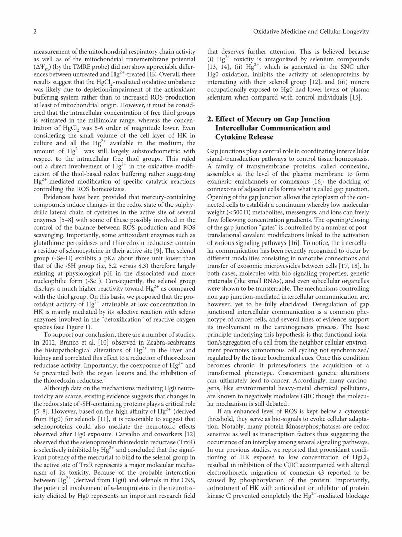
Evidences have been provided that mercury-containingcompounds induce changes in the redox state of the sulphy-drilic lateral chain of cysteines in the active site of severalenzymes [5–8] with some of these possibly involved in thecontrol of the balance between ROS production and ROSscavenging. Importantly, some antioxidant enzymes such asglutathione peroxidases and thioredoxin reductase containa residue of selenocysteine in their active site [9]. The selenolgroup (-Se-H) exhibits a pKa about three unit lower thanthat of the -SH group (i.e, 5.2 versus 8.3) therefore largelyexisting at physiological pH in the dissociated and morenucleophilic form (-Se−). Consequently, the selenol groupdisplays a much higher reactivity toward Hg2+ as comparedwith the thiol group. On this basis, we proposed that the pro-oxidant activity of Hg2+ attainable at low concentration inHK is mainly mediated by its selective reaction with selenoenzymes involved in the “detoxification” of reactive oxygenspecies (see Figure 1).
To support our conclusion, there are a number of studies.In 2012, Branco et al. [10] observed in Zeabra-seabreamsthe histopathological alterations of Hg2+ in the liver andkidney and correlated this effect to a reduction of thioredoxinreductase activity. Importantly, the coexposure of Hg2+ andSe prevented both the organ lesions and the inhibition ofthe thioredoxin reductase.
Although data on the mechanisms mediating Hg0 neuro-toxicity are scarce, existing evidence suggests that changes inthe redox state of -SH-containing proteins plays a critical role[5–8]. However, based on the high affinity of Hg2+ (derivedfrom Hg0) for selenols [11], it is reasonable to suggest thatselenoproteins could also mediate the neurotoxic effectsobserved after Hg0 exposure. Carvalho and coworkers [12]observed that the selenoprotein thioredoxin reductase (TrxR)is selectively inhibited by Hg2+ and concluded that the signif-icant potency of the mercurial to bind to the selenol group inthe active site of TrxR represents a major molecular mecha-nism of its toxicity. Because of the probable interactionbetween Hg2+ (derived from Hg0) and selenols in the CNS,the potential involvement of selenoproteins in the neurotox-icity elicited by Hg0 represents an important research field
that deserves further attention. This is believed because(i) Hg2+ toxicity is antagonized by selenium compounds[13, 14], (ii) Hg2+, which is generated in the SNC afterHg0 oxidation, inhibits the activity of selenoproteins byinteracting with their selenol group [12], and (iii) minersoccupationally exposed to Hg0 had lower levels of plasmaselenium when compared with control individuals [15].
2. Effect of Mecury on Gap JunctionIntercellular Communication andCytokine Release
Gap junctions play a central role in coordinating intercellularsignal-transduction pathways to control tissue homeostasis.A family of transmembrane proteins, called connexins,assembles at the level of the plasma membrane to formexameric emichannels or connexons [16]; the docking ofconnexons of adjacent cells forms what is called gap junction.Opening of the gap junction allows the cytoplasm of the con-nected cells to establish a continuum whereby low molecularweight (<500D) metabolites, messengers, and ions can freelyflow following concentration gradients. The opening/closingof the gap junction “gates” is controlled by a number of post-translational covalent modifications linked to the activationof various signaling pathways [16]. To notice, the intercellu-lar communication has been recently recognized to occur bydifferent modalities consisting in nanotube connections andtransfer of exosomic microvesicles between cells [17, 18]. Inboth cases, molecules with bio-signaling properties, geneticmaterials (like small RNAs), and even subcellular organelleswere shown to be transferrable. The mechanisms controllingnon gap junction-mediated intercellular communication are,however, yet to be fully elucidated. Deregulation of gapjunctional intercellular communication is a common phe-notype of cancer cells, and several lines of evidence supportits involvement in the carcinogenesis process. The basicprinciple underlying this hypothesis is that functional isola-tion/segregation of a cell from the neighbor cellular environ-ment promotes autonomous cell cycling not synchronized/regulated by the tissue biochemical cues. Once this conditionbecomes chronic, it primes/fosters the acquisition of atransformed phenotype. Concomitant genetic alterationscan ultimately lead to cancer. Accordingly, many carcino-gens, like environmental heavy-metal chemical pollutants,are known to negatively modulate GJIC though the molecu-lar mechanism is still debated.
If an enhanced level of ROS is kept below a cytotoxicthreshold, they serve as bio-signals to evoke cellular adapta-tion. Notably, many protein kinase/phosphatases are redoxsensitive as well as transcription factors thus suggesting theoccurrence of an interplay among several signaling pathways.In our previous studies, we reported that prooxidant condi-tioning of HK exposed to low concentration of HgCl2resulted in inhibition of the GJIC accompanied with alteredelectrophoretic migration of connexin 43 reported to becaused by phosphorylation of the protein. Importantly,cotreatment of HK with antioxidant or inhibitor of proteinkinase C prevented completely the Hg2+-mediated blockage
2 Oxidative Medicine and Cellular Longevity

of the GJIC [19]. On this basis, we proposed a mechanisticmodel whereby exposure of HK to Hg2+ causes enhancedROS production by inhibition of selenocysteine-containingantioxidant enzyme. This stimulates member of the proteinkinase C family, proved to be redox sensitive, which in turnhampers/closes the GJIC by phosphorylating connexin 43.The physiological rationale of this adaptive mechanism to aprooxidative setting remains to be fully understood. How-ever, it might be a protective mechanism evolutionaryselected to limit the spread of potentially harmful speciestoward in-contact neighbor cells.
Recent studies have advanced our understanding that theregulation of immune responses is not only confined toimmunocompetent cells. Upon stimulation, keratinocytesare capable of releasing various factors and expressing pat-tern recognition receptors (PRRs) that are significantlyinvolved in the innate immune response. Indeed, in responseto challenge with microbes or microbial-derived substances,the activation and nuclear translocation of NF-κB and theproduction of nitric oxide (NO) and inflammatory cytokinesoccur in keratinocytes, in a TLR-dependent manner [20]. Onthis basis, we have investigated the impact of Hg2+ on theLPS-mediated immune activation of HK. We found thatnanomolar concentrations of HgCl2 significantly reducedthe release of TNF-α and IL-1β in LPS-stimulated cells andthat this effect was redox sensitive as it was abrogated by
antioxidant cotreatment [21]. Although the mechanism link-ing mercury-mediated ROS accumulation to inhibition ofcytokine production remains to be detailed, neverthelessour finding supports the long known immunosuppressiverole of mercury compounds on immunocompetent cells [22].
3. Mercury as Cancer Promoter: a Sensu LatoEpigenetic Modifier
Epigenetics is defined as a complex of events leading to thecontrol and regulation of gene expression without theinvolvement of any change in the genetic sequence. The pro-cesses, which can be altered in the epigenetic modification,include DNA methylation, histone modification, RNA regu-lation, DNA repair, transcription, RNA stability, alternativeRNA splicing, protein degradation, gene copy number, andtransposon activation. Pollutants such as heavy metals, aswell as pharmaceuticals, hormones, nutrition, and behavior,can all modify the expression of genes. The pattern of the epi-genetic alterations can be both transient and permanent to betransmitted to offsprings.
Epigenetic effects of heavy metals have been investigatedextensively, particularly arsenic, cadmium, cobalt, chro-mium, nickel, and mercury. Regarding arsenic, numerousauthors observed global hypomethylation, but also globaland gene-specific hypermethylation particularly P53, plus
-Se− -Se−Hg+
-Se−OH-Se−SG
H2O2
OH−
GSHH2O
GSSG + H+
GSH
O2 O2‧− H2O2
Inactive enzyme
Hg2+
SOD2RC
Glutathione peroxidase
Cellular redoxhomeostasischanges
-Se−-SH
-Se-S
NADPH
NADP+
TrxSHSH
TrxSS
SHSH
SS
2 H2O
-Se−Hg+-SH
Inactive enzyme
Thioredoxinreductase
P
P
Figure 1: Proposed mechanism for the prooxidative action of Hg(II). Mercury ion (Hg2+) is shown to bind to the dissociated form of theselenol (-Se−) moiety of the catalytic selenocysteine residue of glutathione peroxidase and thioredoxin reductase thereby inactivating theenzymes; the result is an enhanced level of reactive oxygen species because of their lower inactivation. The catalytic cycles of the twoantioxidant enzymes are also shown: glutathione peroxidase converts H2O2 in 2 H2O molecules at expense of 2 reduced glutathionemolecules (GSH), which are oxidized to GSSG; thioredoxin reductase reduces oxidized thioredoxin (Txr) at the expense of NADPHthereby enabling reduced Txr to preserve the redox state of protein cysteines (P) from the H2O2-mediated oxidation. Mitochondria isillustrated as a major intracellular producer of ROS generated from electron leaks from the respiratory chain (RC) to O2 to form thesuperoxide anion (O2
•−). This is further converted in H2O2 by the mitochondrial isoform of the superoxide dismutase (SOD2).
3Oxidative Medicine and Cellular Longevity

histone modification (alkylation) and increment of miRNAsas miR-22 or decrement of miR-210 and miR-19a [23].For cadmium, both global hypomethylation and hyperme-thylation have been reported depending on the exposuretime [24–26]. For cobalt, the epigenetic effects publishedconsisted enhanced oxidative stress, proinflammatory effects,and abnormal apoptosis [27]. It has been proved thatchromium caused P16 and Gpt hypermethylation, as wellas histone modifications as alkylation and phosphorylation[28]. Nickel was able to induce hypermethylation of ATF-1,HIF-1 gpt, Rb, and P16, and then histone modification asalkylation and phosphorylation [28–31]. Finally, mercuryinduced global hypomethylation and hypermethylation ofthe signaling G protein GTPase Rnd2 [32, 33].
In addition to the abovementioned modifications,dynamically controlling chromatin remodeling, gene tran-scription can also be indirectly modulated by the availabilityof metabolites functioning as substrates or effectors of epi-genetic modifier enzymes as well as from microRNAs(miRNAs). This emerging notion let us to reconsider epige-netics in a broader sense including all the conditions directlyor indirectly leading to acute or chronic modification of geneexpression. In this acceptation, the intercellular communi-cation may be considered an epigenetic process regulatinggene expression. Indeed, the low molecular weight of the22-nucleotide noncoding miRNAs enables them to be
transferred through gap junction establishing an intercellulargenetic cross-talk. The intercellular genetic communicationis now emerging as an essential requirement for coordinationof cell proliferation and differentiation and has an importantrole in many cellular processes [34].
A promoter carcinogen is by definition any agent able todeterminate an uncontrolled proliferation. This action, in theopinion of several authors, including ourselves, would belinked to inhibition of GJIC. In fact, cells deprived by inhib-itory control of neighboring cells start to proliferate. Thisaspect is, obviously, only one part of the complex and multi-step carcinogenic process where microenvironmental factorsplay conditioning activities. In this context, the cytokine net-work might contribute to determine the destiny of celltowards the death or the survival. Accordingly, numerousobservations proved a link between inflammation and can-cer. If a substance apart from the inhibition of GJIC is alsoable to reduce the release of proinflammatory cytokines, itscarcinogenic potency would result incremented.
In keeping the above defined concepts and on the basis ofthe evidences provided by our experimental work on HK, aswell as by others, we hypothesize that mercury might be anepigenetic carcinogen. Indeed, Hg(II) fulfils both the capacityto induce an inhibition of the GJIC and that to induce immu-nosuppressive effects. This double mechanism of toxicity byHg(II) might overcome the organism defences that exploit
Initiation Latency
Progression
ROS
Hg(II)Selenocysteine-containing antioxidantenzymes
Gap junction intercellularcommunication
Proinflammatory cytokineproduction (TNF-𝛼, IL-1𝛽)
Normal cells
Mitochondria
Promotion
Open GJ (Cx43)
Closed GJ (Cx43)
Figure 2: Proposed role of mercury as epigenetic/promoter carcinogen. The progressive multistep transformation of a normal cell to a cancercell is shown schematically. The indicated phases (i.e., initiation→ latency→ promotion→ progression) are those commonly accepted forcancer development. Mercury (Hg(II)) is indicated to act in the promotion phase by causing an unbalance in the reactive oxygen species(ROS) homeostasis accomplished by selective inhibition of selenocysteine antioxidant enzymes. Mitochondria are also shown as a majorROS generator. The Hg(II)-induced prooxidative state in turn would result in inhibition of the gap junction intercellular communication(GJIC) and of the proinflammatory cytokine release. Both mechanisms might on one hand isolate cells from tissue-specific homeostasispromoting their proliferation and on the other hand tamper the immune system defense/surveillance checkmating the whole organism.The Cx43-related open gap junction is shown as progressively closing in the transitions following the “latent” state.
4 Oxidative Medicine and Cellular Longevity

inflammation to contrast the cell hyperproliferation. Boththese Hg-mediated outcomes are linked to an upstream alter-ation of the intracellular redox tone likely caused by a specificinhibition of antioxidant enzymes containing selenocysteines(see scheme of Figure 2).
The “carcinogen as mutagen” has become a “default”paradigm. This is partly because the carcinogenicity of acompound is commonly assessed by the so-called “genotoxi-city” in vitro assays and tests to evaluate whether epigenetictoxicology are lacking in the routine. Consequently, mercuryhas been classified in group 3 by IARC (International AgencyResearch Cancer) which means “not classifiable as to theircarcinogenicity to humans” [35]. However, if a toxic com-pound inhibits the intercellular communication, it mightpotentially act as a cancer “promoter,” although resultingnongenotoxic. In the case of the GJIC, the impact on it ofchemical agent can be easily assessed as a number of proto-cols have been developed [36, 37]. Most notably, for someof these protocols measuring intercellular fluorescent dyetransfer, either a specialized expertise or particular instru-mentations, is required in making the setup of the assay easilyaccessible to every basic laboratory.
All together, the abovementioned considerations let us toconclude that there is something more beyond the epigeneticeffect. Alyea et al. [38] stated that epigenetic effects causedby metals and toxic compounds always appear at low concen-tration, even below what they called no-observed-adverse-effect level (NOAEL), and then the authors concluded thatthe epigenome dynamic variability is not completely charac-terized. Thus, given the state of our current scientific knowl-edge, an epigenetic change cannot be contextualized asadverse in the absence of a phenotypic anchor. They con-cluded that more research is needed in this area to performadditional epigenetic studies that include apical end pointswith full-dose response curves, in order to gain a more com-prehensive understanding of adverse health outcomes thatcould be causally linked to epigenetic changes. These conclu-sions are useful to approach in a new and unexpected waythe epigenetic effect, trying to evaluate more criticallywhere this event drives. It is known that cells, tissues, organs,apparatuses, and organisms are able to develop feedbackmechanisms that counteract modifications induced by toxicsubstances. Hence, though it is useful to investigate canonicalepigenetic modifications, it will be even more useful in thefuture to ask ourselves what links these epigenetic effects withother epigenetic effects that could be defined “beyond/afterepigenetic effect” to differentiate them. Several authors asAlyea et al. [38] consider inhibition of intercellular commu-nication, enhanced oxidative stress, proinflammatory effects,and abnormal apoptosis/survival as “bona fide” epigeneticeffects. They could be also defined, in our opinion, in a singleword as “metagenetic effects” using an ancient Greek prefixthat means beyond.
Abbreviations
GJIC: Gap junction intercellular communicationPKC: Protein kinase CPKA: Protein kinase A
LPS: Lipopolysaccharide—endotoxinIL-1β: Interleukin 1 betaTNF-α: Tumor necrosis factor alphaNOAEL: No-observed-adverse-effect level.
Data Access
The authors are available to share their data.
Conflicts of Interest
The authors declare that they have no competing interests.
Authors’ Contributions
All authors read and approved the final manuscript.
Acknowledgments
The authors would like to thank Professor Luigi Ambrosifor his scientific advices: these exceeded the limits andinvested our lives, improving it and lighting up the pathtowards future challenges. Local grants are from the Univer-sity of Foggia.
References
[1] T. Syversen and P. Kaur, “The toxicology of mercury and itscompounds,” Journal of Trace Elements in Medicine and Biol-ogy, vol. 26, no. 4, pp. 215–226, 2012.
[2] E.-J. Park and K. Park, “Induction of reactive oxygen speciesand apoptosis in BEAS-2B cells by mercuric chloride,” Toxicol-ogy In Vitro, vol. 21, no. 5, pp. 789–794, 2007.
[3] D. Grotto, J. Valentini, M. Fillion et al., “Mercury exposureand oxidative stress in communities of the Brazilian Amazon,”Science of the Total Environment, vol. 408, no. 4, pp. 806–811,2010.
[4] C. Piccoli, A. D'Aprile, R. Scrima, L. Ambrosi, R. Zefferino,and N. Capitanio, “Subcytotoxic mercury chloride inhibitsgap junction intercellular communication by a redox- andphosphorylation-mediated mechanism,” Free Radical Biology& Medicine, vol. 52, no. 5, pp. 916–927, 2012.
[5] J. Albrecht and E. Matyja, “Glutamate: a potential mediator ofinorganic mercury neurotoxicity,” Metabolic Brain Disease,vol. 11, no. 2, pp. 175–184, 1996.
[6] M. Aschner and J. L. Aschner, “Mercury neurotoxicity: mech-anisms of blood-brain barrier transport,” Neuroscience andBiobehavioral Reviews, vol. 14, no. 2, pp. 169–176, 1990.
[7] N. Brookes and D. A. Kristt, “Inhibition of amino acid trans-port and protein synthesis by HgCl2 and methylmercury inastrocytes: selectivity and reversibility,” Journal of Neurochem-istry, vol. 53, no. 4, pp. 1228–1237, 1989.
[8] M. Yoshida, C. Watanabe, M. Kishimoto et al., “Behavioralchanges in metallothionein-null mice after the cessation oflong-term, low-level exposure to mercury vapor,” ToxicologyLetters, vol. 161, no. 3, pp. 210–218, 2006.
[9] H. Steinbrenner, B. Speckmann, and L. O. Klotz, “Selenopro-teins: antioxidant selenoenzymes and beyond,” Archives ofBiochemistry and Biophysics, vol. 595, pp. 113–119, 2016.
[10] V. Branco, P. Ramos, J. Canário, J. Lu, A. Holmgren, andC. Carvalho, “Biomarkers of adverse response to mercury:
5Oxidative Medicine and Cellular Longevity

histopathology versus thioredoxin reductase activity,” Journalof Biomedicine and Biotechnology, vol. 2012, Article ID359879, 9 pages, 2012.
[11] C. Sasakura and K. T. Suzuki, “Biological interaction betweentransition metals (Ag, Cd and Hg), selenide/sulfide and seleno-protein P,” Journal of Inorganic Biochemistry, vol. 71, no. 3-4,pp. 159–162, 1998.
[12] C. M. L. Carvalho, E. H. Chew, S. I. Hashemy, J. Lu, andA. Holmgren, “Inhibition of the human thioredoxin system.A molecular mechanism of mercury toxicity,” The Journal ofBiological Chemistry, vol. 283, no. 18, pp. 11913–11923, 2008.
[13] M. Farina, F. A. Soares, A. Feoli et al., “In vitro effects of sele-nite and mercuric chloride on liver thiobarbituric acid-reactivesubstances and non-protein thiols from rats: influences of die-tary cholesterol and polyunsaturated and saturated fattyacids,” Nutrition, vol. 19, no. 6, pp. 531–535, 2003.
[14] I. Yamamoto, “Effect of various amounts of selenium on themetabolism of mercuric chloride in mice,” Biochemical Phar-macology, vol. 34, no. 15, pp. 2713–2720, 1985.
[15] A. B. Kobal, M. Horvat, M. Prezelj et al., “The impact of long-term past exposure to elemental mercury on antioxidativecapacity and lipid peroxidation in mercury miners,” Journalof Trace Elements in Medicine and Biology, vol. 17, no. 4,pp. 261–274, 2004.
[16] M. S. Nielsen, L. N. Axelsen, P. L. Sorgen, V. Verma,M. Delmar, and N. H. Holstein-Rathlou, “Gap junctions,”Comprehensive Physiology, vol. 2, no. 3, pp. 1981–2035, 2012.
[17] S. Sisakhtnezhad and L. Khosravi, “Emerging physiologicaland pathological implications of tunneling nanotubes forma-tion between cells,” European Journal of Cell Biology, vol. 94,no. 10, pp. 429–443, 2015.
[18] J. M. Pitt, G. Kroemer, and L. Zitvogel, “Extracellular vesicles:masters of intercellular communication and potential clinicalinterventions,” The Journal of Clinical Investigation, vol. 126,no. 4, pp. 1139–1143, 2016.
[19] R. Zefferino, A. Leone, S. Piccaluga, R. Cincione, andL. Ambrosi, “Mercury modulates interplay between IL-1β,TNF-α, and gap junctional intercellular communication inkeratinocytes: mitigation by lycopene,” Journal of Immunotox-icology, vol. 5, no. 4, pp. 353–360, 2008.
[20] A. Pivarcsi, L. Kemény, and A. Dobozy, “Innate immune func-tions of the keratinocytes. A review,” Acta Microbiologica etImmunologica Hungarica, vol. 51, no. 3, pp. 303–310, 2004.
[21] R. Zefferino, S. Piccaluga, M. Lasalvia, G. D'Andrea,M. Margaglione, and L. Ambrosi, “Role of tumour necrosisfactor alpha and interleukin 1 beta in promoter effect inducedby mercury in human keratinocytes,” International Journal ofImmunopathology and Pharmacology, vol. 19, Supplement 4,pp. 15–20, 2006.
[22] P. Moszczyński, “Mercury compounds and the immunesystem: a review,” International Journal of OccupationalMedicine and Environmental Health, vol. 10, no. 3, pp. 247–258, 1997.
[23] P. Intarasunanont, P. Navasumrit, S. Waraprasit et al., “Effectsof arsenic exposure on DNA methylation in cord blood sam-ples from newborn babies and in a human lymphoblast cellline,” Environmental Health, vol. 11, no. 1, p. 31, 2012.
[24] S. Virani, K. M. Rentschler, M. Nishijo et al., “DNA methyla-tion is differentially associated with environmental cadmiumexposure based on sex and smoking status,” Chemosphere,vol. 145, pp. 284–290, 2016.
[25] K. E. Pelch, E. J. Tokar, B. A. Merrick, and M. P. Waalkes,“Differential DNA methylation profile of key genes inmalignant prostate epithelial cells transformed by inorganicarsenic or cadmium,” Toxicology and Applied Pharmacology,vol. 286, no. 3, pp. 159–167, 2015.
[26] J. Brocato and M. Costa, “Basic mechanics of DNA methyla-tion and the unique landscape of the DNA methylome inmetal-induced carcinogenesis,” Critical Reviews in Toxicology,vol. 43, no. 6, pp. 493–514, 2013.
[27] R. Magaye, J. Zhao, L. Bowman, and M. DING, “Genotoxicityand carcinogenicity of cobalt-, nickel- and copper-based nano-particles,” Experimental and Therapeutic Medicine, vol. 4,no. 4, pp. 551–561, 2012.
[28] J. Brocato and M. Costa, “10th NTES conference: nickel andarsenic compounds alter the epigenome of peripheral bloodmononuclear cells,” Journal of Trace Elements in Medicineand Biology, vol. 31, pp. 209–213, 2015.
[29] Q. Ke, T. Ellen, and M. Costa, “Nickel compounds induce his-tone ubiquitination by inhibiting histone deubiquitinatingenzyme activity,” Toxicology and Applied Pharmacology,vol. 228, no. 2, pp. 190–199, 2008.
[30] X. Zhou, Q. Li, A. Arita, H. Sun, and M. Costa, “Effects ofnickel, chromate, and arsenite on histone 3 lysine methyla-tion,” Toxicology and Applied Pharmacology, vol. 236, no. 1,pp. 78–84, 2009.
[31] L. Ma, Y. Bai, H. Pu et al., “Histone methylation in nickel-smelting industrial workers,” PLoS One, vol. 10, no. 10,article e0140339, 2015.
[32] J. Z. J. Maccani, D. C. Koestler, B. Lester et al., “Placental DNAmethylation related to both infant toenail mercury and adverseneurobehavioral outcomes,” Environmental Health Perspec-tives, vol. 123, no. 7, pp. 723–729, 2015.
[33] J. M. Goodrich, N. Basu, A. Franzblau, and D. C. Dolinoy,“Mercury biomarkers and DNAmethylation among Michigandental professionals,” Environmental and Molecular Mutagen-esis, vol. 54, no. 3, pp. 195–203, 2013.
[34] L. Zong, L. Zhou, Y. Hou et al., “Genetic and epigeneticregulation on the transcription of GABRB2: genotype-dependent hydroxymethylation and methylation alterationsin schizophrenia,” Journal of Psychiatric Research, vol. 88,pp. 9–17, 2017.
[35] http://monographs.iarc.fr/ENG/Classification/ClassificationsGroupOrder.pdf.
[36] I. V. Budunova and G. M. Williams, “Cell culture assays forchemicals with tumor-promoting or tumor-inhibiting activitybased on the modulation of intercellular communication,” CellBiology and Toxicology, vol. 10, no. 2, pp. 71–116, 1994.
[37] J. E. Klaunig and Y. Shi, “Assessment of gap junctional inter-cellular communication,” Current Protocols in Toxicology,2009, Chapter 2, Unit 2.17.
[38] R. A. Alyea, N. P. Moore, M. J. LeBaron, B. B. Gollapudi,and R. J. Rasoulpour, “Is the current product safety assessmentparadigm protective for epigenetic mechanisms?,” Journal ofPharmacological and Toxicological Methods, vol. 66, no. 3,pp. 207–214, 2012.
6 Oxidative Medicine and Cellular Longevity

Submit your manuscripts athttps://www.hindawi.com
Stem CellsInternational
Hindawi Publishing Corporationhttp://www.hindawi.com Volume 2014
Hindawi Publishing Corporationhttp://www.hindawi.com Volume 2014
MEDIATORSINFLAMMATION
of
Hindawi Publishing Corporationhttp://www.hindawi.com Volume 2014
Behavioural Neurology
EndocrinologyInternational Journal of
Hindawi Publishing Corporationhttp://www.hindawi.com Volume 2014
Hindawi Publishing Corporationhttp://www.hindawi.com Volume 2014
Disease Markers
Hindawi Publishing Corporationhttp://www.hindawi.com Volume 2014
BioMed Research International
OncologyJournal of
Hindawi Publishing Corporationhttp://www.hindawi.com Volume 2014
Hindawi Publishing Corporationhttp://www.hindawi.com Volume 2014
Oxidative Medicine and Cellular Longevity
Hindawi Publishing Corporationhttp://www.hindawi.com Volume 2014
PPAR Research
The Scientific World JournalHindawi Publishing Corporation http://www.hindawi.com Volume 2014
Immunology ResearchHindawi Publishing Corporationhttp://www.hindawi.com Volume 2014
Journal of
ObesityJournal of
Hindawi Publishing Corporationhttp://www.hindawi.com Volume 2014
Hindawi Publishing Corporationhttp://www.hindawi.com Volume 2014
Computational and Mathematical Methods in Medicine
OphthalmologyJournal of
Hindawi Publishing Corporationhttp://www.hindawi.com Volume 2014
Diabetes ResearchJournal of
Hindawi Publishing Corporationhttp://www.hindawi.com Volume 2014
Hindawi Publishing Corporationhttp://www.hindawi.com Volume 2014
Research and TreatmentAIDS
Hindawi Publishing Corporationhttp://www.hindawi.com Volume 2014
Gastroenterology Research and Practice
Hindawi Publishing Corporationhttp://www.hindawi.com Volume 2014
Parkinson’s Disease
Evidence-Based Complementary and Alternative Medicine
Volume 2014Hindawi Publishing Corporationhttp://www.hindawi.com