post-sepsis state induces tumor-associated macrophage...
TRANSCRIPT

ROLE OF TAM IN POST-SEPSIS TUMOR PROGRESSION
1
Title: Post-sepsis state induces tumor-associated macrophage accumulation through
CXCR4/CXCL12 and favors tumor progression in mice
Authors: José M Mota 1,2, Caio A Leite 2, Lucas E Souza 3, Paulo H Melo 2, Daniele C
Nascimento 2, Virginia M de-Deus-Wagatsuma 1,4, Jessica Temporal 4, Florêncio Figueiredo
5, Houtan Noushmehr 4 *, José C Alves-Filho 2, Fernando Q Cunha 2, Eduardo M Rego 1.
Authors’ Affiliations: 1Hematology/Oncology Division and Center for Cell-Based Therapy,
Department of Internal Medicine, Ribeirão Preto Medical School, University of São Paulo;
2Laboratory of Inflammation and Pain, Department of Pharmacology, Ribeirão Preto Medical
School, University of São Paulo; 3Laboratory of Gene Transfer, Department of Internal
Medicine, Ribeirão Preto Medical School, University of São Paulo; 4OMICS Laboratory,
Department of Genetics, Ribeirão Preto Medical School, University of São Paulo, 5Laboratory
of Pathology, Department of Pathology, University of Brasilia
*Current Address
Houtan Noushmehr
Ribeirão Preto Medical School, University of São Paulo.
OMICS Laboratory, Department of Genetics
Ribeirão Preto, São Paulo, Brazil
Running title: Role of TAM in post-sepsis tumor progression
Key words: tumor progression, post-sepsis, tumor-associated macrophage, CXCL12, CXCR4
Corresponding author: Prof. Dr. Eduardo Magalhães Rego. Hematology/Oncology
Division, Department of Internal Medicine, Ribeirão Preto Medical School, University of São
on February 1, 2019. © 2016 American Association for Cancer Research. cancerimmunolres.aacrjournals.org Downloaded from
Author manuscripts have been peer reviewed and accepted for publication but have not yet been edited. Author Manuscript Published OnlineFirst on January 27, 2016; DOI: 10.1158/2326-6066.CIR-15-0170
ROLE OF TAM IN POST-SEPSIS TUMOR PROGRESSION
2
Paulo. 3900 Bandeirantes Ave. Ribeirão Preto, São Paulo, Brazil. 14048-900. Phone: +55 16
21019361. Email: [email protected].
Financial support: E.M. Rego has been awarded a grant from São Paulo Research
Foundation (FAPESP) under grant agreements 2013/14228-3 and 2013/08135-2 and
University of São Paulo – Nucleo Apoio Pesquisa (NAP).
Disclosure of Potential Conflicts of interest: No conflicts of interest by authors.
Total word count (excluding Abstract and References): 4185
Number of figures: 6
Number of tables: 1
Number of supplementary figures: 7
on February 1, 2019. © 2016 American Association for Cancer Research. cancerimmunolres.aacrjournals.org Downloaded from
Author manuscripts have been peer reviewed and accepted for publication but have not yet been edited. Author Manuscript Published OnlineFirst on January 27, 2016; DOI: 10.1158/2326-6066.CIR-15-0170
ROLE OF TAM IN POST-SEPSIS TUMOR PROGRESSION
3
ABSTRACT
Survivors from sepsis are in an immunosuppressed state that is associated with higher long-
term mortality and risk of opportunistic infections,, Whether this contributes to neoplastic
proliferation, however, remains unclear. Tumor-associated macrophages (TAMs) can support
malignant cell proliferation, survival, and angiogenesis. We addressed the relationship
between the post-sepsis state, tumor progression and TAM accumulation, and phenotypic and
genetic profile, using a mouse model of sepsis resolution then B16 melanoma in mice. In
addition, we measured the serum concentrations of TNFα, TGFβ, CCL2, and CXCL12, and
determined the effect of in vivo CXCR4/CXCL12 inhibition in this context. Mice that
survived sepsis showed increased tumor progression both in the short and long term and
survival times were shorter. TAM accumulation, TAM local proliferation, and serum
concentrations of TGFβ, CXCL12, and TNFα were increased. Naïve mice inoculated with
B16 together with macrophages from post-sepsis mice also had faster tumor progression and
shorter survival. Post-sepsis TAMs had less expression of MHC-II and leukocyte activation-
related genes. Inhibition of CXCR4/CXCL12 prevented the post-sepsis–induced tumor
progression, TAM accumulation, and TAM in situ proliferation. Collectively, our data show
that the post-sepsis state was associated with TAM accumulation through CXCR4/CXCL12,
which contributed to B16 melanoma progression.
on February 1, 2019. © 2016 American Association for Cancer Research. cancerimmunolres.aacrjournals.org Downloaded from
Author manuscripts have been peer reviewed and accepted for publication but have not yet been edited. Author Manuscript Published OnlineFirst on January 27, 2016; DOI: 10.1158/2326-6066.CIR-15-0170

ROLE OF TAM IN POST-SEPSIS TUMOR PROGRESSION
4
INTRODUCTION
Sepsis is a worldwide leading cause of mortality (1), and despite the fact that
implementation of early intervention-based approaches have improved outcomes (2), patients
who survive from a first sepsis episode remain at a higher relative risk of death (3, 4) and of
opportunistic infections (5) for at least 5 to 8 years. Post-sepsis states have been associated
with an immunosuppressive phenotype, such as increased T regulatory lymphocytes (Tregs)
(6), macrophages, and dendritic cells (7, 8). Döcke and colleagues reported
monocyte/macrophage dysfunction after sepsis, characterized by human leukocyte antigens
(HLA) down-regulation and low tumor necrosis factor (TNF) production (9). Accordingly,
Takahashi and colleagues showed that post-sepsis induced alternatively activated
macrophages (M2-polarized) with no antibacterial activity in mice (10).
One important issue is to determine if the post-sepsis immunosuppressive state
increases the incidence, or affects the evolution of, neoplasms, similarly to what has been
observed in organ recipients under immunosuppressive treatments (11). Cavassani and
colleagues reported that the post-sepsis state resulted in increased tumor expansion in a Lewis
carcinoma model (12). Post-sepsis shares several mechanisms implicated in the development
of the protumor microenvironments described in cancer (13). This supports the idea of a link
between increased tumor progression in post-sepsis and microenvironmental changes.
The tumor inflammatory stroma, which contains tumor-associated macrophages
(TAMs), neutrophils, dendritic cells, lymphocytes, pericytes, and fibroblasts provides a
number of distinct and complex mediators and mechanisms that promote tumor growth,
adjacent tissue invasion, and metastasis (14–16). In this context, TAMs act as major players
of tumor progression, stimulating cell proliferation, survival, angiogenesis, and immune
escape (17). Clinically, TAM accumulation is a predictor of mortality in several cancer types
(18).
on February 1, 2019. © 2016 American Association for Cancer Research. cancerimmunolres.aacrjournals.org Downloaded from
Author manuscripts have been peer reviewed and accepted for publication but have not yet been edited. Author Manuscript Published OnlineFirst on January 27, 2016; DOI: 10.1158/2326-6066.CIR-15-0170
ROLE OF TAM IN POST-SEPSIS TUMOR PROGRESSION
5
Among the mechanisms associated with TAM accumulation, CCL2 is classically
implicated in early monocyte recruitment to the tumor microenvironment (19). In addition,
Tymoszuk and colleagues report the contribution of in situ TAM proliferation in a mouse
model of spontaneous breast cancer (20). On the other hand, the CXCR4/CXCL12 axis keeps
TAMs in a tumor’s hypoxic areas, thus contributing to neoangiogenesis and oxygen delivery
(21, 22).
To evaluate the relationship between the post-sepsis state, tumor progression, and
TAMs, we analyzed intratumoral macrophage accumulation, and phenotype and genetic
profiles using a model of B16 melanoma expansion after the resolution of cecal and ligation-
induced sepsis in mice.
on February 1, 2019. © 2016 American Association for Cancer Research. cancerimmunolres.aacrjournals.org Downloaded from
Author manuscripts have been peer reviewed and accepted for publication but have not yet been edited. Author Manuscript Published OnlineFirst on January 27, 2016; DOI: 10.1158/2326-6066.CIR-15-0170
ROLE OF TAM IN POST-SEPSIS TUMOR PROGRESSION
6
MATERIALS AND METHODS
Mice
6-8-wk-old male C57BL/6 mice were obtained from the animal facility of the Ribeirão
Preto Medical School, kept in appropriate cages in temperature-controlled rooms with 12 h
dark-light cycles and received food and water ad libitum. The experimental protocols and
procedures were previously approved by the local Ethics Committee (protocol number:
141/2012) and were in accordance with the Declaration of Helsinki and the Guide for Care
and Use of Laboratory Animals (National Institutes of Health, Bethesda, MD, USA).
Cells
B16-F10-luc (B16, Caliper Lifesciences, Hopkinton, MA, USA) was cultured in
RPMI containing 10% heat-inactivated FCS (Gibco, Grand Island, NY, USA), 100 UI/mL
penicillin (Gibco), 0,1 mg/mL streptomycin (Gibco) and amphotericin B 2 μg/mL (Gibco).
Prior to use, cells were detached with trypsin-EDTA 0,25% (Gibco) with 70%-80% of
confluence and washed in PBS for two times. B16-F10 cell line was obtained in 2013 from
ATCC and used at the third passage. Authentication was not made.
Sepsis and post-sepsis model
Severe sepsis was induced by cecal and ligation puncture (CLP), as described
elsewhere (23). To induce survival and post-sepsis state, animals were treated with ertapenem
(20 mg/kg, ip., 6 h after surgery, and each 12 h for three days), as described (6). The controls
received the same regimen of ertapenem.
Assays to evaluate tumor progression
on February 1, 2019. © 2016 American Association for Cancer Research. cancerimmunolres.aacrjournals.org Downloaded from
Author manuscripts have been peer reviewed and accepted for publication but have not yet been edited. Author Manuscript Published OnlineFirst on January 27, 2016; DOI: 10.1158/2326-6066.CIR-15-0170
ROLE OF TAM IN POST-SEPSIS TUMOR PROGRESSION
7
Naïve or post-sepsis mice (15 days after CLP) were subcutaneously inoculated in the
right flank with B16 melanoma. Different inoculums were tested (100,000; 30,000 or 10,000
cells) to determine the most suitable to our model. Tumor volumes were calculated as
described (24). Tumor burden was measured through bioluminescence quantification (IVIS
Lumina, Caliper Lifesciences) after D-luciferin (150 mg/kg, ip.). Overall survival was
monitored for 50 days.
Assays to evaluate metastasis in post-sepsis mice
Mice received 30,000 B16 melanoma cells subcutaneously (s.c.) 15 days after CLP
induction. Animals were euthanized after 21 days and organs were harvested and scrutinized
using magnification of 100X in optical microscope (Zeiss, Germany) to detect spontaneous
metastasis. We evaluated the rate of metastasis per organ and per mice. Also, we counted the
number of metastatic sites in the lungs. In another set of experiments, post-sepsis (15 days
after CLP) or naïve mice were inoculated with 30,000 cells (s.c., right flank). Fourteen days
after inoculation, primary tumors were surgically removed Animals were then followed to
assess mortality due to metastasis, as described (25). Additionally, post-sepsis or naïve mice
were submitted to lung colonization assay (30,000 cells, injected i.v. through retro-orbital
plexus). At the 18th day after inoculation with B16 cells (D+18), mice were euthanized and ex
vivo pulmonary tumor burden was measured through bioluminescent signal.
Tissue digestion and flow cytometry
Tumor-bearing post-sepsis or naïve mice were euthanized at D+14 and tumor was
harvested. Tumors were digested using collagenase type II 1 mg/mL (Sigma) and DNAse type
I 0.1 mg/mL (Sigma) to prepare single cell suspensions. Immunostaining was made with
antibodies to CD45 (eBiosciences, CA, USA), F4/80 (eBiosciences) and CD206 (Abd
on February 1, 2019. © 2016 American Association for Cancer Research. cancerimmunolres.aacrjournals.org Downloaded from
Author manuscripts have been peer reviewed and accepted for publication but have not yet been edited. Author Manuscript Published OnlineFirst on January 27, 2016; DOI: 10.1158/2326-6066.CIR-15-0170
ROLE OF TAM IN POST-SEPSIS TUMOR PROGRESSION
8
Serotec, UK) for assessment of TAM accumulation through flow cytometry (FACS CANTO,
BD Biosciences, NJ, USA). CXCR4 expression in TAM was evaluated using anti-CXCR4
(BD Biosciences). For measurement of TAM proliferation an antibody to Ki67 (BioLegend,
CA, USA) was used. The data were analyzed using FCS Express 3.0 (De Novo software, CA,
USA).
Immunofluorescence
TAM accumulation was also measured by immunofluorescence assay (Leica
DMI6000 B, Leica microsystems) with antibodies to F4/80 (rat anti-mouse, 1:50, Caltag
Laboratories, UK), followed by anti-rat Alexa Fluor 488 (1:100, Life Technologies, NY,
USA). Nuclei were revealed with Hoechst 33342 (1 μg/mL, Life Technologies). Total F4/80+
cells per field (magnification of 100 X) were quantified using Image J 1.44 (National
Institutes of Health, Bethesda, Maryland, USA).
ELISA
Serum and tumor and inguinal lymph nodes were harvested to detect IFNγ, IL10,
TNFα, TGFβ, CXCL12, and CCL2 by ELISA (Duo Set, R&D Systems kits, MN, USA).
Results were expressed in pg/mL (serum or lymph node supernatant) or pg/protein (tumor).
TAM isolation
Single cell suspensions from tumors were prepared as described above. Next, tumor
extracts were carefully layered onto a Percoll (Sigma) gradient (70%/30%) and centrifuged
(1800 RPM, 23 min, 4o C). The lymphomononuclear correspondent layer was isolated,
washed in PBS, diluted in 1% FBS supplemented-RPMI, and cultured for 40 minutes at 37o
C. Three vigorous washes ensured only the adherent cells remained in the plate. Prior to use,
on February 1, 2019. © 2016 American Association for Cancer Research. cancerimmunolres.aacrjournals.org Downloaded from
Author manuscripts have been peer reviewed and accepted for publication but have not yet been edited. Author Manuscript Published OnlineFirst on January 27, 2016; DOI: 10.1158/2326-6066.CIR-15-0170
ROLE OF TAM IN POST-SEPSIS TUMOR PROGRESSION
9
adherent cells were removed using a cell scraper. TAMs (CD45+F4/80+) were 70 to 80% pure,
as confirmed through flow cytometry.
RNA isolation from TAM
RNA was isolated using a specific kit (Quick-RNATM MicroPrep, Zymo Research,
CA, USA), following manufacturer instructions. RNA concentrations and absorbance ratios
(A260/A230 and A260/A280) were determined using NanoVue spectrophotometer (GE
Healthcare Life Sciences, UT, USA). The four RNA samples with A260/280 ratio greater
than 1.9 and closest to 2.0 from the 12 samples available of each studied group were selected
for further processing in a microarray analysis.
RNA preparation for microarray
RNA preparation for microarray assay was made following manufacturer instructions
(Agilent Technologies, Santa Clara, CA, USA). To isolate Cy-3–labeled cRNA, we used
RNeasy Mini Kit (Qiagen, CA, USA). Quantifications were made using NanoVue (GE
Healthcare Lifesciences). The concentrations of Cy3 (pmol/μL), cRNA (ng/μL) and 260/280
ratio were determined. cRNA were hybridized in microarray slides following two random
sequential lotteries (Sure Print G3 Mouse Gene Expression 8x60k Microarray, G4852A,
Agilent Technologies, Slide 1: 252800516673, Slide 2: 252800516674, Slide 3:
252800516788). Slides were scanned (G4900DA SureScan Microarray Scanner, Agilent
Technologies) and data was extracted through Agilent Feature Extraction Software (Agilent
Technologies).
Microarray data analysis
on February 1, 2019. © 2016 American Association for Cancer Research. cancerimmunolres.aacrjournals.org Downloaded from
Author manuscripts have been peer reviewed and accepted for publication but have not yet been edited. Author Manuscript Published OnlineFirst on January 27, 2016; DOI: 10.1158/2326-6066.CIR-15-0170
ROLE OF TAM IN POST-SEPSIS TUMOR PROGRESSION
10
The microarray data was imported and analyzed using the open source software tool,
Bioconductor 3.0 (http://www.bioconductor.org). All statistical tests were done using R
(version 3.1.1). We used limma package (26) to perform quality, normalization, and
differential analysis. Normalization was done applying quantile normalization, to ensure the
distribution of probe intensities for each sample in a set of arrays are the same. One sample
replicate from TAM naïve group (TAM.naive, rep2) was identified as an outlier due to low
RNA concentrations and poor genetic material quality and therefore was removed from
subsequent downstream analysis. Principal component analysis (PCA) methods were used to
identify the relationship between groups of samples. A list of differentially expressed genes
was defined with a log2 fold of 0.58 or greater (for upregulated genes) and -0.58 and less (for
downregulated genes) with a false discovery rate (FDR) < 0.01. Pathway analyses were
analyzed using Nextbio database (http://www.nextbio.com). Hierarchical heatmaps were
made using default parameters. All data were deposited for public access in Gene Expression
Ombibus (GEO). The accession code is GSE64498.
Quantitative real-time polymerase chain reaction (qRT-PCR)
qRT-PCR was conducted for specific genes related with macrophage polarization (M1
- Nos2 and Tnf; M2 - Arg1 and Mrc1). Briefly, 50 ng of total RNA was transcribed to cDNA
by reverse transcriptase enzyme action Improm Pre-II® (Promega). qRT-PCR reaction was
done on ABI Prism® 7500 Sequence Detection System (Applied Biosystems), using System
SYBR® green fluorescence (Applied Biosystems) for quantifying the amplification. The level
of each gene was normalized to the levels of the housekeeping gene Gapdh. The results were
analyzed by quantitative relative expression 2-ΔΔCT. Sequences of the primers used in this
study are Gapdh (Forward: CATCTTCTTGTGCAGTGCCA, Reverse:
CGGCCAAATCCGTTCAC), Nos2 (Forward: CTCACTGGGACAGCACAGAA, Reverse:
on February 1, 2019. © 2016 American Association for Cancer Research. cancerimmunolres.aacrjournals.org Downloaded from
Author manuscripts have been peer reviewed and accepted for publication but have not yet been edited. Author Manuscript Published OnlineFirst on January 27, 2016; DOI: 10.1158/2326-6066.CIR-15-0170
ROLE OF TAM IN POST-SEPSIS TUMOR PROGRESSION
11
TGGTCAAACTCTTGGGGTTC), Tnf (Forward: GGCTTGTCACTCGAATTTTGAGA,
Reverse: AGGGATGAGAAGTTCCCAAATG), Arg1 (Forward:
GGTCCACCCTGACCTATGTG, Reverse: GCAAGCCAATGTACACGATG) and Mrc1
(Forward: GTAGTACCGGAGGGTGCAGA, Reverse: TTTTCAGGCCTCAATCCAAC).
Bone marrow-derived macrophage co-inoculation with B16 cells
Animals were euthanized by anesthetic overdose 15 days after sepsis and bone
marrow cells were collected using a 26G syringe and 2 washes of 3 mL of RPMI through
lower leg bones. Red blood cells were lysed and the reminiscent cells were cultured in 10 mL
L929 medium (RPMI supplemented with 10% FBS and 20% of L929 supernatant). In the
third day, 10 mL of L929 medium were added to the cultures. At the seventh day, the plates
were washed with PBS to remove debris and dead cells, and adherent cells were removed
using a cell scraper. Then, bone marrow-derived macrophages (BMDM) were subcutaneously
co-inoculated with B16 cells in a proportion of 1:3 (10,000 and 30,000 cells, respectively) in
naïve recipients. Tumor progression was assessed by tumor volumes and bioluminescent
signal after D-luciferin (150 mg/kg, ip.) injection at D+21. Survival was evaluated daily until
D+50.
M1 and M2 macrophages polarization
M1- and M2-polarized macrophages were obtained as references for the microarray
assessment. BMDM were isolated from naïve mice as aforementioned. M1-polarization was
induced by supplementing the medium at the 3rd day of culture with IFNγ (50 ng/mL) and
lipopolysaccharide (LPS, 10 ng/mL). For M2-polarization, BMDM were supplemented with
IL4 (20 ng/mL), IL13 (20 ng/mL) and IL10 (20 ng/mL) at the same time point. Macrophage
on February 1, 2019. © 2016 American Association for Cancer Research. cancerimmunolres.aacrjournals.org Downloaded from
Author manuscripts have been peer reviewed and accepted for publication but have not yet been edited. Author Manuscript Published OnlineFirst on January 27, 2016; DOI: 10.1158/2326-6066.CIR-15-0170

ROLE OF TAM IN POST-SEPSIS TUMOR PROGRESSION
12
differentiation and polarization were confirmed by flow cytometric analysis after staining
with anti-F4/80, anti-MHC-II (M1 marker, eBiosciences) and anti-CD206 (M2 marker).
Role of CXCR4/CXCL12 in TAM accumulation
In order to inhibit CXCR4/CXCL12 signaling, we designed an experiment in which
15-day CLP animals were inoculated with B16 (30,000 cells, s.c.). Then, the animals were
administered with the specific inhibitor AMD3100 (5 mg/kg, i.p., at D+10 and D+15). Tumor
progression, overall survival, TAM accumulation, and TAM extra medullar proliferation at
D+14 were measured.
Statistical analysis
All nonmicroarray data were analyzed with GraphPad Prism v.5.0 (GraphPad
software, CA, USA). Parametric data were tested with ANOVA followed by the Bonferroni
post-test or Student’s t test, when appropriate. Nonparametric data were analyzed using the
Fisher’s exact test and Kaplan-Meier curves were analyzed by log-rank test. Statistical
significance was set at p < 0.05.
on February 1, 2019. © 2016 American Association for Cancer Research. cancerimmunolres.aacrjournals.org Downloaded from
Author manuscripts have been peer reviewed and accepted for publication but have not yet been edited. Author Manuscript Published OnlineFirst on January 27, 2016; DOI: 10.1158/2326-6066.CIR-15-0170

ROLE OF TAM IN POST-SEPSIS TUMOR PROGRESSION
13
RESULTS
Increased tumor progression
In order to determine the number of cells needed to induce tumors, naïve and post-
sepsis mice were inoculated with 10,000, 30,000, or 100,000 B16 melanoma cells 15 days
after cecal and ligation puncture (CLP, see Methods). We observed tumor development in all
mice that received 30,000 or 100,000 cells. Only 40% of naïve mice developed macroscopic
tumors by D+42 when inoculated with 10,000 B16 cells, in contrast to post-sepsis groups, in
which 80% developed tumors (Fisher’s exact test: P = 0.02). Based on these results, we
selected 30,000 B16 cells as the inoculum for further assays.
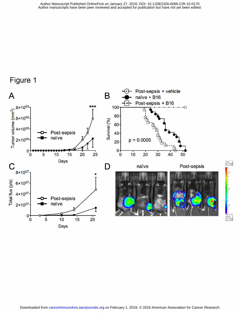
Post-sepsis mice showed increased tumor volumes and reduced overall survival (data
not shown for 10,000 and 100,000 B16 cells). When inoculated with 30,000 B16 cells,
significantly larger tumor volumes were detected in post-sepsis mice (Fig. 1A), which had
shorter survival times than naïve tumor-bearing controls (Fig. 1B). The results were also
confirmed by bioluminescence measurement (Fig. 1C and D).
Moreover, the long-lasting effects of sepsis were evaluated by subcutaneously
inoculating mice with 30,000 B16 cells 30 (Supplementary Fig. S1A and B) or 60 days
(Supplementary Fig. S1C and D) after CLP. In both situations, post-sepsis groups presented
larger tumors and died sooner.
Higher metastatic burden
Supplementary Fig. S2 depicts the experimental protocols used to evaluate metastasis
and lung colonization. An increased number of metastatic lesions in the lungs was observed at
D+21 after inoculation in post-sepsis mice (Supplementary Fig. S2B). Mortality due to
metastasis was evaluated by removing the primary tumors and following up the length of
survival (See Methods). Mice did not present with local recurrences of melanoma lesions.
on February 1, 2019. © 2016 American Association for Cancer Research. cancerimmunolres.aacrjournals.org Downloaded from
Author manuscripts have been peer reviewed and accepted for publication but have not yet been edited. Author Manuscript Published OnlineFirst on January 27, 2016; DOI: 10.1158/2326-6066.CIR-15-0170
ROLE OF TAM IN POST-SEPSIS TUMOR PROGRESSION
14
Increased mortality due to metastasis in post-sepsis group was detected (Supplementary Fig.
S2C). In addition, lung colonization after 30,000 B16 cells intravenous injection was also
increased in post-sepsis mice (Supplementary Fig. S2D), as evaluated by bioluminescence
quantification at D+18.
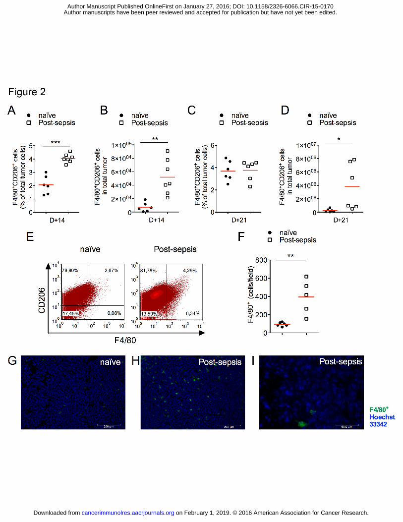
Increased numbers of TAMs
First, we observed that the percentage of leukocytes (CD45+) was increased in tumor
samples from sepsis-surviving animals. Post-sepsis mice had higher percentages of TAMs at
D+14 (Fig. 2A) as well as increased absolute numbers of TAMs (Fig. 2B). Differences in
TAM percentages were not detected at D+21 (Fig. 2C), in contrast to the absolute numbers
(Fig. 2D). Representative plots are presented in Fig. 2E. Immunofluorescence staining for
F4/80+ cells in frozen tumor sections further demonstrated the higher TAM accumulation in
post-sepsis mice (Fig. 2 F–I).
The spleen and draining lymph nodes of post-sepsis tumor bearing mice also had
increased percentages of Tregs, but no differences in intratumoral Tregs (Supplementary Fig.
S3A-C). Differences in CD3+CD4+ and CD3+CD8+ T cell intratumoral subpopulations were
not detected between the groups (Supplementary Fig. S4 A-D).
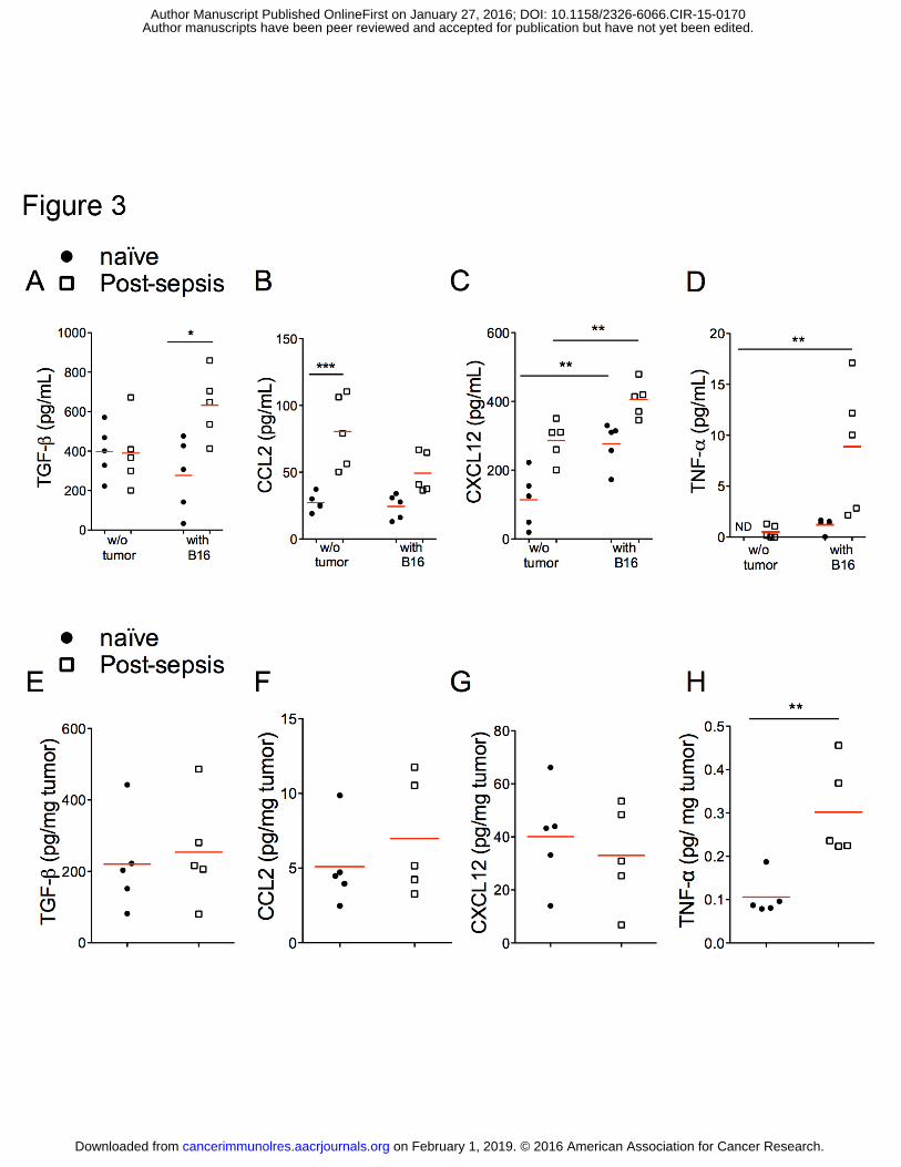
Increased concentrations CXCL12, TNFα, and TGFβ
In an attempt to understand the mechanism underlying the post-sepsis increase of
TAM accumulation, we screened chemokines and cytokines related to the inflammatory
process and to macrophage recruitment (Fig. 3). The concentrations of CCL2 (Fig. 3B) and
CXCL12 (Fig. 3C) were increased in the serum of post-sepsis mice injected with vehicle in
comparison to naïve mice. The presence of the tumor by itself also increased CXCL12 (Fig.
3C). Post-sepsis tumor-bearing mice had higher TNFα (Fig. 3D), TGFβ (Fig. 3A) and
on February 1, 2019. © 2016 American Association for Cancer Research. cancerimmunolres.aacrjournals.org Downloaded from
Author manuscripts have been peer reviewed and accepted for publication but have not yet been edited. Author Manuscript Published OnlineFirst on January 27, 2016; DOI: 10.1158/2326-6066.CIR-15-0170
ROLE OF TAM IN POST-SEPSIS TUMOR PROGRESSION
15
CXCL12 compared to naïve injected with tumor cells (Fig. 3C). We also quantified the same
chemo/cytokines within the tumor mass of naïve and post-sepsis mice. Only TNFα
concentrations were increased in tumor masses from post-sepsis mice (Fig. 3E-H).
Assessments of serum chemokines and cytokines at 30 days after sepsis induction were also
performed. TGFβ was increased in post-sepsis mice after 30 days. Differences in serum
concentrations of CCL2, CXCL12, and TNFα were not detected (data not shown).
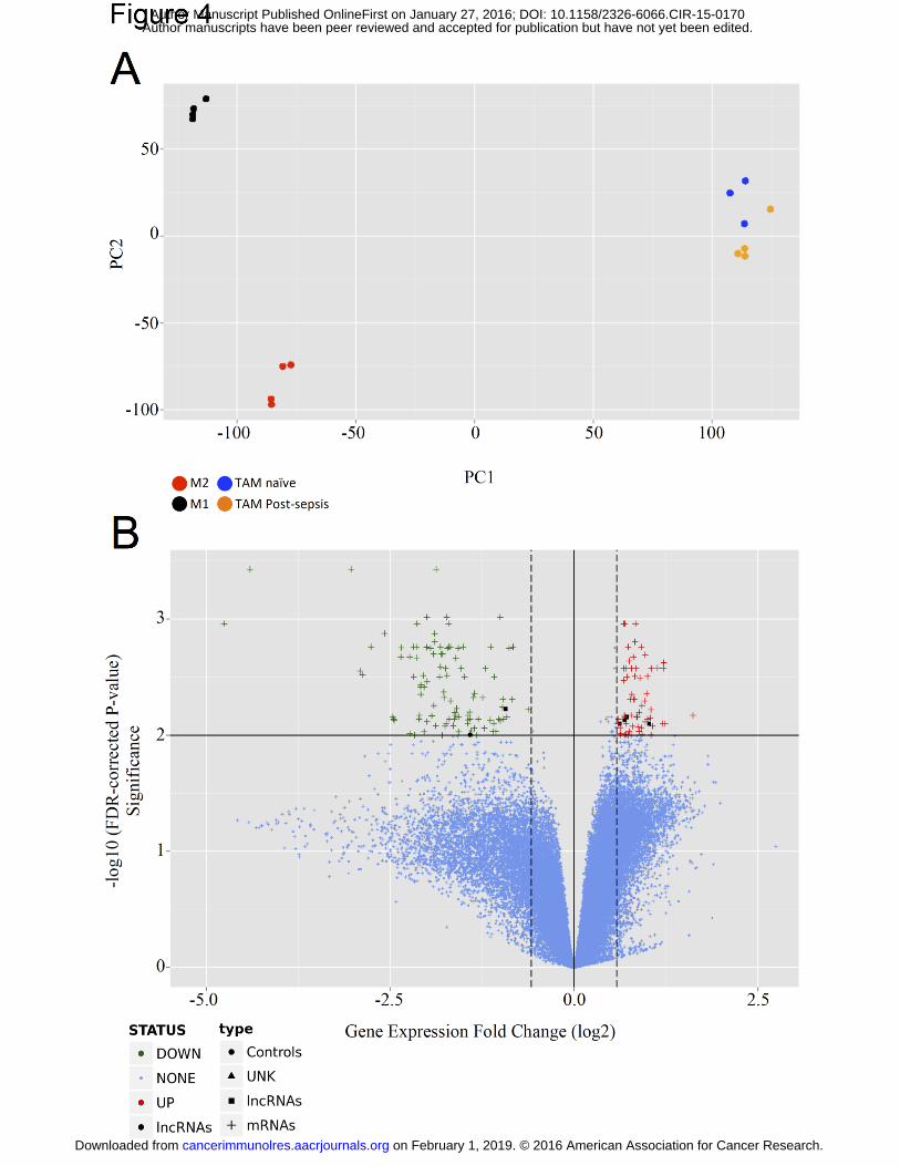
Differences in gene expression
Since we have demonstrated a quantitative early increase of TAM accumulation in
post-sepsis mice, we decided to compare the global gene expression of TAM from naïve and
post-sepsis mice using Agilent microarrays with almost 60,000 probes (39,430 mRNA and
16,251 long noncoding RNAs). M1 and M2-polarized macrophages were used for
comparison.
Figure 4A depicts the principal component analysis for the aforementioned groups,
using an unsupervised approach. The differences between TAM from naïve and TAM from
post-sepsis were only mild and their gene expression profiles were distinct from M1 and M2-
macrophages.
Then, we focused our analysis in the differences between TAM from post-sepsis and
TAM from naïve mice. We identified 61 genes to be up-regulated and 98 genes to be down-
regulated (Table 1) using log2 fold cutoffs at |0.58| and adjusted P value (False Discovery
Rate) < 1% (Fig. 4B). Among the down-regulated genes we detected genes associated with
leukocyte activation (e.g., Cd83, which is a marker of dendritic cell maturation; Cd86, which
is a marker of macrophage classic or M1 activation). Also, genes related to major
histocompatibility complex type II (e.g., H2-Eb1 and H2-Ab1) were down-regulated. Among
on February 1, 2019. © 2016 American Association for Cancer Research. cancerimmunolres.aacrjournals.org Downloaded from
Author manuscripts have been peer reviewed and accepted for publication but have not yet been edited. Author Manuscript Published OnlineFirst on January 27, 2016; DOI: 10.1158/2326-6066.CIR-15-0170

ROLE OF TAM IN POST-SEPSIS TUMOR PROGRESSION
16
chemokines-related genes, Ccl5 and Cxcr4 were down-regulated in TAM from post-sepsis
mice.
A qRT-PCR analysis of specific genes related with macrophage polarization was
carried out. Post-sepsis-derived TAM exhibited reduced gene expression of Nos2 and a trend
for higher gene expression of Tnf, Arg1 and Mrc1 was detected (Supplementary Fig. S5A-D).
Post-sepsis macrophages facilitated tumor progression
In order to evaluate whether macrophages derived from post-sepsis mice could
contribute to tumor progression, we co-inoculated bone marrow–derived macrophages
(BMDMs) from naïve or post-sepsis mice with B16 cells in naïve recipients (Fig. 5A).
BMDMs from post-sepsis or naïve mice were primarily in a M0 as indicated by the low flow
cytometric expression of CD206 and TNFα.
BMDMs from post-sepsis led to larger tumor volumes (Fig. 5B) and shorter overall
survival (Fig. 5C) of the recipients. All animals co-inoculated with BMDM from post-sepsis
mice died within 50 days, whereas approximately 60% of animals co-inoculated with BMDM
from naïve survived in the same time span. Tumor burden increase was confirmed by
bioluminescence quantification at D+21 (Fig. 5D and E).
CXCR4/CXCL12 inhibition
Based on the increased CXCL12 detected in the serum of post-sepsis mice, we
investigated if the pharmacological inhibition of this pathway could reverse the effect of post-
sepsis on tumor progression. AMD3100, a specific antagonist of the CXCR4/CXCL12
pathway, was administered at D+10 and D+15 after B16 cell inoculation. As shown in Fig.
6A, AMD3100 reverted the increase of post-sepsis-induced tumor volumes increase but
resulted in nonsignificant changes in tumor volumes in the naïve group. Post-sepsis and naïve
on February 1, 2019. © 2016 American Association for Cancer Research. cancerimmunolres.aacrjournals.org Downloaded from
Author manuscripts have been peer reviewed and accepted for publication but have not yet been edited. Author Manuscript Published OnlineFirst on January 27, 2016; DOI: 10.1158/2326-6066.CIR-15-0170
ROLE OF TAM IN POST-SEPSIS TUMOR PROGRESSION
17
tumor-bearing mice that received AMD3100 survived longer than vehicle-treated controls
(Fig. 6B). The percentage of TAMs expressing CXCR4 was similar between naïve and post-
sepsis groups (Fig. 6C). CXCR4/CXCL12 blockade through AMD3100 inhibited the TAM
accumulation associated with post-sepsis state (Fig. 6D and E). Additionally, AMD3100
administration inhibited the ability of post-sepsis BMDM to increase tumor growth
(Supplementary Fig. S6).
The percentage of TAMs expressing Ki67 was increased in post-sepsis mice tumors
and the inhibition of CXCR4/CXCL12 by AMD3100 reverted this finding. Of note, Ki67
levels were not increased in CD45– cells (Supplementary Fig. S7A-C).
on February 1, 2019. © 2016 American Association for Cancer Research. cancerimmunolres.aacrjournals.org Downloaded from
Author manuscripts have been peer reviewed and accepted for publication but have not yet been edited. Author Manuscript Published OnlineFirst on January 27, 2016; DOI: 10.1158/2326-6066.CIR-15-0170

ROLE OF TAM IN POST-SEPSIS TUMOR PROGRESSION
18
DISCUSSION
Here we describe the increase of melanoma B16 progression in sepsis-surviving mice,
which was associated with tumor microenvironmental TAM accumulation through
CXCR4/CXCL12 signaling. In addition, post-sepsis state was associated with an increased
pulmonary metastatic burden and increased lung colonization. The effect of sepsis on B16
melanoma tumor progression proved to be long-lasting, since it was observed even when the
neoplastic cells were inoculated 30 or 60 days after CLP-induced sepsis. Accordingly,
Weycker and colleagues have shown that patients with sepsis present an increased long-term
mortality (3). Also, Otto and colleagues proved that sepsis survivors are at higher risk of
opportunistic infections after a period between 16 to 150 days following sepsis resolution (5).
If clinical post-sepsis state is associated with higher tumor incidence, tumor progression or
metastasis still remains elusive.
TAM accumulation is a relevant marker of unfavorable prognosis in cancer (18). It has
been associated to increased tumor progression, neoangiogenesis, and immune escape in
several tumor types (17, 27). We assessed TAMs (F4/80+CD206+) through flow cytometry
and immunofluorescence and found they accumulated heavily in post-sepsis mice tumors at
an early stage of tumor progression (D+14) when the tumor sizes were not different between
the groups. In a later phase (D+21) the relative amounts of TAMs show no difference
between the groups.
Classically, TAM accumulation depends on blood stream-derived monocyte
infiltration mediated by CCL2, CCL7, VEGF, and other cytokines (27–29). However,
Tymoszuk and colleagues demonstrated in situ proliferation of CD11bloF4/80hi cells through
BrdU labeling and Ki67 staining in a model of spontaneous breast cancer (20). This suggests
an important contribution of in situ TAM proliferation. Corroborating these findings, we
observed that Ki67 positivity in TAM cells was increased in post-sepsis mice. This could
on February 1, 2019. © 2016 American Association for Cancer Research. cancerimmunolres.aacrjournals.org Downloaded from
Author manuscripts have been peer reviewed and accepted for publication but have not yet been edited. Author Manuscript Published OnlineFirst on January 27, 2016; DOI: 10.1158/2326-6066.CIR-15-0170
ROLE OF TAM IN POST-SEPSIS TUMOR PROGRESSION
19
represent a contribution of local macrophage proliferation for TAM accumulation in the post-
sepsis state.
We confirmed the importance of post-sepsis macrophages to tumor progression by co-
inoculating BMDM together with B16 cells in naïve mice. Post-sepsis–derived BMDM co-
inoculation resulted in higher tumor progression and less overall survival. Cho and colleagues
found that the inoculation of M2-polarized macrophage provoked increased progression and
metastasis in a breast cancer model, possibly due to increased angiogenesis (30).
In order to check whether TAM from post-sepsis mice have acquired a M2-like
phenotype, the global gene expression profile of isolated TAM was analyzed. The principal
component analysis showed a distinct clustering of TAM from naïve and TAM from post-
sepis mice. The microarray analysis revealed few similarities between TAM and M2-
macrophages in contrast to the reported in literature (31). One could speculate this could have
occurred due to suboptimal purity of TAM isolation (70 to 80%), when compared to bone-
marrow derived macrophages (almost 100%).
Sixty-one genes were up-regulated and 98 genes were down-regulated in post-sepsis-
derived TAM compared to naïve-derived TAM. Genes related to M2-phenotype, such as Il10,
Arg1 and Fizz1, were not differentially expressed in the microarray analysis. However, we
detected a reduced expression of Nos2, a classic M1-marker, in TAM from post-sepsis mice
in additional qRT-PCR assays. Of note, Marco, a gene up-regulated in IL10-induced M2-
macrophages (32), showed higher expression in TAM from post-sepsis mice. In spite of this,
it is unclear whether Marco is a M2-marker or a marker of innate macrophage activation (33).
On the other hand, reduced expression of leukocyte activation and major histocompatibility
complex type II (MHC-II)-related genes were detected in these cells. In addition, Cd86, which
is a co-stimulatory molecule-related gene, was down-regulated. The latter two molecules are
more likely associated with M1 rather than M2-polarization (32).
on February 1, 2019. © 2016 American Association for Cancer Research. cancerimmunolres.aacrjournals.org Downloaded from
Author manuscripts have been peer reviewed and accepted for publication but have not yet been edited. Author Manuscript Published OnlineFirst on January 27, 2016; DOI: 10.1158/2326-6066.CIR-15-0170
ROLE OF TAM IN POST-SEPSIS TUMOR PROGRESSION
20
Since TGFβ has been implicated in monocyte recruitment, monocyte-to-macrophage
differentiation and acquisition of protumoral functions by TAM (34), we assessed its serum
concentrations, which were increased in post-sepsis tumor bearing mice. In addition, CXCL12
was increased in the serum of these mice. Along this line, Wang and colleagues reported that
TGFβ increases the response of CXCL12 chemotactic signaling (35). Based on these results,
we tested whether CXCR4/CXCL12 inhibition through AMD3100 administration would
affect tumor progression and TAM accumulation. When signaling through CXCR4 was
blocked in vivo, this reversed the effect of post-sepsis on tumor size and also improved overall
survival.
These findings were also associated with the reduction of TAM accumulation.
CXCR4/CXCL12 is a known pathway implicated in TAM accretion in hypoxic areas of
tumor microenvironments (22). Our results are in agreement with Beider and colleagues, who
described that multiple myeloma cells recruit monocytes to their microenvironmental niche,
differentiate them to macrophages that then polarize into M2-like phenotype through
CXCR4/CXCL12 (36). The impediment of TAM accumulation was, at least in part,
dependent on the reduction of TAM extramedullary proliferation, since we observed reduced
numbers of Ki67+F4/80+ cells.
Other cells may take part in the post-sepsis-associated tumor progression. In this
regard, Cavassani and colleagues had previously assessed post-sepsis neoplastic expansion
and demonstrated the Treg-dependent increase in tumor burden using a heterotopic Lewis
lung carcinoma model (12). Zhou and colleagues have demonstrated the frequent association
of Tregs and TAMs within the tumor microenvironment and reported that Treg/TAM co-
localization is associated with worse outcome in patients with hepatocelular cancer (37). This
could be relevant in the context of our findings, since Tregs have a role in the post-sepsis
on February 1, 2019. © 2016 American Association for Cancer Research. cancerimmunolres.aacrjournals.org Downloaded from
Author manuscripts have been peer reviewed and accepted for publication but have not yet been edited. Author Manuscript Published OnlineFirst on January 27, 2016; DOI: 10.1158/2326-6066.CIR-15-0170
ROLE OF TAM IN POST-SEPSIS TUMOR PROGRESSION
21
immunosuppressive state (6). However, we could not detect significant differences in Treg
numbers in the tumor microenvironment of post-sepsis mice.
In summary, sepsis has a long-lasting effect, which may favor neoplastic expansion. In
our model, TAM accumulated in the tumor microenvironment of post-sepsis subjects in an
early phase, which is, at least in part, dependent on CXCR4/CXCL12 signaling. Overall, our
data indicate at least three potential mechanisms of post-sepsis-induced tumor progression in
mice: a possible direct effect of CXCL12 in tumor progression, increased TAM accumulation
and proliferation (perhaps in response to increased CXCL12 in serum) and intrinsically
altered bone-marrow-derived macrophages (and so potentially TAM). The evaluation of such
phenomena in the clinical setting should be a matter of future concern. Specifically, it is
important to determine if sepsis-surviving patients have a higher risk of cancer development,
progression, or mortality, and if CXCR4/CXCL12 blockade could reverse this effect.
on February 1, 2019. © 2016 American Association for Cancer Research. cancerimmunolres.aacrjournals.org Downloaded from
Author manuscripts have been peer reviewed and accepted for publication but have not yet been edited. Author Manuscript Published OnlineFirst on January 27, 2016; DOI: 10.1158/2326-6066.CIR-15-0170
ROLE OF TAM IN POST-SEPSIS TUMOR PROGRESSION
22
ACKNOWLEDGMENTS
The authors gratefully acknowledge Giuliana Bertozi, Priscila Scheucher and Amélia
Goes de Araújo for their technical assistance. We would like to dedicate this work to the
memory of Professor Ronaldo de Albuquerque Ribeiro, MD, PhD.
on February 1, 2019. © 2016 American Association for Cancer Research. cancerimmunolres.aacrjournals.org Downloaded from
Author manuscripts have been peer reviewed and accepted for publication but have not yet been edited. Author Manuscript Published OnlineFirst on January 27, 2016; DOI: 10.1158/2326-6066.CIR-15-0170
ROLE OF TAM IN POST-SEPSIS TUMOR PROGRESSION
23
REFERENCES
1. Beale R, Reinhart K, Brunkhorst FM, Dobb G, Levy M, Martin G, et al. Promoting Global Research Excellence in Severe Sepsis (PROGRESS): lessons from an international sepsis registry. Infection. 2009;37:222–32.
2. Dellinger RP, Levy MM, Rhodes A, Annane D, Gerlach H, Opal SM, et al. Surviving sepsis campaign: international guidelines for management of severe sepsis and septic shock: 2012. Crit Care Med. 2013;41:580–637.
3. Weycker D, Akhras KS, Edelsberg J, Angus DC, Oster G. Long-term mortality and medical care charges in patients with severe sepsis. Crit Care Med. 2003;31:2316–23.
4. Quartin AA, Schein RM, Kett DH, Peduzzi PN. Magnitude and duration of the effect of sepsis on survival. Department of Veterans Affairs Systemic Sepsis Cooperative Studies Group. JAMA. 1997;277:1058–63.
5. Otto GP, Sossdorf M, Claus RA, Rödel J, Menge K, Reinhart K, et al. The late phase of sepsis is characterized by an increased microbiological burden and death rate. Crit Care.2011;15:R183.
6. Nascimento DC, Alves-Filho JC, Sônego F, Fukada SY, Pereira MS, Benjamim C, et al. Role of regulatory T cells in long-term immune dysfunction associated with severe sepsis. Crit Care Med. 2010;38:1718–25.
7. Munoz C, Carlet J, Fitting C, Misset B, Blériot JP, Cavaillon JM. Dysregulation of in vitro cytokine production by monocytes during sepsis. J Clin Invest. 1991;88:1747–54.
8. Carson WF, Cavassani K a., Dou Y, Kunkel SL. Epigenetic regulation of immune cell functions during post-septic immunosuppression. Epigenetics. 2011;6:273–83.
9. Döcke WD, Höflich C, Davis KA, Röttgers K, Meisel C, Kiefer P, et al. Monitoring temporary immunodepression by flow cytometric measurement of monocytic HLA-DR expression: a multicenter standardized study. Clin Chem. 2005;51:2341-7.
10. Takahashi H, Tsuda Y, Takeuchi D, Kobayashi M, Herndon DN, Suzuki F. Influence of systemic inflammatory response syndrome on host resistance against bacterial infections. Crit Care Med. 2004;32:1879–85.
11. Dahlke E, Murray CA, Kitchen J, Chan A-W. Systematic review of melanoma incidence and prognosis in solid organ transplant recipients. Transplant Res. 2014;3:10.
on February 1, 2019. © 2016 American Association for Cancer Research. cancerimmunolres.aacrjournals.org Downloaded from
Author manuscripts have been peer reviewed and accepted for publication but have not yet been edited. Author Manuscript Published OnlineFirst on January 27, 2016; DOI: 10.1158/2326-6066.CIR-15-0170
ROLE OF TAM IN POST-SEPSIS TUMOR PROGRESSION
24
12. Cavassani KA, Carson WF, Moreira AP, Wen H, Schaller MA, Ishii M, et al. The post sepsis-induced expansion and enhanced function of regulatory T cells create an environment to potentiate tumor growth. Blood. 2010;115:4403–11.
13. Hotchkiss RS, Monneret G, Payen D. Sepsis-induced immunosuppression: from cellular dysfunctions to immunotherapy. Nat Rev Immunol. Nature Publishing Group; 2013;13:862–74.
14. Hanahan D, Weinberg R a. Hallmarks of cancer: the next generation. Cell. 2011;144:646–74.
15. Cavallo F, De Giovanni C, Nanni P, Forni G, Lollini P-L. 2011: the Immune Hallmarks of Cancer. Cancer Immunol Immunother. 2011;60:319–26.
16. Coussens LM, Werb Z. Inflammation and cancer. Nature. 2010;420:860–7.
17. Biswas SK, Allavena P, Mantovani A. Tumor-associated macrophages: functional diversity, clinical significance, and open questions. Semin Immunopathol. 2013;35:585-600.
18. Zhang Q, Liu L, Gong C, Shi H, Zeng Y, Wang X, et al. Prognostic significance of tumor-associated macrophages in solid tumor: a meta-analysis of the literature. PLoS One.2012;7:e50946.
19. Cortez-Retamozo V, Etzrodt M, Newton A, Rauch PJ, Chudnovskiy A, Berger C, et al. Origins of tumor-associated macrophages and neutrophils. Proc Natl Acad Sci. 2012;109:2491–6.
20. Tymoszuk P, Evens H, Marzola V, Wachowicz K, Wasmer M-H, Datta S, et al. In situ proliferation contributes to accumulation of tumor-associated macrophages in spontaneous mammary tumors. Eur J Immunol. 2014;44:2247–62.
21. Schioppa T, Uranchimeg B, Saccani A, Biswas SK, Doni A, Rapisarda A, et al. Regulation of the chemokine receptor CXCR4 by hypoxia. J Exp Med. 2003;198:1391–402.
22. Knowles H, Harris AL. Macrophages and the hypoxic tumour microenvironment. Front Biosci. 2007;12:4298–314.
23. Hubbard WJ, Choudhry M, Schwacha MG, Kerby JD, Rue LW, Bland KI, et al. Cecal Ligation and Puncture. Shock. 2005;24:52–7.
on February 1, 2019. © 2016 American Association for Cancer Research. cancerimmunolres.aacrjournals.org Downloaded from
Author manuscripts have been peer reviewed and accepted for publication but have not yet been edited. Author Manuscript Published OnlineFirst on January 27, 2016; DOI: 10.1158/2326-6066.CIR-15-0170
ROLE OF TAM IN POST-SEPSIS TUMOR PROGRESSION
25
24. Jacob D, Davis J, Fang B. Xenograftic tumor models in mice for cancer research, a technical review. Gene Ther Mol Biol. 2004;8:213–9.
25. Ketcham A, Kinsey D, Wexler H, Mantel N. The development of spontaneous metastases after the removal of a “primary” tumor. II. Standardization protocol of 5 animal tumors. Cancer. 1961;14:875–82.
26. Ritchie ME, Phipson B, Wu D, Hu Y, Law CW, Shi W, et al. limma powers differential expression analyses for RNA-sequencing and microarray studies. Nucleic Acids Res. 2015;1–13.
27. Solinas G, Germano G, Mantovani a, Allavena P. Tumor-associated macrophages (TAM) as major players of the cancer-related inflammation. J Leukoc Biol. 2009;86:1065–73.
28. Loberg RD, Ying C, Craig M, Yan L, Snyder L a., Pienta KJ. CCL2 as an Important Mediator of Prostate Cancer Growth In Vivo through the Regulation of Macrophage Infiltration. Neoplasia. 2007;9:556–62.
29. Qian B, Li J, Zhang H, Kitamura T, Zhang J, Campion LR, et al. CCL2 recruits inflammatory monocytes to facilitate breast-tumour metastasis. Nature. 2011;475:222–5.
30. Cho HJ, Jung JI, Lim DY, Kwon GT, Her S, Park JH, et al. Bone marrow-derived, alternatively activated macrophages enhance solid tumor growth and lung metastasis of mammary carcinoma cells in a Balb/C mouse orthotopic model. Breast Cancer Res. 2012;14:R81.
31. Mantovani a, Sozzani S, Locati M, Allavena P, Sica a. Macrophage polarization: tumor-associated macrophages as a paradigm for polarized M2 mononuclear phagocytes. Trends Immunol. 2002;23:549–55.
32. Martinez FO, Gordon S. The M1 and M2 paradigm of macrophage activation: time for reassessment. F1000Prime Rep. 2014;6:13.
33. Martinez FO, Sica A, Mantovani A, Locati M. Macrophage activation and polarization. Front Biosci. 2008;13:453–61.
34. Bierie B, Moses HL. Transforming growth factor beta (TGF-beta) and inflammation in cancer. Cytokine Growth Factor Rev. 2010;21:49–59.
on February 1, 2019. © 2016 American Association for Cancer Research. cancerimmunolres.aacrjournals.org Downloaded from
Author manuscripts have been peer reviewed and accepted for publication but have not yet been edited. Author Manuscript Published OnlineFirst on January 27, 2016; DOI: 10.1158/2326-6066.CIR-15-0170
ROLE OF TAM IN POST-SEPSIS TUMOR PROGRESSION
26
35. Wang J, Guan E, Roderiquez G, Calvert V, Alvarez R, Norcross M a. Role of tyrosine phosphorylation in ligand-independent sequestration of CXCR4 in human primary monocytes-macrophages. J Biol Chem. 2001;276:49236–43.
36. Beider K, Bitner H, Leiba M, Gutwein O, Koren-Michowitz M, Ostrovsky O, et al. Multiple myeloma cells recruit tumor-supportive macrophages through the CXCR4/CXCL12 axis and promote their polarization toward the M2 phenotype. Oncotarget. 2014;5:11283–96.
37. Zhou J, Ding T, Pan W, Zhu L-Y, Li L, Zheng L. Increased intratumoral regulatory T cells are related to intratumoral macrophages and poor prognosis in hepatocellular carcinoma patients. Int J cancer. 2009;125:1640–8.
Figure 1. Post-sepsis state was associated to increased tumor progression. (A) Post-sepsis
mice developed increased tumor volumes in comparison to naïve mice after the s.c.
inoculation of B16 melanoma cells. n = 6 (naïve) or n = 7 (post-sepsis). Graph is
representative of four independent experiments. (B) Overall survival was reduced in post-
sepsis tumor-bearing mice. n = 17 (naïve inoculated with B16 cells), n = 19 (post-sepsis
inoculated with B16 cells), or n = 7 (post-sepsis injected with vehicle). Results calculated
from four independent experiments. (C) The post-sepsis-related tumor progression was
confirmed through bioluminescence measurement (total flux = photons/second). n = 7 (naïve)
or n = 8 (post-sepsis). Graph is representative of two independent experiments. (D) Three
representative mice of each group analyzed at D+21 for the tumor bioluminescence
quantification. As the scale indicates, the redder the signal, the greater is the tumor burden.
Data are represented as the means ± S.E.M. or Kaplan-Meier curves when appropriate. *
indicates P < 0.05; ** indicates P < 0.01; *** indicates P < 0.001.
Figure 2. TAM accumulation was increased in the tumor microenvironment of sepsis
surviving mice. (A-B) TAM (F4/80+CD206+) infiltrates were increased in post-sepsis tumor
on February 1, 2019. © 2016 American Association for Cancer Research. cancerimmunolres.aacrjournals.org Downloaded from
Author manuscripts have been peer reviewed and accepted for publication but have not yet been edited. Author Manuscript Published OnlineFirst on January 27, 2016; DOI: 10.1158/2326-6066.CIR-15-0170

ROLE OF TAM IN POST-SEPSIS TUMOR PROGRESSION
27
samples at D+14 when compared to naïve group. n = 6 (naïve, D+14), n = 7 (post-sepsis,
D+14). (C-D) By D+21, the relative difference was not detected, in contrast to the difference
in the absolute numbers of TAM. n = 6 (naïve, D+21) and n = 6 (post-sepsis, D+21). Graph is
representative of two independent experiments. (E) Representative dots plots of flow
cytometry analysis performed at D+14. (F) Immunofluorescent analysis of TAM
accumulation at D+14. n = 6 (naïve), n = 5 (post-sepsis). Graph is representative of two
independent experiments. (G-I) Representative microphotographs of tumors from (G) naïve or
(I-H) post-sepsis mice at D+14. (H) Detail of the extracellular F4/80+ staining in two cells.
Bars indicate the scales. Green: F4/80+ (macrophage, TAM) and Blue: Hoechst 33342
(nuclei). The horizontal red lines represent the mean and individual data are presented as
scattered dot plots. Groups were compared using the Student’s t test. * indicates P < 0.05; **
indicates P < 0.01; *** indicates P < 0.001.
Figure 3. Differential cytokine profiles in serum and tumor samples of post-sepsis and
naïve mice. Samples were collected 14 days after tumor inoculation. (A) Post-sepsis tumor
bearing mice presented increased TGFβ concentrations in serum. (B) CCL2 serum
concentrations were higher in post-sepsis mice injected with vehicle, but not in the other
groups. (C) CXCL12 serum concentrations were increased in post-sepsis mice injected with
vehicle and in naïve tumor bearing mice when compared to naïve mice injected with vehicle.
Post-sepsis tumor bearing mice showed higher amounts of CXCL12 compared to all groups.
(D) TNF-α serum concentrations were higher in post-sepsis tumor bearing, in comparison to
the remaining experimental groups. (E to G) Differences in TGFβ, CCL2, and CXCL12
intratumoral concentrations were not statistically significant among groups. (H) TNF-α
intratumoral concentrations were higher in post-sepsis tumor bearing mice, in comparison to
the other groups. n = 5 per group. Graphs are representative of two independent experiments.
on February 1, 2019. © 2016 American Association for Cancer Research. cancerimmunolres.aacrjournals.org Downloaded from
Author manuscripts have been peer reviewed and accepted for publication but have not yet been edited. Author Manuscript Published OnlineFirst on January 27, 2016; DOI: 10.1158/2326-6066.CIR-15-0170
ROLE OF TAM IN POST-SEPSIS TUMOR PROGRESSION
28
Filled circles indicate naïve mice; unfilled squares indicate post-sepsis mice. The horizontal
red lines represent the mean and individual data are presented as scattered dot plots. *
indicates P < 0.05; ** indicates P < 0.01; *** indicates P < 0.001.
Figure 4. Comparison between the gene expression profiles of TAM from post-sepsis
and naïve mice. (A) First two principal components are plotted elucidating the group
relationship between TAM from naïve and from post-sepsis mice. (B) Volcano plot
comparing TAM from post-sepsis to TAM from naïve mice. Fold change was set at 0.58 for
up-regulated and 0.58 for down-regulated gene expression. Significance was set at FDR <
0.01. 98 genes were down-regulated (green) and 61 were up-regulated (red). mRNAs:
messenger RNAs, lncRNAs: long non-coding RNAs, UNK: unknown RNA, FDR: false
discovery rate, M1: M1-polarized macrophage, M2: M2-polarized macrophage.
Figure 5. Bone marrow (BM)–derived macrophages (MØs) from post-sepsis mice
increased tumor progression. (A) Experimental protocol. (B). Recipients of Post-sepsis
MØs presented increased tumor progression in comparison to controls. n = 10 (naïve MØs)
and n = 9 (Post-sepsis MØs). (C) Mice injected with Post-sepsis MØs + B16 cells presented
increased mortality. n = 10 (naïve MØs) and n = 9 (Post-sepsis MØs). (D) Increased tumor
burden in Post-sepsis MØs group was confirmed at D+21 through bioluminescent signal
quantification after D-luciferin (150 mg/kg, ip.) administration. n = 7 (naïve MØs). and n = 5
(Post-sepsis MØs). (E) Representative mice of each group. Graphs are representative of two
independent experiments. The horizontal red lines represent the mean and individual data are
presented as scattered dot plots. * indicates P < 0.05; ** indicates P < 0.01; *** indicates P <
0.001.
on February 1, 2019. © 2016 American Association for Cancer Research. cancerimmunolres.aacrjournals.org Downloaded from
Author manuscripts have been peer reviewed and accepted for publication but have not yet been edited. Author Manuscript Published OnlineFirst on January 27, 2016; DOI: 10.1158/2326-6066.CIR-15-0170
ROLE OF TAM IN POST-SEPSIS TUMOR PROGRESSION
29
Figure 6. CXCR4/CXCL12 blockade reverted the post-sepsis effect on tumor
progression and TAM accumulation. (A) The tumor volumes of post-sepsis treated with
AMD3100 were not different from untreated naïve mice. n = 6 (naïve plus vehicle), n = 10
(naïve plus AMD3100), n = 5 (post-sepsis plus vehicle), and n = 12 (post-sepsis plus
AMD3100). The arrows indicate the days in which AMD3100 was administered. (B) Overall
survival was improved after CXCR4/CXCL12 blockade. Both naïve and post-sepsis mice that
received AMD3100 showed survival improvement compared to their untreated counterparts.
n = 5 (post-sepsis without tumor), n = 6 (naïve plus vehicle), n = 11 (naïve plus AMD3100), n
= 7 (post-sepsis plus vehicle), and n = 13 (post-sepsis plus AMD3100). (C) TAM
(CD45+F4/80+) from naïve and post-sepsis mice expressed the same levels of CXCR4. n = 5
(naïve) and n = 6 (post-sepsis). (D) AMD3100 reverted the TAM accumulation in tumors of
post-sepsis mice at D+14. n = 5 (naïve plus vehicle), n = 5 (naïve plus AMD3100), n = 5
(post-sepsis plus vehicle), and n = 5 (post-sepsis plus AMD3100). (E) Representative dot
plots of post-sepsis plus vehicle group and post-sepsis plus AMD3100 group. All graphs are
representative of two independent experiments. The horizontal red lines represent the mean
and individual data are presented as scattered dot plots. * indicates P < 0.05; ** indicates P <
0.01; *** indicates P < 0.001.
on February 1, 2019. © 2016 American Association for Cancer Research. cancerimmunolres.aacrjournals.org Downloaded from
Author manuscripts have been peer reviewed and accepted for publication but have not yet been edited. Author Manuscript Published OnlineFirst on January 27, 2016; DOI: 10.1158/2326-6066.CIR-15-0170
Table 1 - Differentially expressed genes in Post-sepsis TAM compared with naïve TAM.
Genes related to leukocyte activation, macrophage polarization and MHC type II complex
Gene Fold-change p Function Reference
Cd2 -2.91 0.0028 Adhesive properties;
co-stimulatory molecule
Bimal et al., 2012
Ccr7 -2.76 0.0017
CCL19 and CCL21 receptor; related to classic macrophage
activation
Oh et al., 2012
H2-DMb1 -2.14 0.0017 MHC class II-related molecule Cho et al., 1991
Cd24a -2.10 0.0067 Cell adhesion molecule; DC marker Qu et al., 2014
Cd74 -1.82 0.0026 MIF receptor; related to classic macrophage
activation Leng et al., 2003
H2-Eb1 -1.82 0.0017 MHC class-II-related Widera and Flavell, 1984
Cd86 -1.80 0.002
Co-stimulatory molecule, related to classic macrophage
activation
Cavnar et al., 2013
Selplg -1.70 0.0017
P-selectin glycoprotein ligand;
cell adhesion molecule
Tchernychev et al., 2003
H2-Ab1 -1.68 0.0027 MHC class-II-related molecule Lacaze et al., 2009
Cxcr4 -1.62 0.0064 CXCL12 receptor;
TAM accumulation in hypoxic areas
Solinas et al., 2009 Schioppa et al., 2003
Marco 1.62 0.0067 Scavenger receptor; related to alternative
macrophage activationTomioka et al., 2012
H2-Ob -1.60 0.0059 MHC class-II-related molecule
Karlsson and Peterson, 1992
Il2rb -1.51 0.0017
IL-2 receptor β chain; IL-2 is a cofactor for macrophage classic
activation
Han et al., 1999
Ccl5 -1.44 0.0074 Pro-inflammatory chemokine; M1
macrophage marker
Sica and Mantovani, 2012
Cd209a -1.42 0.0063 C-type lectin;
mediates recognition and phagocytosis
Xi-Jiang Lu, 2013
on February 1, 2019. © 2016 American Association for Cancer Research. cancerimmunolres.aacrjournals.org Downloaded from
Author manuscripts have been peer reviewed and accepted for publication but have not yet been edited. Author Manuscript Published OnlineFirst on January 27, 2016; DOI: 10.1158/2326-6066.CIR-15-0170
Il6ra -1.41 0.0072
IL-6 receptor; modulates
macrophage phenotype
Mauer et al., 2014
Differences in gene expression comparison between TAM from post-sepsis and TAM
from naïve mice were only mild. It was considered a fold-change of 0.58 to up
regulated genes and -0.58 to down regulated genes. Statistical significance was set at
p < 0.01. Among all, 61 genes were found to be up-regulated and 98 down-regulated.
This table brings only the selected genes that could be in such a way related to TAM
functions. Genes associated with leukocyte activation, macrophage polarization and
MHC-II were depicted here. MHC-II, major histocompatibility complex type II. MIF,
macrophage migration inhibitory factor. TAM, tumor associated macrophage. DC,
dendritic cell.
on February 1, 2019. © 2016 American Association for Cancer Research. cancerimmunolres.aacrjournals.org Downloaded from
Author manuscripts have been peer reviewed and accepted for publication but have not yet been edited. Author Manuscript Published OnlineFirst on January 27, 2016; DOI: 10.1158/2326-6066.CIR-15-0170
Published OnlineFirst January 27, 2016.Cancer Immunol Res Jose M Mota, Caio A Leite, Lucas E. B. Souza, et al. progression in miceaccumulation through CXCR4/CXCL12 and favors tumor Post-sepsis state induces tumor-associated macrophage
Updated version
10.1158/2326-6066.CIR-15-0170doi:
Access the most recent version of this article at:
Material
Supplementary
http://cancerimmunolres.aacrjournals.org/content/suppl/2016/01/27/2326-6066.CIR-15-0170.DC1
Access the most recent supplemental material at:
Manuscript
Authoredited. Author manuscripts have been peer reviewed and accepted for publication but have not yet been
E-mail alerts related to this article or journal.Sign up to receive free email-alerts
Subscriptions
Reprints and
To order reprints of this article or to subscribe to the journal, contact the AACR Publications
Permissions
Rightslink site. Click on "Request Permissions" which will take you to the Copyright Clearance Center's (CCC)
.http://cancerimmunolres.aacrjournals.org/content/early/2016/01/27/2326-6066.CIR-15-0170To request permission to re-use all or part of this article, use this link
on February 1, 2019. © 2016 American Association for Cancer Research. cancerimmunolres.aacrjournals.org Downloaded from
Author manuscripts have been peer reviewed and accepted for publication but have not yet been edited. Author Manuscript Published OnlineFirst on January 27, 2016; DOI: 10.1158/2326-6066.CIR-15-0170