post surgical complications of hirschsprung's disease and
TRANSCRIPT

POST SURGICAL COMPLICATIONS OF
HIRSCHSPRUNG'S DISEASE AND THEIR
MANAGEMENT AT KENYATTA NATIONAL HOSPITAL:
A f O-YEAR RETROSPECTIVE STUDY
BY
DR. FRANCIS NJOROGE KURIA (MBChB)
MEDICAL LIBRARY •HIVERSITY OF NAIROW
A DISSERTATION SUBMITTED IN PART FULFILMENT FOR THE
DEGREE OF MASTER OF MEDICINE (SURGERY) OF THE UNIVERSITY
OF NAIROBI, 2003
Library

DECLARATION
I, Francis Njoroge Kuria, do declare that this dissertation is my original work
and has not been presented for a degree in any other University.
This Dissertation has been submitted for the degree of Master of Medicine
(Surgery) with our approval.
n
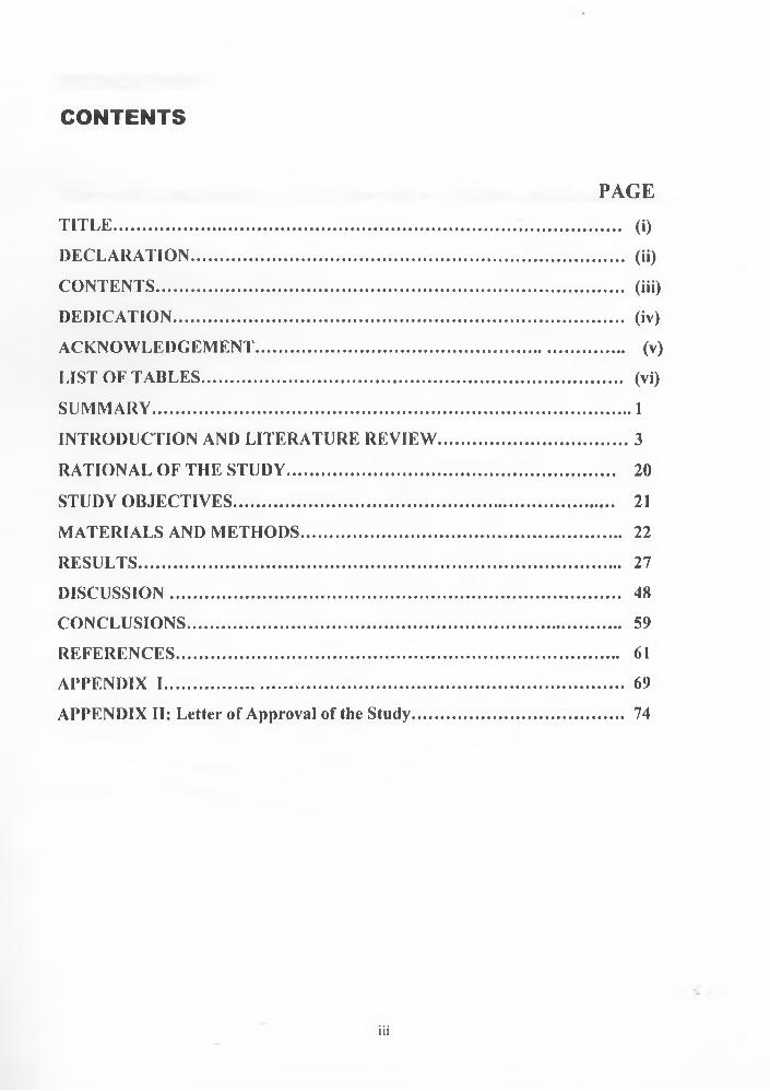
C O N TEN TS
PAGE
TITLE........................................................................................................... (i)DECLARATION............................................................................................ (ii)CONTENTS................................................................................................... (iii)DEDICATION................................................................................................ (iv)ACKNOWLEDGEMENT.............................................................................. (v)LIST OF TABLES......................................................................................... (vi)SUMMARY....................................................................................................1INTRODUCTION AND LITERATURE REVIEW....................................... 3
RATIONAL OF THE STUDY..................................................................... 20
STUDY OBJECTIVES................................................................................ 21MATERIALS AND METHODS................................................................... 22RESULTS...................................................................................................... 27DISCUSSION............................................................................................... 48CONCLUSIONS........................................................................................... 59REFERENCES............................................................................................. 61APPENDIX 1................................................................................................. 69APPENDIX II: Letter of Approval of the Study............................................ 74
iii

DEDICATION
This work is dedicated to my wife Jane and my children Jeniffer and Lilian.
IV

ACKNOW LEDGEM ENTS
I would like to thank all those who made the completion of this study possible.
In particular I feel deeply indebted to the following.
Prof. G.A.O Magoha, IOM, MBBS, F1BA, FWACS, FICS, FMCS (Urol), FCS (ECSA)
Professor of Surgery, Consultant Urologist and Deputy Vice Chancellor,
Administration and Finance, University of Nairobi, and Mr. J. M. Ndungu,
Senior lecturer and Consultant Peadiatric Surgeon, for their supervisory role in
this study and their willingness to work hand in hand to support me.
Kenyatta National Hospital Ethical and Research Committee for giving
approval for this study.
The technical staff of the Department of Medical Records of Kenyatta
National Hospital for assisting in retrieving of the patients’ records.
Mr. Muniu of KEMRI for his assistance and guidance in statistical details of the
study.
Mr. S. M. Macharia and Miss Susan Ruguru for their invaluable secretarial and
printing services.
All those who directly or indirectly assisted me in this study.

LIS T OF TAB LES Pages
Table 1 Classification of patients based on the length of aganglionic colon involved as seen at Kenyatta National Hospital 28
Table 2 Surgical procedures performed in the management of Hirschsprung’s disease at Kenyatta National Hospital. 28
Table 3 Early post-surgical complications of Hirschsprung’s disease observed at Kenyatta National Hospital. 30
Table 4 Late post-surgical complications of Hirschsprung’s disease observed at Kenyatta National Hospital. 31
Table 5(a) Early post surgical complications observed against the level of aganglionosis 32
Table 5(b) Late post-surgical complications observed against the level of aganglionosis. 33
Table 6(a) Early post-surgical complications observed against the major operative procedures. 34
Table 6(b) Late post-surgical complications observed against the major operative procedures. 35
Table 7(a) Early complications observed with different stagings of correction of Hirschsprung’s disease. 36
Table 7(b) Late complications observed with different stagings of correction of Hirschsprung's disease. 37
Table 8(a) Early complications observed against the histological reports of proximal ends of resected segments of colon. 38
Table 8(b) Late complications observed against the histological reports of proximal ends of resected segments of colon. 39
VI

Table 9(a) Early complications observed against the age at definitivesurgery. 40
Table 9(b) Late complications observed against the age at definitiveSurgery. 41
Table 10 Causes of re-operations. 42
Table 11 The main operations performed at re-operation. 43
Table 12 Causes of re-do pull-through procedures. 44
Table 13(a) Early complications of re-do pull-through procedures. 45
Table 13(b) Late complications of re-do pull-through procedures. 45
Table 14 Duration of follow-up after surgery for Hirschsprung’sdisease. 46
Table 15 Outcome of management of Hirschsprung’s disease atKenyatta National Hospital. 47
vii

SUMMARY
This is a retrospective study of the post-surgical complications of Hirschsprung’s
disease and their management at Kenyatta National Hospital during the period
January 1991 to December 2000. Medical records of 96 patients who underwent
surgery for histologically proven Hirschsprung’s Disease at Kenyatta National
Hospital and any associated colostomies closed were reviewed. Aganglionosis
extended upto the rectosigmoid region in 75 (78.1%), proximal to the internal anal
sphincter in 15 (15.6%) and proximal to the splenic flexure in 6 (6.3%)
The surgical procedures employed included Swenson’s pull-through in 54 (56.3%),
Soave-Boley endorectal pull-through in 39 (40.6%) and myectomy in 3 (3.1%). No
patient had definitive surgery in the neonatal period, while 16 (16.7%) had surgery
between one month and one year and 80 (83.3%) after one year of life.
Seventeen (17.7%) of the definitive procedures were one-stage, 17(17.7%), 2- stage
and 62 (64.%) were 3-stage procedures.
Early post-operative complications occurred in 17(17.7%) and late complications in
70 (72.9%). The main complications observed were persistent constipation in 25
(26.0%), ano-rectal stenosis in 20 (20.8%), faecal incontinence in 21 (21.9%) and
enterocolitis in 11(11.5%).
Anorectal stenosis was commonest after a Soave-Boley procedure (55%) compared
with Swenson’s (45%). Persistent constipation and faecal incontinence occurred
almost equally following any of the surgical procedures but were commoner in
those undergoing surgery at a later age and in those undergoing staged operations.
l

The incidence of postoperative enterocolitis was higher in children who underwent
surgery at an age of less than one year. Recurrent symptoms were commoner after
Swenson’s pull-through and in those with long segment disease but equally
common among those in whom the histology of the proximal end of the resected
segment was reported as irregular or aganglionic compared with those in whom it
was reported as regular.
Nineteen patients underwent repeat pull through. The most common indications
for redo procedures were anorectal stenosis and incomplete resection of the
aganglionic colon. The Swenson’s procedure was the preferred procedure in
twelve patients, Soave-Boley in two and myectomy in five.
2

Ganglion cells are necessary for the relaxation of the bowel so that proximal
contents are accepted and passed on by the process of peristalsis. The absence of
transmural ganglia and of the non-adrenergic inhibitory fibres interferes with the
normal relaxation mechanism of peristalsis and with internal sphincter relaxation.
This in turn interferes with the normal propulsive peristalsis. Spasm, lack of
propulsive peristalsis or both could account for the functional obstruction but lack
of peristalsis is the most consistent cause of obstruction and pressure studies of the
anal sphincter mechanism in patients with Hirschsprung’s disease have disclosed
that rather than distention of the rectum causing reflex internal sphincter relaxation
contraction and increased internal sphincter pressure results (3).
Hirschsprung’s disease results from arrested caudal migration of neuroblasts in the
alimentary tract. As such no skip lesion exists with the aganglionic portion always
located distally. The length of the segment varies hence the various types of
megacolon (2,4).
a) Rectosigmoid (classic variation) - occurring in 70% - 85% of the cases
involves the rectum and the sigmoid colon.
b) Long segment aganglionosis - occurring in 10% of the cases may extend
to any level between the hepatic flexure and the descending colon.
c) Total colonic aganglionosis - occurring in 10% of the cases may involve
a variable length of the terminal ileum.
d) Short segment aganglionosis - occurring in 5% of the cases involves the
area just proximal to the internal sphincter mechanism (2 4).
4

Presentation of Hirschsprung’s disease starts at birth with delayed passage of
meconium and subsequent constipation, and evacuation may occur after a rectal
examination. When the passage of the first meconium is delayed beyond 48 hrs in
a term otherwise healthy infant, aganglionic megacolon should be suspected (5).
Infants with Hirschsprung’s disease, however, may follow different clinical courses
ranging from complete intestinal obstruction at birth with vomiting, abdominal
distention, failure to pass meconium and radiological evidence of low intestinal
obstruction, to that of only mild constipation. In a certain group with initial mild
symptoms of constipation there is an abrupt onset of enterocolitis with diarhoea,
abdominal distention, fever and prostration (2).
The most serious complication in the neonatal period is ischaemic enterocolitits
caused by ischaemic necrosis of the mucosa of the bowel above the aganglionic
segment often extending to the small intestine. Intestinal pneumatosis, pericolic
abscess, perforation and septiceamia commonly lead to death. It is the most usual
cause of death in infants with untreated obstruction due to Hirschsprung’s disease
(6). It can occur before and after definitive operation for Hirschsprung’s disease and
before or after colostomy fashioning. When present in the neonate it almost always
recurs during the post-operative period (6).
Presumptive diagnosis of Hirschsprung’s disease is usually made through the
careful assessment of presenting history and the clinical findings of the patient. The
diagnosis is, however, made after a series of investigations including plain
abdominal radiographs, barium enema examination, suction rectal biopsy and full
thickness rectal biopsy. Plain radiographs may show evidence of distended or
dilated air filled colon with features of low intestinal obstruction. Barium enema
examination may show an area of transition from dilated proximally obstructed
bowel to the narrow distal segment. However 20% of cases will not have a5

demonstrable transition zone especially in the newborn and in total colonic
aganglionosis. Overall, radiologic diagnosis is 83% accurate, but it is inaccurate in
total colonic aganglionosis, short segment disease and patients with a colostomy. In
these groups of patients, anorectal manometry is superior(2). Anorectal manomerty
based on the anorectal inhibitory reflex is more reliable for the diagnosis of ultra-
short segment disease. Overall its diagnostic reliability is 85% <2). The definitive
diagnosis of Hirschsprung’s disease rests on the demonstration of the absence of
trans-mural ganglion cells and of the presence of excess non-myelinated nerve
trunks in the distal intestine in an adequate rectal biopsy. The accuracy of rectal
biopsy in making a diagnosis is about 95% (2).
Suction biopsy using the Dobbin suction biopsy technique <7) obtains a specimen of
mucosa and submucosa. The identification of nerve cells in the submucosal
(Meissner) plexus is more difficult than in the myenteric plexus. Besides, the
interpretation of ganglion cells rests on the identification of nerve units rather than
large irregular ganglion cells and because nerve cells mature as the infant grows the
presence as well as the number of ganglion cells is altered, as the infant grows
older. The suction biopsy technique may therefore not be very helpful in making a
diagnosis in a neonate (8). Full thickness rectal biopsy from the posterior wall of the
rectum taken 2-3 cm above the dentate line offers a more reliable definitive
diagnosis (8).
Definitive surgical treatment of aganglionic megacolon involves bringing normal
bowel as low in the rectum as is technically possible by resecting or bypassing the
aganglionic bowel. In the recent years the management of Hirschsprung’s disease
has been changing from multistage treatment to one stage pull-through without a
preliminary diverting colostomy.
6

Benefits o f one stage treatment include avoidance o f multiple operations, and
elimination o f complications associated with a colostomy, shorter duration o f
hospital stay and completion o f treatment at an earlier age (9, l0).
The timing of the definitive procedure also has been changing from delayed pull-
through in infants to early neonatal surgery. The decrease in incidence and severity
of enterocolitis with early diagnosis and prompt decompression has encouraged
earlier definitive operation often without preliminary colostomy (2). The functional
results are comparable with staged procedures; there are fewer complications and
shorter hospital stay(ll).
In Kenyatta National Hospital as in other developing countries staged procedures
are still the main option <l2), mainly due to the non-availability of laboratory
facilities. A diverting colostomy is fashioned to decompress dilated colon, relief of
obstruction and to protect the coloanal anastomosis after the definitive procedure.
At the time of fashioning of the colostomy, full thickness serial biopsies are taken
from the peritoneal reflection, the transition zone, and the dilated zone and at the
site of colostomy. The histological appearance of the specimens aids in establishing
the exact level of aganglionosis thereby ensuring that the pulled-through colon is
normally innervated. Although staged procedures are the main practice, one-stage
pull-through operations have also been performed, but the complications and
outcomes of these have not been evaluated as y e t<l2).
The first successful definitive surgical management of Hirschsprung’s disease was
described by Swenson and Bill in 1948 (l3). Swenson’s operation was adopted by
other surgeons and over the years even in the most capable hands a regularly
recurring pattern of complications became apparent. Their dissatisfaction with the
7
post-operative results led to the development of other procedures such as the
Duhamel(l4), Soave pull-through <l5) and the Rehbein-States operation(l6).
Swenson’s Procedure
Involves resection of all of the aganglionic bowel, freeing the rectum by precise
dissection close to the rectal wall down to the sphincteric mechanism, temporary
closure of both the distal aganglionic rectum and the proximal normally innervated
colon, eversion of the closed rectal stump, through the anus and a precise two
layered anastomosis of the pulled through colon to the everted rectum performed
perineally. The resection should leave at least 1.5cm of rectal wall anteriorly and
almost none posteriorly so as actually to perform a posterior sphincterotomy(l3).
The Duhamel’s Procedure
Devised by Duhamel in 1956, the technique intended to obviate the extensive
pelvic dissection in the Swenson’s procedure confining the pelvic dissection to the
posterior or presacral plane and retains the aganglionic anterior rectum to which the
normal bowel is anastomosed.
The aganglionic intestine is resected down to the peritoneal reflection and the
rectum is sutured closely. The proximal normal intestine is then brought through a
retrorectal tunnel and through an incision in the posterior half of the circumference
of the distal rectal wall, the posterior wall of the rectum above this level and the
anterior wall of the pulled through colon then apposed by a crushing clamp which
results in a wide anastomosis of the end of the colon to the posterior wall of the
rectum (l4).
8

Snave’s operation
Introduced by Soave in 1964. It confines the entire pelvic dissection within the
rectum. It involves removal of the mucosa of the distal bowel by submucosal
dissection to the anus and the passage of the normally innervated colon through the
remaining seromuscular tube.
The original procedure allowed a segment of the pulled through colon to protrude
well beyond the skin margin for removal at a second stage two weeks later. Boley
later modified the procedure to a one-stage procedure by primary anastomosis of
the pull through colon to the seromuscular cuff at the dentate line (l5).
Rehbein-State’s operation
Involves extended anterior resection of the colon upto well proximal to mid-
transverse colon and primary anastomosis of the transverse colon to the upper
rectum. The rectum is retained and remains in continuity without alterations.
Rehbein modified the original state’s operation by resecting a greater portion of the
rectum (l6).
Post - operative complications
Conditions essential for successful treatment of Hirschsprung’s disease are those
common to any successful colonic operation and include adequate blood supply to
the two anastomosed ends of intestine, lack of tension on the suture line, good
haemostasis and adequate resection of the aganglionic intestine <2).
9

All the surgical procedures have been used successfully by their proponents and
modifications of their original procedures have been tried by others, yet each has
failed to attain the goal of complete reconstruction. Each has been attended by a
recurring pattern of complications (4).
Post - operative intestinal obstruction from adhesions, volvulus or intussusception
is common to all the operative procedures. Adhesion results from excessive intra-
peritoneal scarring which can be avoided by gentle handling of tissues, avoidance
of the use of dry pads and sponges, elimination of foreign bodies (glove powder,
excessive length of sutures) and avoidance of gross ligatures of omentum and
mesentery which produce nodules of necrotic tissue that invite adhesions (2).
Kleinhaus (l7> reporting on an analysis of 1196 children with Hirschsprung’s disease
conducted by the Surgical Section of the American Academy of Paediatricians to
evaluate the long term complications of major operative procedures for colonic
aganglionosis excluding total colonic involvement, reported this to occur in 71% of
patients undergoing the Duhamel procedure compared to 9% with Swenson’s and
4.8% with the endorectal pull-through procedures. Swenson et al in their
comprehensive review of 483 patients undergoing the Swenson’s procedure
reported this to occur in 2.7% of the patients (l8).
Wound sepsis was observed in 6.5% of patients in the Swenson’s et al report.
Factors that predispose to wound sepsis include malnutrition, prolonged operation
time, intra-operative hypothermia, inadequate bowel preparation, extensive tissue
damage, inadequate hemostasis and presence of remote infection (2).
Anastomotic leakage occurred in 5.3% of the patients in the Klienhaus report (l7)
and was commonest following the Swenson’s procedure occurring in 11.2% of the
10

patients who underwent the procedure compared with 2.4% with the Duhamel
procedure, 1.1% with the original Soave and 5.8% with the Boley modification of
Soave procedure <l7). Swenson et al in their series reported a 5% rate of anastomotic
leaks <,8).
Adequate preoperative bowel preparation, proper nutritional preparation and
meticulous technique ensuring lack of tension, adequate blood supply and
prevention of infection are important in preventing the development of anastomotic
leaks and fistulae. Anastomotic leakage is particularly hazardous in the Swenson’s
procedure since the anastomosis is above the levator mechanism but below the
peritoneal floor and therefore resulting abscess may not be apparent before rupture
into the peritoneum. Anastomotic leakage in the endorotectal pull through results
in a sleeve abscess between the pulled through colon and the seromuscular cuff of
rectum. This may be difficult to diagnosis until chronic sepsis or fistula indicates
the source of trouble <2).
Post - operative enterocolitis(POE) resulting in significant morbidity and mortality
occurred in 8.3% of the patients following pull-through in the Kleinhaus report <l7).
The incidence of POE according to many series ranges between 2% to 33% (l9,20,21-
22). The survey in Klienhaus concluded that Swenson’s procedure was followed by
the highest incidence (15%) of POE and that the incidence of this complication was
lower with the other operative procedures (1, l7). In the Swenson et al review (l8)
enterocolitisis occurred immediately post-operatively in 79 (16.4%) and late in 100
(27%) of the patients comparing well with the Kleinhaus report(l7).
The most common explanation of this complication is the presence of a persistently
hypertonic anal sphincter resulting in a functional obstruction, leading to infection
and inflammation of the proximal bowel, (22,23,24■25). Late postoperative enterocolitis
n

may mimic symptoms of untreated Hirschsprung’s disease (2). To treat POE,
sphincterotomy and anorectal myomectomy have been used with good, long-term
results in most cases(26t27).
Wilson-Storey et al by assessing immunological mucosal defence in patients who
had enterocolitis showed that these patients had a marked deficiency in transfer of
secretory IgA across the gastrointestinal mucosal cell, and thus were prone to
mucosal invasion of both pathogenic and commensal organisms. The abnormalities
were detected upon initial investigation (at presentation) prior to onset of
enterocolitis, and persisted into later life. This observation helped to explain why
enterocolitis can occur many years after definitive surgery <28).
Rectal stenosis/stricture is associated with all major procedures (2). Several series
quote this to be commonest following the Soave procedure (l7,22,29) but less frequent
with the Boley modification. Other series quote this to be commonest with the
Swenson’s procedure (2). Swenson et al in their review (l8) reported it in 6.2% of
their patients.
j Constipation persisting after pull-through can be functional with spontaneous
improvement on long term follow up (2>. It has been well established that the
Duhamel procedure even with a modification to eliminate the rectal pouch is
significantly more constipating than other as seen in several series (25-29,30'31-32,33). This
is felt to be caused by enlargement and inadequate emptying of the partially
aganglionic rectal pouch with subsequent stenosis, fecoloma and soiling(33).
Faecal incontinence/soiling occurred in 13.3% of the patients following the
Swenson’s procedure in his review(l8). This is believed to be due to extensive pelvic
dissection and destruction of sensory nerves of the rectum undertaken in the

Swenson’s procedure (2). Severe perianal excoriation related to frequent bowel
motions are known to be common after an endorectal pull through and felt to be
caused by the small capacity of the neorectum created in the procedure (20'29'34). With
time however this has been shown to improve (2,l8’30).
Ano-rectal achalasia may persist after pull-through and cause ineffective bowel
evacuation with persistent constipation, faecal retention and frequent bouts o f
enterocolitis (33,35). Anorectal achalasia is believed to occur because o f inadequate
division o f the internal sphincter o f the anorectum(36).
The problem with all the procedures for Hirschsprung’s disease whether Duhamel,
Swenson’s or Soave is that a third of the abnormal internal sphincter that lies below
the dentate line is left intact below the anastomosis (27), and majority of the children
continue to have a relatively non-relaxing internal sphincter and persistent
anorectal achalasia is not uncommon leading to abdominal distention constipation
and frequent bouts of POE <17-20 25-37'38).
It has been reported by several investigators that achalasia of the internal sphincter
and the rectum as well as failure of peristalsis in the aganglionic colonic segment
are responsible for severe constipation <39'40). Thus eliminating anorectal achalasia as
well as resecting the anganglionic colon segment are crucial for treating
Hirschsprung’s disease (33'39).
Kasai-et a l (39) first attempted intra-operative internal sphincterotomy at the time of
colectomy and the procedure allowed the function of the anorectum be almost
normal.
13
Marks (4I) reported on the endorectal split sleeve pull through procedure in which a
posterior median proctomyotomy was performed from above to within one
centimetre o f the dentate line to accomplish partial internal sphincterotomy.
Kimura et al described five post-Soave pull through patients in whom a posterior
sagittal internal sphincterotomy was curative for persistent achalasia<42).
Takeshi Minayo et al modified the Soave pull-through by performing near-total
sphincterotomy using a sagittal incision of the posterior portion of the rectum in the
midline just distal to the dentate line. In a retrospective study of 43 patients
undergoing the modified Soave pull through they noted a relatively low incidence
(5%) of postoperative enterocolitis (43).
The surgical management of patients with Hirschsprung’s disease at Kenyatta
National Hospital has been changing over the years as in other places. These
changes have been attributed to the need for using surgical procedures with fewer
complications and the increasing number of specialist paediatric surgeons each
with differing preferences for the definitive surgical procedure (l2).
Barrak in a ten year rectospective study of 162 patients seen with Hirschsprung’s
disease in Kenyatta National Hospital revealed that the surgical procedures carried
out were Soave (Boley modification) in 88 patients, Rehbein -S tate’s operation in
56 patients, myectomy in 9 patients and Duhamel in 9 patients. The immediate post
operative complications observed were wound infections, in 11%, anastomotic
leaks, in 2.6% pelvic abscesses, in 2.6% and intestinal obstruction in 1.9% of the
patients. Late complications included rectal stricture in 11% and temporary soiling
in 8%. The highest incidence of postoperative complications was with Duhamel
procedure and the best results were seen with the Soave -Boley procedure02’.14
Serial biopsies taken at the time of colostomy are a significant tool in the
histological diagnosis of Hirschsprung’s disease and in establishing the exact level
of aganglionosis, especially in instances where the transition zone delieneated by
radiological examination is not certain.
The histological demarcation of the transition from aganglionic to normally
innervated colon is not always dramatic and requires good pathological
interpretation, yet this demarcation sets the limits for resection in the definitive
procedure. This pathological differentiation of normal and aganglionic bowel is
also necessary while examining the proximal ends of the resected bowel segments
at the time of definitive surgery.
Schulter et al (44) reporting on the role of the histomorphological findings in the
proximal segment of the resected bowel specimen in predicting post-operative
functions reported a strong link between the two. A histologicaly regular proximal
bowel segment generally predicted good post-operative bowel function. In patients
with intestinal neuronal dysplasia (IND) of the proximal segment, the overall
clinical result remained unchanged with a rise in constipation rate and in additional
encopresis. The distinct group with aganglionosis of the proximal segment follows
a complicated post-operative course with secondary bowel resection and recurrent
episode of enterocolitis. However, these findings become less important whenever
an extensive resection more than left hemicolectomy is required.
Total colonic involvement fortunately is a rare form of Hirschsprung’s disease
accounting for 5-8% of all cases. Signs and symptoms of total aganglionosis are
more severe. Mortality rate has been reported at 65% although in recent years it
has significantly been reduced due to the introduction of total parenteral nutrition
and new surgical procedures <45,46). There are complications peculiar to the surgical15
correction of total colonic aganglionosis. Initially the stools are predominantly
liquid but over time become formed. Recurrent bouts of abdominal distension are
common if the anastomosis is created too high on the posterior rectal wall, while
incontinence is common if the anastomosis is too low for which a posterior
approach and plication of the terminal ileum gives satisfactory results. Protracted
diarrhoea due to regrowth of the septum between the small bowel and the rectum
can be corrected by resection of the septum(2,4).
Short segment aganglionosis has for many years been managed by anorectal
myectomy since it was described by Lynn 1966. The outcome depends on the
resection extending well proximally to areas with normal ganglion cells other wise
complications follow related to retained aganglionic segment or inadequate
resection of the internal sphincter with features of anoretal achalasia (2,4).
Secondary Procedures
A secondary procedure is undertaken when the original procedure has failed or is
attended by unrelenting complications. The common indications for secondary
procedures are, incomplete resection of the aganglionic segment, anastomotic leak,
anorectal stenosis/stricture, fistula, recurrent rectal septum, rectal prolapse,
persisting incontinence and anorectal achalisia or failed pull-through (25,36).
The commonly undertaken secondary procedures include:- Myectomy,rectal
septum division, diverting colostomy, sphincterotomy, fistulectomy, revision of
rectal prolapse and repeat pull-through (25,36).
On repeat pull-through procedures, a more difficult pelvic dissection is encountered
due to adhesions and difficulties in mobilising already shortened bowel while16

avoiding tension and ensuring adequate blood supply to the pulled through bowel.
There is, therefore, a high risk of damage to more pelvic structures especially the
nerves to the vas deferens leading to infertility; and the sensory nerves to the
rectum with more incontinence. Sphincter mechanism stenosis and stricture are also
quite common following repeat pull-through (47).
Recurrent constipation and faecal incontinence as complications of a remote post
operative period take place more frequently than incontinence of faeces.
Improvement and later complete recovery is common, so that early functional
evaluation may be erroneous. A follow up period of less than five years is
insignificant for the final evaluation of the various operative procedures (2'30). One of
the causes of constipation after radical operation for Hirschsprung’s disease may be
a long hypoganglionic zone of the distal portion of the colon. To solve the question
about re-operation of children with Hirschsprung’s disease evidence of
aganglionosis in the distal bowel should be demonstrated in full thickness rectal
biopsy (2). Manometry should also be performed to rule out achalasia of the internal
anal sphincter and intestinal neuronal dysplasia (42).
Although inadequate resection of the aganglionic bowel is the most common
explanation of this aganglionosis after an otherwise well performed procedure,
acquired aganglionosis must be borne in mind. Acquired aganglionosis is a rare but
documented occurrence following pull-through for Hirschsprung’s disease <48).
West et al (4g) confirmed the presence of normal ganglion cells on the biopsies of the
pulled-through segment at the initial operation. With recurrence of symptoms full
column barium enema studies revealed a transition zone or narrow area in the
rectosigmoid or descending colon and repeat full thickness rectal biopsy at 3cm
17

above the anal verge in the pulled-through segment confirmed the absence of
ganglion cells.
Vascular compromise of the distal bowel segments at the time of initial pull-
through procedure may contribute to the selective loss of ganglion cells post-
operatively as neural tissues are most sensitive to hypoxia(48).
Even after a seemingly well-performed secondary procedure, symptoms may
persist in some patients. In such patients a possible cause of failure of treatment is
Intestinal Neuronal Dysplasia (IND) (49). This owes credit to recent evidence that
Hirschsprung’s disease is only a part of a spectrum of conditions representing
disorders of bowel innervation and ganglion distribution, that can co-exist and
includes IN D (50).
IND is a distinct clinical entity, which can be clearly proven histologicaly. Patients
with IND not only have abnormalities of submucosal and myenteric plexuses but
also defective innervation of the muscle and neuromuscular junction as well as the
internal sphincter (50). The frequency of IND co-existing with Hirschsprung’s
disease has been reported to vary between 20% to 66% (50).
Precise options for therapy have not been clearly established but it is worth noting
that clinical regression and objective histological improvement have been reported
* \ Banani et al reporting on 11 out of 20 patients with recurrent symptoms who
did not respond to posterior anorectal myectomy noted that all had histological
signs ol IND in previously pulled through colon. There was, however, satisfactory
howel movement in a 10-54 months follow up period following subtotal colectomy
of the descending and transverse colon and pull-through of the right colon after
clockwise derotation of the entire bowel. He felt that this procedure would be18

myectomy
ve when symptoms persist after conservative therapy or posterior anorectal(49)
S .t <
RATIONAL o f t h e s t u d y
In Kenyatta National Hospital Hirschsprung’s Disease has been managed for over
30 years. Every year approximately 11 patients with Hirschsprung’s Disease
undergo surgical management in the hospital. The results of that treatment are not
uniformly successful and complications are noted in about 12 percent of these
patients. Some have had long hospital readmission and others have even required
repeat pull through procedures. The management of these complications poses an
enormous challenge to the surgeon, the patient, the relative/parent and the health
resources. This study is therefore aimed at critically evaluating the complications
following the surgical treatment of Hirschsprung’s Disease at Kenyatta National
Hospital.
20

STUDY O B JE C TIV E S
Broad Objectives
To critically review the post surgical complications of Hirschsprung’s disease and
their management at Kenyatta National Hospital between January 1991 and
December 2000.
Specific objectives
1. To study the types and rates of complications following surgical management of
Hirschsprung’s disease.
2. To determine whether the types and rates of complications are related to the
type of surgical procedure performed, the length of aganglionic colon segment
involved, the histological appearance of the proximal end of the resected
colonic segment, the timing of the definitive procedure, and the staging of the
definitive procedure.
3. To study the causes of, the types of, complications and outcomes of redo
procedures.
21

MATERIALS a n d m e t h o d s
This is a descriptive retrospective study of patients who had a pull-through or other
relevant surgical procedure for histologicaly proven Hirschsprung’s disease over a
period of 10 years from l sl January 1991 to 31st December 2000. Case records of
all patients with histologically proven Hirschsprung’s disease treated in Kenyatta
National Hospital within the study period were retrieved from records department
of Kenyatta National Hospital. Information was extracted from records of those
who have undergone pull-through and fit in the admission criteria. The information
obtained from these records including age, details of clinical examinations,
radiologic, histopathological and intra-operative findings was reviewed to
determine the type of aganglionosis, the nature of operative management, the
complications and the outcome of treatment in these patients.
Histological findings of biopsies were classified as: -
a) aganglionic, if ganglion cells were entirely absent;
b) regular, if ganglion cells were present and normal in size,
maturation and distribution; and,
c) irregular, if ganglion cells were present but abnormal such as
hypoganglionosis, immature, distorted or sparsely distributed.
22

Complications in this study were classified as: -
a) early : if occurring within one month of the definitive surgery.
b) late : if occurring later than one month after the definitive surgery.
A secondary procedure is any procedure performed after the initial intended
definitive procedure either due to failure of the first operation or due to
complications arising from it..
The outcomes of secondary procedures (re-operations) and overall management of
Hirschsprung’s disease were evaluated as:-
a) good, when there was significant improvement. The patients developed
voluntary bowel movements without constipation, soiling or recurrence
of symptoms.
b) average, when some improvement occurred after operation but the
patient developed mild complications. The patients developed minimal
soiling (once or twice per week in minimal amounts) giving no social
problem to the patients; or constipation managed by change of diet or
laxatives.
c) poor, when no improvement was noted or the patient developed severe
complications: constipation requiring enemas; soiling that is constant and
representing a social problem to the patient; recurrence of symptoms; or
other related complications requiring surgical correction.
23

Constipation is defined as the incapacity to empty the rectum spontaneously
(without help) every day.
Soiling is defined as the involuntary leakage o f small amounts o f stool, which
provokes smearing o f the underwear.
Eligibility criteria
Inclusion
• All patients operated on at Kenyatta National Hospital for histologically
proven Hirschsprung’s disease and any colostomies closed within the
period of study.
• All patients must have had 6 months follow-up after closure of
colostomy.
Exclusion
• All patients without adequate medical records for example missing
histopathology reports.
• All patients whose colostomies were still open after pull-through or
awaiting definitive surgery.
24

Study Limitations
a) Records of some of the patients were incomplete and with some of the
clinical details missing for example physical findings on examination,
intra-operative findings etc.
b) Some of the patients records could not be traced
c) Loss of patients to follow up.
d) Misinterpretation of patients' symptoms.
[ Ethical considerations
This was a retrospective study and the data was obtained from patients’ records.
I The information obtained was handled in a manner to ensure privacy and protect
I confidentiality. The strategies for protecting confidentiality included:-
a) storing data in locked file cabinets,
b) limiting access to the research data,
c) coding data to hide identity of subjects, and
d) ensuring that the individual subjects cannot be identified when the findings
are published.
The study was conducted after obtaining approval from Kenyatta National Hospital
Epical and Research committee.
25

The data was extracted using a predesigned proforma questionaire (appended) then
entered into a computer and analysed using SPSS/pc+ for windows version 10.05.
Analysis involved descriptive statistics like means, standard deviations, medians,
frequency distributions and cross tabulations. For categorical data where
comparisons were made between groups, Chi-square statistics or Fisher’s exact
probability (where applicable, in case chi-square was not valid) were used. The
level of significance of 5% (p<0.05) was used.
26

RESULTS
In the study, medical records of 96 patients who underwent surgery for
histologically proven Hirschsprung’s disease and were followed up at Kenyatta
National Hospital, during the period of study January 1991 to December 2000 were
reviewed. Fourteen were females and 82 were males with a sex ratio, females to
males of 1: 6 The age at diagnosis ranged from 3 days to 15 years with a mean age
at diagnosis of 2.7 years and standard deviation of 2.95 years (mode =3 years and
median =1.71 years). Only four (4.2%) of the patients were diagnosed in the
neonatal period and all were males. Of the other males 28 (34.1%) were diagnosed
below one year of age, 40 (48.8%) between one year and five years, and only
14 (17.1%) after five years of age. All the females were diagnosed at an age of
below four years with 8 (57.1%) being diagnosed in infancy. Using either,
radiological, histopathological or intra-operative findings the patients were
classified according to the length of aganglionic colon involved. They were also
classified according to the age at the time of definitive surgery, the staging of
operations, the surgical technique employed and the histological findings of the
resected segment of colon.
27

Table 1
Classification of patients based on the length of aganglionic colon involved
as seen at Kenyatta National Hospital.
TyPe no. of patients percentage
"Rectosigmoid 75 78.1
'Short segment 15 15.6
Tong segment 6 6.3
Total 96 100.0
Rectosigmoid (classical) disease occurring in 78.1% o f the patients was the
commonest variant encountered.
Table 2
Classification of patients based on the surgical procedures performed in the
management of Hirschsprung’s disease at Kenyatta National Hospital.
Type of operation No. of patients Percentage
Swenson’s procedure 54 56.3
Soave — Boley 39 40.6
Myectomy 3 3.1
Total 96 100
Only three types of procedures were performed, Swenson’s pull-through, Soave -
Boley pull-through and myectomy. The Swenson’s pull-through was the most
preferred procedure.28
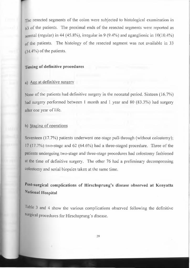
The resected segments of the colon were subjected to histological examination in
6 3 of the patients. The proximal ends of the resected segments were reported as
normal (regular) in 44 (45.8%), irregular in 9 (9.4%) and aganglionic in 10(10.4%)
of the patients. The histology of the resected segment was not available in 33
(34.4%) of the patients.
Timing of definitive procedures
a) Age at definitive surgery
None of the patients had definitive surgery in the neonatal period. Sixteen (16.7%)
had surgery performed between 1 month and 1 year and 80 (83.3%) had surgery
after one year of life.
b) Staging of operations
Seventeen (17.7%) patients underwent one-stage pull-through (without colostomy);
17 (17.7%) two-stage and 62 (64.6%) had a three-staged procedure. Three of the
patients undergoing two-stage and three-stage procedures had colostomy fashioned
at the time of definitive surgery. The other 76 had a preliminary decompressing
colostomy and serial biopsies taken at the same time.
Post-surgical complications of Hirschsprung’s disease observed at Kenyatta
National Hospital
Table 3 and 4 show the various complications observed following the definitive
surgical procedures for Hirschsprung’s disease.
29

Early complications
Out of the 96 patients who underwent surgery, only 17 (17.7%) developed
complications in the immediate post-operative period while 79 (82.3%) had no
complications. The main complications observed are shown in table 3.
Table 3
Complication No. of patients with
complication
Percentage
Wound Infection 8 8.3
Enterocolitis 4 4.1
Anastomotic leak 2 2.0
Pelvic abscess 3 3.1
Intestinal Obstruction 3 3.1
Wound dehiscence 2 2.0
Peritonitis 1 1.0
Some patients had more than one complication. Wound infection was the
commonest early complication
30
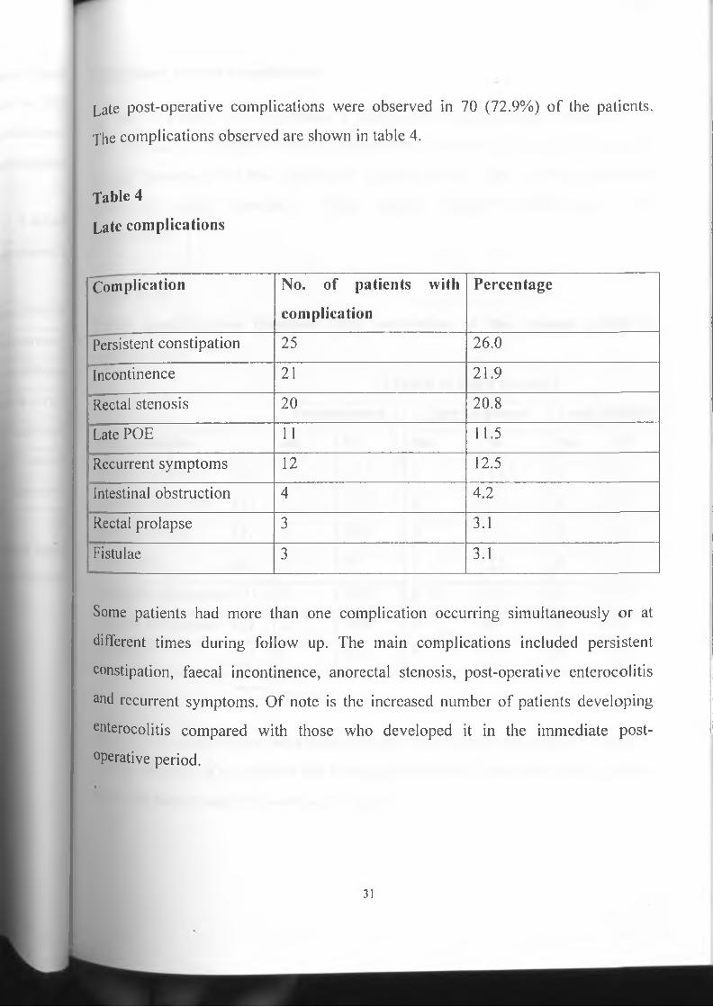
Late post-operative complications were observed in 70 (72.9%) of the patients.
The complications observed are shown in table 4.
Table 4
Late complications
"Complication No. of patients with
complication
Percentage
Persistent constipation 25 26.0
Incontinence 21 21.9
Rectal stenosis 20 20.8
Late POE 11 11.5
Recurrent symptoms 12 12.5
Intestinal obstruction 4 4.2
Rectal prolapse 3 3.1
Fistulae 3 3.1
Some patients had more than one complication occurring simultaneously or at
different times during follow up. The main complications included persistent
constipation, faecal incontinence, anorectal stenosis, post-operative enterocolitis
and recurrent symptoms. Of note is the increased number of patients developing
enterocolitis compared with those who developed it in the immediate post
operative period.
31

Colostomy related complications
Of the 76 patients who underwent a preliminary decompressing colostomy; 51
(67.1%) had no colostomy related complications. However, 20 (26.3%) developed
stomal prolapse, 3(3.9%) developed stomal stenosis and 2(2.6%) developed
colostomy stomal bleeding. Three patients required refashioning of the
colostomies.
Table 5 (a)
Early complications observed after correction of the various levels of
aganglionosis
Length of colon involved
rectosigmoid Short segment Long segment
Complications no. % No. % No. %
Wound infection (8) 7 87.5 1 12.5 0 0
Anastomotic leak (2) 2 100 0 0 0 0
Pelvic abscess (3) 3 100 0 0 0 0
Early POE (4) 3 75 1 25 0 0
Intestinal obstruction (3) 3 100 0 0 0 0
Wound dehiscence (2) 2 100 0 0 0 0
Peritonitis (1) 1 100 0 0 0 0
Most of the complications were observed after correction of rectosigmoid disease
though majority of the patients had rectosigmoid disease. Due to the small numbers
involved further statistical tests were invalid.
32

rfable 5(b)
Late complications observed after correction of the various levels of
aganglionosis
Length of colon involved
rectosigmoid short segment Long segment
Complications no. % No. % No. %
Ano-rectal stenosis (20) 18 90 1 5.6 1 5.6
Persistent constipation (25) 20 80 4 16 1 4
Incontinence (21) 13 61.9 7 33.3 1 4.8
Late POE (11) 9 81.8 2 18.2 0 0
Recurrent symptoms (12) 7 58.3 2 16.7 3 25.0
Intestinal obstruction (4) 3 75 1 25 0 0
Rectal prolapse (3) 3 100 0 0 0 0
Fistulae (3) 3 100 0 0 0 0
Majority of the complications occurred among those patients with rectosigmoid
disease. The statistical significance of this was however difficult to test because
both Chi-square and Fisher’s exact tests were invalid.
Twenty-five percent (25%) of recurrent symptoms were in the patients with long
segment disease. However, this represented a significantly large number of patients
(50%) among those with long segment disease and this was found to be statistically
significant ( p value = 0.01).
33

Table 6 (a)
Early complications observed against the major operative procedures
Surgical technique
Swenson’s Soave-Boley Myectomy
Complications no. % No. % No. %
Wound infection (8) 3 37.5 5 62.5 0 0
Anastomotic leak (2) 0 0 2 100 0 0
Pelvic abscess (3 ) 0 0 3 100 0 0
Early POE (4) 3 75 1 25 0 0
Intestinal obstruction (3) 2 66.7 1 33.3 0 0
Wound dehiscence (2) 1 50 1 50 0 0
Peritonitis (1) 1 100 0 0 0 0
The Soave-Boley procedure had a higher complication rate, compared with other
procedures. Since there were no complications noted among the myectomy group,
comparison was done between the Swenson’s and the Soave-Boley groups.The
Soave-Boley group had a higher complication rate compered with the Swenson’s
group but the differences for individual complications were statistically
insignificant (Fisher’s exact probability, p value >0.07 in all).
34

Late complications observed against the major operative procedures
Table 6 (b)
Surgical technique
Swenson’s Soave-Boley Myectomy
Complications no. % No. % No. %
Rectal stenosis (20) 9 45 11 55 0 0
Persistent constipation (25) 12 48 12 48 1 4
Incontinence (21) 11 52.4 10 47.6 0 0
Late PO E (11) 6 54.5 5 45.5 0 0
Recurrent symptoms (12) 8 66.7 3 25 1 8.3
Intestinal obstruction (4) 3 75 1 25 0 0
Rectal prolapse (3) 0 0 3 100 0 0
Fistulae (3) 0 0 3 100 0 0
Persistent constipation and faecal incontinence were almost equaly as frequent in
the Swenson’s and the Soave-Boley groups.
Recurrent symptoms were however commoner in the Swenson’s group, but the
statistical significance of this could not be evaluated because the Chi-square test
was invalid. WEDTCAL LIBRAR1UNIVERSITY OF NAIROBI
Rectal stenosis was commoner after the Soave-Boley procedure (55%) compared
with the Swenson’s procedure (45%). However, this was statistically insignificant
(Fisher’s exact probability, p value = 0.18).
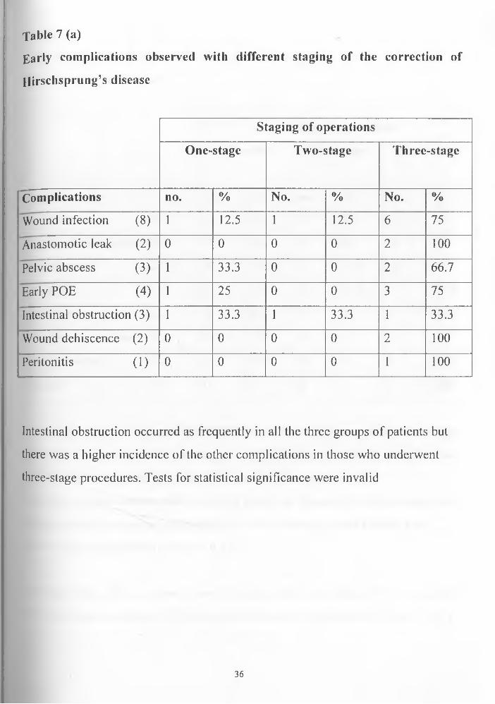
Table 7 (a)
Early complications observed with different staging of the correction of
Hirschsprung’s disease
Staging of operations
One-stage Two-stage Three-stage
Complications no. % No. % No. %
Wound infection (8) 1 12.5 1 12.5 6 75
Anastomotic leak (2) 0 0 0 0 2 100
Pelvic abscess (3) 1 33.3 0 0 2 66.7
Early POE (4) 1 25 0 0 3 75
Intestinal obstruction (3) 1 33.3 1 33.3 1 33.3
Wound dehiscence (2) 0 0 0 0 2 100
Peritonitis (1) 0 0 0 0 1 100
Intestinal obstruction occurred as frequently in all the three groups of patients but
there was a higher incidence of the other complications in those who underwent
three-stage procedures. Tests for statistical significance were invalid
36
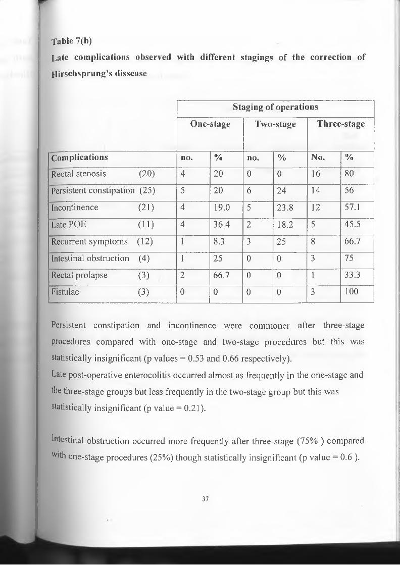
■Table 7(b)
Late complications observed with different stagings of the correction of
Hirschsprung’s dissease
Staging of operations
One-stage Two-stage Three-stage
Complications no. % no. % No. %
Rectal stenosis (20) 4 20 0 0 16 80
Persistent constipation (25) 5 20 6 24 14 56
Incontinence (21) 4 19.0 5 23.8 12 57.1
Late POE (11) 4 36.4 2 18.2 5 45.5
Recurrent symptoms (12) 1 8.3 3 25 8 66.7
Intestinal obstruction (4) 1 25 0 0 3 75
Rectal prolapse (3) 2 66.7 0 0 1 33.3
Fistulae (3) 0 0 0 0 3 100
Persistent constipation and incontinence were commoner after three-stage
procedures compared with one-stage and two-stage procedures but this was
statistically insignificant (p values = 0.53 and 0.66 respectively).
Late post-operative enterocolitis occurred almost as frequently in the one-stage and
the three-stage groups but less frequently in the two-stage group but this was
statistically insignificant (p value = 0.21).
Intestinal obstruction occurred more frequently after three-stage (75% ) compared
Wlth one-stage procedures (25%) though statistically insignificant (p value = 0.6 ).
37

fable 8(a)
Early complications observed against the histological reports of the proximal
ends of resected segments of colon
Histological reports
Regular Irregular AganglionicNot
available
Complication no. % no. % no. % no. %
Wound infection (8) 6 75 0 0 0 0 2 25
Anastomotic leak (2) 1 50 0 0 0 0 1 50
Pelvic abscess (3) 1 33.3 0 0 0 0 2 66.7
Early POE (4) 1 25 0 0 0 0 3 75
Intestinal obstruction (3) 1 33.3 0 0 1 33.3 1 33.3
Wound dehiscence (2) 2 100 0 0 0 0 I T 0
Peritonitis (1) 1 100 0 0 0 0 0 0
The histological reports of the proximal ends of resected segments bore no
relevance to the development of early complications and these occurred almost
randomly in those with regular histology and those in whom the reports were not
available.
38

Table 8 (b)
Late complications observed against the histological reports of the proximal
ends of resected segments of colon
Histological reports
Regular Irregular Aganglionic Not
available
Complication No. % No. % No. % No. %
Rectal stenosis (20) 12 60 1 5 2 10 5 25
Persistent constipation (25) 9 36 1 4 1 4 14 56
Incontinence (21) 11 52.4 3 14.3 1 4.8 6 28.6
Late POE (11) 4 36.4 2 18.2 1 9.1 4 36.4
Recurrent symptoms (12) 4 33.3 2 16.7 2 16.7 4 33.3
Intestinal obstruction (4) 3 75 0 0 0 0 1 25
Rectal prolapse (3) 0 0 0 0 1 33.3 2 66.7
Fistulae (3) 1 33.3 1 33.3 1 33.3 0 0
Persistent constipation occurred more frequently in those in whom the histological
reports were not available (56 %) compared with those in whom the reports were
available (44%). This was however not statistically significant (Fisher’s exact
probability, p value = 0.07).
Recurrent symptoms developed equally in those with regular histological reports
a'id those in whom the reports were not available (33.3%); and even in those in
whom it was reported as irregular and aganglionic combined. Though statistically
lnsignificant (p value = 0.4), it is concerning to note that in such a large proportion
39

of patients developing complications (33.3%), histological reports of proximal
ends of the resected segments were not available.
Post operative enterocolitis occurred as frequently in those with regular reports and
in those in whom the reports were not available. This is understandable bearing in
mind the multiplicity of factors involved in the pathogenesis of enterocolitis of
Hirschsprung’s disease.
Table 9 (a)
Early complications observed against age at definitive surgery
Age at definitive surgery
1 month - 1 year More than 1 year
Complications No. % No. %
Wound infection (8) 0 0 8 100
Anastomotic leak (2) 0 0 2 100
Pelvic abscess (3) 0 0 3 100
Early POE (4) 0 0 4 100
Intestinal obstruction (3) 1 33.3 2 66.7
Wound dehiscence (2) I T 0 T ~ 100
Peritonitis (1) 0 0 1 100
Most of the complications occurred in those operated on after 1 year of life though
majority (83.3%) of the patients were operated after 1 year of life.
40

Late complications observed against age at definitive surgery
Table 9 (b)
Age at definitive surgery
1 month - 1 year More than 1 year
Complications No. % No. %
Rectal stenosis (20) 2 10 18 90
Persistent constipation (25) 5 20 20 80
Incontinence (21) 2 9.5 19 90.5
Late P O E (11) 4 36.4 7 63.6
Recurrent symptoms (12) 2 16.7 10 83.3
Intestinal obstruction (4) 0 0 4 100
Rectal prolapse (3 ) 2 66.7 1 33.3
Fistulae (3) 0 0 3 100
Persistent constipation and incontinence occurred more frequently in those who
underwent definitive surgery at an age of more than one year (80% and 90.5%
respectively) compared with those who underwent definitive surgery at an earlier
age (20% and 19% respectively), though these were statistically insignificant (p
values = 0.4 and 0.2 respectively).
An appreciable percentage of late post-operative enterocolitis developed in those
who had surgery performed at an age of more than one year of life (63.6%)
compared with those who had surgery at an earlier age (36.4%) but this was
statistically insignificant (p value = 0.08)
41
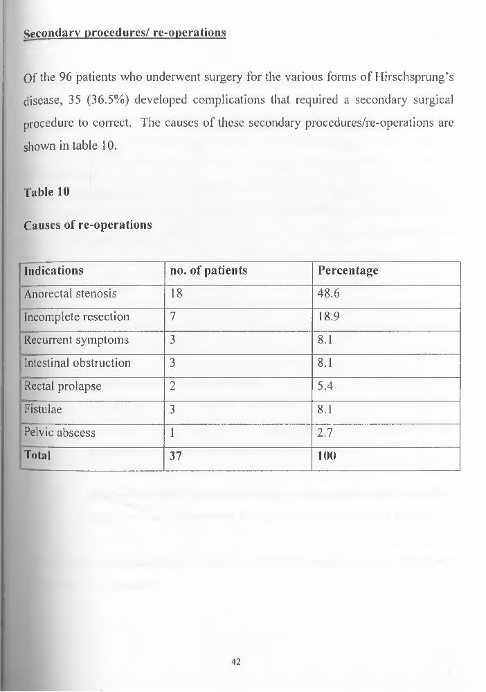
.Secondary procedures/ re-operations
Of the 96 patients who underwent surgery for the various forms of Hirschsprung’s
disease, 35 (36.5%) developed complications that required a secondary surgical
procedure to correct. The causes of these secondary procedures/re-operations are
shown in table 10.
Table 10
Causes of re-operations
Indications no. of patients Percentage
Anorectal stenosis 18 48.6
Incomplete resection 7 18.9
Recurrent symptoms 3 8.1
Intestinal obstruction 3 8.1
Rectal prolapse 2 5.4
Fistulae 3 8.1
Pelvic abscess 1 2.7
Total 37 100
42
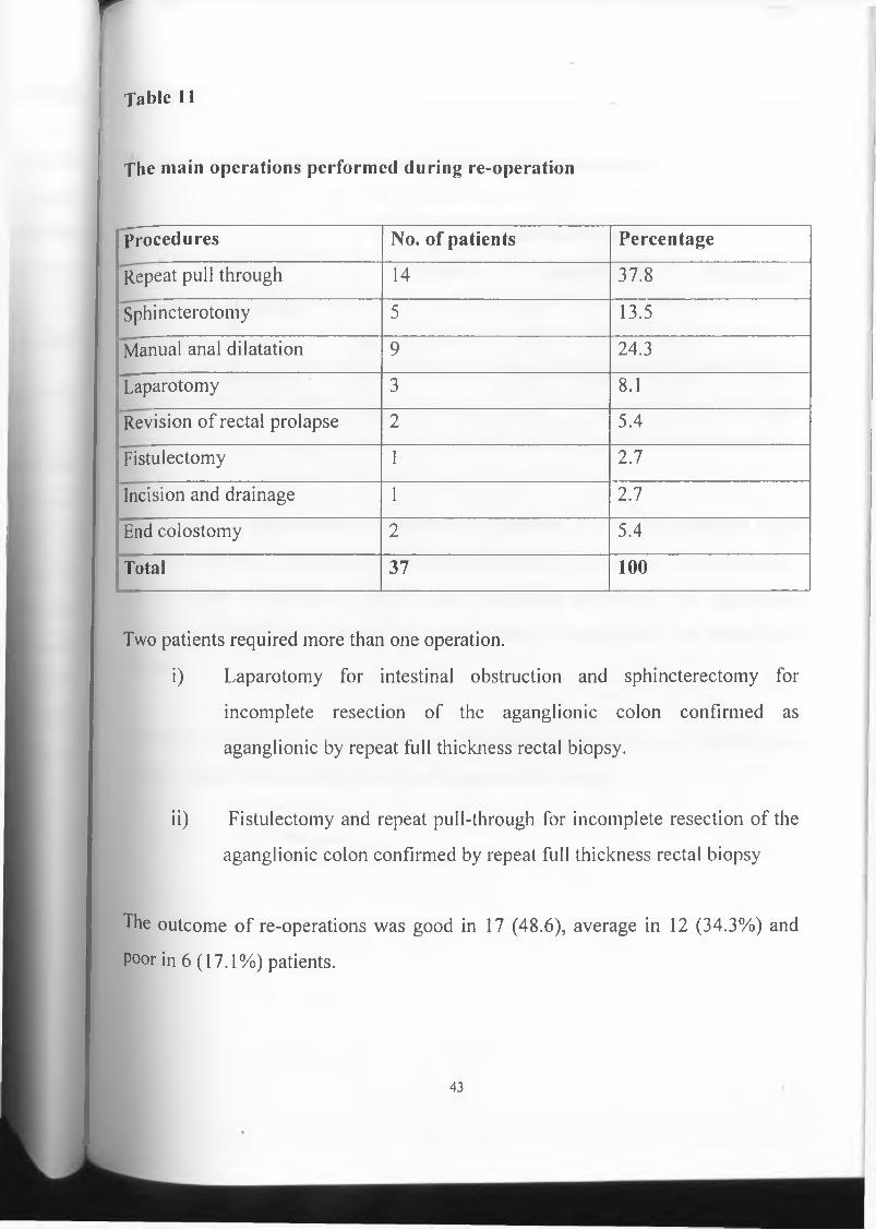
rTable 11
The main operations performed during re-operation
procedures No. of patients Percentage
Repeat pull through 14 37.8
Sphincterotomy 5 13.5
Manual anal dilatation 9 24.3
Laparotomy 3 8.1
Revision of rectal prolapse 2 5.4
Fistulectomy 1 2.7
Incision and drainage 1 2.7
End colostomy 2 5.4
Total 37 100
Two patients required more than one operation.
i) Laparotomy for intestinal obstruction and sphincterectomy for
incomplete resection of the aganglionic colon confirmed as
aganglionic by repeat full thickness rectal biopsy.
ii) Fistulectomy and repeat pull-through for incomplete resection of the
aganglionic colon confirmed by repeat full thickness rectal biopsy
The outcome of re-operations was good in 17 (48.6), average in 12 (34.3%) and
Poor in 6 (17.1%) patients.
43
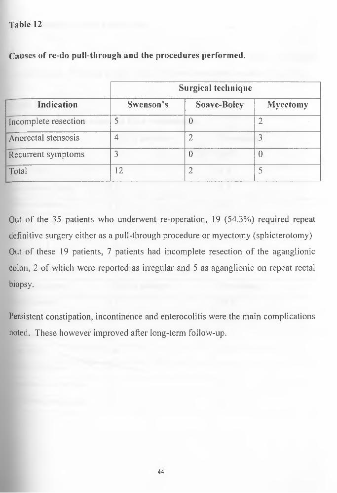
Table 12
Causes of re-do pull-through and the procedures performed.
Surgical technique
Indication Swenson’s Soave-Boley Myectomy
Incomplete resection 5 0 2
Anorectal stensosis 4 2 3
Recurrent symptoms 3 0 0
Total 12 2 5
Out of the 35 patients who underwent re-operation, 19 (54.3%) required repeat
definitive surgery either as a pull-through procedure or myectomy (sphicterotomy)
Out of these 19 patients, 7 patients had incomplete resection of the aganglionic
colon, 2 of which were reported as irregular and 5 as aganglionic on repeat rectal
biopsy.
Persistent constipation, incontinence and enterocolitis were the main complications
noted. These however improved after long-term follow-up.
44

Complications of re-do procedures
rNineteen patients underwent re-do procedures, 14 of whom developed
complications. The main complications observed are shown on table 13 (a & b).
Table 13 (a)
Early complications observed after re-operation
Complications No. of patients Percentage
Wound infection 5 26.3
Early POE 1 5.3
Anastomotic leak 1 5.3
Pelvic abscess 1 5.3
Table 13 (b)
Late complications observed after re-operation
Complications No. of patients Percentage
In co n tin en ce 7 36.8
P ersis ten t c o n s tip a tio n 3 15.8
F is tu lae -in -an o 4 21.1
Late POE 4 21.1
Rectal p ro la p se 1 5.3
Intestinal o b s tru c tio n 1 5.3
R ecurren t sy m p to m s 1 5.3
45
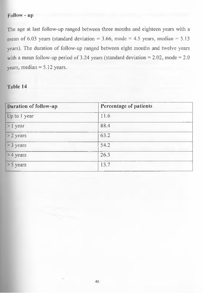
Follow - up
The age at last follow-up ranged between three months and eighteen years with a
mean of 6.03 years (standard deviation = 3.66, mode = 4.5 years, median = 5.13
years). The duration of follow-up ranged between eight months and twelve years
with a mean follow-up period of 3.24 years (standard deviation = 2.02, mode = 2.0
years, median = 5.12 years.
Table 14
Duration of follow-up Percentage of patients
Up to 1 year 11.6
> 1 year 88.4
> 2 years 63.2
> 3 years 54.2
> 4 years 26.3
> 5 years 13.7
46

Final outcome of management of Hirschsprung’s disease at Kenyatta National
Hospital
Table 15
Outcome Number of patients Percentage
Good 70 72.92
Average 21 21.87
Poor 5 5.21
Total 96 100
Among those with average outcomes the main complication was either residual
faecal incontinence or persistent constipation that were expected to improve on
continued follow-up. All those with poor outcomes needed another re-do
procedure.
47
DISCUSSION
Given the complexities of etiology and genetics, pathology and pathophysiology of
Hirschsprung’s disease it is not surprising that what appears to be well-defined and
anatomicaly based surgical therapy still leads to inconsistent and poorly
predictable outcomes. Complications after treatment for Hirschsprung’s disease
are not uncommon. Various factors influencing these outcomes can be found in
the literature including age of the child at the time of treatment, extent of the
disease, associated anomalies (eg Trisomy 21), the type of surgical procedure
performed with their associated complications and the age of the child at follow
up.
It is generally felt that re-establishment of continuity of the gastrointestinal tract at
an earlier age in childhood results in children having better control of defaecation
(30). Theoretically, early sensation of passage of feaces through the anal canal
should allow early reestablishment of the anorectal reflex and other neural curcuits
required for optimal control of defaecation (30).
Most of the patients (83.3%) had definitive surgery after one year of life and there
was no patient who underwent definitive surgery in the neonatal period. Overall
there was no significant difference in the complication rates among the age groups.
However, there was a higher rate of persistent constipation and incontinence in the
patients undergoing surgery at an age of more than one year. This is however
expected to improve with time on follow-up,(2I8 30).
Most (78.1%) of the patients had the rectosigmoid variant of Hirschsprung’s
disease while 15.6% had short segment and 6.3% had long segment aganglionosis.
48
None of the patients had total colonic involvement. The incidence of complications
was almost equal in all groups of patients. However almost half of the patients
with long segment disease had recurrent symptoms after seemingly thorough
surgical correction. The proximal end of the resected segment of colon was
reported as aganglionic in one but normal in the other two. At repeat biopsy the
patient with an earlier on aganglionic pulled through colon remained aganglionic
while the other two turned to be irregular. It was difficult to ascertain whether this
was due to pathological misreporting or truly cases of acquired aganglionosis,
which though rare has been documented (48).
The most common surgical procedure used was the Swenson’s pull-through
(56.3%). In an earlier report(ll), the commonest procedure employed in Kenyatta
National Hospital was the Soave-Boley endorectal pull-through and no patient
underwent the Swenson procedure. This was attributed to a change in the
preference of procedures used earlier due to associated complications and
surgeons’ preferences. In this study there was no significant difference in the
overall complications with the different surgical procedures. There was however a
higher incidence of pelvic abscesses and anorectal stenosis following the Soave-
Boley procedures than in other procedures.Recurrent symptoms were on the other
hand more frequent following the Swenson’s procedure while persistent
constipation and incontinence occurred almost as frequently following either the
Swenson’s or the Soave-Boley procedures. All the patients who developed rectal
prolapse had a Soave-Boley procedure.
In staged procedures colostomy related problems were frequently seen. Langer et
aI-(5I) described stoma related complications in 26% of their two stage procedures
when comparing the outcomes to a series of primary pull-throughs. In this study
49

32.9% of the patients undergoing staged procedures had colostomy related
complications mainly stomal prolapse in 26.3% and stomal stenosis (3.9%).
Indeed avoidance of an initial colostomy is one argument used to support primary
pull-through procedures. Persistent constipation, incontinence post-operative
intestinal obstruction developed more frequently following three-stage procedures.
Successful treatment of Hirschsprung’s disease is largely determined by adequate
resection of the aganglionic segment of colon as confirmed by the demonstration
of normal ganglion cells at the proximal end of the resected bowel. In 44 (45.8%)
of the patients the pull-through colon had normal ganglion cells. In 9 (9.4%) the
ganglion cells were present but reported as either immature, sparsely distributed or
distorted and in another 10 (10.4%) the pull-through end was reported as
aganglionic. In another 33% of the patients the histology reports were not
available.
Late post-operative enterocolitis occurred as frequently in those in whom the
histological reports of proximal ends of resected segments were regular (36.4%)
and in those in whom the reports were not available (36.4%).
Recurrent symptoms also occurred equally as frequently in those with regular
histological reports and those in whom the reports were not available (33.3%).
Though not statistically significant, this finding may seem to explain the need for
getting histology on all the resected segments.
Post-operative enterocolitis occurred even in those in whom the pulled through
colon had normal ganglion cells. Infact it was observed that the incidence of late
Post-operative enterocolitis was higher than that of early enterocolitis among the
Patients with normal pulled through colon.This could be explained by the
distention of the the neorectum that occurs with time after definitive surgery
50

resulting in faecal stasis and invasion by pathogenic organisms especially if there
is achalasia of the internal anal sphincter <22 23 2425». Furthermore, there are other
factors to explain the development of enterocolitis in Hirschsprung’s disease
including the relative immunodefficiency among those who develop enterocolitis (28)
Though there was a no significant difference in the complication rates in the
different categories of patients based on the histology of the proximal ends of the
resected segments, its worth noting that 73% of the patients who developed
complications were in the category with either an abnormal or absent histology.
It is interesting to note that among the ten patients in whom the pull-through colon
was reported as aganglioninc, only a half of them developed complications with
some patients developing more than one complication. Two developed recurrent
symptoms and repeat rectal biopsy confirmed incomplete resection of the
aganglionic segment and after repeat pull-through they did well. Another two had
repeat pull-through due to persistent constipation attributed to anorectal stenosis
and repeat biopsy for histology not performed. It would be expected that a repeat
rectal biopsy be performed in all those who had been reported as having
aganglionic pull-through ends to remove uncertainty about the credibility of the
histology reports but this was not the case in this study. Repeat biopsy was
performed in only 2 patients after they developed recurrent symptoms and it
actually did confirm incomplete resection.
Anastomotic leakage occurred in 2(2%) of the patients compared with 5.3% in the
Kleinhaus report (l7). Both had rectosigmoid disease and underwent 3-stage Soave-
51
Boley endorectal pull-through. Soave reported it in 1.1% and 5.8% in the Boley
modification (l7).
Though commonest in the Swenson’s procedure (l7) it was not reported in any of the
patients who underwent Swenson’s pull-through in this study inspite of this being
the preferred procedure. In one patient anastomotic leak led to the development of
pelvic abscess and later fistulae-in-ano accompanied by rectal prolapse and the
patient was managed with an end colostomy. In the other patient the leak closed
on conservative management but the patient later developed anorectal stenosis that
improved after serial manual anal dilatations.
The diagnosis of post operative enterocolitis at Kenyatta National Hospital was
mainly clinical defined as the occurrence of a clinical syndrome consisting of
diarhoea, fever, abdominal distention, cramping abdominal pain and lethergy. The
incidence of POE according to many reports ranges between 2% to 33% (l9,20,2,,22).
Kleinhaus <l7) reported that Swenson’s procedure was followed by a higher
incidence (15%) of POE and that the incidence was lower in the other procedures.
In this study early POE occurred in 4(4.1%) and lately in 11(11.5%) of the
patients. Early POE was commoner in the Swenson’s pull-through group
occurring in 75% compared with 25% in the Soave-Boley group.Late POE was
also commoner in the Swenson’s group (54.5%) compared with Soave-Boley
group (45.5% ). Overall there was no statistically significant difference noted
between the patients undergoing the different procedures.
There was no relationship between the development of POE and the staging of
surgery. It was however commoner among those undergoing pull-through at an
age of more than one year at the time of surgery. It was also commoner in
rectosigmoid disease and if the pulled-through colon had abnormal or no ganglion
52

cells. Overall however, the difference was either not statistically significant or
could not evaluated.
Though there are changing trends from delayed pull-through in infants to early
primary neonatal surgery the relative immuno-incompetence of the neonate and the
younger children may result in a more profound septic state compared to the older
child (30). This should raise some element of concern with primary surgical
correction in the neonatal period. Almost all the patients who developed
enterocolitis responded to medical management without need for surgical
intervention.
Twelve patients developed recurrent symptoms. Though commoner in patients
who underwent the Swenson’s procedure (66.7%), it was not related to the age at
definitive surgery or the staging of the operations. These occurred more frequently
in those in whom the histological reports were not available compared with the
group with regular reports. This could be explained by the presence of a retained
segment of aganglionic colon. Out of the twelve patients only seven had
histologicaly demonstrable retained aganglionic segment of colon. Two had
anorectal stenosis and three were due to unexplained reasons and were assumed to
be due to anorectal achalasia. However, all of these had a repeat pull-through.
Persistent constipation even after a seemingly successful definitive procedure can
be functional with spontaneous improvement on long term follow-up (2). It
occurred in 25 (26%) of the patients. Upon digital rectal examination 20 of these
•tad anorectal stenosis. The other five had functional constipation and on long-
lenn follow-up improved without need for surgical invention. Majority did well on
53

laxatives. Infact ano-rectal stenosis may not necessary have been the cause for the
constipation given that in some it persisted even after correction of the stenosis.
It occurred as frequently (48%) following both the Soave-Boley and Swenson’s
procedures. Conversely it was commoner in children undergoing surgery after one
year of age (80%) compared with the younger child (20%) and in three-staged
procedures.
Ano-rectal stenosis occurred in 20 (20.8%) of the patients. It was commonest after
the Soave-Boley pull-through (55%) compared with Swenson’s pull-through
(45%). Swenson <l8) reported it only 6.2% of his patients. Several series quote this
to be commonest following the Soave procedure<l7 22,29)’ while others quote this to
be commonest after Swenson’s pull-through t2). Eighteen of the patients needed a
secondary procedure because of either persistent constipation or recurrent
symptoms. Nine were put through a programme of serial manual anal dilatations
and improved. Six underwent repeat pull-through and three underwent myectomy.
The introduction of a programme of dilatations after wound healing may be
beneficial to avoid the development of a stenosing ring at the anastomotic line.
Faecal incontinence occurred in 21 (21.9%) of the patients. Swenson in his series
reported it in 13.3% of his patients. In this study the incidence of faecal
incontinence postoperatively was almost equal in those undergoing either
Swenson’s or Soave -Boley procedures. Destruction of the sensory nerves in the
rectum due to the extensive pelvic dissection undertaken in the Swenson’s
Procedure (2) and the small capacity of neorectum created in the endorectal pull-
through procedure <2029J4) are possible explanations of incontinence.Though
54
insignificant it was common after staged pull-through and in those undergoing
surgery after one year of life, may be due to re-establishment of gastro intestinal
continuity later. Assessed objectively majority of the patients will achieve normal
bowel function as they reach adulthood. In this study majority improved on
follow-up.
Post operative intestinal obstruction from adhesions, intususception or volvulus is
common to all the operative procedures (3). In this study it occurred in the early
post operative period in three patients, and in the late post-operative period in four
patients.lt occurred more commonly following three-stage procedures. Of these,
only three required laparatomy, the others being managed conservatively with
success. At laparotomy one had intususception and the other two had adhesions.
Pelvic abscess followed the correction of rectosigmoid disease in 3 patients. In
one it was a consequence of anastomotic leak following Soave-Boley pull-through.
The patient later developed fistulae-in-ano and rectal prolapse for which an end
colostomy was fashioned. He benefited later from a posterior sagittal
anorectoplasty (PSARP) and pull-through. The other two patients who developed
pelvic abscess after Swenson’s and Soave-Boley procedures respectively did well
on intravenous antibiotics with incision and drainage performed on only one of
them.
Three patients developed fistulae-in-ano all after a Soave-Boley pull-through.
Fistulectomy was performed in one. The other two had end colostomies fashioned.
One of the two later developed sepsis and intestinal obstruction and inspite of
'aparoptomy died of sepsis. The other had PSARP and pull-through later and did
well at follow-up though still incontinent of stool at the last follow-up visit.
55
Three patients developed rectal prolapse after Soave-Boley procedure. This is not
a commonly described problem in the literature.
Secondary Procedures
Thirty-five patients (36.5%) required surgery for the management of complications
arising after pull-through. Common indications for secondary procedures were
anorectal stenosis (48.6), incomplete resection of the aganglionic colon (21.6%),
intestinal obstruction (8.1%) and recurrent symptoms (5.4%)
Persistent constipation and faecal retention quite common as in other studies was
noted in 25 (26%) of the patients. Upon digital rectal examination, anorectal
stenosis was observed in 20 of these. Of these 20 patients, 18 had significant
stenosis requiring surgical intervention. Manual anal dilatation was successftil in
9, while 6 underwent repeat pull through procedure and 3 myectomy.
Twelve patients had recurrent symptoms of Hirschsprung’s disease. In 2 of these
anorectal stenosis was detected on digital rectal examination and after myectomy
had good outcomes. Repeat rectal biopsy was performed in only 7 of these and all
were noted to have an element of incomplete resection of the aganglionic colon.
The retained segment in some was quite short as shown by supplementary barium
enema examination. Two of these had favourable outcomes following myectomy
while 5 underwent a repeat pull-through procedure.
Another nine patients underwent a repeat pull-through procedure, six of them due
to anorectal stenosis and three due to recurrent symptoms.In those who developed
^current symptoms, digital rectal examination did not reveal anorectal stenosis. A
56

repeat full thickness rectal biopsy was not performed and yet a repeat pull-through
was performed. The recurrence of symptoms could have been explained as due to
achalasia of the internal anal sphincter. These would have benefited more from an
internal sphicterotomy as described by Kasai-et-al<39) without the need for a repeat
pull-through.
Overall nineteen redo procedures were performed but these were justified by
demonstration of aganglionosis in full thickness rectal biopsy in only seven. In
two of these redo procedures the outcome was poor.
The Swenson’s procedure was the preferred procedure in 12, Soave in 2 and
myectomy in 5 patients. Technically the Swenson’s procedure is regarded as easier
as it avoids the difficult submucosal disection in already scarred tissues expected in
a Soave procedure.
The outcome of re-do operations was good in 5 (26.3%) of the 19 patients. Re-do
operations in addition to being technically more difficult are associated with higher
complication rates. The main complication noted was faecal incontinence though
in a majority of the patients in resolved spontaneously.
Five patients had poor outcomes requiring additional surgical procedures to correct
these. One patient had myectomy after repeat full thickness rectal biopsy was
reported as aganglionic. He developed recurrent symptoms with persistent
constipation and recurrent bouts of enterocolitis. Sphincteromyotomy solved the
Problem. The other four patients had repeat pull-through procedures.
Unfortunately none had a repeat full thickness rectal biopsy prior to the repeat pull-
through. All had serious complications ranging from persistent constipation (2),
ano-rectal stenosis (1) fistulae-in-ano (3), and gangrene of the pulled through colon
57

(1). They all had repeat pull-through procedures, either as Swenson’s or Soave-
j3oley procedures. The outcomes of these additional pull-throughs were not
evaluated.
/U the end ol follow-up, 26(27.08%) of the patients had a poor or average outcome
the main complications being persistent constipation and soiling. The outcome
vvas however regarded as good in 70(72.92%) of the patients. Persistent
constipation and soiling however improve on long term follow-up but this requires
long periods of follow-up (2'30). In the study though, only 13.7% of the patients were
followed up for more than 5 years with a mean follow-up period of 3.2years.
58

CONCLUSIONS
The surgical management of Hirschsprung’s disease at Kenyatta National Hospital
as in many other centres is associated with a recurring pattern of complications and
at almost comparable rates. The types and rates of these complications cannot
however be associated with the length of colon involved, the surgical technique
employed in the correction of the disease, the age at definitive surgery or the
staging of the operations. Since patients are diagnosed at different ages in Kenyatta
National Hospital and there is no procedure that can be claimed to be superior to
the other in terms of risks of developing any of the many different post-operative
complications the choice of procedure to employ or the staging of operations,
regardless of the age of the patient should be at the discretion of the individual
paediatric surgeon.
Following a seemingly well performed operation a significant number of
specimens of the resected colon were not subjected to histological examination to
confirm that the pulled through colon is normal and therefore that there is adequate
resection. This creates confusion when complications arise and the decision to re
operate is then not fully justified. Repeat full thickness rectal biopsy would then
be of help coupled with other relevant clinical and radiological observations to
make a rational decision to re-operate. The rate of repeat rectal biopsy was notable
low. There should be a policy to make sure that all properly tagged specimens of
resected gut are subjected to histological examination and the report availed before
discharge of the child from hospital. Preferably the specimen should be examined
by a pathologist inclined towards paediatric gastroenterology to avoid certain
infusing reports such as distorted ganglion cells without a clear conclusion.
59

'
Some of the indications for repeat pull-through are not justified. Anorectal
stenosis was a common indication for repeat pull-through. Sphincteromyotomy
and/or incision of the stenosis ring may have sufficed especially consideringjhat
most of the patients presented with persistent constipation that could as well be
functional. It should be defined clearly what patients require repeat pull-through
especially noting the complications that accompany repeat pull-throughs.
60

REFERENCES
1. Shaw, H. W. (Josh Billings) His sayings.ImJo/w, D ., H a ze l, E ., R o sa lin d , F.
e t a l (ieds). M a cm illa n d ic tio n a ry o f q u o ta tio n s;2 n d edition , 1989 p g 17.
M a cm illa n P u b lish in g C om pany, N ew York.
2. William K. Sieber. Hirschsprung’s disease. \n :K en n e th , J. W., Ju d so n ,G . R.,
M a rk , M . R .{eds). P a ed ia tr ic S u rgery; 4 th E dition , 1986, Vol. 2, C h a p ter 106
P g 9 9 5 -1 0 2 0 .Y ear B o o k M ed ica l P u b lish ers ,C h ica g o .
3. Tobon, F., Nigel, C. R., Reed, W., et. al. Non-surgical test for the diagnosis
of Hirschsprung’s disease. N . Engl. J. M ed. 1968, 278:188 .
4. Shackelford, R. T., Zuidema, G. D. Congenital aganglionosis
(Hirschsprung’s disease; megacolon) In:R ichard ,T .S ., G eorge,Z .D .
(eds.) S u rg e ry o f the a lim en ta ry tract, 2 n d edition , 1982, ch a p te r 5
P g 75- 90. W. B S a u n d ers,P h ila d e lp h ia .
5. Swenson, O., Sherman, J. O., Fisher,J. H. Diagnosis of congenital
megacolon. An analysis of 501 patients. J. P aed ia tr. Surg. 1973,
8:58 .
6. Bill, A. H.,Chapman, N. D.The enterocolitis of Hirschsprung’s disease
its natural history and treatment. Am . J. Surg. 1962, 103:70,.
61
7. Dobbins, W. O., Bill, A. H. Diagnosis of Hirschsprung’s disease excluded
by rectal suction biopsy. N. E ngl. J. M ed. 1965, 272: 990.
8. Smith, B. Pre and post-natal development of the ganglion cell of the rectum
and its surgical implication. J. Pciediatr. Surg. 1968, 3: 386.
9. Ramesh, J. C., Ramanujamu, T. M., Yik, Y. T., et al. Management of
Hirschsprung’s disease with reference to one stage pull-through without
colostomy. J. P eadia tr. Surg . 1999, 34 (11): 1691 -94.
10. Langer, J. C.,Fitzgerald,P. C.,Winthrop,A. L.et al. One-stage versus two-
stage pull-through in the first year of life. J. P aed ia tr. Surg. 1 9 9 6 ,3 1 :3 3 -3 7
11. Sidler, D., Miller, A. J., Rode, H., et al. Neonatal Soave endorectal pull-
through for Hirschsprung’s disease. S. A fr. J. Surg . 1999, 37: 4 7 -50.
I
12. Barrack, S. M. A fifteen year study of Hirschsprung’s disease at Kenyatta
National Hospital 1974 -1988. M. M ed. Surg. D isserta tion , U .O .N , 1990.
13. Swenson, O., Bill, A. H. Resection of rectum and restosignoid with
preservation of the sphincter for benign spastic lession producing
megacolon. An experimental study. Surgery. 1948 ,24:212 -220 .
14. Duhamel, B. Rectorectal and transanal pull-through procedure for the
treatment of Hirschsprung’s disease. D is. C olon R ectum . 1964, 7:
4550 .
62
15. Soave F. Hirschsprung’s disease. A new surgical technique.
A rch . D is. C h ild 1964, 39:116 .
16. Rehbein, F., Morger, R . , Knudert, J. G.,et al. Surgical problems in congenital
megacolon(Hirschsprung’s disease). J. P aed ia tr. Surg. 1966,1: 526.
17. Kleinhaus, S., Boley, S. J., sheran, M., et. al. Hirschsprung’s
disease. A survey of the members of the surgical section of the
America Academy of Paediatricians. J. P aed ia tr. Surg. 1979,
1 4 .5 8 8 - 5 9 7 .
18. Swenson, O., Sherman, J.O., Fisher, J. FI., et. al. The treatment and
post-operative complications of congenital megacolon .A 25yr follow-
up.A nn. Surg . 1975, 1 8 2 :2 6 6 -2 7 3 .
19. Engum, S. A., Grosfeld, J. L. Hirschsprung’s disease: Duhamel pull-through;
In: S tringer, M. D., o /dham , K. 7 , M ouriguand , P .D . E. e t al.
(ed s)P a ed ia tr ic su rg ery a n d uro logy: L o n g Term O utcom es. 1st edition,
1998, p p 32 9 - 339. W. B. Saunders,L o n d o n .
20. Tariq, G. M., Brereton, R. J., Wright, V. M. Complications of endorectal
pull through for Hirschsprung’s disease . J. P aed ia tr . Surg. 1991, 26: 1202 -
1206.
63

21. Blane, C. E., Elhalaby, E., Coran A. G. Enterocolitis following
endorectal pull- through in children surgery for Hirschsprung’s
diseasq.Pa ed ia tr ic s 1995, 96: 118 -1 2 1 .
22. Marty, T. L., Matlak, M. E., Hendrickson, M. et al.Unexpected death from
enterocolitis after surgery for Hurschsprung’s disease. P a ed ia tr ic s 1995,
96:118121 .
23. Skinner, M. A. Hirschsprung’s disease: current problems. Surg . 1996, 33:
39 4 - 460.
24. Elhalaby, E. A., Coran, A. G., Blane, C., et. al. Enterocolitis associated with
Hirschsprung’s disease. A clinical - radiological characterisation based on
168 patients. J. P aedia tr. S u rg 1995, 30: 76 - 83.
25. Fortuna, R. S., Weber, T. R., Tracy, T. F, Jnr, et. al. Critical
analysis of the operative treatment of Hirschsprung’s disease. A rch.
Surg. 1996, 131: 5 2 0 - 525.
26. Blair, G. K., Murphy, J. J., Fraser, G. C. Internal sphincterotomy in post
pull through Hirschsprung’s disease. J. P a ed ia tr surg. 1996, 3 1 :8 4 3 - 845.
27. Weber, T. R., Fortuna, R. S., Silen, M. L., et al. Re- operation for
Hirschsprung’s disease. J. P aed ia tr. Surg. 1999, 34: 153 - 157.
64

28. Wilson,G., Storey, D., Scobie, W. G. Impaired gastrointestinal
mucosal defense in Hirschsprung’s disease: a clue to the pathogenesis
of enterocolitis. J. p a ed ia tr . Surg. 1989, 24: 462 - 464.
29. Moore, S. W., Albertyn, R., Cywes, S. Clinical outcomes and long
term quality of life after surgical correction of Hirschsprung’s
disease. J. P aed ia tr. Surg . 1996, 31: 1496 - 1502.
30. Natalie, L.Y., Pierre, S. Long-term outcome after Hirschsprung’s
disease: patients' perspectives. J. P eadia tr. Surg . 1999, 34: 1152-
1160. W. B. Sa u n d ers.
31. Mary, T. L., Seo, T., Matlak, M. E., et al. Gastrointestinal
function after surgical correction of Hirschsprung’s disease: long term
follow-up in 135 patients. J. P aed ia tr. Surg. 1995, 30: 655.
32. Rescorla, F. J., Morrison, A. N., Eugles, D., et.al.Hirschsprung’s
disease: evaluation of mortality and long term function in 260 cases.
A rch . Surg . 1992, 127: 9 34 - 942.
33. Davies, M. R. Q., Cywes, S. Inadequate pouch emptying following Martins
pull-through procedure for intestinal aganglionosis. J. P ead ia tr . surg . 1983,
18: 14- 20.
34. Khan, O., NixonH. H. Results following surgery for Hirschsprung’s
disease: a review of three operations with reference to neorectal
capacity. Br. J. surg. 1980, 67: 43 6 - 438.
65
35. Velcek, F. T., Klotz, D. H., Friedman, A., et. al. Operative failure
and secondary repair In Hirschsprung’s disease. J. P a ed ia tr surg.
1982, 17: 719 - 785.
36. Langer, J. C. Repeat pull-through surgery for complicated
Hirschsprung’s disease : indications, techniques and results. J.
P ead ia tr . surg. 1999, 34 (7): 1136 - 41.
37. Foster, P., Cowan, G., wren, E. L. Twenty- five years experience
with Hirschsprung’s disease. J. P a ed ia tr surg. 1990, 25: 5 31 - 534.
38. Joseph, V. T., Sim, C. K. Problems and pitfalls in the management
of Hirschsprung’s disease. J. p a ed ia tr . Surg. 1988, 23: 39 8 - 402.
39. Kasai, M., Suzuki, H., Watanabe, K. Rectal myotomy with
colectomy: a new radical operation for Hirschsprung’s disease. J.
P aed ia tr. Surg. 1971, 6 :3 6 - 4 1 .
40. Lawson, J. O. N., Nixon, H. H. Anal canal pressures in the diagnosis
of Hirschsprung’s disease. J. P aed ia tr. Surg. 1967, 2: 544 - 552.
41. Marks, R. M. Endorectal split sleeve pull-through procedure for
Hirschsprung’s disease. Surg. G ynecol. O bste t.1973 , 136: 62 7 - 728.
66

42. Kimura, K., Inomata, Y., Soper, R. T. Posterior sigittal myectomy
for persistent rectal achalasia after the Soave procedure
Hirschsprung’s disease. J. P adia tr. Surg. 1993, 28: 1200 - 1201.
43. Takeseii, M., Atsuyuki, y., Masahiku, U., et. al. Modified Soave
pull-through for Hirschsprung’s disease ;intra operative sphincterotomy.
J. P aed ia tr . Surg. 1999, 34: 15999-1602 .
44. Schulten, D., Holschneider, A. N., Meier - Ruge, N. Proximal
segment histology of resected bowel in Hirschsprung’s disease
predicts post-operative bowel function. Eur. J. p a ed ia tr . Surg . 2000,
10(6) : 3 7 8 -8 1 .
45. Strugel, G. Extensive intestinal aganglionosis including ileum:
a new surgical technique. J. P eaditr . Surg. 1986, 21: 6 6 7 - 670.
46. Ikeda, K., Goto, S. Total colonic aganglionosis with or without small
bowel involvement: an analysis of 137 patients. J. p a ed ia tr . Surg . 1986,
21 : 3 1 9 - 3 2 2 .
47. Wilcox, D. T., Kiely, E. M. Repeat pull-through for Hirschsprung’s
disease. J. pa ed ia tr . Surg. 1998, 33: 1507 - 1509.
48. West, K. W., Grosfeld, J. C., Rescorla, F. J., Vane, D. W.Acquired
aganglionosis, a rare occurrence following pull-through procedure for
Hirschsprung’s disease. J. p a e d ia tr surg. 1990, 25: 104 - 108.
67

49. Banani, S. A., Forootan, H. R., Kumar, P. V. Intestinal neuronal
dysplasia as a cause of surgical failure in Hirschsprung’s disease
new modality of surgical treatment. J. p a ed ia tr . Surg. 1996, 31
5 72- 4.
50. Pena, A. Paediatric surgical problems. In: C orm an, M. L., (ed)
C olon a n d R ec ta l Surgery . 3 rd E d itio n ,1993, C h a p ter 8 P g - 332
346. L ip p in co t-R a ven P ub lishers, P h iladelph ia .
68
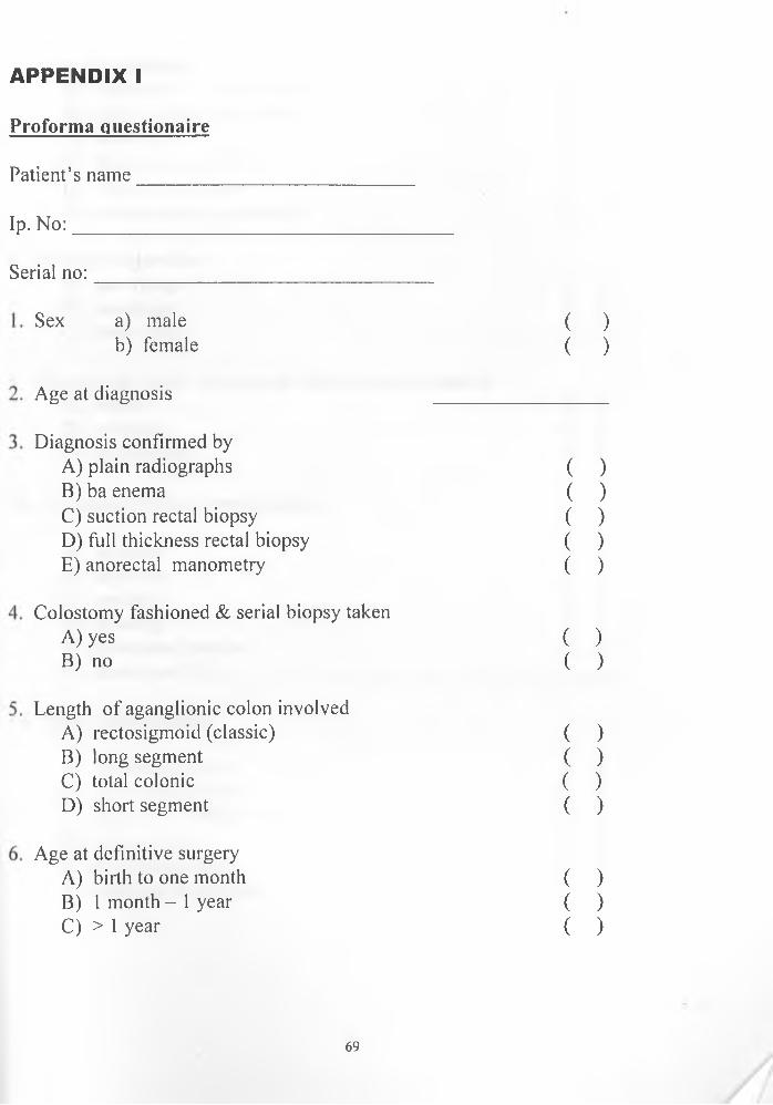
APPENDIX I
Proforma questionaire
Patient’s nam e_______
Ip. N o :______________
Serial no:
Sex a) male ( )b) female ( )
Age at diagnosis
Diagnosis confirmed byA) plain radiographs ( )B) ba enema ( )C) suction rectal biopsy ( )D) full thickness rectal biopsy ( )E) anorectal manometry ( )
Colostomy fashioned & serial biopsy takenA) yes ( )B) no ( )
Length of aganglionic colon involvedA) rectosigmoid (classic) ( )B) long segment ( )C) total colonic ( )D) short segment ( )
Age at definitive surgeryA) birth to one month ( )B) 1 month - 1 year ( )C) > 1 year ( )
69

7. Type of operationA) swenson’s procedure ( )B) soave - boley procedure ( )C) myectomy ( )D) duhamel procedure ( )E) martin’s procedure ( )F) rehbein-state’s operation ( )
8. Staging of operationA) one - stage ( )B) two-stage ( )C) three-stage ( )
9. Histological report of proximal end of resected segmentA) regular ( )B) irregular ( )C) aganglionic ( )
10. Colostomy related complications
A) prolapse ( )B) retraction ( )C) stenosis ( )D) bleeding ( )E) parastomal hernias ( )F) others specify_________________________________________
11. Early complications
A) wound infections ( )B) wound dehiscence ( )C) anastomotic leak ( )D) pelvic abcess ( )e) early enterocolitis ( )F) intestinal obstruction ( )
70

12. L a te c o m p l i c a t i o n s
A) late post-operative enterocolitis ( )B) persistent constipation ( )C) faecal incontinence/soiling ( )D) rectal prolapse ( )E) recurent rectal septum ( )F) fistulae ( )g) rectal stenosis/stricture ( )H) recurrent symptoms ( )I) intestinal obstruction ( )
Secondary procedures needed
A) yes ( )B) no ( )
Histology report of repeat biopsy if done
A) regular ( )B) irregular ( )C) aganglionic ( )
Indications for secondary procedure
A) ano-rectal stenosis ( )B) persistent constipation ( )C) retained aganglionic sement ( )D) recurrent symptoms ( )E) fecal incontinence ( )F) fistulae ( )G) rectal prolapse ( )H) recurrent rectal septum ( )I) intestinal obstruction ( )
71

16. T y p e o f p r o c e d u r e p e r f o r m e d
A) laparatomy for intertinal obstruction ( )B) myectomy ( )C) division of rectal septum ( )D) revision of rectal prolapse ( )E) fistulectomy ( )F) repeat pull-through procedure ( )G) others (specify)
17. Type of pull-through performed in seconday procedure
A) swenson’s procedure ( )B) soave-boley procedure ( )C) Myectomy/sphincterotomy ( )D) Duhamel procedure ( )D) rehbein-state’s operation ( )E) others (specify)
18. Early complications
A) wound infection ( )B) wound dehiscence ( )C) anastomotic leak ( )C) pelvic abscess ( )D) early enterocolitis ( )E) intestinal obstructionF) others (specify)
( )
72
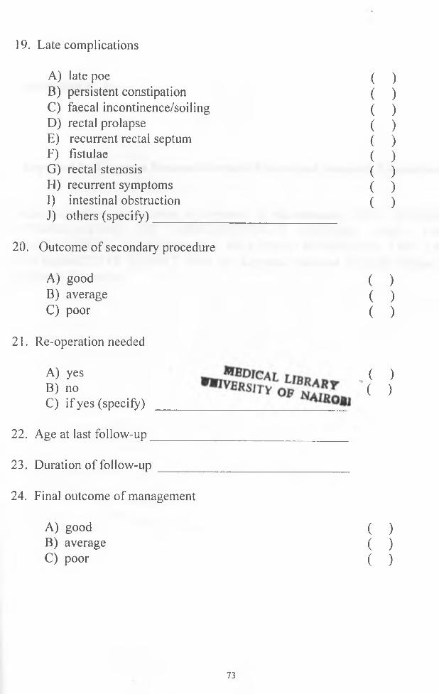
19. L a t e c o m p l i c a t i o n s
A) late poe ( )B) persistent constipation ( )C) faecal incontinence/soiling ( )D) rectal prolapse ( )E) recurrent rectal septum ( )F) fistulae ( )G) rectal stenosis ( )H) recurrent symptoms ( )I) intestinal obstruction ( )J) others (specify)
20. Outcome of secondary procedure
A) goodB) averageC) poor
21. Re-operation needed
A) yesB) noC) if yes (specify)
( ) ( ) ( )
. ( ) ( )
22. Age at last follow-up
23. Duration of follow-up
24. Final outcome of management
A) good ( )B) average ( )C) poor ( )
73

APPENDIX II
Approval by Kenyatta National Hospital Ethical and Research Committee
Attached below is the letter of approval of the research “POST SURGICAL COMPLICATIONS OF HIRSCHSPRUNG’S DISEASE AND THEIR MANAGEMENT AT KENYATTA NATIONAL HOSPITAL:A TEN YEAR RETROSPECTIVE STUDY” from the Kenyatta National Hospital Ethical and Research Committee.
74

Tel: 726300-19 726550 - 9 726562 - 6 726450 - 9 726581 -2
Fax: 725272
KENYATTA NATIONAL HOSPITAP.O. Bo v 20723,NAIROBI.
Email: k n h ( a h e a l t h n e t o r .k e
Ref: K N H - E R C / 0 1 / 1 3 5 6 16 M a y 2002
Dr. F. N j o r o g e K u r i a Dept, of Surgery F a c u l t y of M e d i c i n e U n i v e r s i t y of N a i r o b i
Dear Dr. Kuria,
RESEARCH PROPOSAL " C O M P L I C A T I O N S F O L L O W I N G SURG I C A L M A N A G E M E N T OF H I R S C H S P R U N G S D I S E A S E IN K E N Y A T T A N A T I O N A L HOSPITAL: A TEN Y E A RR E T R O S P E C T I V E STUDY"_______________________________________________ (P13/1/2002)
This is to info r m you that the K e n y a t t a N a t i o n a l H o s p i t a l E t h i c a l and R e s e a r c h C o m mittee has revi e w e d and a p p r o v e d the r e v i s e d v e r s i o n of your above cited r e s e a r c h proposal.
O n behalf of the Co m m i t t e e I w i s h you fruitful r e s e a r c h and look forward to re c e i v i n g a s u mmary of the r e s e a r c h findings u p o n compl e t i o n of the study.
This i n f o r m a t i o n w i l l form part of da t a base that w i l l be c o n sulted in future w h e n p r o c e s s i n g r e l a t e d r e s e a r c h study so as to m i n i m i z e chances of study duplication.
T h a n k you.
Y o u r s faithfully,
c.c. Prof. K.M. Bhatt,Chairman, KNH-ERC,Dept, of Medicine, U0N.
D e p u t y D i r e c t o r (CS),K e n y a t t a N. Hospital.S u p e r v i s o r s : Prof. G.A.O. Magoha, Dept, of Surgery, UON
Mr. J.M. Ndungu, Dept, of Surgery, UONThe Chairman, Dept, of Surgery, UON The Dean, F a culty of Medicine, UON