powerpoint presentation · • scope of iris analysis as a health assessment tool • iris analysis...
TRANSCRIPT
HMCL312
www.endeavour.e
du.au
Session 2
Clinical Skills 2
Naturopathic Medicine Department

© Endeavour College of Natural Health www.endeavour.edu.au 2
Iris Analysis: IntroductionSession Summary
• Scope of iris analysis as a health assessment tool
• Iris analysis & Naturopathic principles, Therapeutic Order and
Process of Disease and Health
• Anatomy of the eye and iris zones

© Endeavour College of Natural Health www.endeavour.edu.au 3

© Endeavour College of Natural Health www.endeavour.edu.au 4
Iris Analysis as a Health
Assessment Tool
Iris analysis/Iridology – the study & analysis of
the neuro-optic reflex, which is thought to have
the potential to reveal disharmonies in the body
ranging from pathological, structural and
functional to psychological & emotional.
Sarris, J & Wardle, J 2010 Clinical Naturopathy

© Endeavour College of Natural Health www.endeavour.edu.au 5
Iris Analysis as a Health
Assessment Tool
Structural patterns & colours in the iris and
indicators in the sclera of the eye can indicate:
• Inherent strengths & weaknesses of systems,
organs and tissues in the body
• Assimilation of nutrients, toxicity,
inflammation, circulatory & lymphatic
irregularities

© Endeavour College of Natural Health www.endeavour.edu.au 6
Iris Analysis as a Health
Assessment Tool
• Iridology may help to determine pathological,
structural, functional & emotional disturbances
& predispositions within the individual.
• Chemical (physiological) & structural
(anatomical) changes to body tissues are
observable in the iris

© Endeavour College of Natural Health www.endeavour.edu.au 7
Contrasting Iris Structures
How would you describe the differences of appearance of
these two irises?

© Endeavour College of Natural Health www.endeavour.edu.au 8
• An analytical tool for gathering information about an
individual’s condition of health & wellbeing
• A means of determining the reflex conditions of body
tissues, organs & systems
• A means of identifying areas of inflammation,
congestion & xenobiotic accumulations
• A tool for determining constitutional strengths &
weaknesses
Iris Analysis as a Health
Assessment Tool

© Endeavour College of Natural Health www.endeavour.edu.au 9
Tissue changes in the body are reflected in the iris
Iris analysis:
• Provides an insight into the development of sub-acute
& chronic health conditions
• Provides a way of gauging the efficacy of
treatment/prevention of health conditions (acute, sub-
acute, chronic, degenerative)
Iris Analysis as a Health
Assessment Tool

© Endeavour College of Natural Health www.endeavour.edu.au 10
Process of Disease and
HealingNormal Health
Disturbing Discharge
Factors Process
Disturbance of Function
Reaction
(fever, inflammation, etc.)
Chronic Reaction (Structure Disturbance)
Degeneration
Adapted from Zeff, J.L., Snider, P., & Myers, S. (2006)
Acute
Sub-Acute
Chronic
Degenerative

1. Establish the Conditions for Health
By addressing the Determinants of Health:
a) Identify and remove disturbing factors (obstacles to cure)
b) Institute a more healthful regimen
2. Stimulate the Vis Medicatrix Naturae
3. Tonify Weakened Systems
4. Correct Structural Integrity
5. Address Pathology:
a) Natural Substances
b) Pharmacologic or Synthetic Substances
6. Suppress or Surgically Remove Pathology
NATUROPATHIC THERAPEUTIC ORDER

© Endeavour College of Natural Health www.endeavour.edu.au 12
o Iris analysis is also a means of making meaningful contact
with clients
• making a connection
• assisting to establish a client-practitioner (therapeutic)
relationship
o Compliance is usually improved where some form of
diagnostic/analytical technique/method is incorporated in a
consultation or treatment – particularly where that technique
may be used to monitor progress
Iris Analysis as a Health
Assessment Tool

© Endeavour College of Natural Health www.endeavour.edu.au 13
Two approaches to utilising iris analysis in clinical practice:
1. “Diagnostic” approach - conduct iris analysis before anything
else & inform client of findings
2.Analytic approach - take the case, gain information about
client’s health condition; then conduct iris analysis & look for
indications that provide information about their health
condition (e.g constitution, tissue strengths and weaknesses,
areas of possible pathology)
• What iris reveals will suggest possible lines of enquiry
to follow for further investigation
Iris Analysis as a Health
Assessment Tool
© Endeavour College of Natural Health www.endeavour.edu.au 14
o Rather than using iris analysis as a definitive diagnostic
tool/method, use it to gain more information that will help
refine your health analysis and better understand the client’s
health from a holistic perspective.
o Rule of Three – look for 3 examples of evidence before
finalising your health analysis of a client
• e.g. iris signs/symptoms & signs of pathology; nail analysis;
tongue analysis; clinical examination/path. lab test results;
systematic & comprehensive case taking/interviewing
Iris Analysis as a Health
Assessment Tool
© Endeavour College of Natural Health www.endeavour.edu.au 15
Historical Background
• The science and practice of iris analysis is not new.
The oldest records uncovered thus far have shown
that a form of iris interpretation was used in ancient
China as far back as 1,000 BC.
• In 1670 the physician Philippus Meyens, in his
book, ‘Chiromatica Medica’, described the division
of the Iris, according to body regions.
• A quote from the Viennese ophthalmologist Dr.
Beer, in his publication of 1813, ‘Textbook of Eye
Diseases’, states that; "Everything that affects the
organism of an individual cannot remain without
effect on the eye, and vice versa."

© Endeavour College of Natural Health www.endeavour.edu.au 16
Historical Background• In 1881 an Hungarian physician, Dr. Ignaz von
Peczely, published ‘Discovery in Natural History
and Medical Science: A Guide to the Study and
Diagnosis from the Eye’ which achieved
international renown. Von Peczely is considered the
father of modern iridology/iris analysis.
• In modern times, doctors and scientists, primarily
from Europe (Josef Deck & Josef Angerer) and
the United States (Dr. Bernard Jensen, David
Pesek, Denny Johnson) have further brought iris
analysis/iridology into worldwide recognition. Refer to prescribed reading: Jensen, B 1982, History
of iridology and chart development

Dr Ignatz
von Peczely
www.iridology.ie/globaliridologyresearch.org
www.altertv.org/tv

Nils Liljequistwww.iridology.ie
www.iridologycollege.org
Pastor Felkewww.felke-institut.de
Dr John Christopherwww.schoolofnaturalhealing.com
Josef Angererwww.ausbildung-zum-heilpraktiker.de

© Endeavour College of Natural Health www.endeavour.edu.au 19
Other Pioneers of Iris
Analysis
o Rudolph Schnabel (1952)
o Theodore Kreige (1969)
o Bernard Jensen (1974)
o Jim Jenks (1978)
o Dorothy Hall* (1980)
o Denny Johnson (1984 -Rayid Iridology)
o David Pesek (1990s -)
o Father Robert Felke(1856-1922)
o Johannes Theil (1905)
o Nils Liljequist (1911)
o Dr. Edward Lahn (1914)
o Dr. Collins (1918)
o Dr. Henry Lindlahr (1919)
o Dr. Haskel Kritzer (1924)
* Hall was an Australian Naturopath/Herbalist who developed
her own approach to iris analysis

© Endeavour College of Natural Health www.endeavour.edu.au 20
Bernard
Jensen
Dorothy Hall
David
Pesek
Denny
Johnson

© Endeavour College of Natural Health www.endeavour.edu.au 21
Iris Analysis – the evidence-base
1. Research-based evidence
• 26 references to iridology in science literature
• 1 is a literature review
• 13 are clinical studies
• others are updates or editorials
(naturalstandards.com/databases)

© Endeavour College of Natural Health www.endeavour.edu.au 22
Iris Analysis – the evidence-base
•Preliminary studies suggest iridology may assist in identification of individual
predispositions for vascular diseases (hypertension)
•Preliminary study shows a correlation between iris signs and individuals with
Diabetes mellitus
•Limited available data supporting iridology as a diagnostic tool in cancer
•Preliminary study suggests no evidence supporting iridology for diagnosis of
gallbladder disease
•Preliminary study suggests no evidence supporting iridology as diagnostic tool in
kidney disease
(naturalstandards.com/databases)

© Endeavour College of Natural Health www.endeavour.edu.au 23
Iris Analysis – the evidence-base
2. Empirical/Traditional evidence
• in use for over 3000 years
• over 300 years use as a systematised approach to health analysis
• majority of the iris charts currently in use are consistent
• significant anecdotal evidence of its clinical efficacy
o Well-designed and appropriate research studies are required to determine
the research-based evidence for iris analysis.
o Iris analysis is an officially accepted diagnostic method in the former Soviet
Union, Belarus & South Korea
Sarris, J & Wardle, J 2010 Clinical
Naturopathy

emedtravel.files.wordpress.com/2011
Basic Eye Anatomy

© Endeavour College of Natural Health www.endeavour.edu.au 25
Basic Eye Anatomy
• Conjunctiva: Membrane overlaying the eyeball
and inside of the eyelids
• Cornea: Membrane overlaying the pupil and the
iris
• Pupil: Aperture through which light enters the eye
• Iris: Coloured portion of the eye; consists of two
layers: the front pigmented fibrovascular known as
a stroma and, beneath the stroma, pigmented
epithelial cells
• Sclera: Outer, tough layer of the eye. The ‘white
of the eye’

© Endeavour College of Natural Health www.endeavour.edu.au 26
Basic Eye Anatomy
• Choroid: Middle, blood-rich layer of the eye
• Retina: Inner, light-sensitive layer of the eye
• Lens: Sits behind the pupil and refracts light
onto the retina
• Aqueous Humor & Vitreous Humor:
Viscous fluids that fill the chambers of the
eye

© Endeavour College of Natural Health www.endeavour.edu.au 27
Basic Eye Anatomy
Iris anatomy:• Iris is comprised of layers of tissues
– 4 main layers
o Anterior endothelium – single
layer of flattened cells located at
the anterior surface of the iris
(continuation of posterior surface
of cornea)
o Anterior border layer – just
beneath anterior endothelium;
contains pigment that gives iris
its colour; in a blue/lighter colour
iris this layer is thin, in a brown
iris this layer is thick & densely
pigmented; within this layer are
intertwining connective tissue
and pigment cells
Hogan MJ, Alvarado JA, Weddell JE. Histology of
the Human Eye—An Atlas and Textbook.

© Endeavour College of Natural Health www.endeavour.edu.au 28
Basic Eye Anatomyo Stroma – behind anterior border
layer; constitutes most of the
iris; made up of blood vessels
enmeshed with connective
tissue & nerves; each blood
vessel wrapped in a collagen
sheath – these vessels are the
iris fibres/ trabeculae
o Posterior membrane – a thin
layer of muscle fibres (dilator
muscle) that draws back the
pupil ruff/border causing the
pupil to dilate
o Posterior epithelium – heavily
pigmented layer lining back of
iris & curls around pupil ruff;
protects posterior chamber from
light penetration
Hogan MJ, Alvarado JA, Weddell JE. Histology of
the Human Eye—An Atlas and Textbook.
© Endeavour College of Natural Health www.endeavour.edu.au 29
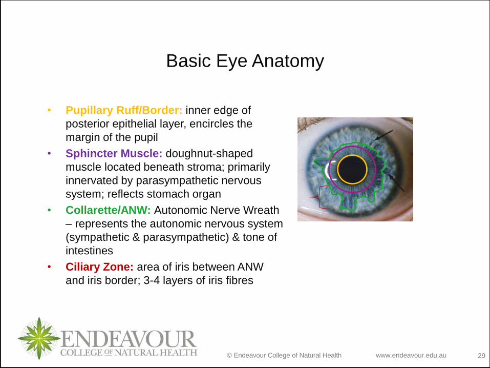
Basic Eye Anatomy
• Pupillary Ruff/Border: inner edge of
posterior epithelial layer, encircles the
margin of the pupil
• Sphincter Muscle: doughnut-shaped
muscle located beneath stroma; primarily
innervated by parasympathetic nervous
system; reflects stomach organ
• Collarette/ANW: Autonomic Nerve Wreath
– represents the autonomic nervous system
(sympathetic & parasympathetic) & tone of
intestines
• Ciliary Zone: area of iris between ANW
and iris border; 3-4 layers of iris fibres

Brown iris – denser
pigmentation
Blue iris – less dense
pigmentation
Blood vessels & nerves
Muscle fibres
Posterior membrane &
epithelium
Layers of the Iris
http://www.ijser.org/paper/Segmentation_Techniques_for_Iris_Recognition_System/Image_013.jpg

© Endeavour College of Natural Health www.endeavour.edu.au 31
How Does Iris Analysis Work?• The iris is connected to the
dura mater of the brain via
28,000 nerve endings that
form part of the the optic
nerve (part of central nervous
system);
• The iris is connected to the
sympathetic &
parasympathetic nervous
systems
• Each stromal cell &
chromatophore in the iris
receives it’s own nerve
supply that enters the root of
the iris via the ciliary body
www.meddean.luc.edu/
www.images.missionforvisionusa.org

© Endeavour College of Natural Health www.endeavour.edu.au 32
How Does Iris Analysis Work?• 33 separate arteries supply the tissues of the eye
• Through association with the brain and nervous
system the iris is indirectly and directly connected
with every, tissue, gland and organ of the body
• Via electrical & chemical impulses from the nerves
and circulatory system, the eye and iris receive
stimuli from the whole of the body (neuro-optic
reflex)
• Embryonically the iris develops from the mesoderm
and the neuroectoderm (same tissue that forms the
brain and spinal chord)
• Muscles of the iris are only body muscles derived
from the neuroectoderm Tart-Jensen, E 2012 Techniques in iris
analysis

© Endeavour College of Natural Health www.endeavour.edu.au 33
How Does Iris Analysis
Work?• As a fetus develops, nerve impulses & genetic
information are recorded in the structures of the
iris (genotype); health problems/changes in body
chemistry, physiology and structure that occur
throughout life can be refelected in the iris, pupil,
sclera and convunctiva of the eye.
• By examining the iris colour, structure and
pigmentation, the pupil, and the sclera, information
relating to an individual’s anatomical and
physiological health may be obtained.Tart-Jensen, E 2012 Techniques in iris analysis
© Endeavour College of Natural Health www.endeavour.edu.au 34
Iris Maps• The association of signs/indicators in the iris with
health problems relating to specific parts of the
body led to a mapping of areas in the iris to
tissues, organs and systems of the body.
• Over the centuries iris maps have evolved as the
result of fine-tuning adjustment due to the
interchange of information globally.
• Of the contemporary iris maps used
internationally there is at least 85% consistency.
• The iris map we will use as a reference in the
course is the Jensen chart* (see following slide)* Available via the LMS
© Endeavour College of Natural Health www.endeavour.edu.au 35
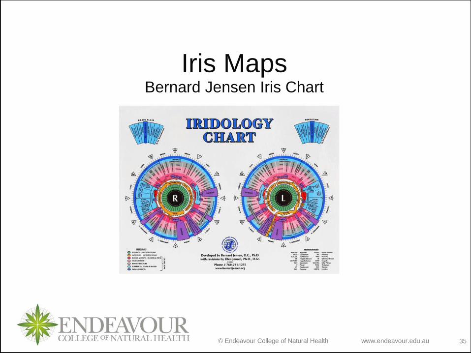
Iris MapsBernard Jensen Iris Chart
© Endeavour College of Natural Health www.endeavour.edu.au 36
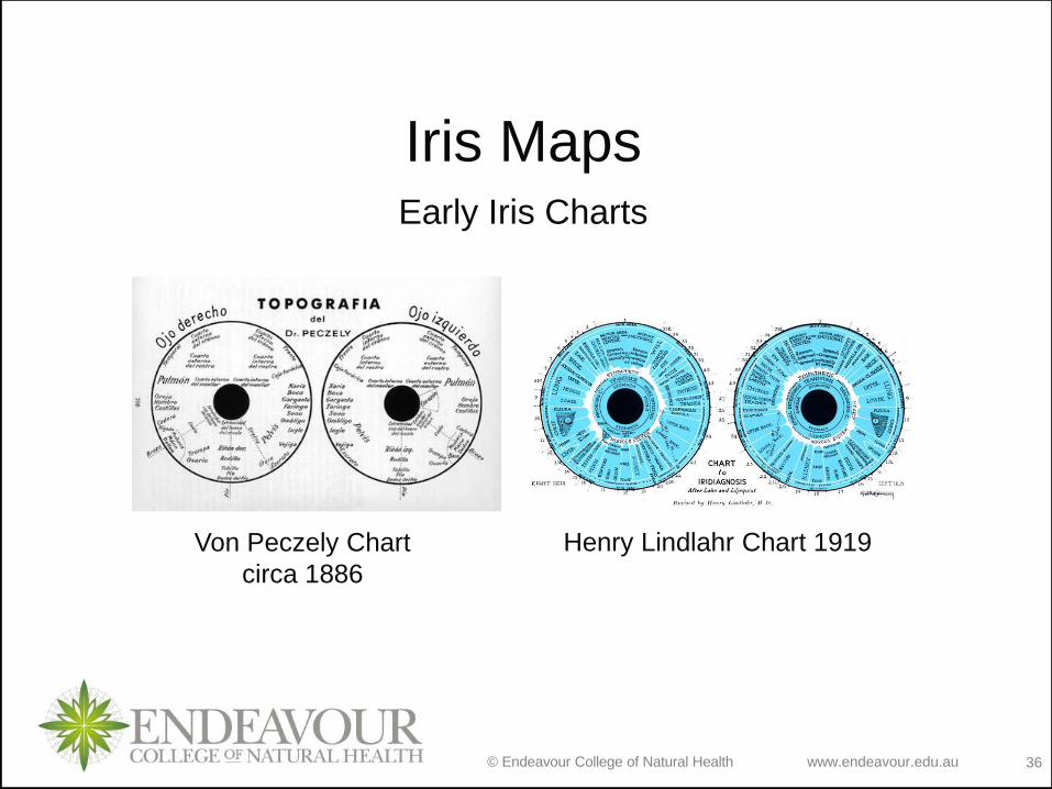
Iris MapsEarly Iris Charts
Von Peczely Chart
circa 1886
Henry Lindlahr Chart 1919
© Endeavour College of Natural Health www.endeavour.edu.au 37
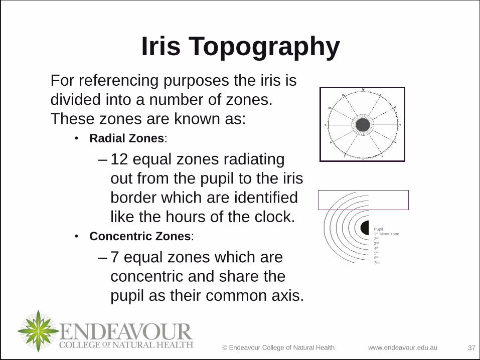
Iris TopographyFor referencing purposes the iris is
divided into a number of zones.
These zones are known as:• Radial Zones:
– 12 equal zones radiating
out from the pupil to the iris
border which are identified
like the hours of the clock.• Concentric Zones:
– 7 equal zones which are
concentric and share the
pupil as their common axis.
Pupil
1st Minor zone
2nd
3rd
4th
5th
6th
7th
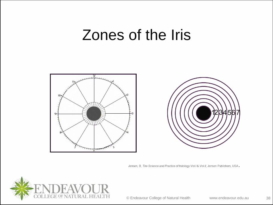
© Endeavour College of Natural Health www.endeavour.edu.au 38
Zones of the Iris

© Endeavour College of Natural Health www.endeavour.edu.au 39
Radial Zones
o Radial zones assist in referencing the reflex
position of body organs, for example;• The liver is situated between 7:30 and 7:40 in the right
iris.
o Radial zones also assist the practitioner in
referencing specific areas in the iris, for
example;• A practitioner may want to convey to another practitioner
the whereabouts of an iris sign which may appear in a
specific radial zone.

© Endeavour College of Natural Health www.endeavour.edu.au 40
Concentric Zoneso There are seven (7) Concentric zones in the Iris;
o Concentric zones are evenly distributed out from the
pupil border (Zone 1) to the Iris border (Zone 7);
o Concentric zones are zones in which specific body
systems and body organs generally tend to be located,
for example:
• Zone 1 = The Stomach Zone (Zone of Digestion)
• Zone 2 = The Intestinal Zone (Zone of Absorption)
• Zone 3 = The Humoral Zone (Blood & Lymph)
• Zone 4 = The Muscle Zone (Zone of Utilization)
• Zone 5 = The Skeletal Zone (Zone of ultimate Utilization)
• Zone 6 = The Lymph Zone (Zone of Detoxification)
• Zone 7 = The Skin Zone (Zone of Elimination)
Nutritive zone

© Endeavour College of Natural Health www.endeavour.edu.au 41
The Stomach Zone (Zone 1)
o The ‘Stomach Zone’ represents the integrity and function
of stomach and digestion (green on the iris chart)
© Endeavour College of Natural Health www.endeavour.edu.au 42
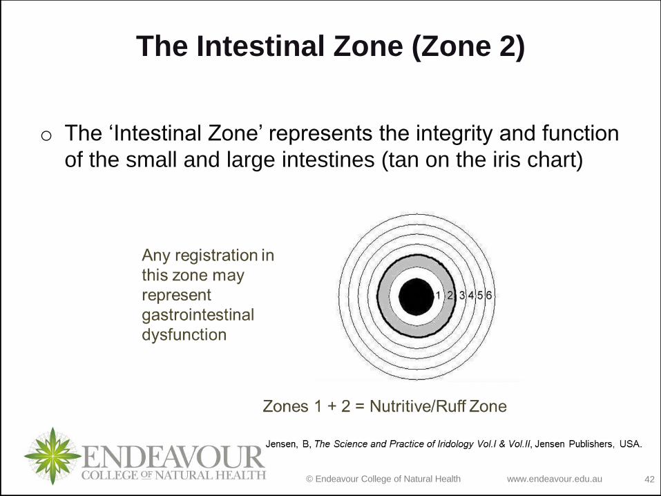
The Intestinal Zone (Zone 2)
o The ‘Intestinal Zone’ represents the integrity and function
of the small and large intestines (tan on the iris chart)

© Endeavour College of Natural Health www.endeavour.edu.au 43
The Humoral Zone (Zone 3)
o The ‘Humoral Zone’ (blood & lymph zone) represents the
dynamics of the transformation and distribution of
nutrients; major blood & lymph vessels, the heart &
some glands are located in this zone (dark pink on the
iris chart)

© Endeavour College of Natural Health www.endeavour.edu.au 44
The Muscular Zone (Zone 4)
o The ‘Muscular Zone’ represents where nutrients are
been distributed in the body. The first zone of nutrient
utilization and nourishment; registrations in this zone
may also relate to poor nutritional status of the
organ/tissue concerned (light pink on the iris chart)
© Endeavour College of Natural Health www.endeavour.edu.au 45
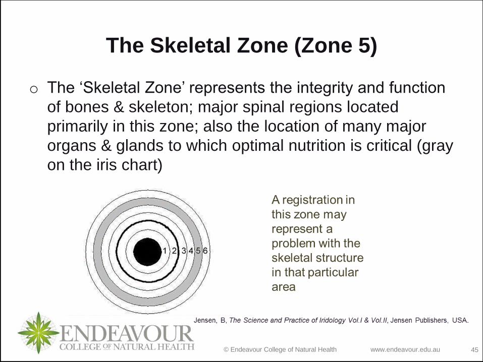
The Skeletal Zone (Zone 5)
o The ‘Skeletal Zone’ represents the integrity and function
of bones & skeleton; major spinal regions located
primarily in this zone; also the location of many major
organs & glands to which optimal nutrition is critical (gray
on the iris chart)
© Endeavour College of Natural Health www.endeavour.edu.au 46
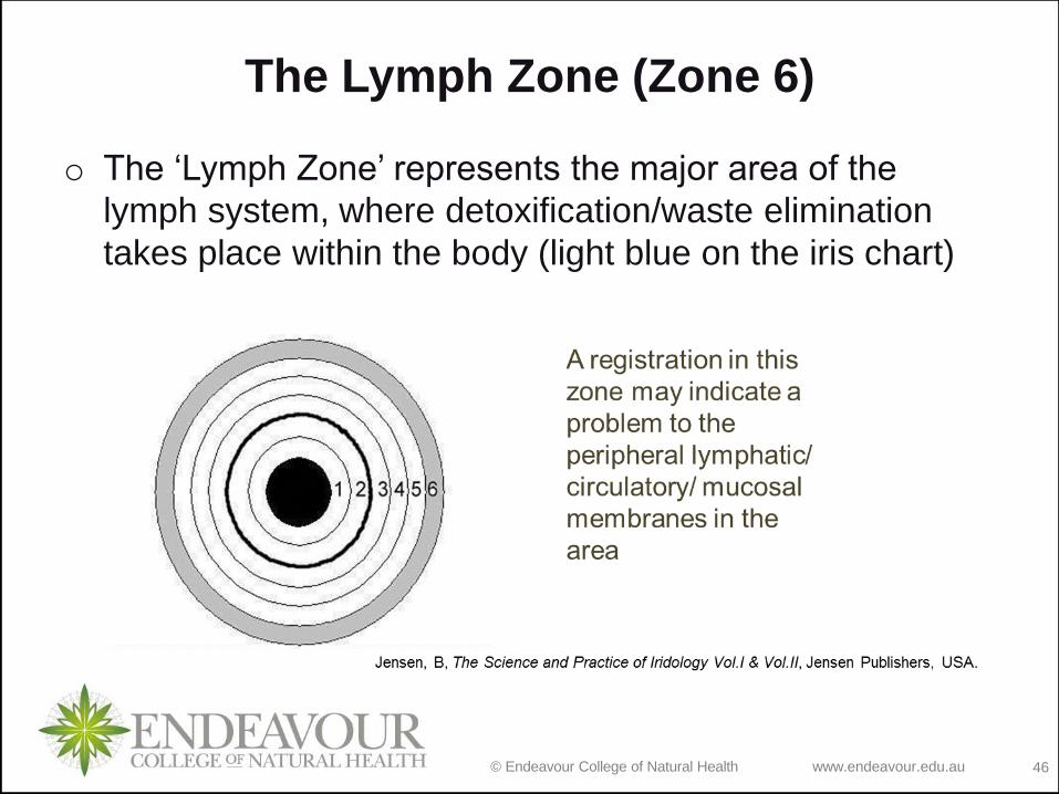
The Lymph Zone (Zone 6)
o The ‘Lymph Zone’ represents the major area of the
lymph system, where detoxification/waste elimination
takes place within the body (light blue on the iris chart)

© Endeavour College of Natural Health www.endeavour.edu.au 47
The Skin Zone (Zone 7)
7
Jensen, B, The Science and Practice of Iridology Vol.I & Vol.II, Jensen Publishers, USA.
Represents the skin, the zone of elimination, includes orifices
of the body. Located inside the iris border (dark blue on the
iris chart)
Any registration
in this zone
may represent
a skin problem
or dysfunction
of eliminatory
functions
© Endeavour College of Natural Health www.endeavour.edu.au 48
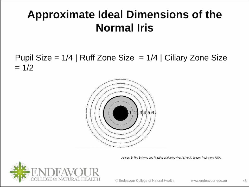
Approximate Ideal Dimensions of the
Normal Iris
Pupil Size = 1/4 | Ruff Zone Size = 1/4 | Ciliary Zone Size
= 1/2

© Endeavour College of Natural Health www.endeavour.edu.au 49
The Ruff/Nutritive Zone (Zones 1 & 2)
o The ‘Ruff Zone’ represents the integrity and function of
stomach and Intestinal tissue. It’s size is an indication of
the digestive function, and therefore ‘nutritional status’,
of an individual.

© Endeavour College of Natural Health www.endeavour.edu.au 50
The Ruff/Nutritive Zone contd
o An abnormal registration in this zone may represent
dysfunction of the processes of digestion, absorption
& assimilation in the stomach and intestines
o Normal size Ruff = good digestion, absorption &
assimilation
o Very small Ruff = hyperactivity of GIT functions
o Large Ruff = poor digestion, absorption &
assimilation
© Endeavour College of Natural Health www.endeavour.edu.au 51
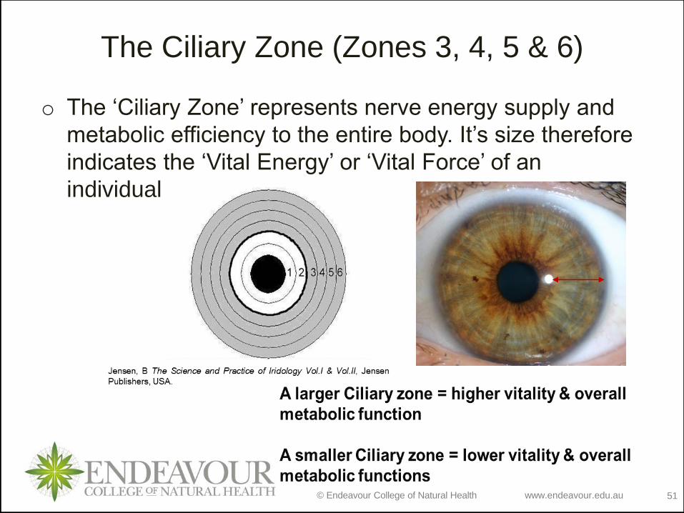
The Ciliary Zone (Zones 3, 4, 5 & 6)
o The ‘Ciliary Zone’ represents nerve energy supply and
metabolic efficiency to the entire body. It’s size therefore
indicates the ‘Vital Energy’ or ‘Vital Force’ of an
individual.
© Endeavour College of Natural Health www.endeavour.edu.au 52
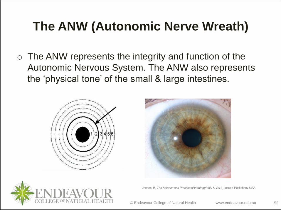
The ANW (Autonomic Nerve Wreath)
o The ANW represents the integrity and function of the
Autonomic Nervous System. The ANW also represents
the ‘physical tone’ of the small & large intestines.

© Endeavour College of Natural Health www.endeavour.edu.au 53
Variations of ANWs

© Endeavour College of Natural Health www.endeavour.edu.au 54
Abnormalities in the Iris
Abnormalities in the physical iris anatomy
represent dysfunction in the organs and
tissue of the body and can be seen in:• The Pupil (Size and shape)
• The ANW (Size, shape and colour)
• The Ruff Zone (Size)
• The Ciliary Zone (Size)
Pupil (Size and Shape):
The pupil represents the Central Nervous
System (Mostly the Spinal column)
© Endeavour College of Natural Health www.endeavour.edu.au 55
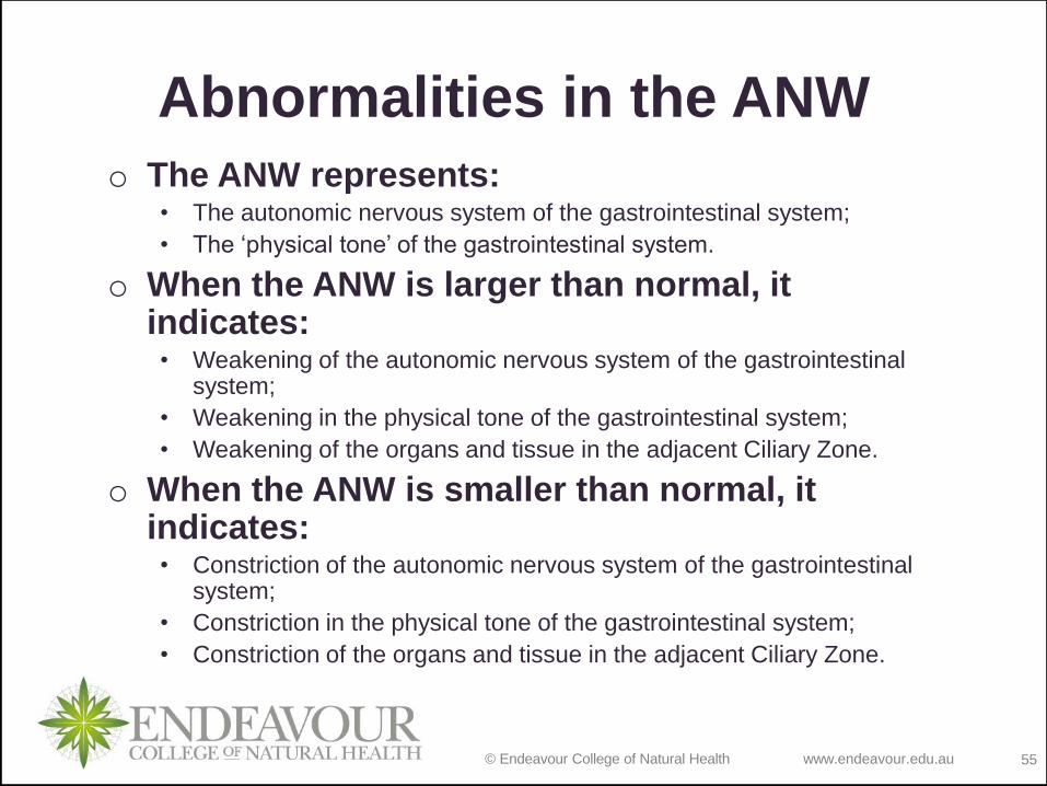
Abnormalities in the ANW
o The ANW represents: • The autonomic nervous system of the gastrointestinal system;
• The ‘physical tone’ of the gastrointestinal system.
o When the ANW is larger than normal, it indicates: • Weakening of the autonomic nervous system of the gastrointestinal
system;
• Weakening in the physical tone of the gastrointestinal system;
• Weakening of the organs and tissue in the adjacent Ciliary Zone.
o When the ANW is smaller than normal, it indicates:• Constriction of the autonomic nervous system of the gastrointestinal
system;
• Constriction in the physical tone of the gastrointestinal system;
• Constriction of the organs and tissue in the adjacent Ciliary Zone.

© Endeavour College of Natural Health www.endeavour.edu.au 56
Abnormalities of the ANW

© Endeavour College of Natural Health www.endeavour.edu.au 57
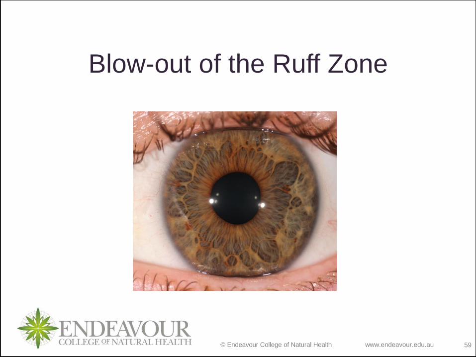
Abnormalities in the Ruff
Zone o The Ruff Zone indicates:
• Integrity and function of the Stomach and Intestines;
• The ‘Nutritional status’ of the individual.
o A small Ruff Zone indicates:• Constriction of the ANS to the GIT (Bowel);
• Constriction of the physical tone of the Bowel;
• Poor nutritional status.
o A large Ruff Zone indicates:• Lowered ANS energy in the Bowel;
• Poor physical tone of the Bowel (Blown-out);
• Poor nutritional status.

© Endeavour College of Natural Health www.endeavour.edu.au 58
Constriction of the Ruff Zone
© Endeavour College of Natural Health www.endeavour.edu.au 59
Blow-out of the Ruff Zone
© Endeavour College of Natural Health www.endeavour.edu.au 60
Abnormalities in the
Ciliary Zone
o The Ciliary Zone indicates:• Integrity and function of all body organs and systems;
• The ‘Vital Energy’ of the individual.
o A small Ciliary Zone indicates:• General lowered metabolic function of the body;
• Poor ‘Vital Energy’ of the individual.
o A large Ciliary Zone indicates:• General increased metabolic function of the body;
• Good ‘Vital Energy’ of the individual.

© Endeavour College of Natural Health www.endeavour.edu.au 61
Be sure to bring your iris
torch/magnifiers and iris
chart to the next class!
© Endeavour College of Natural Health www.endeavour.edu.au 62
ReferencesJensen B 1952. Iridology; the science and practice in the healing arts vol
1. Bernard Jensen Publisher, Escondido
Jensen B 1982. Iridology; the science and practice in the healing arts vol
2. Bernard Jensen Publisher, Escondido
Miller, T 2008.The integrated iridology textbook. Inter Health Australia,
Lake Munmorah, Australia
Sarris, J & Wardle, J 2010. Clinical Naturopathy, An evidence-based
guide to practice. Elsevier, Sydney
Sharan, F 1989. Iridology: a complete guide to diagnosing through the
iris and to related forms of treatment. Thorsons, Wellingborough, UK
Tart-Jensen, E 2013. Techniques in iris analysis: a textbook in iridology.
Infinite Iris Publishers, USA

© Endeavour College of Natural Health www.endeavour.edu.au 63
COMMONWEALTH OF AUSTRALIA
Copyright Regulations 1969
WARNING
This material has been reproduced and communicated to you by or on
behalf of the Australian College of Natural Medicine Pty Ltd (ACNM)
trading as Endeavour College of Natural Health, FIAFitnation, College
of Natural Beauty, Wellnation - Pursuant Part VB of the Copyright Act
1968 (the Act).
The material in this communication may be subject to copyright under
the Act. Any further reproduction or communication of this material by
you may be the subject of copyright protection under the Act.
Do not remove this notice.

© Endeavour College of Natural Health www.endeavour.edu.au 64
Tutorial Activity Session 2
• In pairs, observe the photo of the iris in the following slide &
discuss with your partner what you see in light of the information
covered in today’s session (10 mins).
• Come back into the main group & take turns reporting back
the main things you observed from your observation of the
iris.

© Endeavour College of Natural Health www.endeavour.edu.au 65

© Endeavour College of Natural Health www.endeavour.edu.au 66
Tutorial Activity Session 2 contd
• In layman’s terms explain as you would to a client how iris analysis works.
Make reference to basic eye anatomy and to some of the health indications
that iris patterns and registrations may suggest.
o
• Consider how iris analysis may assist in determining an appropriate
treatment/health management strategy for clients. Make reference to the
Naturopathic Therapeutic Order when framing your response.
o
• Naturopathic philosophy states that the three primary causes of disease are:
o Lowered vitality
o Abnormal composition of blood & lymph (poor nutritional status)
o Accumulation of toxins/xenobiotics and metabolic wastes in the system
• Based purely on what you have learned in this session, identify which of
the 7 concentric zones of the iris may provide information about these
three primary disease causes.